imaging workspace an overview and roadmap eliot l. siegel, md imaging workspace lead sme january 23,...
TRANSCRIPT

Imaging Workspace
An Overview and Roadmap
Eliot L. Siegel, MD
Imaging Workspace Lead SME
January 23, 2008

Introduction
• Imaging has been separate island with its own standards (DICOM)• DICOM allows high level of
interoperability among clinical imaging modalities such as CT and MRI
• Patient centered and very limited for data mining and research
• Completely separate from world of XML, service oriented architecture, Grid Computing
• Different vendors algorithms provide different results with no standards for annotation and image mark-up

caBIG IMG: A Brief History
The In Vivo Imaging Workspace was added to the caBIG program in April of 2005:
1.To advance imaging informatics for treatment of patients with cancer
2.To leverage caBIG technology such as the caGRID, Internet tools such as XML, and existing DICOM standards by creating “middleware,” to facilitate sharing of images in a variety of settings
3.To strive towards a standardized way to evaluate and annotate images, especially for evaluation of tumor burden and response
4.To facilitate secure and easy sharing of images and image analysis & visualization algorithms with an emphasis on the cancer community

caBIG IMG: A Brief History
Adoption activities
defined (CTTI/NTROI)
Interoperability of caBIG
Imaging tools
demonstrated
2005 2006 2007 2008
caBIG Imaging WS established
(110 years after the discovery of the
x-ray)
SMEs selected
NCIA development began/LIDC-use
case
Strategic plan set
XIP, Middleware, AIM
development began
First RSNA presentation
Adoption activities
planned
Next phase of
development
NLST IP

National Cancer Imaging Archive
• The NCIA network-accessible "in vivo image repository" provides image archives to assist development and validation of imaging software tools
• Multiple image libraries currently stored on archive including LIDC
• Not only repository, but software for NCIA is free and open source
• Federated system so single inquiry can produce responses from multiple repositories
• Includes visualization tool

What are the Imaging Workspace “Products”?

eXtensible Imaging Platform (XIP) Allows Easy Sharing of Image Enhancement/Analysis/Visualization Algorithms
XIP Application
(Can be replaced with any DICOM WG23-compatible Host)
XIP Host Adapter
XIP ModulesHost Independent
WG23
XIP HostWG23
WG23
Web-based Application
Medical Imaging Workstation
Standalone Application
Distribute
Distribute
DICOM, HL7, & otherservices per IHE
caGRID Services viaImaging Middleware
XIP Application Builder
XIP Class Library Auto Conversion Tool
Host-Specific Plug-in Libs
WG23
Distribute
ITK
VTK
XIP
LIB
. . .
A free and open source platform that facilitates the sharing not of images and other patient data but of image display, processing, and analysis algorithms themselves.

Imaging Middleware (including GridCAD and Virtual PACS)
• Middleware provides connection between DICOM and Grid computing
• The GRID has tremendous potential to promote interoperability, improve security, and support more efficient sharing of image data and software algorithms
• Middleware projects such as CAD (computer aided detection) for lung nodules on CT scans demonstrate the power and potential of GRID computing

Annotations and Imaging Markup (AIM)
The first project of its kind that we’re aware of to propose/create a “standard” means of adding information/knowledge to an image in a clinical environment in which there is currently chaos in order to create a future in which image content can be easily and automatically searched.

Algorithm Validation Tools (AVT)
BaselineMax Diameter 36.2mmVolume 6.1cm3
Baseline +20 weeksMax Diameter 32.6mmVolume 9.48cm3 55% increase
The purpose of AVT is to provide a set of tools capable of generating measurements using validated and consistent methods for detecting change; and to associcate information including clincial outcome data that would be helpful in assessing the performance of image-based change assessment tools
DICOM Ontology

DICON Ontology
• A single common reference information model for DICOM
• Unify and make explicit all the key entities and relations in DICOM in a human-usable but machine-processable format.
• Represent the existing DICOM model, whether it be implicit or explicit, as an ontology.

Query Formulation
• Create an Imaging Query Formulation tool
• Automates the creation of ontology-based queries to image resources
• Query Formulation Engine will translate user queries that are formulated using the QueryTool UI into an ontology-based query graph
“Mary J.”
Breast Cancer
Chemo-therapy
“Mary J’s CT Images”
Progressive Disease (RECIST)
PATIENT
DISEASE
TREATMENT
DISEASE STATUS
DISEASE IMAGING
has Disease Assessment
has RECIST Assessmenthas Disease
has TreatmentPhysical
Exam
has Disease Assessment
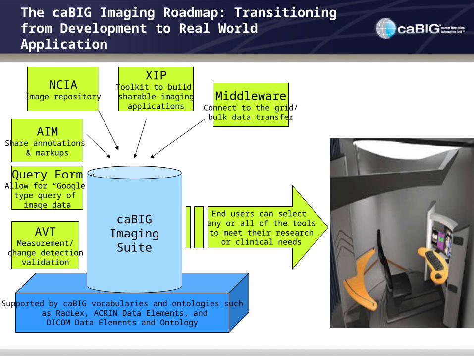
The caBIG Imaging Roadmap: Transitioning from Development to Real World Application
NCIAImage repository
XIPToolkit to build
sharable imagingapplications
MiddlewareConnect to the grid/bulk data transfer
AIMShare annotations
& markups
AVTMeasurement/
change detectionvalidation
Supported by caBIG vocabularies and ontologies such as RadLex, ACRIN Data Elements, and
DICOM Data Elements and Ontology
caBIGImaging
Suite
End users can select any or all of the tools to meet their research
or clinical needs
Query FormAllow for “Google”
type query of image data

In The Near Future: Practical Applications of Workspace Tools
Open Source
Imaging Viewer
Clinical Trials Tool
Integration
Other Adoption
Activities