imaging osteosarcoma & surgical...
TRANSCRIPT

Imaging Osteosarcoma & Surgical Outcomes
William R. Silveira, HMS IIIGillian Lieberman, M.D.Harvard Medical School
Beth Israel Deaconess HospitalNovember 12, 2007

Patient 1: Clinical presentation
He now reports…Moderate pain at restSevere pain when bearing weightFrequent waking at night with knee pain
He works out “once in a while”He denies a history of trauma and infectionHe denies previous problems with his left kneeNo significant past medical history or family historyNo fever, chills, nausea, or vomiting
15 year old boy presents with…“Pain in the upper part of my left knee”
The pain became progressively worse over 3 months

Patient 1: Clinical presentation
Physical ExamNo evidence of injuryMarked tenderness to palpationDiffuse swelling of the distal femurFirm, circumferential, fixed mass
Limited range of motion (painful!)Intact sensation & motor functionStrong pulses throughout extremityNo lymphadenopathy
15 year old boy presents with…“Pain in the upper part of my left knee”

Patient 1: Differential diagnosis: Atraumatic
knee pain in an adolescent
Juvenile rheumatoid arthritisReactive arthritisInfectious arthritisOsteomyelitisOsteochondritis dessicansDegenerative joint diseaseTendonitis, bursitisChondromalaciaReferred pain
MalignancyEwing’s sarcoma OsteosarcomaRhabdomyosarcomaLymphoid neoplasmMetastasis
M. M. Reeder and B Felson, Gamuts
in Radiology, Springer-Verlag Telos, 3rd edition, 1993S. Kahan and E. G. Smith, Signs & Symptoms, Blackwell publishing, Massachusetts, 2004
Consider tumor, inflammation & infection

Where to begin? Menu of tests: Atraumatic
knee pain in an adolescent
1.
RadiographAnteroposterior (AP)LateralNotch or tunnel viewAxial view Ipsolateral hip films (AP/frog leg lateral)
2. Nuclear study: radionuclide bone scan 3. Ultrasound (US) 4. Computed tomography (CT) 5. Magnetic resonance imaging (MRI) 6. Aspiration/arthrogram7. CT postarthrogram
“Mandatory workup”ACR guidelines
American College of Radiology, ACR Appropriateness Criteria, date of origin 1995, last reviewed 2005, http://www.acr.org

Patient 1: Radiograph of the left knee –
AP viewSoft tissue
1. Increased amorphous density2. Diffuse spiculated mass3. Continuous with metaphysis
Bone from far4. Osteosclerotic distal femur 5. Highest density at the metaphysis6. Wide zone of transition
Bone from near7. Ill defined cortical/medullary border8. Periosteal reaction:
lamellar & spiculated9. Physis open, difficult to assess
Joint 10. Difficult to assess
Image courtesy of Dr. Jim Wu
1
3
2

Patient 1: Radiograph of the left knee –
AP viewSoft tissue
1. Increased amorphous density2. Diffuse spiculated mass3. Continuous with metaphysis
Bone from far4. Osteosclerotic distal femur 5. Highest density at the metaphysis6. Wide zone of transition
Bone from near7. Ill defined cortical/medullary border8. Periosteal reaction:
lamellar & spiculated9. Physis open, difficult to assess
Joint 10. Difficult to assess
Image courtesy of Dr. Jim Wu
4
5
6

Patient 1: Radiograph of the left knee –
AP viewSoft tissue
1. Increased amorphous density2. Diffuse spiculated mass3. Continuous with metaphysis
Bone from far4. Osteosclerotic distal femur 5. Highest density at the metaphysis6. Wide zone of transition
Bone from near7. Ill defined cortical/medullary border8. Periosteal reaction:
lamellar & spiculated9. Physis open, difficult to assess
Joint 10. Difficult to assess
Image courtesy of Dr. Jim Wu
7
9
8
10
8

Patient 1: Radiograph of the left knee –
lateral viewSoft tissue
1. Amorphous & focal densities
Bone from far2. Osteosclerotic distal femur with a wide zone of transition
Bone from near3. Poor cortical/medullary border4. Periosteal reaction:
lamellar & spiculated5. Classic “Codman triangle”
Joint6. Well aligned
Image courtesy of Dr. Jim Wu
1
4
3
5
2
6
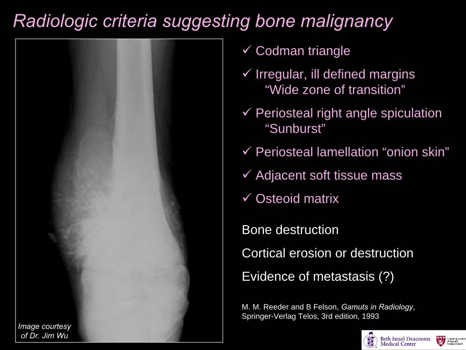
Radiologic criteria suggesting bone malignancyCodman triangle
Irregular, ill defined margins“Wide zone of transition”
Periosteal right angle spiculation“Sunburst”
Periosteal lamellation “onion skin”
Adjacent soft tissue mass
Osteoid matrix
Bone destruction
Cortical erosion or destruction
Evidence of metastasis (?)
Image courtesy of Dr. Jim Wu
M. M. Reeder and B Felson, Gamuts
in Radiology, Springer-Verlag Telos, 3rd edition, 1993

Patient 1:
Narrowing the differential for… “…an aggressive appearing, sclerotic bone lesion of
the distal femur with a significant periosteal
reaction”
Ranked differential diagnosis1. Osteosarcoma (ages 10-25)2. Ewing’s sarcoma (ages 5-20)3. Metastasis
→ PlanMagnetic Resonance ImagingRadionuclide studyChest radiographCT guided biopsy

Patient 1: MRI –
Coronal STIR of the left kneeExtent of disease for surgical planningExcellent marrow & soft tissue contrastDetailed anatomy
STIR suppresses signal from fatSensitive to edema and bone pathologyNormal marrow and fat: darkFluid & edema: bright
Bone findings:1. Increased signal in the medullary canal2. Irregular pattern in the metaphysis3. Ill defined cortical outline4. Extension past the physis
Soft tissue findings:5. High signal around distal femur, suggesting edema and growth into the surrounding tissue
Image courtesy of Dr. Jim Wu
1
2
3
4
5

Patient 1: MRI –
Axial STIR of distal femur
Image courtesy of Dr. Jim Wu
High signal in the medullary canal (1) and soft tissue (2)
2 1

Companion patient 1, osteosarcoma: An example of bone scintigraphy
Courtesy of Dr. Jim Wu
Evaluation of metabolic activity
Technetium, 99Tcm, coupled to methylene diphosphate
IV administration
Radioactive decay yields a 142.7 keV γ-ray
2D projections constructed with a scintillation camera
Extent of disease & metastasis
“Hot spots” arise in fractures & tumors
Significant uptake in the distal femur
G. Friedlander, Nuclear and Radiochemistry, 3rd edition, John Wiley & Sons, 1981.

Osteosarcoma: CT guided bone biopsy
1. Formation of new, abnormal bone with a coarse lacelike architecture
2. Variable tumor cell size & shape, with hyperchromatic nuclei and mitoses
Histology confirmed radiological suspicion of osteosarcoma in the distal femur of patient 1
PACS, BIDMCV. Kumar, A. K. Abbas, N. Fausto, Robbins and Cotran
Pathologic Basis of Disease, 7th edition, Elsevier, 2005
1
1
2
Companion patient #2An example of CT guided biopsy

Now for a brief discussion of osteosarcoma

What is osteosarcoma?
V. Kumar, A. K. Abbas, N. Fausto, Robbins and Cotran
Pathologic Basis of Disease, 7th edition, Elsevier, 2005
2nd most common primary malignant bone tumorPeak incidence in males 10-20 years of age3 cases/million population/year
Metaphysis of long bones: 60% about the knee
Older patients: associated with Paget’s disease, radiation, bone infarcts, or chondrosarcomas
Potential etiologies include viral, mutations (RB), p53, ionizing radiation
Clinical findings: Pain, soft tissue swelling, fracture
Hematogenous spread to the lungs, liver & brain
definition: a tumor of the skeleton in which cancerous cells produce bone matrix
Courtesy of Dr. Jim Wu
Courtesy of Dr. Jim Wu

OsteosarcomaGross appearance
Large tumorsGritty & grayish-white HemorrhageCystic degeneration Cortical destruction Spreads – medullary canalSoft tissue masses
V. Kumar, A. K. Abbas, N. Fausto, Robbins and Cotran
Pathologic Basis of Disease, 7th edition, Elsevier, 2005.
Courtesy of Dr. Jim Wu

Classification of osteosarcoma
Classic osteosarcoma (75%): Affects long tubular bones, particularly the metaphysis, with mixed osteolysis & osteosclerosis and marked periosteal reaction
Telangiectatic osteosarcoma (2.5-12.5%): Lytic tumors consisting of large cystic cavities filled with blood
Small cell osteosarcoma (1-4%): Similar to classic osteosarcoma, primarily osteolytic with a poor prognosis
Surface osteosarcoma (4-10%): Marrow is uninvolved, often low grade with a good prognosis
Tumor stage: Based on grade, extension, and metastases
V. Kumar, A. K. Abbas, N. Fausto, Robbins and Cotran
Pathologic Basis of Disease, 7th edition, Elsevier, 2005.S. Suresh and A. Saifuddin, Clinical Radiology
(2007) 62, 314-323.

Treatment options for classic osteosarcoma
Surgery alone: 20% cure rateSurgery & chemotherapy: 60-80% cure rateRadiation therapy, rarely
Radical surgical treatment• Amputation• Limb salvage (used in 80-90% of all cases)
– Bone replaced with a bone allograft or a prosthesis• Arthrodesis: joint fusion
Complete resection of pulmonary & other metastasisClosely monitor primary and metastatic disease
“Bone Tumors: Surgical Options” from Children’s Hospital Bostonhttp://www.childrenshospital.org/az/Site646/mainpageS646P0.htmlS. Suresh and A. Saifuddin, Clinical Radiology
(2007) 62, 314-323.

Now, returning to patient 1….

Patient 1, osteosarcoma –
PA chest radiograph10-20% of patients have pulmonary metastases at the time of diagnosis
Multiple focal densities in both lungs, more numerous and dense toward the bases
In those who die from the disease, most have metastases to the lung, bones, or brain
Unfortunately, patient 1 was unsuccessfully treated with chemotherapy & surgery
The following three slides show the progression of the lung metastasesImage courtesy
of Dr. Jim Wu
P. Staddon et al., The Oncologist
(2002) 7, 144-153.

Patient 1, osteosarcoma –
Chest CT
Image courtesy of Dr. Jim Wu
Axial CT at the level of the carina, lung window
Multiple pulmonary nodules found at initial diagnosis of osteosarcoma

Patient 1, osteosarcoma –
Chest CT
Image courtesy of Dr. Jim WuImage courtesy of Dr. Jim Wu
Several months after initial presentation
Growth of the metastases and formation of a pneumothorax
(arrow)

Patient 1, osteosarcoma –
Chest CT
Image courtesy of Dr. Jim Wu
Growth of the metastases and formation of a pneumothorax
(arrow)
Several months after initial presentation

Next, we have a patient with osteosarcoma and a much better
prognosis & surgical outcome

Patient 2, osteosarcoma
Radiograph (AP) of left knee T2 weighted MRI
22 year old man with osteosarcoma of the proximal tibiaInitial clinical presentation: knee pain
PACS, BIDMC PACS, BIDMC

Patient 2, osteosarcoma
Radiograph (AP) of left knee T2 weighted MRI
22 year old man with osteosarcoma of the proximal tibiaInitial clinical presentation: knee pain
PACS, BIDMC PACS, BIDMCHigh density
Low density
Low Signal
Soft tissue changes

Patient 2, osteosarcoma –
Limb salvage surgery
PACS, BIDMC PACS, BIDMC
AP Lateral

Patient 2, osteosarcoma –
Limb salvage surgery
PACS, BIDMC PACS, BIDMC
AP Lateral
Allograph
Allograph

Finally, we have a patient with osteosarcoma with very interesting
surgical outcome

Patient 3, osteosarcoma –
AP & lateral radiographsHPI: 13 year-old boy On a skiing trip with his familySwerved to miss a poleFractured left femur
No prior traumaPreviously healthy
“Occasional ache” “I thought it was just a muscle”
Workup: Plain film, MRI, bone scintigraphy, and biopsy confirmed osteosarcoma
AP Lateral PACS, CHB
Simple oblique fracture of the distal femur with anterior translation of proximal femur, pathologic

Patient 3, osteosarcoma –
Scintigraphy
Images from PACS, CHB
R L
After the bone was set, this radionuclide scan
was obtained
Increased uptake in the distal femur is consistent
with bone tumor

Images from PACS, CHB
Patient 3, osteosarcoma –
Saggital
MRI T1W
MRI shows the location of the fracture, as well as the extent of disease along the medullary canal (arrows). Normal marrow is characterized by high signal, while the malignancy is characterized by low signal. Abnormal, weak cortex surrounds the pathologic marrow.

The tumor was resected…

PACS, BIDMC PACS, BIDMC PACS, BIDMC
Images courtesy of Anne C. Kim, M.D.
Patient 3, osteosarcoma –
Status post rotation-plastyAP radiographs of left femur, tibia and fibula

Images from “Bone Tumors: Surgical Options” from Children’s Hospital Boston
http://www.childrenshospital.org/az/Site646/mainpageS646P0.html
Rotation-plasty
• When limb-sparing not possible• Most nervous function preserved• Ankle joint functions as knee
Distal limb rotated 180º

Clinical Anatomy 17:345–353 (2004)
oblique incisions
Rotation-plasty
–
surgical incisions & resection

The New England Journal of Medicine 351;8John P. Dormans and Sumeet Gargwww.nejm.org August 19, 2004
Patient 3, osteosarcoma –
Rotation-plasty
Patient 3 outcome:Oncologist: “He’s doing very well.”Follow-up radiographs every 6 months
Higher function
Increased participation in hobbies/sports

Acknowledgments
Gillian Lieberman, MDJim Wu, MDAnne C. Kim, MDPrachi Dubey, MDMaria LevantakisFargol Booya, MD

ReferencesM. M. Reeder and B Felson, Gamuts
in Radiology, 3rd edition, Springer-Verlag Telos, 1993.
S. Kahan and E. G. Smith, Signs & Symptoms, Blackwell publishing, Massachusetts, 2004.
G. Friedlander, Nuclear and Radiochemistry, 3rd edition, John Wiley & Sons, 1981.
“Bone Tumors: Surgical Options” from Children’s Hospital Boston. http://www.childrenshospital.org/az/Site646/mainpageS646P0.html.
American College of Radiology, ACR Appropriateness Criteria, date of origin 1995, last reviewed 2005. http://www.acr.org.
V. Kumar, A. K. Abbas, N. Fausto, Robbins and Cotran
Pathologic Basis of Disease, 7th edition, Elsevier, 2005.
S. Suresh and A. Saifuddin, Clinical Radiology
(2007) 62, 314-323.
P. Staddon et al., The Oncologist
(2002) 7, 144-153.
J. P. Dormans and S. Garg, The New England Journal of Medicine
351;8, August 19, 2004, http://www.nejm.org.