imaging of perineural tumor spread in head and … of perineural tumor spread in head and neck...
TRANSCRIPT

Imaging of Perineural Tumor Spread in Head and Neck Cancer
Lawrence E. Ginsberg, MD
Departments of Diagnostic Radiology and Head and Neck Surgery
University of Texas M.D. Anderson Cancer Center
Houston, Texas
Perineural Tumor Spread (PNS)
Definition: dissemination of tumor from the primary site along tissue planes of the neural sheath
Small vs. large nerve PNS
Clinical settings:
salivary gland-parotid, minor salivary gland (palate)
mucosal (SCCa)-palate/RMT, nasopharynx (via MS, PPF)
skin (SCCa, desmoplastic melanoma)
previously treated/forgotten disease*
Symptoms: pain, paresthesias, motor denervation, but up to 40% may be asymptomatic
Implications: serious finding associated with decreased survival. Detection will often affect treatment
Perineural Tumor Spread (PNS)
Failure to recognize PNS (big problem)
common pitfall in head and neck imaging
good way to get sued
guarantees disease recurrence
Because may be asymptomatic, up to
radiologist to think of PNS and detect it
Rarely, site to which tumor spreads
perineurally may present prior to detection of
primary cancer. Therefore, must consider PNS
whenever lesion is seen in Meckel’s cave,
pterygopalatine fossa, or cavernous sinus
PNS Anatomic Considerations
Trigeminal nerve V
V1 ophthalmic
V2 maxillary
V3 mandibular
Facial nerve VII
Connections between V and VII
Uncommon routes
Perineural Tumor Spread-Imaging Widening/destruction of or excessive
enhancement within neural foramina (ovale, rotundum, palatine, stylomastoid foramen/descending facial canal, vidian canal)
CT better for bone destruction (late finding)
Loss of normal fat density (CT)/T1 signal intensity (MR) or excessive enhancement/widening of the pterygopalatine fossa
Enlargement/excessive enhancement within cavernous sinus or Meckel’s cave
MR technique: 16-18 cm FOV, 3 mm slices, fat-suppressed, post contrast T1-weighted images
What should a normal pterygopalatine fossa look like?

What should a normal Meckel’s Cave look like? Ophthalmic Nerve V1
Provides sensory innervation to the eye, lacrimal gland, conjunctiva, and skin of the nose, supraorbital region, and frontal scalp
Course-cavernous sinus to SOF, orbit, divides into branches-lacrimal, nasociliary, frontal
Lacrimal branch also carries parasympathetic innervation originating in the facial nerve, via the GSPN and ultimately a small twig from the zygomaticotemporal branch of V2
Main nerve involved in PNS is frontal nerve, which divides into (or is formed by the joining of) the supratrochlear and supraorbital branches
58-y/o man, s/p resection SCCa left medial forehead, locally recurrent
5 months later, with PNS on supratrochlear branch V1, extending to
frontal nerve
72-y/o man presented with left forehead numbness, subsequently developing fullness.
Imaging (Brain MR) allegedly normal. Biopsy of left eyebrow region SCCa. Post-bx
developed diplopia attributable clinically to left abducens palsy. Note tumor going
through superior orbital fissure.
(PNS) Anatomic Considerations Maxillary nerve, V2-sensory to the mid-face,
palate, sinonasal region, upper oral cavity. Common pathway to PPF, foramen rotundum, cavernous sinus, Meckel’s cave
Mandibular nerve, V3-sensory to lower face and oral cavity, motor innervation to muscles of mastication. Common pathway to foramen ovale, Meckel’s cave
Antegrade PNS-Meckel’s cave to cavernous sinus or downward along V3, Cavernous sinus anteriorly along V2, PPF along palatine or infraorbital nerves
Facial nerve, generally from primary parotid lesions or lesions that secondarily extend into the parotid

Fair-skinned 74-y/o male with left cheek melanoma, and V2 hypesthesia
62-y/o man, now with left V2 paresthesias following Mohs surgery for
left cheek SCCa. Recurrence with infraorbital nerve PNS
Palatal/maxillary alveolar ridge ACCa, PNS to PPF, rotundum
greater
palatine
foramen
64-y/o female presents with
sudden onset of left cheek
numbness. Unsuccessfully
treated for sinusitis. PE
confirmed V2 sensory
abnormality and was
otherwise normal

(PNS) Anatomic Considerations Maxillary nerve, V2-sensory to the mid-face, palate,
sinonasal region, upper oral cavity. Common pathway to PPF, foramen rotundum, cavernous sinus, Meckel’s cave
Mandibular nerve, V3-sensory to lower face and oral cavity, motor innervation to muscles of mastication. Common pathway to foramen ovale, Meckel’s cave
Antegrade PNS-Meckel’s cave to cavernous sinus or downward along V3, Cavernous sinus anteriorly along V2, PPF along palatine or infraorbital nerves
Facial nerve, generally from primary parotid lesions or lesions that secondarily extend into the parotid
good ovale bad ovale
Recurrent SCCa in the right masticator space, growing up V3
Prior left buccal SCCa, now recurrent to masticator space, and then
PNS along V3, into Meckel’s cave PNS in Nasopharyngeal Carcinoma
Requires extension into:
pterygopalatine fossa (V2 PNS)
direct extension through pterygoid plates
anterior extension into nasal cavity and laterally through the sphenopalatine foramen
both
masticator space (V3 PNS)
lateral extension
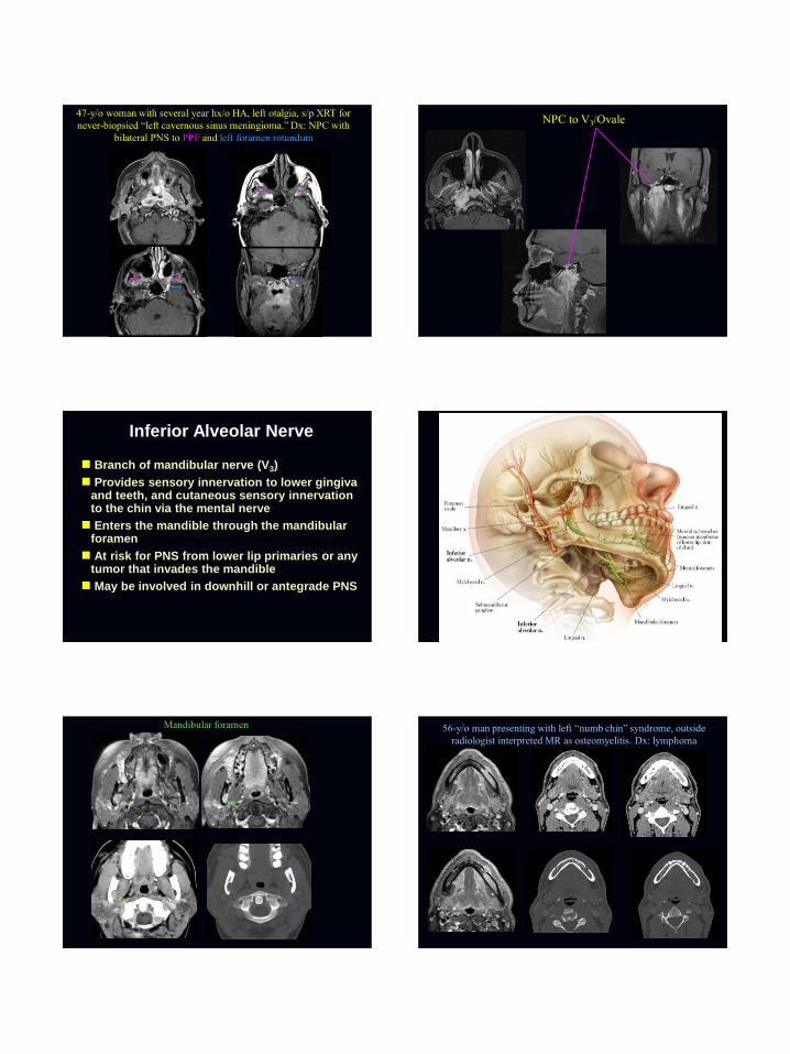
47-y/o woman with several year hx/o HA, left otalgia, s/p XRT for
never-biopsied “left cavernous sinus meningioma.” Dx: NPC with
bilateral PNS to PPF and left foramen rotundum
NPC to V3/Ovale
Inferior Alveolar Nerve
Branch of mandibular nerve (V3)
Provides sensory innervation to lower gingiva and teeth, and cutaneous sensory innervation to the chin via the mental nerve
Enters the mandible through the mandibular foramen
At risk for PNS from lower lip primaries or any tumor that invades the mandible
May be involved in downhill or antegrade PNS
Mandibular foramen 56-y/o man presenting with left “numb chin” syndrome, outside
radiologist interpreted MR as osteomyelitis. Dx: lymphoma

40-y/o man, s/p WLE left lower lip desmoplastic melanoma late 90’s,
and resection of recurrence 2/05. Did well until 4/06 when he started
experiencing numbness in the left lower teeth. Patient required
hemimandibulectomy. Intramandibular inferior alveolar nerve
perineural recurrence
2/05
4/06
Recurrent left lower lip SCCa with proven PNS along the
mental/inferior alveolar nerve to level of main trunk V3
Auriculotemporal Nerve Branch of mandibular nerve (V3)
Arises just below foramen ovale
Provides cutaneous innervation to lateral face, preauricular, external ear, and TMJ
Also acts as conduit for post-ganglionic parasympathetic fibers (originating as LSPN), that provide secretomotor innervation to the parotid gland
ATN and therefore V3 at risk for PNS in 1° or 2° malignancies of parotid gland and skin cancers in its cutaneous distribution
Evaluation of all parotid malignancies should include foramen ovale and proximal course of V3
Auriculotemporal nerve
Pre-auricular carcinoma, invasive of parotid, with PNS along
auriculotemporal nerve to foramen ovale

66-y/o man developed right-sided trigeminal pain, progressing to numbness, unresponsive
to steroids and acyclovir. Shortly thereafter developed facial neuropathy. History notable for
removal of several skin cancers including left medial canthus and right nasal dorsum
(SCCa) and several left facial BCCs.
Initial outside imaging
showed tumor along right
V3 but precise site or
origin unclear
Repeat imaging at MDACC
Scans read as likely right temporal subcutaneous primary (or recurrence), with ATN PNS.
Surgeon did not read report and patient was on table for craniotomy, Meckel’s cave biopsy,
which was aborted when soft tx biopsy proved SCCa. There is retrograde spread onto the main
trigeminal trunk, and antegrade spread into foramen rotundum
PNS-Anatomic Considerations-Facial Nerve
Generally related to parotid pathology, either primary parotid malignancy, or lesions, generally skin cancers, that secondarily invade the parotid, at diagnosis or at recurrence
Less commonly, skin cancers that have not yet invaded the parotid
Beware the subdermal skin cancer, that is difficult to detect clinically
When is “Bell’s Palsy” a Bell’s Palsy?
How Can Cancer Access the Facial Nerve Perineurally?
Via peripheral branches and back into main trunk
Directly into the stylomastoid foramen
Back along GSPN (to follow)
59-y/o man with multiple recurrences right facial SCCa, now with
parotid region recurrence and facial palsy

79-y/o man with recurrent scalp SCCa left parotid
Outside brain,
radiologist told
“Bell’s Palsy,” no
mention of prior
skin cancer
Facial nerve
enhancement
attributed to
“Bell’s
Palsy.”
No mention
of parotid
met
Relationship Between CN 5 and 7
Distal branches of V serve as conduits for small branches of VII and IX. These represent real or potential routes of PNS
Potential sources of PNS:
chorda tympani-tongue, SM/SL glands
LSPN (runs with auriculotemporal nerve)-parotid gland
Greater superficial petrosal nerve (GSPN)
Branch of CN7 originating in nervus intermedius
Preganglionic fibers, motor root of SP ganglion
Post ganglionic supply to palate, nasal, lacrimal
Potential for perineural tumor spread quite real
Course of the GSPN
Geniculate
ganglion
Facial hiatus
Foramen
lacerum
Vidian canal

rotundum
vidian
Foramen rotundum
rotundum
vidian
43-y/o woman with right facial pain and numbness. Dx: ACCa of the
hard palate with PNS to the PPF and vidian nerve
69-y/o man with ACCa left nasomaxillary. Spread to PPF facilitates
PNS to vidian and rotundum
* * *
62-y/o woman with several year history of left ear discomfort and placement of
tympanostomy tube for treatment of eustachian tube dysfunction. More recently
developed left trigeminal sensory neuropathy, and oh yeah, just noticed she’s
NOT TEARING FROM THE LEFT EYE. Outside brain MR read as cavernous
sinus meningioma, patient referred for proton therapy.
Repeat imaging obtained primarily for XRT planning. How to make a
very long story short…?

Dx: ACC NP
Unusual Pathways of PNS
Nasociliary nerve
Great auricular nerve
Supraclavicular nerve
Other cranial nerve
Nasociliary Nerve
Branch of V1
Provides cutaneous sensory fibers to skin of lateral nose, and sensory innervation from the frontal dura, sphenoid and ethmoid sinus mucosa, nasal mucosa, and medial canthus
Shah K, Esmaeli B, Ginsberg LE. Perineural tumor spread along the
nasociliary branch of the ophthalmic nerve: imaging findings. J Comput
Assist Tomogr 37(2):282-5, Mar-Apr, 3/2013.
77-year old woman with recurrent SCCa, left medial canthal region
Great Auricular Nerve (GAN)
Superficial branch of the superficial cervical plexus.
GAN provides sensory innervation to the skin over the parotid and lower pre-auricular region.
GAN leaves plexus, courses over and around the SCM (Erb’s point) and then upward toward the ear.
Has communicating branches with the facial nerve within the parotid gland, and with the auricular branch of the vagus nerve
GAN at risk for PNS in its cutaneous distribution
Ginsberg LE, Eicher SA. Great auricular nerve: anatomy and imaging of
perineural tumor spread. AJNR 21: 568-571, 2000.

68-y/o man with prior skin resections, now with recurrent SCCa left
parotid, and PNS along auriculotemporal and greater auricular nerve
72-y/o man with multiply recurrent SCCa left face, now with
recurrence over the left lower parotid region
Alsarraf L, Shah K, Hessel A, Williams M, Ginsberg LE. Perineural spread along the intermediate
branch of the supraclavicular nerve- A case report. Neurographics. In Press.
Supraclavicular Nerves
Branches of cervical plexus
Formed by twigs from C3, C4 spinal nerve ventral rami
Provide sensory innervation to skin over the clavicle, anteromedial shoulder, upper chest
Anterior, posterior, intermediate branches

61-year-old man with recurrent SCCa, left supraclavicular region.
Rare Nerves-advanced, slow growing malignancies can spread to 3rd, 6th cranial nerves, maybe others, if cavernous
sinus involved
Advanced recurrent lacrimal ACCa
Advanced recurrent facial melanoma, already had cavernous
sinus disease. Progressive 3rd nerve involvement
Conclusion
Perineural tumor spread is a very serious and potentially life-threatening complication of head and neck cancer
Because it may be asymptomatic at presentation or masked at recurrence due to prior therapy, it is critical that the radiologist make the diagnosis
Diagnosing PNS requires careful attention to imaging technique and a solid understanding of the relevant neuro-anatomy