imaging of atherosclerosis in apoliprotein e knockout mice...
TRANSCRIPT
Imaging of Atherosclerosis in ApoliproteinE Knockout Mice: Targeting ofa Folate-Conjugated Radiopharmaceuticalto Activated Macrophages
Wilfredo Ayala-Lopez, Wei Xia, Bindu Varghese, and Philip S. Low
Department of Chemistry, Purdue University, West Lafayette, Indiana
Early detection of heart disease is essential for the implementa-tion of intervention strategies that reduce the risk of cardiovascu-lar events. Radioimaging methods that have been explored forthis purpose include 18F-FDG, which measures sites of elevatedmetabolic activity; 99mTc-annexin A5, which reveals regions ofenhanced apoptosis and thrombosis; and 99mTc-labeled anti–lectinlike oxidized low-density lipoprotein receptor 1 antibody,which detects the lectinlike oxidized low-density lipoproteinreceptor 1 that is overexpressed on a variety of vasculature-associated cells. In this study, we examine the use of a folate-targeted chelate of 99mTc, termed 99mTc-EC20, for imaging offolate receptor (FR)–expressing macrophages that accumulatein atherosclerotic plaques, internalize cholesterol-rich lipopro-tein particles, and evolve into foam cells that form componentsof vulnerable atherosclerotic lesions. Methods: 99mTc-EC20was injected into apoliprotein E knockout (apoE2/2) mice feda normal or Western (high-fat) diet for 25 wk and imaged byg-scintigraphy. Treated mice were also dissected, and radioac-tivities in excised aortas were quantified by g-counting andimaged by autoradiography. The role of FR-expressing macro-phages in uptake of 99mTc-EC20 was also examined by compar-ing images of apoE2/2 mice before and after treatment withclodronate liposomes to deplete tissue macrophages, compar-ing the sites of 99mTc-EC20 enrichment with sites of macrophageaccumulation in thin sections of atherosclerotic tissues, andexamining the expression of FRs on atherosclerotic plaque–derived macrophages by flow cytometry. Results: ApoE2/2 miceon Western chow exhibited significantly greater accumulationof 99mTc-EC20 in atherosclerotic lesions than their counterpartson normal chow. The aortas of apoE2/2 mice on a Western dietdemonstrated greater numbers of FR-positive macrophages byflow cytometry than did those of apoE2/2 mice on a normaldiet. Clodronate liposome treatment significantly reduced theaccumulation of 99mTc-EC20 in atherosclerotic tissues, suggest-ing that macrophages or monocytes are responsible for uptakeof the folate-linked radioimaging agent. Histologic and autora-diographic analysis of tissue sections demonstrated that macro-phage accumulation correlated with regions of 99mTc-EC20
uptake. Conclusion: 99mTc-EC20 can be used for the imagingof atherosclerosis by selectively targeting FR-positive activatedmacrophages.
Key Words: atherosclerosis-associated macrophages; imagingof vulnerable plaque; folate receptor targeting; EC20
J Nucl Med 2010; 51:768–774DOI: 10.2967/jnumed.109.071324
Cardiovascular events associated with progressiveatherosclerosis constitute the main cause of death inWestern societies today (1). Atherosclerosis originatesfrom chronic inflammation of the arteries characterized byenhanced infiltration of leukocytes, uptake of lipoproteinparticles, proliferation of smooth muscle and endothelialcells, and apoptosis of foam cells (2). Myocardial infarc-tion, the most serious of the atherosclerotic sequelae, canresult from expansion and destabilization of atheroscle-rotic lesions, leading to plaque rupture and subsequentthrombotic events (2). Prevention of serious cardiovascu-lar events has, therefore, not surprisingly been linked toearly diagnosis and detection of vulnerable atheroscleroticlesions (3).
Vascular properties that are thought to differ betweenvulnerable and quiescent atherosclerotic lesions generallyinclude the inflammatory status, morphology, degree ofstenosis, cap thickness and stability, and proteinase activityof the atherosclerotic plaque (3). Attempts to quantitatethese differences as a means of assessing cardiovascularrisk have focused on the design of noninvasive imagingagents that detect the accumulation of immune cells (4),expression of metalloproteinase activity (5), heightenedconsumption of glucose (6), and apoptosis of cells withinthe vascular bed (7). Although such strategies show con-siderable promise for identification of vulnerable plaque,they also suffer from some degree of nonspecificity becauseeach strategy also images healthy cells involved in otherprocesses.
Received Oct. 4, 2009; revision accepted Jan. 26, 2010.For correspondence or reprints contact: Philip S. Low, Department of
Chemistry, Purdue University, 560 Oval Dr., West Lafayette, IN 47907.E-mail: [email protected] ª 2010 by the Society of Nuclear Medicine, Inc.
768 THE JOURNAL OF NUCLEAR MEDICINE • Vol. 51 • No. 5 • May 2010
by on June 22, 2018. For personal use only. jnm.snmjournals.org Downloaded from

In an effort to add another tool to the arsenal of methodsfor the detection of vulnerable plaque, we have undertakenthe imaging of sites of accumulation of activated macro-phages within the vasculature. Activated macrophages arethought to constitute key players in the development ofvulnerable plaque (8,9), because they accumulate withinthe intima at sites of vascular damage; take up largequantities of lipoprotein particles, eventually becomingmacrophage foam cells; discharge inflammatory cytokinesand growth factors that promote the influx and proliferationof other cells; and release metalloproteinases and reactiveoxygen species that can cause plaque weakening andrupture (2). For this reason, the macrophage has emergedas an important diagnostic and therapeutic target foratherosclerosis (9,10).
A major distinction between activated and quiescentmacrophages lies in their expression of a folate receptor(FR) (11,12). Thus, FR b, a glycosylphosphatidylinositol-anchored glycoprotein, is expressed on the surface ofactivated macrophages (11–13) but essentially absent fromthe surfaces of resting macrophages and other immunecells. As a consequence, FR-targeted imaging agents havebeen used to image sites of inflammation in a variety ofinflammatory diseases in both animals and humans. 99mTc-EC20, a folate-targeted radiopharmaceutical, has beenparticularly useful for imaging sites of macrophage accu-mulation in both rheumatoid arthritis and osteoarthritis(12,14,15). In this study, however, we evaluate for the firsttime—to our knowledge—whether 99mTc-EC20 can beused as an effective imaging agent for atherosclerosis.Using apolipoprotein E knockout (apoE2/2) mice raisedon both normal and high-fat diets, we documented selectiveuptake of 99mTc-EC20 by macrophages present withinatherosclerotic lesions, and we demonstrated the ability todetect atherosclerosis in live mice by whole-animal radio-imaging.
MATERIALS AND METHODS
AnimalsAll animal procedures were approved by the Purdue Animal
Care and Use Committee in accordance with guidelines from theNational Institutes of Health. ApoE2/2 breeding trios (JacksonLaboratories) were maintained in a temperature- and humidity-controlled room on a 12-h dark–light cycle. Female offspring wereweaned at 3 wk of age and either maintained on normal rodentchow or transferred at 5 wk of age to a Western diet consisting of2% cholesterol and 21.2% fat (Harlan-Teklad), as indicated in thefigure legends.
Preparation of 99mTc-EC20EC20 vials were a kind gift from Endocyte, Inc. 99mTc-EC20
was prepared as described elsewhere (16). Briefly, vials containinglyophilized EC20 were heated at 100�C for 5 min, after which2 mL of a 925 MBq/mL solution of sodium pertechnetate(Cardinal Health) was added, and the vial was heated for anadditional 15 min. After dilution with the desired volume ofsaline, mice were injected intraperitoneally with either 400 mL of
imaging agent (18.5 MBq, ;250 nmol of EC20 per kilogram) orthe same volume of imaging agent supplemented with a 100-foldmolar excess of free folic acid (to compete for unoccupied FRs).Unbound 99mTc-EC20 was allowed to clear from the tissues for 4 hbefore imaging.
Imaging and Assessment of 99mTc-EC20 Accumulationin ApoE2/2 Mice
ApoE2/2 mice on a normal or Western diet were anesthetizedwith 3% isoflurane and imaged using a Kodak Image Stationoperated with Kodak molecular imaging software (version 4.5;Carestream Molecular Imaging). Abdomens were shielded witha 5-mm-thick lead shield to mask radioactivities emanating fromthe kidneys and the bladder. Both radiographic images andradioimages had a focus setting of 7 mm and a field of view of200 · 200 mm. Radioimages were acquired for 1 min usinga radioisotopic phosphor screen (Carestream Molecular Imaging),no illumination source, a 4 · 4 binning setting, and an f-stop of 0.Radiographic images were acquired for 55 s using a Kodakradiographic phosphor screen (Carestream Molecular Imaging)and used to coregister anatomic structures with radioisotopicsignals during overlays. The following settings were used forradiographs: energy of 35 kVp, current of 149 mA, no x-ray filter,no illumination source, and f-stop of 4. The signal was quantitatedusing region-of-interest analysis. Net intensities were recordedand plotted using GraphPad Prism software (version 4; GraphPadSoftware).
For quantitation of the accumulation of 99mTc-EC20 in mouseaortas and heart tissues, mice were euthanized and thoracic aortasexcised. Radioactivities were counted for 2 min using a g-counter(Packard). Results are reported as percentage injected dose pergram of tissue.
Autoradiography and HistologyTo image areas of accumulation of 99mTc-EC20 in atheroscle-
rotic aortas, apoE2/2 mice on a normal or Western diet wereinjected with 99mTc-EC20 and euthanized, and thoracic aortaswere excised. For cross-sections, aortic arches were embedded inoptimal-cutting-temperature compound and frozen in liquid nitro-gen. Serial sections were cut with a Leica CM1800 cryostat andplaced on polylysine-coated microscope slides (Thermo Scien-tific). Either whole aortas or aortic arch cross-sections (40 mm)were exposed to a phosphor screen for 18 h at 4�C. The phosphorscreen was read using a Typhoon phosphor imager (GE Health-care) at a resolution of 50 mm.
Aortic tissue sections (10 mm thick) directly adjacent to thoseused for autoradiography were used for histology. Hematoxylinand eosin staining was performed to visualize lesion morphology.Staining with a macrophage-specific monoclonal antibody (Mac-3/CD107b; eBioscience Inc.) was conducted as follows. Aorticarch sections were fixed with zinc-buffered formalin for 10 min,and endogenous biotin and peroxidase activity were blocked.Sections were incubated with antimouse CD107b antibody (1:50dilution) for 1 h; after washing, sections were then incubated withgoat antirat biotinylated antibody (KPL Protein Research Prod-ucts) at a 1:500 dilution for 30 min. After the second washing,streptavidin–horseradish peroxidase (BD Pharmingen) was addedfor an additional 30 min. Slides were developed with diamino-benzidine (BD Pharmingen) according to the manufacturer’sinstructions. Negative controls consisted of slides developed inthe absence of primary antibody. An Olympus BH-2 microscope
FOLATE TARGETING IN ATHEROSCLEROSIS • Ayala-Lopez et al. 769
by on June 22, 2018. For personal use only. jnm.snmjournals.org Downloaded from
coupled with a charge-coupled device camera was used to obtainall photomicrographs.
Treatment of Mice with Clodronate LiposomesPhosphate-buffered saline (PBS) and clodronate liposomes
were synthesized as described (17). Briefly, 86 mg of eggphosphatidylcholine plus 8 mg of cholesterol were dissolved in1:1 chloroform:methanol. Solvent was evaporated using a roto-evaporator for 15 min, and the resulting film was rehydrated withPBS or a 0.6 M solution of clodronate (Sigma) in PBS for 2 h.Resulting multilamellar vesicles were sonicated for 3 min andallowed to swell for 2 h at 25�C. Liposomes were washed 3 timeswith PBS by centrifugation at 100,000g for 30 min and resus-pended in 4 mL of PBS. Liposomes were extruded 5 times throughboth a 400- and 200-nm-pore-size polycarbonate filter and storedat 4�C until use. The resulting liposomes consisted of a 7:1.3molar ratio of egg phosphatidylcholine:cholesterol. The efficiencyof clodronate entrapment using this method is 7.8% (17).
For systemic elimination of macrophages, apoE2/2 mice werefed a Western diet for 8 wk, after which 200 mL of PBS orclodronate liposomes (4 mg of clodronate per dose) were injectedintraperitoneally daily for 5 d. After treatment, mice were injectedintraperitoneally with 99mTc-EC20 and imaged, as describedabove.
Digestion of Aortas and Flow CytometryApoE2/2 mice on a normal or Western diet for 25 wk were
euthanized, and their thoracic aortas were dissected. Aortas weretransferred to folate-deficient RPMI 1640 (Invitrogen) containing12.5% fetal bovine serum, 1% phosphatidylserine, 1 mg ofcollagenase type II (Sigma) per milliliter, and 1 mg of elastasetype IV (Sigma) per milliliter (18). Aortas were incubated for 2 hat 37�C, with gentle swirling of the suspension every 30 min. Cellswere washed 3 times with fresh folate-deficient RPMI 1640 andresuspended in the same medium in preparation for flow cyto-metric analyses.
The resulting cell suspensions were incubated for 1 h at 37�C ina 1:50 dilution of polyclonal rabbit anti-FR antibody (FL-257;Santa Cruz Biotechnologies). After washing, a 1:100 dilution of
fluorescein isothiocyanate–conjugated antirabbit antibody (Sigma)and a 1:100 dilution of tricolor anti-F4/80 monoclonal antibody(eBioscience) were added and incubated for an additional hourat 37�C. Cells were washed, resuspended in PBS, and analyzedin a FACSCalibur flow cytometer (BD Bioscience). Cells wereanalyzed using CellQuant software (version 3.5; BD Biosciences).
Statistical AnalysisStatistical significance among experimental groups was calcu-
lated using t tests. Values of P less than 0.05 were consideredsignificant.
RESULTS
99mTc-EC20 Targets Atherosclerotic Aortas of ApoE2/2Mice by Binding to FR
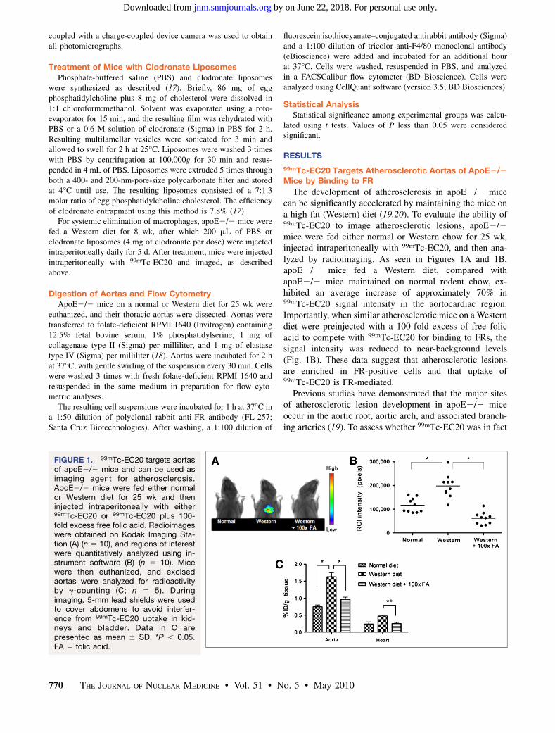
The development of atherosclerosis in apoE2/2 micecan be significantly accelerated by maintaining the mice ona high-fat (Western) diet (19,20). To evaluate the ability of99mTc-EC20 to image atherosclerotic lesions, apoE2/2mice were fed either normal or Western chow for 25 wk,injected intraperitoneally with 99mTc-EC20, and then ana-lyzed by radioimaging. As seen in Figures 1A and 1B,apoE2/2 mice fed a Western diet, compared withapoE2/2 mice maintained on normal rodent chow, ex-hibited an average increase of approximately 70% in99mTc-EC20 signal intensity in the aortocardiac region.Importantly, when similar atherosclerotic mice on a Westerndiet were preinjected with a 100-fold excess of free folicacid to compete with 99mTc-EC20 for binding to FRs, thesignal intensity was reduced to near-background levels(Fig. 1B). These data suggest that atherosclerotic lesionsare enriched in FR-positive cells and that uptake of99mTc-EC20 is FR-mediated.
Previous studies have demonstrated that the major sitesof atherosclerotic lesion development in apoE2/2 miceoccur in the aortic root, aortic arch, and associated branch-ing arteries (19). To assess whether 99mTc-EC20 was in fact
FIGURE 1. 99mTc-EC20 targets aortasof apoE2/2 mice and can be used asimaging agent for atherosclerosis.ApoE2/2 mice were fed either normalor Western diet for 25 wk and theninjected intraperitoneally with either99mTc-EC20 or 99mTc-EC20 plus 100-fold excess free folic acid. Radioimageswere obtained on Kodak Imaging Sta-tion (A) (n 5 10), and regions of interestwere quantitatively analyzed using in-strument software (B) (n 5 10). Micewere then euthanized, and excisedaortas were analyzed for radioactivityby g-counting (C; n 5 5). Duringimaging, 5-mm lead shields were usedto cover abdomens to avoid interfer-ence from 99mTc-EC20 uptake in kid-neys and bladder. Data in C arepresented as mean 6 SD. *P , 0.05.FA 5 folic acid.
770 THE JOURNAL OF NUCLEAR MEDICINE • Vol. 51 • No. 5 • May 2010
by on June 22, 2018. For personal use only. jnm.snmjournals.org Downloaded from
targeting these regions of enhanced atherosclerosis, tho-racic aortas and hearts were dissected, and accumulation of99mTc-EC20 in the resected tissues was quantitated byg-counting. As shown in Figure 1C, 99mTc-EC20 uptakewas 3-fold lower in the hearts than in the aortas, andaccumulation in the aortas was approximately 120% higherin mice on Western chow than on a normal diet. Moreover,competition with excess folic acid, compared with non-competed controls, decreased 99mTc-EC20 retention in theaortas by 41% (Fig. 1C).
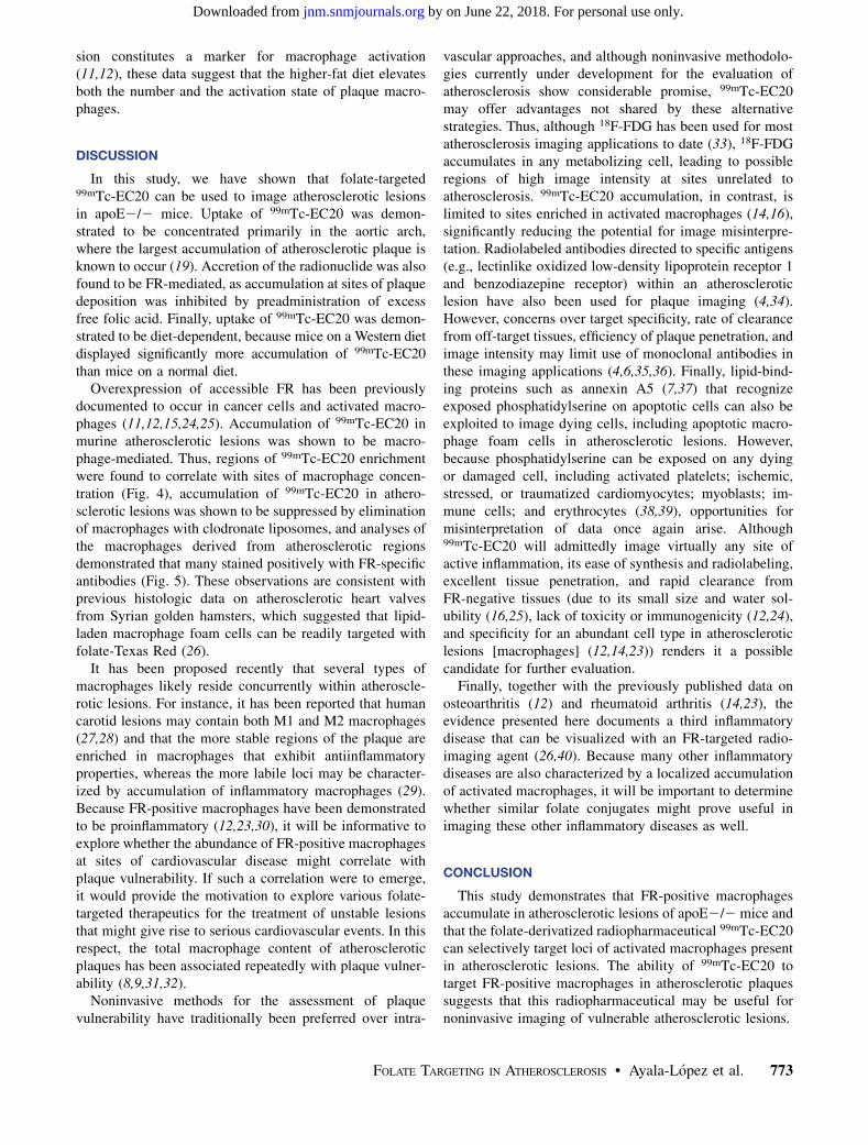
To further assess the specificity of 99mTc-EC20 intargeting atherosclerotic aortas, uptake of 99mTc-EC20 inthe aortas of a different set of similarly treated apoE2/2mice was examined by autoradiography. As shown inFigure 2, aortas of mice on a Western diet showedsignificantly greater uptake in the aortic root and arch thanthose of mice fed a normal diet. However, aortas fromapoE2/2 mice on normal chow also exhibited uptake intheir aortic roots, albeit at a lower level—that is, consistentwith the observation that apoE2/2 mice spontaneouslydevelop atherosclerotic lesions even on a normal diet(19,20). Also, as seen previously, when mice fed theWestern diet were administered a 100-fold greater dose offree folic acid than 99mTc-EC20, the radioactivity in theaortic root and arch was significantly reduced (Fig. 2),suggesting again that uptake is FR-mediated.
Activated FR-Positive Macrophages Are Responsible forUptake of 99mTc-EC20 in Atherosclerotic Plaque
Because accumulation of activated macrophages consti-tutes a central step in the pathogenesis of atherosclerosis(2,21), and because activated macrophages express highlevels of FR (11–13), we explored whether systemicdepletion of macrophages might reduce uptake of 99mTc-EC20 in atherosclerotic lesions. For this purpose, apoE2/2mice maintained on a Western diet were treated with
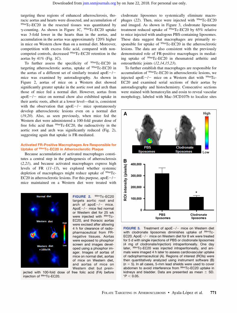
clodronate liposomes to systemically eliminate macro-phages (22). Then, mice were injected with 99mTc-EC20and imaged. As shown in Figure 3, clodronate liposometreatment reduced uptake of 99mTc-EC20 by 65% relativeto mice injected with analogous PBS-containing liposomes.These data suggest that macrophages are primarily re-sponsible for uptake of 99mTc-EC20 in the atheroscleroticlesions. The data are also consistent with the previouslydemonstrated role of FR-positive macrophages in mediat-ing uptake of 99mTc-EC20 in rheumatoid arthritic andosteoarthritic joints (12,14,15,23).
To further establish that macrophages are responsible foraccumulation of 99mTc-EC20 in atherosclerotic lesions, weinjected apoE2/2 mice on a Western diet with 99mTc-EC20 and examined serial sections of their aortas byautoradiography and histochemistry. Consecutive sectionswere stained with hematoxylin and eosin to reveal vascularmorphology, labeled with Mac-3/CD107b to localize sites
FIGURE 2. 99mTc-EC20targets aortic root andarch of apoE2/2 mice.ApoE2/2 mice fed normalor Western diet for 25 wkwere injected with 99mTc-EC20, and thoracic aortaswere excised after allowing4 h for clearance of radio-pharmaceutical from FR-negative tissues. Aortaswere exposed to phosphorscreen and images devel-oped using a phosphor im-ager. Images of aortas ofmice on normal diet, aortasof mice on Western diet,and aortas of mice onWestern diet but prein-
jected with 100-fold dose of free folic acid (FA) beforeinjection of 99mTc-EC20.
FIGURE 3. Treatment of apoE2/2 mice on Western dietwith clodronate liposomes diminishes uptake of 99mTc-EC20. ApoE2/2 mice on Western diet for 8 wk were treatedfor 5 d with single injections of PBS or clodronate liposomes(4 mg of clodronate/injection) intraperitoneally. One daylater, 99mTc-EC20 was injected intraperitoneally, and ani-mals were imaged 4 h later to assess cardiovascular uptakeof radiopharmaceutical (A). Regions of interest (ROIs) werethen quantitatively analyzed using instrument software (B)(n 5 5). In all cases, 5-mm lead shields were used to coverabdomen to avoid interference from 99mTc-EC20 uptake inkidneys and bladder. Data are presented as mean 6 SD.*P , 0.05.
FOLATE TARGETING IN ATHEROSCLEROSIS • Ayala-Lopez et al. 771
by on June 22, 2018. For personal use only. jnm.snmjournals.org Downloaded from

of macrophage enrichment, and imaged by autoradiographyto identify locations of 99mTc-EC20 accumulation. As seenin Figure 4 and Supplemental Figure 1 (supplemental ma-terials are available online only at http://jnm.snmjournals.org), areas of high macrophage content and atheroscleroticlesion formation invariably corresponded with loci ofelevated 99mTc-EC20 uptake.
Macrophages Isolated from Aortas of ApoE2/2 MiceExpress FR
To confirm by yet another method that FR-positivemacrophages play the major role in mediating accumula-tion of 99mTc-EC20 in the aortas of apoE2/2 mice,thoracic aortas were digested to obtain single-cell suspen-sions, and cells expressing a macrophage marker (F4/80)were analyzed by flow cytometry for simultaneous expres-sion of FR. As seen in Supplemental Figure 2, F4/80-positive macrophages were found to comprise 1.1% and3.0% of all cells in the thoracic aortas of mice fed a normaldiet and Western diet, respectively. This diet-dependentincrease in macrophage content was not unexpected,because an increase in monocyte or macrophage infiltrationhas been established to constitute a hallmark of atherogen-esis (2,10,21). More important, macrophages from mice feda normal diet were only 11% FR-positive, whereas macro-
phages from mice on the Western diet were 33% FR-positive, suggesting that the high-fat diet not only increasedtotal macrophage content but also tripled the percentage ofFR-positive macrophages (Fig. 5). Given that FR expres-
FIGURE 4. 99mTc-EC20 preferentially accumulates inareas of high macrophage content within atheroscleroticplaques of apoE2/2 mice. ApoE2/2 mice on Western dietfor 25 wk were injected with 99mTc-EC20. After 4-h tissueclearance period, aortas were dissected and embedded inoptimal-cutting-temperature medium. Sections of ascendingaorta and brachiocephalic artery were prepared andexposed to phosphor screen for 18 h. Images were takenusing phosphor imager. Consecutive sections were used forMac-3 immunohistochemistry and hematoxylin and eosinstaining (Supplemental Fig. 1). Bar 5 100 mm.
FIGURE 5. Percentage increase in FR-positive macrophagenumbers in apoE2/2 mice on Western diet. ApoE2/2 micewere fed normal or Western diet for 25 wk. Mice wereeuthanized and thoracic aortas excised and digested withcollagenase and elastase. Resulting cell suspensions wereanalyzed by flow cytometry after incubation with tricolor-conjugated F4/80 antibody (macrophage marker) and rabbitanti-FR primary antibody followed by fluorescein isothio-cyanate–conjugated (FITC) antirabbit IgG secondary anti-body.
772 THE JOURNAL OF NUCLEAR MEDICINE • Vol. 51 • No. 5 • May 2010
by on June 22, 2018. For personal use only. jnm.snmjournals.org Downloaded from
sion constitutes a marker for macrophage activation(11,12), these data suggest that the higher-fat diet elevatesboth the number and the activation state of plaque macro-phages.
DISCUSSION
In this study, we have shown that folate-targeted99mTc-EC20 can be used to image atherosclerotic lesionsin apoE2/2 mice. Uptake of 99mTc-EC20 was demon-strated to be concentrated primarily in the aortic arch,where the largest accumulation of atherosclerotic plaque isknown to occur (19). Accretion of the radionuclide was alsofound to be FR-mediated, as accumulation at sites of plaquedeposition was inhibited by preadministration of excessfree folic acid. Finally, uptake of 99mTc-EC20 was demon-strated to be diet-dependent, because mice on a Western dietdisplayed significantly more accumulation of 99mTc-EC20than mice on a normal diet.
Overexpression of accessible FR has been previouslydocumented to occur in cancer cells and activated macro-phages (11,12,15,24,25). Accumulation of 99mTc-EC20 inmurine atherosclerotic lesions was shown to be macro-phage-mediated. Thus, regions of 99mTc-EC20 enrichmentwere found to correlate with sites of macrophage concen-tration (Fig. 4), accumulation of 99mTc-EC20 in athero-sclerotic lesions was shown to be suppressed by eliminationof macrophages with clodronate liposomes, and analyses ofthe macrophages derived from atherosclerotic regionsdemonstrated that many stained positively with FR-specificantibodies (Fig. 5). These observations are consistent withprevious histologic data on atherosclerotic heart valvesfrom Syrian golden hamsters, which suggested that lipid-laden macrophage foam cells can be readily targeted withfolate-Texas Red (26).
It has been proposed recently that several types ofmacrophages likely reside concurrently within atheroscle-rotic lesions. For instance, it has been reported that humancarotid lesions may contain both M1 and M2 macrophages(27,28) and that the more stable regions of the plaque areenriched in macrophages that exhibit antiinflammatoryproperties, whereas the more labile loci may be character-ized by accumulation of inflammatory macrophages (29).Because FR-positive macrophages have been demonstratedto be proinflammatory (12,23,30), it will be informative toexplore whether the abundance of FR-positive macrophagesat sites of cardiovascular disease might correlate withplaque vulnerability. If such a correlation were to emerge,it would provide the motivation to explore various folate-targeted therapeutics for the treatment of unstable lesionsthat might give rise to serious cardiovascular events. In thisrespect, the total macrophage content of atheroscleroticplaques has been associated repeatedly with plaque vulner-ability (8,9,31,32).
Noninvasive methods for the assessment of plaquevulnerability have traditionally been preferred over intra-
vascular approaches, and although noninvasive methodolo-gies currently under development for the evaluation ofatherosclerosis show considerable promise, 99mTc-EC20may offer advantages not shared by these alternativestrategies. Thus, although 18F-FDG has been used for mostatherosclerosis imaging applications to date (33), 18F-FDGaccumulates in any metabolizing cell, leading to possibleregions of high image intensity at sites unrelated toatherosclerosis. 99mTc-EC20 accumulation, in contrast, islimited to sites enriched in activated macrophages (14,16),significantly reducing the potential for image misinterpre-tation. Radiolabeled antibodies directed to specific antigens(e.g., lectinlike oxidized low-density lipoprotein receptor 1and benzodiazepine receptor) within an atheroscleroticlesion have also been used for plaque imaging (4,34).However, concerns over target specificity, rate of clearancefrom off-target tissues, efficiency of plaque penetration, andimage intensity may limit use of monoclonal antibodies inthese imaging applications (4,6,35,36). Finally, lipid-bind-ing proteins such as annexin A5 (7,37) that recognizeexposed phosphatidylserine on apoptotic cells can also beexploited to image dying cells, including apoptotic macro-phage foam cells in atherosclerotic lesions. However,because phosphatidylserine can be exposed on any dyingor damaged cell, including activated platelets; ischemic,stressed, or traumatized cardiomyocytes; myoblasts; im-mune cells; and erythrocytes (38,39), opportunities formisinterpretation of data once again arise. Although99mTc-EC20 will admittedly image virtually any site ofactive inflammation, its ease of synthesis and radiolabeling,excellent tissue penetration, and rapid clearance fromFR-negative tissues (due to its small size and water sol-ubility (16,25), lack of toxicity or immunogenicity (12,24),and specificity for an abundant cell type in atheroscleroticlesions [macrophages] (12,14,23)) renders it a possiblecandidate for further evaluation.
Finally, together with the previously published data onosteoarthritis (12) and rheumatoid arthritis (14,23), theevidence presented here documents a third inflammatorydisease that can be visualized with an FR-targeted radio-imaging agent (26,40). Because many other inflammatorydiseases are also characterized by a localized accumulationof activated macrophages, it will be important to determinewhether similar folate conjugates might prove useful inimaging these other inflammatory diseases as well.
CONCLUSION
This study demonstrates that FR-positive macrophagesaccumulate in atherosclerotic lesions of apoE2/2 mice andthat the folate-derivatized radiopharmaceutical 99mTc-EC20can selectively target loci of activated macrophages presentin atherosclerotic lesions. The ability of 99mTc-EC20 totarget FR-positive macrophages in atherosclerotic plaquessuggests that this radiopharmaceutical may be useful fornoninvasive imaging of vulnerable atherosclerotic lesions.
FOLATE TARGETING IN ATHEROSCLEROSIS • Ayala-Lopez et al. 773
by on June 22, 2018. For personal use only. jnm.snmjournals.org Downloaded from

ACKNOWLEDGMENT
This work was supported by a research grant fromEndocyte, Inc.
REFERENCES
1. Ross R. Atherosclerosis: an inflammatory disease. N Engl J Med. 1999;340:115–
126.
2. Libby P. Inflammation in atherosclerosis. Nature. 2002;420:868–874.
3. Langer HF, Haubner R, Pichler BJ, Gawaz M. Radionuclide imaging:
a molecular key to the atherosclerotic plaque. J Am Coll Cardiol. 2008;52:1–12.
4. Fujimura Y, Hwang PM, Trout Iii H, et al. Increased peripheral benzodiazepine
receptors in arterial plaque of patients with atherosclerosis: an autoradiographic
study with [3H]PK 11195. Atherosclerosis. 2008;201:108–111.
5. Hartung D, Schafers M, Fujimoto S, et al. Targeting of matrix metalloproteinase
activation for noninvasive detection of vulnerable atherosclerotic lesions. Eur J
Nucl Med Mol Imaging. 2007;34(suppl 1):S1–8.
6. Laurberg JM, Olsen AK, Hansen SB, et al. Imaging of vulnerable atherosclerotic
plaques with FDG-microPET: no FDG accumulation. Atherosclerosis. 2007;192:
275–282.
7. Zhao Y, Kuge Y, Zhao S, et al. Comparison of 99mTc-annexin A5 with 18F-FDG
for the detection of atherosclerosis in apoE2/2 mice. Eur J Nucl Med Mol
Imaging. 2007;34:1747–1755.
8. Halvorsen B, Otterdal K, Dahl TB, et al. Atherosclerotic plaque stability: what
determines the fate of a plaque? Prog Cardiovasc Dis. 2008;51:183–194.
9. Tiwari RL, Singh V, Barthwal MK. Macrophages: an elusive yet emerging
therapeutic target of atherosclerosis. Med Res Rev. 2008;28:483–544.
10. Choudhury RP, Lee JM, Greaves DR. Mechanisms of disease: macrophage-
derived foam cells emerging as therapeutic targets in atherosclerosis. Nat Clin
Pract Cardiovasc Med. 2005;2:309–315.
11. Nakashima-Matsushita N, Homma T, Yu S, et al. Selective expression of folate
receptor beta and its possible role in methotrexate transport in synovial
macrophages from patients with rheumatoid arthritis. Arthritis Rheum. 1999;42:
1609–1616.
12. Xia W, Hilgenbrink AR, Matteson EL, Lockwood MB, Cheng JX, Low PS. A
functional folate receptor is induced during macrophage activation and can be
used to target drugs to activated macrophages. Blood. 2009;113:438–446.
13. van der Heijden JW, Oerlemans R, Dijkmans BA, et al. Folate receptor b as
a potential delivery route for novel folate antagonists to macrophages in the
synovial tissue of rheumatoid arthritis patients. Arthritis Rheum. 2009;60:12–21.
14. Turk MJ, Breur GJ, Widmer WR, et al. Folate-targeted imaging of activated
macrophages in rats with adjuvant-induced arthritis. Arthritis Rheum. 2002;46:
1947–1955.
15. Matteson EL, Lowe VJ, Prendergast FG, et al. Assessment of disease activity in
rheumatoid arthritis using a novel folate targeted radiopharmaceutical Folates-
can. Clin Exp Rheumatol. 2009;27:253–259.
16. Leamon CP, Parker MA, Vlahov IR, et al. Synthesis and biological evaluation of
EC20: a new folate-derived, 99mTc-based radiopharmaceutical. Bioconjug Chem.
2002;13:1200–1210.
17. Love WG, Camilleri JP, Williams BD. Efficient clodronate entrapment within
multilamellar and unilamellar liposomes. J Pharmacol Toxicol Methods. 1992;
27:185–189.
18. Ray JL, Leach R, Herbert JM, Benson M. Isolation of vascular smooth muscle
cells from a single murine aorta. Methods Cell Sci. 2001;23:185–188.
19. Nakashima Y, Plump AS, Raines EW, Breslow JL, Ross R. ApoE-deficient mice
develop lesions of all phases of atherosclerosis throughout the arterial tree.
Arterioscler Thromb. 1994;14:133–140.
20. Jawien J, Nastalek P, Korbut R. Mouse models of experimental atherosclerosis.
J Physiol Pharmacol. 2004;55:503–517.
21. Shashkin P, Dragulev B, Ley K. Macrophage differentiation to foam cells. Curr
Pharm Des. 2005;11:3061–3072.
22. Buiting AM, Zhou F, Bakker JA, van Rooijen N, Huang L. Biodistribution of
clodronate and liposomes used in the liposome mediated macrophage ‘suicide’
approach. J Immunol Methods. 1996;192:55–62.
23. Paulos CM, Varghese B, Widmer WR, Breur GJ, Vlashi E, Low PS. Folate-
targeted immunotherapy effectively treats established adjuvant and collagen-
induced arthritis. Arthritis Res Ther. 2006;8:R77.
24. Low PS, Henne WA, Doorneweerd DD. Discovery and development of folic-
acid-based receptor targeting for imaging and therapy of cancer and
inflammatory diseases. Acc Chem Res. 2008;41:120–129.
25. Reddy JA, Xu LC, Parker N, Vetzel M, Leamon CP. Preclinical evaluation of99mTc-EC20 for imaging folate receptor-positive tumors. J Nucl Med. 2004;45:
857–866.
26. Antohe F, Radulescu L, Puchianu E, Kennedy MD, Low PS, Simionescu M.
Increased uptake of folate conjugates by activated macrophages in experimental
hyperlipemia. Cell Tissue Res. 2005;320:277–285.
27. Bolick DT, Skaflen MD, Johnson LE, et al. G2A Deficiency in mice promotes
macrophage activation and atherosclerosis. Circ Res. 2009;104:318–327.
28. Charo IF. Macrophage polarization and insulin resistance: PPARg in control.
Cell Metab. 2007;6:96–98.
29. Bouhlel MA, Derudas B, Rigamonti E, et al. PPARg activation primes human
monocytes into alternative M2 macrophages with anti-inflammatory properties.
Cell Metab. 2007;6:137–143.
30. Varghese B, Haase N, Low PS. Depletion of folate-receptor-positive macro-
phages leads to alleviation of symptoms and prolonged survival in two murine
models of systemic lupus erythematosus. Mol Pharm. 2007;4:679–685.
31. San Miguel Hernandez A, Inglada-Galiana L, Garcia Iglesias R, Alonso
Castillejos N, Martin Gil FJ. Soluble CD40 ligand: a potential marker of
cardiovascular risk [in Spanish]. Rev Clin Esp. 2007;207:418–421.
32. Broz P, Ben-Haim N, Grzelakowski M, Marsch S, Meier W, Hunziker P.
Inhibition of macrophage phagocytotic activity by a receptor-targeted polymer
vesicle-based drug delivery formulation of pravastatin. J Cardiovasc Pharmacol.
2008;51:246–252.
33. Nahrendorf M, Zhang H, Hembrador S, et al. Nanoparticle PET-CT imaging
of macrophages in inflammatory atherosclerosis. Circulation. 2008;117:379–387.
34. Ishino S, Mukai T, Kuge Y, et al. Targeting of lectinlike oxidized low-density
lipoprotein receptor 1 (LOX-1) with 99mTc-labeled anti-LOX-1 antibody:
potential agent for imaging of vulnerable plaque. J Nucl Med. 2008;49:1677–
1685.
35. de Wolf FA, Brett GM. Ligand-binding proteins: their potential for application in
systems for controlled delivery and uptake of ligands. Pharmacol Rev. 2000;52:
207–236.
36. Laitinen I, Marjamaki P, Haaparanta M, et al. Non-specific binding of [18F]FDG
to calcifications in atherosclerotic plaques: experimental study of mouse and
human arteries. Eur J Nucl Med Mol Imaging. 2006;33:1461–1467.
37. Laufer EM, Reutelingsperger CP, Narula J, Hofstra L. Annexin A5: an imaging
biomarker of cardiovascular risk. Basic Res Cardiol. 2008;103:95–104.
38. van Genderen HO, Kenis H, Hofstra L, Narula J, Reutelingsperger CP.
Extracellular annexin A5: functions of phosphatidylserine-binding and two-
dimensional crystallization. Biochim Biophys Acta. 2008;1783:953–963.
39. Boersma HH, Kietselaer BL, Stolk LM, et al. Past, present, and future of annexin
A5: from protein discovery to clinical applications. J Nucl Med. 2005;46:2035–
2050.
40. Chen WT, Mahmood U, Weissleder R, Tung CH. Arthritis imaging using a near-
infrared fluorescence folate-targeted probe. Arthritis Res Ther. 2005;7:R310–
R317.
774 THE JOURNAL OF NUCLEAR MEDICINE • Vol. 51 • No. 5 • May 2010
by on June 22, 2018. For personal use only. jnm.snmjournals.org Downloaded from
Doi: 10.2967/jnumed.109.071324Published online: April 15, 2010.
2010;51:768-774.J Nucl Med. Wilfredo Ayala-López, Wei Xia, Bindu Varghese and Philip S. Low Folate-Conjugated Radiopharmaceutical to Activated MacrophagesImaging of Atherosclerosis in Apoliprotein E Knockout Mice: Targeting of a
http://jnm.snmjournals.org/content/51/5/768This article and updated information are available at:
http://jnm.snmjournals.org/site/subscriptions/online.xhtml
Information about subscriptions to JNM can be found at:
http://jnm.snmjournals.org/site/misc/permission.xhtmlInformation about reproducing figures, tables, or other portions of this article can be found online at:
(Print ISSN: 0161-5505, Online ISSN: 2159-662X)1850 Samuel Morse Drive, Reston, VA 20190.SNMMI | Society of Nuclear Medicine and Molecular Imaging
is published monthly.The Journal of Nuclear Medicine
© Copyright 2010 SNMMI; all rights reserved.
by on June 22, 2018. For personal use only. jnm.snmjournals.org Downloaded from