image formation introduction the invisible and visible image image characteristics
TRANSCRIPT

IMAGE FORMATION
IntroductionThe Invisible and Visible ImageImage Characteristics

2
By the end of this Lecture the student will be able to:
Define the terms, image and identify its types and methods of viewing
Identify the stages of image formation
Differentiate between the invisible (latent) and visible image
List and define the basic image characteristics
Differentiate between density and contrast and state their relationship
Learning objectives

3
References• John Ball& Tony Price; Chesney's Radiographic Imaging
Websites
• http://www.e-radiography.net/
IMAGE FORMATION

4
What is an Image? The term image describe a recognizable pattern carrying information
Other meaning An optical appearance A mental representation A form or semblance An idea or conception
Visual images are of two types Real images : Those have real existence such as photographic or radiographic images -Static ( RADIOGRAPH OR PHOTOGRAPH- Dynamic ( On a television screen)
Mental images: Pictures those generated within our minds
IMAGE FORMATION

Medical imaging is essentially the extraction of anatomical and physiological information from the patient and the interpretation of these information for the purpose of diagnosing diseases
The flow of the information from the patient to the observer is through three stages: The formation of the invisible image The conversion of the invisible image into a visible light image Interpretation of the visible image
IMAGE FORMATION

6
Scatter
Stage.1 The formation of the invisible image During radiographic examination X-ray passes through the patient As x-rays penetrate through the body tissues it become modified
each part of the beam is attenuated in a degree which depend on The tissue type The intensity of the beam Thickness of the tissue
Transmit Absorb
IMAGE FORMATION

Primary Radiation – The beam of photons, interacts with the pt’s body.Remnant Radiation – The resulting beam that is able to exit from the patient.Scatter Radiation – Radiation that interacts with matter & only continues in a different direction – not useful for image production.Attenuation – Primary radiation that is changed (partially absorbed) as it travels through the pt.
IMAGE FORMATION
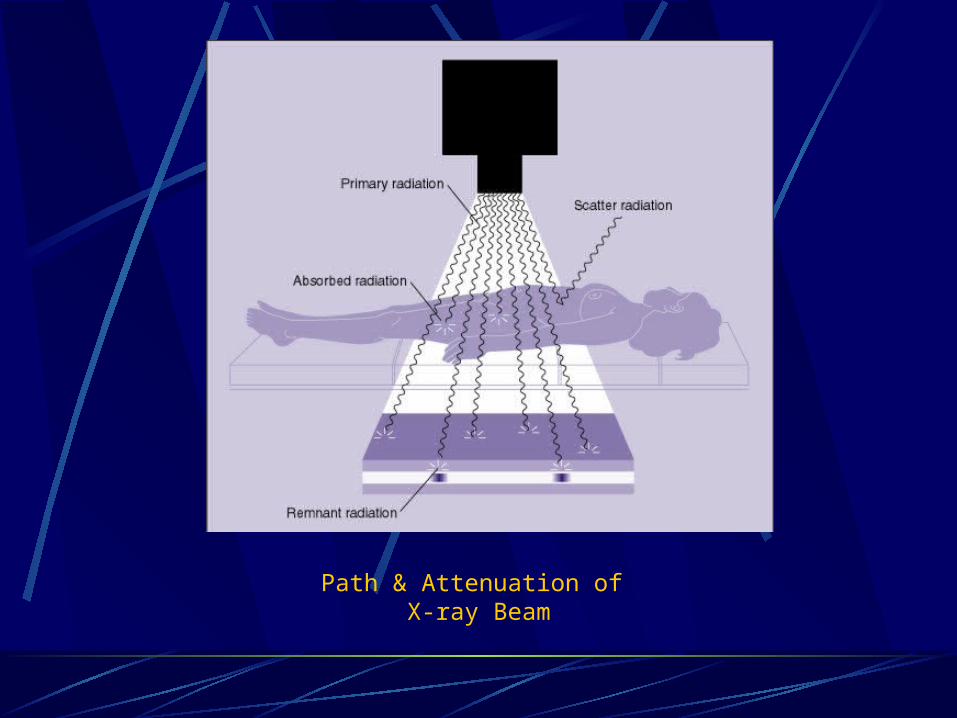
Path & Attenuation of X-ray Beam

9
Stage.1 The formation of the invisible image During radiographic examination X-ray passes through the patient As x-rays penetrate through the body tissues it become modified
each part of the beam is attenuated in a degree which depend on The tissue type The intensity of the beam Thickness of the tissue
Film is the simplest image receptor, but it is usually coupled with intensifying screens, which help reduce the dose to the patient. X-ray film responds to a range of wavelengths and is in fact far more sensitive to light than it is to x-rays. After exposure to x-rays an invisible image known as the latent image is formed.
IMAGE FORMATION
10
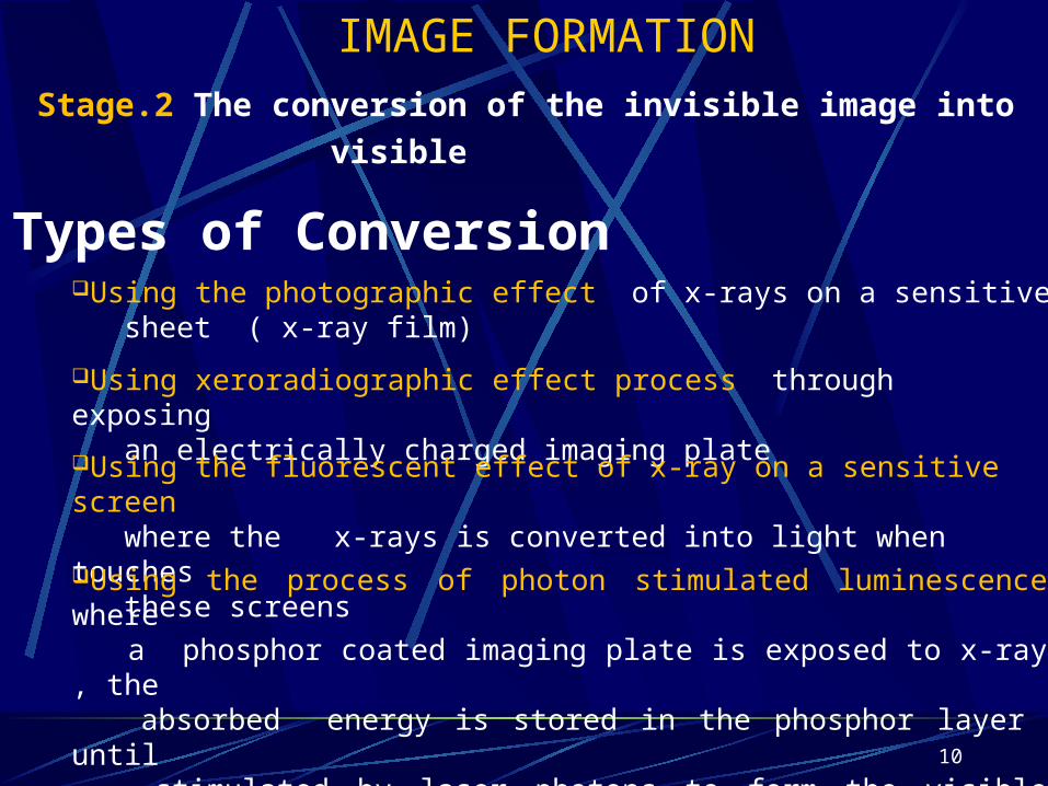
Stage.2 The conversion of the invisible image into
visible
Using the photographic effect of x-rays on a sensitive sheet ( x-ray film)
Using xeroradiographic effect process through exposing an electrically charged imaging plate
Using the fluorescent effect of x-ray on a sensitive screen where the x-rays is converted into light when touches these screensUsing the process of photon stimulated luminescence where a phosphor coated imaging plate is exposed to x-ray , the absorbed energy is stored in the phosphor layer until stimulated by laser photons to form the visible image
IMAGE FORMATION
Types of Conversion
Why you see what you see…
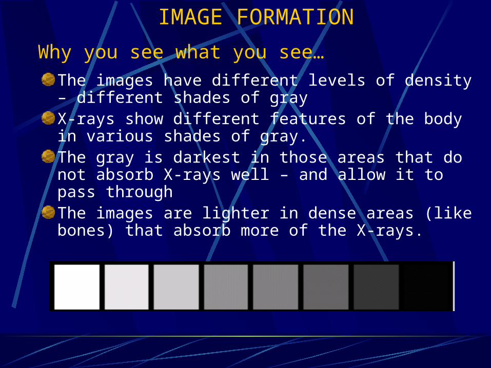
The images have different levels of density – different shades of grayX-rays show different features of the body in various shades of gray. The gray is darkest in those areas that do not absorb X-rays well – and allow it to pass throughThe images are lighter in dense areas (like bones) that absorb more of the X-rays.
IMAGE FORMATION
12
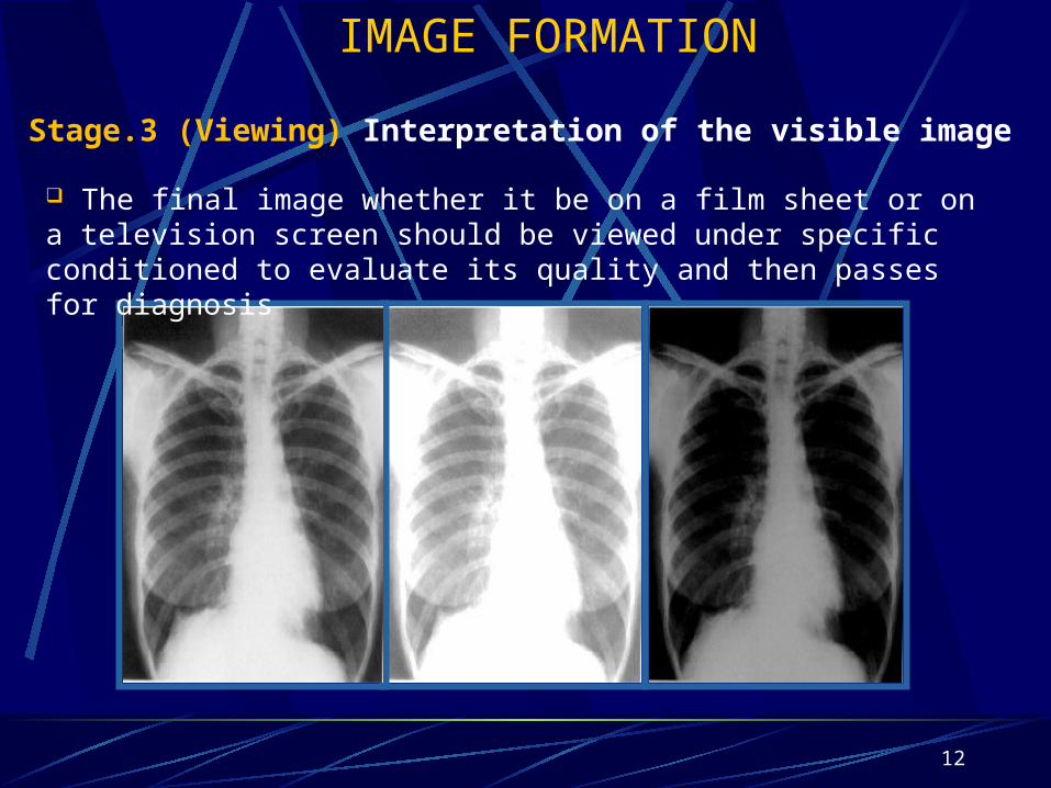
Stage.3 (Viewing) Interpretation of the visible image The final image whether it be on a film sheet or on a television screen should be viewed under specific conditioned to evaluate its quality and then passes for diagnosis
IMAGE FORMATION
13
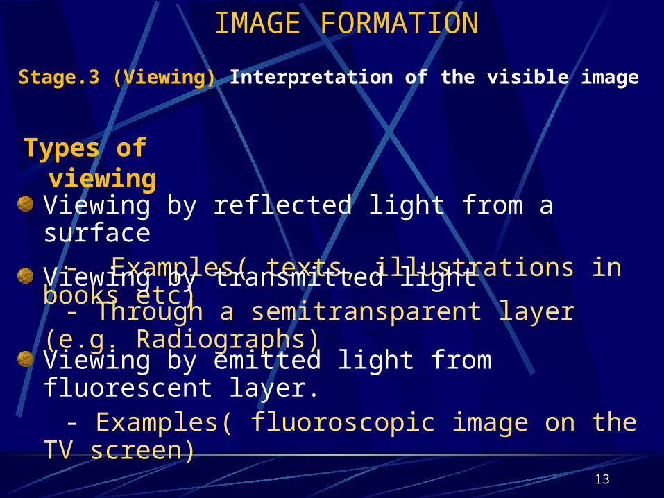
Stage.3 (Viewing) Interpretation of the visible image
IMAGE FORMATION
Viewing by reflected light from a surface - Examples( texts, illustrations in books etc)
Viewing by transmitted light - Through a semitransparent layer (e.g. Radiographs)
Viewing by emitted light from fluorescent layer. - Examples( fluoroscopic image on the TV screen)
Types of viewing
14
IMAGE FORMATION
Image characteristics
Noise
Contrast
Sharpness
Resolution

15
IMAGE FORMATION
Image characteristics
NoiseReal images consist of 2 components - Meaningful pattern (Signal) - Chaotic pattern ( Noise) The details of the structures is affected by noise
+Noise = - Details

16
IMAGE FORMATION
Image characteristics
Signal to Noise Ratio (SNR)
Under optimal condition the magnitude of signal is greater than the magnitude of the noise - SNR is Saied to be high ( More details)
High SNR
When the magnitude of nearest the magnitude of the noise
- SNR is Saied to be Low ( Less details)
Low SNR

17
IMAGE FORMATION
Image characteristics
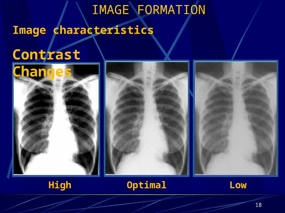
Density and Contrast
Density
The amount of blackening
“darkness” on the radiograph
Contrast
The differences between the blacks to the whites
18
High Optimal Low
Contrast Changes
IMAGE FORMATION
Image characteristics
19
IMAGE FORMATION
Image characteristics
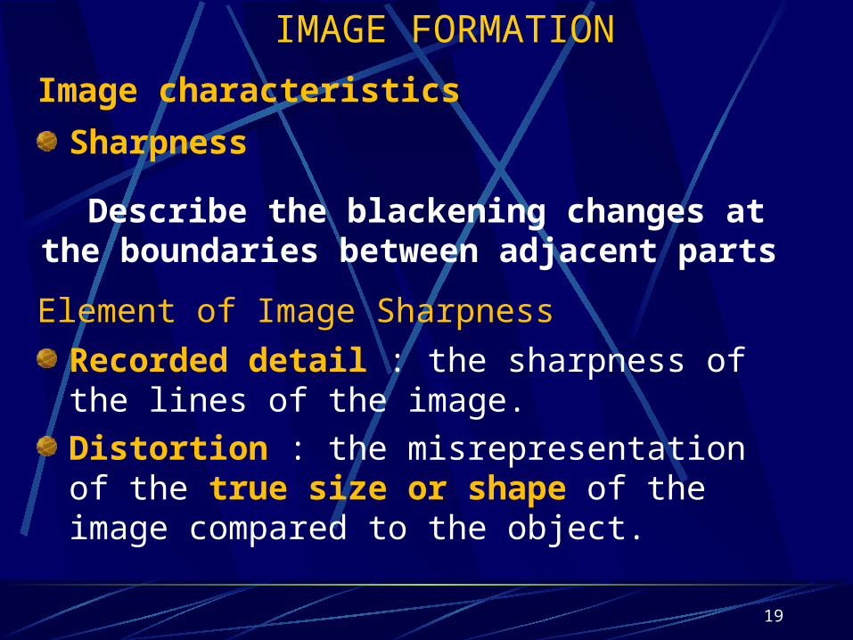
Sharpness
Describe the blackening changes at the boundaries between adjacent parts
Element of Image Sharpness
Recorded detail : the sharpness of the lines of the image.
Distortion : the misrepresentation of the true size or shape of the image compared to the object.

20
True Shape
Elongated
Foreshortened
lengthwidth
IMAGE FORMATION
Image characteristics
Sharpness (SHAPE DISTORTION – TERMS)
21
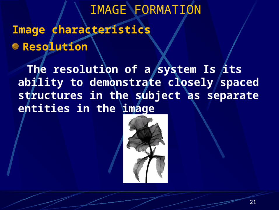
IMAGE FORMATION
Image characteristics
Resolution
The resolution of a system Is its ability to demonstrate closely spaced structures in the subject as separate entities in the image

22
Visibility Sharpness
density contrast detail distortion
size shape
Image characteristics
IMAGE FORMATION
Summary

Unit III 23