illustra card plasmidprep mini spin kit - vwr
TRANSCRIPT

GE Healthcare
illustraplasmidPrep Mini Spin Kit For the rapid extraction and purification of plasmid DNA from small scale cultures of E. coli.
Product booklet
Code: 28-9042-69 (50 purifications) 28-9042-70 (250 purifications)
See back cover for quick reference
protocol card

2
Page finder 1. Legal 3
2. Handling and storage 4 2.1. Safety warnings and precautions 4 2.2. Storage 4 2.3. Expiry 4
3. Components 5 3.1. Kit contents 5 3.2. Materials and equipment to be supplied by user 6 3.3 Equipment needed 6
4. Description 7 4.1. Introduction 7 4.2. The basic principle 8 4.3. Product specifications 10 4.4. Typical output 11
5. Protocols 13 5.1. Preparation of working solutions 14 5.2. Protocol for 1.5 ml & 3 ml culture volumes 14
6. Appendices 22 6.1. RPM calculation from RCF 22 6.2. Isolation of low copy number and large molecular weight plasmids 22 6.3. Factors affecting plasmid DNA yield and purity 22 6.4. Troubleshooting guide 24 6.5. Related products 27
7. References 30
Quick Reference Protocol Cards Back Cover Tear off sheet containing a protocol for the experienced user purifying plasmid DNA from E. coli.

3
1. Legal Restrictions for product use
The illustra™ plasmidPrep Mini Spin Kit has been designed, developed, and sold for research purposes only. They are suitable for in vitro use only. No claim or representation is intended for its use to identify any specific organism or for clinical use (diagnostic, prognostic, therapeutic, or blood banking).
It is the responsibility of the user to verify the use of the illustra plasmidPrep Mini Spin Kit for a specific application range, as the performance characteristics of this product have not been verified for any specific organism.
GE, imagination at work and GE monogram are trademarks of General Electric Company.
illustra, TempliPhi, ECL Direct, Gene Images, GFX, AutoSeq, NAP, Sephacryl, Ready-To-Go, PuReTaq and FideliTaq are trademarks of GE Healthcare companies.
All third party trademarks are the preperty of their respective owners.
© 2006–2008 General Electric Company-All rights reserved. Previously published 2006.
All goods and services are sold subject to the terms and conditions of sale of the company within GE Healthcare which supplies them. A copy of these terms and conditions is available upon request. Contact your GE Healthcare representative for the most current information.
http://www.gehealthcare.com/lifesciences
GE Healthcare UK Limited. Amersham Place, Little Chalfont, Buckinghamshire. HP7 9NA UK.

4
2. Handling and storageWarning: This protocol requires the use of ethanol.
2.2. Storage All kit components should be stored at room temperature (20–25°C).
2.3. Expiry For expiry date please refer to outer packaging label.
2.1. Safety warnings and precautions Warning: For research use only. Not recommended or intended for diagnosis of disease in humans or animals. Do not use internally or externally in humans or animals.All chemicals should be considered as potentially hazardous. Only persons trained in laboratory techniques and familiar with the principles of good laboratory practice should handle these products. Suitable protective clothing such as laboratory overalls, safety glasses and gloves should be worn. Care should be taken to avoid contact with skin or eyes; if contact should occur, wash immediately with water (see Material Safety Data Sheet(s) and/or Safety Statement(s) for specific recommendations).

3. Components 3.1. Kit contents
Identification Pack Size
Cat. No.
10
purifications
Sample Pack
50
purifications
28-9042-69
250
purifications
28-9042-70
Lysis buffer type 7 (Red
colored cap)
3 ml 15 ml 60 ml
Lysis buffer type 8 (Clear colored cap)
3 ml 15 ml 60 ml
Lysis buffer type 9 (Blue colored cap)
10 ml 50 ml 220 ml
Wash buffer type 1 (Yellow colored cap)
1 ml
Add 4 ml
ethanol
before use
7 ml
Add 28 ml
ethanol
before use
26 ml
Add 104 ml
ethanol
before use
Elution buffer type 4 (Silver colored cap)
2 ml 10 ml 35 ml
illustra™ plasmid mini
columns
10 50 250
Collection tubes
10 50 250
Refer to the Certificate of Analysis for a complete list of kit components.
5

6
GE Healthcare supplies a wide range of buffer types across the illustra nucleic acid purification and amplification range. The composition of each buffer has been optimized for each application and may vary between kits. Care must be taken to only use the buffers supplied in the particular kit you are using and not use the buffers supplied in other illustra kits e.g. the Lysis buffers supplied in the illustra plasmidPrep Mini Spin Kit are not the same as the Lysis buffers supplied in the illustra plasmidPrep Midi Flow Kit.
In order to avoid confusion and the accidental switching of buffers between kits, a numbering system has been adopted that relates to the entire range of buffers available in the illustra purification range. For example there are currently 14 Lysis buffers in the illustra range, 6 Wash buffers and 8 Elution buffers, denoted by Lysis buffer type 1–14, Wash buffer type 1–6 and Elution buffer type 1–8, respectively. Please ensure you use the correct type of Lysis, Wash and Elution buffer for your purification.
Note: Lysis buffer type 7 (red colored cap) contains RNase A. The Elution buffer type 4 (silver colored cap) consists of 10mM Tris-HCI
(pH 8.0).
3.2. Materials and equipment to be supplied by userDisposables: DNase-free 1.5 ml microcentrifuge tubes (snap cap). One per purification.
Chemicals: Absolute ethanol
3.3. Equipment neededMicrocentrifuge that accommodates 1.5 ml microcentrifuge tubesVortex mixer (optional)

7
4. Description
4.1. Introduction The illustra plasmidPrep Mini Spin Kit is designed for the rapid extraction and purification of plasmid DNA from 1.5 ml and 3 ml cultures of Escherichia coli (E. coli). The procedure can be completed in approximately 9 minutes (2 × cultures) to yield plasmid DNA with a purity and quality compatible with many common molecular biology techniques, including cloning, restriction enzyme digestion, PCR amplification and DNA sequencing (see figures 1–5).
The plasmid DNA yield from a 1.5 ml culture of a freshly grown E. coli strain containing a high copy number plasmid (> 300 copies/cell) and grown to A600 approximately 2.5 is typically 6 to 9 µg (A260/A280 > 1.8).
Users purifying low copy number plasmids (10–20 copies per cell) or large molecular weight plasmids (> 35 kbp) should follow the protocol in section 5.3. with a 3 ml culture of E. coli. After the harvesting of Bacterial Culture step, all further steps are identical to that for harvesting bacteria from 1.5 ml E. coli culture i.e., no extra buffer volumes are required. The illustra plasmidPrep Mini Spin Kit utilizes a simple plasmid DNA purification protocol, employing a modified alkaline cell lysis procedure (1–3) and a novel silica-based membrane.
No organic solvents are used; instead, chaotropic salts are included to denature protein components and promote the selective binding of plasmid DNA to the novel silica membrane (4, 5). Denatured contaminants are easily removed by subsequent washing. The purified plasmid DNA is eluted in a low ionic strength buffer, at a plasmid concentration suitable for most molecular biological applications.

8
4.2. The basic principle
Harvesting of Bacterial Culture
Lysis
Plasmid Binding
Wash (optional-strain dependent)
Plasmid DNA ready for downstream applications
1.
Use of the illustra plasmidPrep Mini Spin Kit involves the following steps:
4.
3.
2.
Wash & Dry5.
Elution 6.

9
Step Comments Component
1. Harvesting of Bacterial Culture
Bacteria are harvested by centrifugation and the spent medium
removed.
Bacteria
2. Lysis Bacterial cells are re-suspended in an isotonic solution containing RNase A.
Lysis buffer type 7
Cells are lysed by alkali treatment; genomic DNA and proteins are
denatured.
Lysis buffer type 8
The pH of the lysate is neutralized with an acetate buffered solution, containing
a chaotropic salt.
Lysate is centrifuged to pellet cellular debris, including genomic DNA, proteins
and lipids.
Lysis buffer type 9
3. Plasmid Binding
The cleared cellular lysate is applied to the illustra plasmid mini column.
Presence of chaotrope promotes binding of plasmid DNA binds to the membrane.
illustra plasmid mini column & Collection tube
4. Wash (optional strain dependent)
An optional wash step removes residual nuclease activity & carbohydrates. Recommended for EndA+ strains.
Lysis buffer type 9
5. Wash & Dry
A combined washing & drying step, with an ethanolic buffer, removes residual
salts and other contaminants.
Wash buffer type 1
6. Elution Plasmid DNA is eluted from the illustra plasmid mini column in a low ionic
strength buffer.
Elution buffer type 4

4.3. Product specifications
Sample type: 1.5 ml processed bacterial culture
3.0 ml processed bacterial culture
Typical A600 2.5–3.0 2.5–3.0
Time/prep* Approximately 9 minutes
Approximately 9 minutes
Yield** 6–9 µg 9–15 µg
purity - A260/A280 > 1.8 > 1.8
purity - A260/A230 > 1.7 > 1.7
* Actual time/prep will vary slightly depending on the user’s experience with the protocol.
** Actual yield will vary depending upon the bacterial strain used, growth conditions and the plasmid type isolated. For example, the values quoted above refer to the isolation of a 6.3 kbp high-copy number plasmid (300–500 copies/cell) extracted from E. coli. strain TOP10 grown overnight in Lysogeny Broth (LB) medium. LB is a nutritionally complex medium, primarily used for the growth of bacteria. (LB can also be known as Luria broth or Luria Bertani broth. Adjust salt levels as appropriate for bacterial strain, culture conditions and salt sensitivity of antibiotic used).
10

11
4.4. Typical outputFigure 1. Undigested plasmid DNA
pCORON1002-EGFP-C1; 400 ng samples were run on a 1% (w/v) agarose gel.
Figure 2. Digested plasmid DNA
pCORON1002-EGFP-C1; 400 ng samples were digested with 1 unit of the salt sensitive restriction enzyme HindIII, at 37°C for 1 hour.
- Supercoiled plasmid DNA
6 kbp - 4 kbp -
2 kbp -
6 kbp - 4 kbp -
2 kbp -

1 – Undigested pDNA2 – NheI (6313 bps)3 – SacI (6313 bps)4 – BamHI (6313 bps)5 – HindIII (4273 & 2040 bps)6 – PsiI (4497, 1252 & 564 bps)7 – HindIII/NheI (4273, 1278 & 762 bp)0.5kbp -
1.0kbp -
2.0kbp -4.0kbp -6.0kbp -
1 2 3 4 5 6 7
Figure 3. Multiple restriction digests
Sample 1 from above; 400 ng aliquots were digested with 5 units of the restriction endonucleases indicated, at 37°C for 1 hour.
Figure 4. End-point PCR using several thermal stable DNA polymerases
Lanes Q & I represent amplification products derived from plasmid (pCORON1002-EGFP-C1) DNA samples extracted using a kit from either an alternative supplier or the illustra plasmidPrep Mini Spin Kit respectively. (-) represents no template control reactions. The numbers 10, 20 & 30 indicate the number of thermo-cycles performed. Aliquots (5 µl) of each reaction were loaded on a 1% (w/v) agarose gel.
12
Taq DNA polymerase Vent DNA polymerase Pfu Turbo DNA polymerase
10 20 30 10 20 30 10 20 30
Q i (-) Q i (-) Q i (-) Q i (-) Q i (-) Q i (-) Q i (-) Q i (-) Q i (-)

13
Figure 5. Phred 20 scores as an indication of plasmid DNA quality.
From 3 separate cultures of E. coli, 6 plasmid DNA (pCORON1002-EGFP-C1) extractions were performed from 1.5 ml culture volumes. All plasmids were subjected to DNA sequencing and the Phred 20 score determined for each reaction.
0
200
400
600
800
1000
Cultu
re 1
Cultu
re 3
Cultu
re 2
Phre
d 20
sco
re
DNA sequencing Phred 20 scores
Culture illustra
1 806 ± 15
2 818 ± 10
3 819 ± 12

5. ProtocolsFactors that may affect plasmid yield and purity are outlined in Section 6.1.
Note: Buffers and mini columns ARE NOT transferable between GE Healthcare kits, e.g., the composition of the Wash buffer in the plasmidPrep Mini Spin Kit is not the same as the Wash buffer in the plasmidPrep Midi Flow Kit and the illustra plasmid mini columns supplied in the plasmidPrep Mini Spin Kit are not the same as the columns provided in the blood genomicPrep Mini Spin Kit.
Use of iconsThe Key below describes the purpose of the icons used throughout the protocol booklet.
This icon is used to highlight particularly critical steps within the protocol that must be adhered to. If this advice is not followed it will have a detrimental impact on results.
This icon is used to highlight technical tips that will enhance the description of the step. These tips may indicate areas of flexibility in the protocol or give a recommendation to obtain optimum performance of the kit.
14
5.1. Preparation of working solutions See section 3.2 and 3.3 for Materials & Equipment to be supplied by user.
Lysis buffer type 8 Ensure no precipitate is visible in the bottle containing Lysis buffer type 8. If necessary, warm the solution in a 37°C water bath for 5 minutes. Lysis buffer type 8 should be stored at room temperature (20–25°C).
Wash buffer type 1 Prior to first use, add absolute ethanol to the bottle containing Wash buffer type 1. Add 28 ml of absolute ethanol to Wash buffer type 1 in kit 28-9042-69 (50 purifications) or add 104 ml to Wash buffer type 1 in kit 28-9042-70 (250 purifications). Mix by inversion. Indicate on the label that this step has been completed. For 10 purifications sample pack size, please add 4 ml absolute ethanol to Wash buffer type 1 prior to use.
Once ethanol has been added, store buffer upright and airtight.
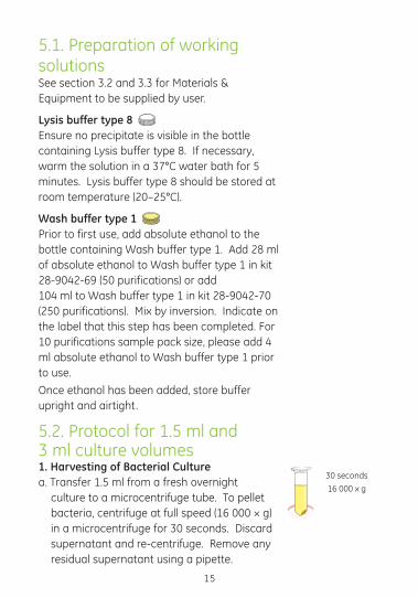
5.2. Protocol for 1.5 ml and 3 ml culture volumes1. Harvesting of Bacterial Culturea. Transfer 1.5 ml from a fresh overnight
culture to a microcentrifuge tube. To pellet bacteria, centrifuge at full speed (16 000 × g) in a microcentrifuge for 30 seconds. Discard supernatant and re-centrifuge. Remove any residual supernatant using a pipette.
30 seconds
16 000 x g
15

b. If processing 3 ml culture volumes, repeat step a. Pelleted DNA can be stored at -20°C if necessary.
Note: If purifying a high molecular weight or low copy number plasmid, process 3 ml culture volume.
2. Lysisa. Cell re-suspension- Add 175 µl Lysis buffer
type 7 to the bacterial pellet and thoroughly re-suspend the pellet.
Note: Cell re-suspension can be achieved by either vortexing, pipetting up and down or by scraping the base of the microcentrifuge tube across the surface of an empty pipette tip box. Incomplete cell re-suspension will result in reduced plasmid DNA recovery.
b. Cell Lysis - Add 175 µl Lysis buffer type 8 and mix immediately by gentle inversion (approximately 5 times) until solution becomes clear and viscous.
Note: Vigorous mixing will shear genomic DNA resulting in contamination of the final purified sample. Do not vortex. Do not allow the lysis reaction to exceed 5 minutes. Lysis buffer type 8 contains NaOH which will denature the plasmid DNA on prolonged incubation.
c. Neutralisation - Add 350 µl Lysis buffer type 9 and mix immediately by gentle inversion
until the precipitate is evenly dispersed.
16
175 µl
Lysis buffer type 7
Re-suspend
bacteria
175 µl
Lysis buffer type 8
Gently invert until
solution clears
350 µl
Lysis buffer type 9
Gently invert
until precipitate is
evenly dispersed

17
Note: The total column loading volume has been reduced to 700 µl compared to that described in the previous protocol. This change has been fully validated and has no impact on kit performance.Note: Cellular debris, genomic DNA and KDS appear as a white flocculent precipitate. Continue gentle inversion until the precipitate is evenly dispersed (approximately 10 times). The precipitate must be effectively dispersed to ensure consistent purity and yield of the isolated plasmid DNA.
Note: Do not shake or mix vigorously since genomic DNA will be sheared and co-purify with the plasmid DNA; mix by gentle inversion.
d. Flocculent spin-Centrifuge at full speed (approximately 16 000 × g) for 4 minutes.
Note: For applications that require less stringent purity (e.g. DNA sequencing) a 3 minute flocculent spin can be performed. For applications that are more salt sensitive, a > 5 minute flocculent spin may be required.
e. During centrifugation, for each purification that is to be performed, place one illustra plasmid mini column in one Collection tube.
Note: If required the snap-on lid can be removed without affecting the performance of the illustra plasmid mini column.
4 minutes
16 000 × g

18
Load supernatant
onto column.
30 seconds
16 000 × g
3. Plasmid Binding a. Column lysate loading-Carefully transfer the cleared supernatant to the mini column (approximately 700 µl). Close the lid of the column gently. Centrifuge at full speed (approximately 16 000 × g) for 30 seconds. Discard the flowthrough by emptying the Collection tube.
Note: When purifying plasmid DNA from 3 ml culture volumes, the user may notice some residual lysate on the column after this centrifugation step. This can be ignored; proceed with the Wash & Dry step below. On washing this residual lysate will generally pass through the column with no effect on the yield or purity of the isolated plasmid DNA.
If significant volumes of residual lysate do remain, this probably indicates that excessive flocculent material is being transferred onto the illustra plasmid mini column, blocking the silica membrane. Increase the time of the flocculent spin above to 6–10 minutes, providing that the culture A600 was approximately 2.5.
Note: It is recommended that a pipette is used but decanting directly can be performed as an alternative. It is important to avoid transferring any cellular debris to the column as this will affect the purity of the isolated plasmid DNA.
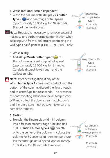
4. Wash (optional-strain dependent)a. Wash the column with 400 µl Lysis buffer
type 9 and centrifuge at full speed (approximately 16 000 × g) for 30 seconds. Discard the flowthrough.
Note: This step is necessary to remove potential nuclease and carbohydrate contamination when isolating DNA from E. coli strains containing the wild type EndA+ gene (e.g. HB101 or JM101) only.
5. Wash & Drya. Add 400 µl Wash buffer type 1 to
the column and centrifuge at full speed (approximately 16 000 × g) for 1 minute. Carefully discard flowthrough and the Collection tube.
Note: After centrifugation, if any of theWash buffer type 1 comes into contact with the bottom of the column, discard the flow through and re-centrifuge for 30 seconds. The presence of contaminating ethanol in the eluted plasmid DNA may affect the downstream applications and therefore care must be taken to ensure its complete removal.
6. Elutiona. Transfer the illustra plasmid mini column
into a fresh microcentrifuge tube and add 100 µl Elution buffer type 4 directly onto the center of the column. Incubate the column for 30 seconds at room temperature. Microcentrifuge at full speed (approximately 16 000 × g) for 30 seconds to recover
Optional step:
400 µl Lysis buffer
type 9
30 seconds
16 000 × g
400 µl Wash buffer
type 1
1 minute
16 000 × g
100 µl Elution
buffer type 4
Room temperature
30 seconds
30 seconds
16 000 x g
19

the plasmid DNA as flowthrough in the microcentrifuge tube.
Note: If a higher yield is required, follow the elution protocol described above, but elute with two successive 50 µl elution volumes.If a more concentrated sample is required, add
a single 50 µl volume of Elution buffer type 4, incubate for 1 minute and centrifuge to elute.
b. Store the purified plasmid DNA at -20°C.
20

6. Appendices6.1. RPM calculation from RCF The appropriate centrifugation speed for a specific rotor can be calculated from the following formula:
RPM=1000 × √(RCF/1.12r)
Where RCF = relative centrifugal force; r = radius in mm measured from the centre of the spindle to the bottom of the rotor bucket; and RPM = revolutions per min.
For example, if an RCF of 735 × g is required using a rotor with a radius of 73 mm, the corresponding RPM would be 3 000.
6.2 Isolation of low copy number and large molecular weight plasmids The procedure described in section 5.2 for the isolation of plasmid DNA from a 3 ml E. coli culture is that recommended for the isolation of either low copy number or large molecular weight plasmids. The 3 ml protocol has been successfully applied to the isolation of both a low copy number (10–20 copies/cell, 11 kb) and a large molecular weight plasmid (10–20 copies/cell, 35 kb). In both instances the purified plasmid DNA samples generated were of sufficient yield, purity, and quality to be compatible with most molecular biology techniques, including restriction enzyme digestion, PCR amplification and DNA sequencing. Using only 1.5 ml culture volumes may result in a prohibitively low yield of plasmid DNA.
6.3. Factors affecting plasmid DNA yield and purity Cell density - The yield and purity of plasmid DNA isolated with the illustra plasmidPrep Mini Spin Kit can be affected by a number of external factors e.g. culture cell density, the type of plasmid (high or low copy number), the size of the insert and the host strain used.
21

22
Cell density is the most important factor and cultures grown to an extremely high density (A600 > 5) can overload the column system and result in the poor recovery of plasmid DNA in terms of yield and purity. If high cell densities are obtained, it is suggested that the user processes smaller culture volumes to ensure no deleterious effect on plasmid recovery.
The A600 of an overnight culture of the E. coli strain TOP10 (transformed with a high copy number plasmid > 300 copies/cell) and grown in LB is approximately 2.5. From a 1.5 ml volume of LB, the illustra plasmidPrep Mini Spin Kit will routinely isolate 6 to 9 µg of high quality plasmid DNA (A260/A280 > 1.8).
Growth conditions -Specific factors which affect culture growth, and ultimately the density of the culture, are listed below.
Inoculation - for 1–3 ml cultures, use a fresh single E. coli colony from an agar plate containing the appropriate antibiotics to inoculate growth medium.
Culture medium - When incubated for an equivalent period of time, cultures grown in enriched media (e.g. 2 × YT and Terrific Broth) tend to give cell densities that are significantly higher than those achieved with LB medium. 2 × YT broth (16 g tryptone, 10 gm yeast extract, 5 gm sodium chloride per liter medium) and Terrific Broth (TB; 12 gm tryptone, 24 gm yeast extract, 4 ml glycerol, 2.31 gm KH2PO4, 12.54 gm K2HPO4) should therefore be used with caution (see Cell density notes above).
Aeration - Cultures should be well-aerated during growth. When growing cultures in a 30 ml universal container, no more than 3 ml of media should be used. Aeration will be poor if cultures are grown in 1.5 or 2.0 ml microcentrifuge tubes. Poor aeration will lead to poor culture growth, and subsequently to low yields of plasmid DNA.
Plasmid copy number - For a given length of incubation and a given medium, low copy number plasmids will give lower yields than high copy number plasmids.

Size of insert - In general, the larger the size of the insert, the lower the cell density, and lower the yield of plasmid DNA from a given culture medium. Insert size in the plasmid used to generate typical data in section 4.4 is 783 bp.
Host strain - Strains which grow poorly or contain large amounts of nucleases or carbohydrates should be avoided. HB101 and its derivatives express endonuclease A (EndA+), which if not inactivated, can digest plasmid DNA. These strains may also release carbohydrates that can inhibit restriction digests (7). Note the E. coli strains DH5α and TOP10 facilitate the extraction of good quality plasmid DNA and are recommended for use with the illustra plasmidPrep Mini Spin Kit.
Length of incubation –For cultures grown in an enriched medium (e.g. 2xYT or TB), the length of the incubation time should not exceed 12 hours. Cultures in LB medium should be grown for at least 9 hours to obtain sufficient cell mass for processing. Cultures (in any medium) should not be grown for more than 16 hours, due to increased rates of cell death, which will affect the yield and quality of extracted plasmid DNA
23

24
6.4. Troubleshooting guide This guide may be helpful in the first instance, however if problems persist or for further information please contact GE Healthcare technical services. Telephone numbers are on the back page. Alternatively log onto http://www.gehealthcare.com/illustra.
Problem: plasmid DNA yield is low
Possible cause Suggestions
The bacterial culture was not fresh.
• A culture should be processed in a timely manner after it has reached the required cell density. Alternatively, bacterial pellets can be stored at -20°C, prior to plasmid DNA extraction with no significant effect on purity or quality.
The total A600 units of the volume of culture processed was too high.
• Measure the A600 of the culture before processing. If the culture density A600 > 5, reduce the volume of culture processed.
The cell pellet was not completely re-suspended in Lysis buffer type 7.
• Cell re-suspension can be achieved by either vortexing, pipetting up/down or alternatively by scraping the base of the microcentrifuge tube across the surface of an empty pipette tip box (6).
Following the addition of Lysis buffer type 9, the sample was not adequately mixed.
• After Lysis buffer type 9 is added, mix by gently inverting the tube until a flocculent precipitate appears. Continue inverting until the precipitate is evenly dispersed (10–20 inversions). The cell lysate must be broken up effectively to ensure consistent yields.

25
Problem: plasmid DNA yield is low (continued)
Possible cause Suggestions
The Wash buffer type 1 was not completely removed.
• After the Wash & Dry centrifugation step, if any of the ethanolic Wash Buffer comes into contact with the bottom of the column discard the flowthrough and re-centrifuge for 30 seconds. The presence of residual ethanol may affect downstream applications and must be carefully removed.
Problem: Plasmid DNA is contaminated with genomic DNA
Possible cause Suggestions
The sample was mixed too vigorously after adding Lysis buffers type 8 and/or 9.
• Mix gently by inverting the sample 10–15 times after adding either of the solutions. Vigorous mixing may cause shearing of genomic DNA thereby facilitating its co-purification with plasmid DNA.
Problem: Agarose gel electrophoresis shows a band migrating faster than supercoiled plasmid DNA. The fast band does not cut with restriction enzymes.
Possible cause Suggestions
Plasmid DNA was irreversibly denatured by Lysis buffer type 8.
• The band migrating slightly faster on the agarose gel is denatured plasmid DNA. It is generated when plasmid DNA is exposed to Lysis buffer type 8 for an excessive amount of time. Plasmid DNA should be exposed to Lysis buffer type 8 for no more than a few minutes prior to the addition of the neutralizing Lysis buffer type 9. Do not allow the cell lysis reaction to proceed for > 5 minutes.

26
Problem: plasmid DNA does not cut to completion or is degraded on incubation at 37ºC.
Possible cause Suggestions
Plasmid DNA irreversibly denatured by Lysis buffer type 8 and therefore will not cut.
• See previous
Host strain possesses carbohydrates (that may interfere with restriction enzyme digestion) or residual nucleases (that are carried over into the final sample and degrade the isolated plasmid DNA).
• Perform the optional Lysis buffer type 9 wash as described in the protocol. This step is necessary to remove any possible nuclease and carbohydrate contamination. If the total A600 unit of the culture used was excessively high, incubate Lysis buffer type 9 on the column for 2–3 minutes to ensure complete nuclease inactivation. Alternatively, use an EndA negative strain such as TOP10.
Wash buffer type 1 was not completely removed and therefore interfered with the restriction digest.
• Discard the column flowthrough by emptying the Collection tube as described in the procedure. If necessary, re-place the column into the Collection tube and re-spin briefly (30 seconds) to remove any residual Wash buffer type 1. Note - if any of the ethanolic Wash buffer comes into contact with the bottom of the column, discard the flow-through and re-centrifuge.

6.5. Related products available from GE HealthcareA full range of Molecular Biology reagents can be found on the GE Healthcare web site and in the catalog http://www.gehealthcare.com/illustraIf you need further information, GE technical services are happy to assist (world-wide phone numbers can be found on the back cover).
Application Product Product code Pack sizes
Purification of transfection quality plasmid DNA from E. coli
illustra plasmidPrep Midi
Flow Kit
28-9042-67 25 purifications
Purification of genomic DNA from small volumes of whole blood and blood cell fractions
illustra blood genomicPrep Mini
Spin Kit
28-9042-64 50 purifications
Purification of genomic DNA from animal tissues and cultured mammalian cells
illustra tissue & cells
genomicPrep Mini Spin Kit
28-9042-75 50 purifications
Purification of genomic DNA from various bacterial strains
illustra bacteria genomicPrep Mini
Spin Kit
28-9042-58 50 purifications
Small scale RNA isolation. High quality RNA from diverse sample types
illustra RNAspin Mini Kit
25-0500-70 20 preparations
27

28
Application Product Product code Pack sizes
Preparation of circular DNA templates
illustra TempliPhi™ 100 Amplification kit
25-6400-10 100 reactions
Kits containing ready-to-use mix for PCR amplification
illustra PuReTaq™
Ready-To-Go™ PCR Beads
27-9557-01 96 reactions
FideliTaq™ PCR Master Mix Plus
E71183 125 units
DNA purification from PCR and enzymes
illustra GFX™ PCR DNA & Gel Band purification kit illustra GFX 96
PCR Purification kit
27-9602-01
25-6902-02
100 columns
10 × 96 well plates
DNA Ligation DNA ligation System
RPN1507 50 reactions
Ligate-IT Rapid Ligation kit
US78400 25 reactions
Ready-To-Go T4 DNA ligase
27-0361-01 50 reactions
Blunt-Ended PCR Cloning
Blunt-ended PCR Cloning Kit
RPN5110 40 reactions
Non-radioactive nucleic acid labeling & detection
Gene Images™ random-prime
DNA Labeling kit
RPN3520 30 reactions
Gene Images 3’-Oligolabeling kit
RPN5770 Labels 1000 pmol

29
Application Product Product code Pack sizes
Non-radioactive nucleic acid labeling & detection
ECL Direct™ Nucleic acid
labeling & Detection System
RPN3000 Labels 5 µg
Gene Images ECF Detection kit
RPN3580 2500 cm2 membrane
Gene Images CDP-Star™
Detection kit
RPN3550 2500 cm2 membrane
Large scale purification of plasmid DNA by gel filtration
Sephacryl™ S-1000SF
17-0476-01 750 ml
Purification of oligonucleotides following synthesis
illustra NAP™-5 Columns
17-0853-01 20 purifications
Dye terminator removal from automated sequencing reactions
illustra AutoSeq™ G-50
27-5340-01 50 columns

30
7. References1. Birnboim, H.C. & Doly, J., Nucl. Acids Res. 7, 1513 (1979).
2. Ish-Horowicz, D. & Burke, J.F., Nucl. Acids Res. 9, 2989 (1981).
3. Sambrook, J., Fritsch, E.F. & Maniatis, T., Molecular cloning: A laboratory Manual, Cold Spring Harbor Laboratory, 2nd eds., (1989).
4. Vogelstein, B. & Gillespie, D., Proc. Natl. Acad. Sci. USA 76, 615 (1979).
5. Marko, M.A., Chipperfield, R. & Birnboim, H.C., Anal. Biochem. 121, 382 (1982).
6. Voo, K.S. & Jacobsen B.M., Biotechniques, 24, 240-243 (1998).
7. Ausubel, F.M. et al., eds., Current Protocols in Molecular Biology 1, 1.68 (1991).

31

28-9042-71PL Rev F 06/2008
For contact information for your local office,
please visit: www.gehealthcare.com/contact
GE Healthcare UK Limited Amersham Place,
Little Chalfont, Buckinghamshire,
HP7 9NA, UK
http://www.gehealthcare.com/illustra
imagination at work
GE Healthcare offices:
GE Healthcare Bio-Sciences AB
Björkgatan 30, 751 84 Uppsala,
Sweden
GE Healthcare Europe GmbH
Munzinger Strasse 5 D-79111 Freiburg,
Germany
GE Healthcare Bio-Sciences Corp
800 Centennial Avenue, P.O. Box 1327,
Piscataway, NJ 08855-1327,
USA
GE Healthcare Bio-Sciences KK
Sanken Bldg. 3-25-1, Hyakunincho,
Shinjuku-ku, Tokyo 169-0073,
Japan

The next four pages are a protocol cards.Please add to the back page as a tear off addition.

28-9
042-
69 (5
0 pu
rific
atio
ns)
28-9
042-
70 (2
50 p
urifi
catio
ns)
Qui
ck R
efer
ence
Pro
toco
l Car
dill
ustr
a™ p
lasm
idPr
ep M
ini S
pin
Kit
Prot
ocol
for
1.5
& 3
ml c
ultu
re v
olum
es
• Ch
eck
appr
opri
ate
volu
me
of e
than
ol a
dded
to
Was
h bu
ffer
type
1
1. H
arve
stin
g of
bac
teri
al c
ultu
re
2. L
ysis
3. P
lasm
id b
indi
ng
:Add
:Spi
n:In
cuba
te
1.5
ml b
acte
rial
cul
ture
3
0 se
cond
s 16
000
× g
•
Pou
r of
f and
dis
card
sup
erna
tant
•
Rep
eat f
or 3
ml c
ultu
re v
olum
e 3
0 se
cond
s 16
000
× g
(for
all
cultu
re v
olum
es)
• R
emov
e re
sidu
al s
uper
nata
nt
1
75 µ
l Lys
is b
uffe
r ty
pe 7
; re-
susp
end
pelle
t
175
µl L
ysis
buf
fer
type
8; g
ently
inve
rt
3
50 µ
l Lys
is b
uffe
r ty
pe 9
; gen
tly in
vert
4
min
utes
16
000
× g
• T
rans
fer
supe
rnat
ant t
o pl
asm
id m
ini c
olum
n
in
side
Col
lect
ion
tube
3
0 se
cond
s 16
000
× g
•
Dis
card
flow
thro
ugh
4
00 µ
l Lys
is b
uffe
r ty
pe 9
3
0 se
cond
s 16
000
× g
•
Dis
card
flow
thro
ugh
4
00 µ
l Was
h bu
ffer
type
1
1 m
inut
e 16
000
× g
•
Dis
card
flow
-thr
ough
and
Col
lect
ion
tube
• T
rans
fer
plas
mid
min
i col
umn
to a
new
DN
ase-
free
mic
roce
ntri
fuge
tube
100
µl E
lutio
n bu
ffer
type
4
30
seco
nds
at ro
om te
mpe
ratu
re
30
seco
nds
16 0
00 ×
g
• R
etai
n el
uant
•
Sto
re p
urifi
ed p
lasm
id D
NA
at -2
0°C

imag
inat
ion
at w
ork
28-9
042-
71PL
Rev
F 0
6/20
08
4. W
ash
(opt
iona
l-sta
in d
epen
dent
)
1.5
ml b
acte
rial
cul
ture
3
0 se
cond
s 16
000
× g
•
Pou
r of
f and
dis
card
sup
erna
tant
•
Rep
eat f
or 3
ml c
ultu
re v
olum
e 3
0 se
cond
s 16
000
× g
(for
all
cultu
re v
olum
es)
• R
emov
e re
sidu
al s
uper
nata
nt
1
75 µ
l Lys
is b
uffe
r ty
pe 7
; re-
susp
end
pelle
t
175
µl L
ysis
buf
fer
type
8; g
ently
inve
rt
3
50 µ
l Lys
is b
uffe
r ty
pe 9
; gen
tly in
vert
4
min
utes
16
000
× g
• T
rans
fer
supe
rnat
ant t
o pl
asm
id m
ini c
olum
n
in
side
Col
lect
ion
tube
3
0 se
cond
s 16
000
× g
•
Dis
card
flow
thro
ugh
4
00 µ
l Lys
is b
uffe
r ty
pe 9
3
0 se
cond
s 16
000
× g
•
Dis
card
flow
thro
ugh
4
00 µ
l Was
h bu
ffer
type
1
1 m
inut
e 16
000
× g
•
Dis
card
flow
-thr
ough
and
Col
lect
ion
tube
• T
rans
fer
plas
mid
min
i col
umn
to a
new
DN
ase-
free
mic
roce
ntri
fuge
tube
100
µl E
lutio
n bu
ffer
type
4
30
seco
nds
at ro
om te
mpe
ratu
re
30
seco
nds
16 0
00 ×
g
• R
etai
n el
uant
•
Sto
re p
urifi
ed p
lasm
id D
NA
at -2
0°C
5. W
ash
& D
ry
6. E
lutio
n


imag
inat
ion
at w
ork
GE,
imag
inat
ion
at w
ork
and
GE
mon
ogra
m a
re tr
adem
arks
of G
ener
al E
lect
ric C
ompa
ny.
illus
tra
is a
trad
emar
k of
GE
Hea
lthca
re C
ompa
nies
© 2
006–
2008
Gen
eral
Ele
ctric
Com
pany
– A
ll rig
hts
rese
rved
. Pr
evio
usly
pub
lishe
d 20
06.
All g
oods
and
ser
vice
s ar
e so
ld s
ubje
ct to
the
term
s an
d co
nditi
ons
of s
ale
of
the
com
pany
with
in G
E H
ealth
care
whi
ch s
uppl
ies
them
. A
copy
of t
hese
term
s an
d co
nditi
ons
is a
vaila
ble
upon
req
uest
. C
onta
ct y
our
GE
Hea
lthca
re
repr
esen
tativ
e fo
r th
e m
ost c
urre
nt in
form
atio
n (s
ee b
ack
cove
r fo
r co
ntac
t det
ails
). ht
tp://
ww
w.g
ehea
lthca
re.c
om/li
fesc
ienc
es
GE
Hea
lthca
re U
K Li
mite
d.
Amer
sham
Pla
ce, L
ittle
Cha
lfont
, Bu
ckin
gham
shire
, HP7
9N
A U
K