il-33 in hepatocytes in murine models of hepatitis 5 … · citation: arshad mi, patrat-delon s,...
TRANSCRIPT

Pathogenic Mouse Hepatitis Virus or Poly(I:C) InduceIL-33 in Hepatocytes in Murine Models of HepatitisMuhammad Imran Arshad1,2,3,6☯, Solène Patrat-Delon1,2,3☯, Claire Piquet-Pellorce1,2,3, AnnieL’Helgoualc’h1,2,3, Michel Rauch1,2,3, Valentine Genet1,2,3, Catherine Lucas-Clerc2,4, Christian Bleau5, LucieLamontagne5, Michel Samson1,2,3*
1 Institut de Recherche Santé Environnement et Travail (IRSET) - U.1085, Institut National de la Santé et de la Recherche Médicale (Inserm), Rennes,Bretagne, France, 2 Université de Rennes 1, Rennes, Bretagne, France, 3 Structure Fédérative BioSit UMS 3480 CNRS-US18 Inserm, Rennes, Bretagne,France, 4 Service de Biochimie CHU Rennes, Université de Rennes 1, Rennes, Bretagne, France, 5 Département des Sciences Biologiques, Université duQuébec à Montréal, Montréal, Québec, Canada, 6 Institute of Microbiology, University of Agriculture, Faisalabad, Pakistan
Abstract
The IL-33/ST2 axis is known to be involved in liver pathologies. Although, the IL-33 levels increased in sera of viralhepatitis patients in human, the cellular sources of IL-33 in viral hepatitis remained obscure. Therefore, we aimed toinvestigate the expression of IL-33 in murine fulminant hepatitis induced by a Toll like receptor (TLR3) viral mimetic,poly(I:C) or by pathogenic mouse hepatitis virus (L2-MHV3). The administration of poly(I:C) plus D-galactosamine (D-GalN) in mice led to acute liver injury associated with the induction of IL-33 expression in liver sinusoidal endothelialcells (LSEC) and vascular endothelial cells (VEC), while the administration of poly(I:C) alone led to hepatocytespecific IL-33 expression in addition to vascular IL-33 expression. The hepatocyte-specific IL-33 expression wasdown-regulated in NK-depleted poly(I:C) treated mice suggesting a partial regulation of IL-33 by NK cells. The CD1dKO (NKT deficient) mice showed hepatoprotection against poly(I:C)-induced hepatitis in association with increasednumber of IL-33 expressing hepatocytes in CD1d KO mice than WT controls. These results suggest that hepatocyte-specific IL-33 expression in poly(I:C) induced liver injury was partially dependent of NK cells and with limited role ofNKT cells. In parallel, the L2-MHV3 infection in mice induced fulminant hepatitis associated with up-regulated IL-33expression as well as pro-inflammatory cytokine microenvironment in liver. The LSEC and VEC expressed inducibleexpression of IL-33 following L2-MHV3 infection but the hepatocyte-specific IL-33 expression was only evidentbetween 24 to 32h of post infection. In conclusion, the alarmin cytokine IL-33 was over-expressed during fulminanthepatitis in mice with LSEC, VEC and hepatocytes as potential sources of IL-33.
Citation: Arshad MI, Patrat-Delon S, Piquet-Pellorce C, L’Helgoualc’h A, Rauch M, et al. (2013) Pathogenic Mouse Hepatitis Virus or Poly(I:C) InduceIL-33 in Hepatocytes in Murine Models of Hepatitis. PLoS ONE 8(9): e74278. doi:10.1371/journal.pone.0074278
Editor: Maria Leite de Moraes, CNRS, France
Received April 3, 2013; Accepted July 30, 2013; Published September 13, 2013
Copyright: © 2013 Arshad et al. This is an open-access article distributed under the terms of the Creative Commons Attribution License, which permitsunrestricted use, distribution, and reproduction in any medium, provided the original author and source are credited.
Funding: This work was supported by Institut National de la Santé et de la Recherche Médicale, the Ministère de l’Education Nationale de la Recherche etde la Technologie, the University of Rennes 1, the Région Bretagne, the “Ligue contre le cancer, comités du Grand Ouest” and by NSERC fromGovernment of Canada. MIA was supported by a Ph.D fellowship from the Government of Pakistan (Higher Education Commission, University ofAgriculture, Faisalabad). CB was supported by a Ph.D. fellowship from Natural Sciences and Engineering Research Council (Canada). The funders had norole in study design, data collection and analysis, decision to publish, or preparation of the manuscript.
Competing interests: The authors have declared that no competing interests exist.
* E-mail: [email protected]
☯ These authors contributed equally to this work.
Introduction
Interleukin-33 (IL-33), a member of IL-1 family also called asIL-1F11, is known to drive immune responses by interactionwith its specific receptors ST2 and IL-RAcP [1,2]. TheIL-33/ST2 axis is crucially involved in diverse inflammatory andimmune mediated pathologies [3,4]. However, limited data isavailable about the association of IL-33 and ST2 expression inviral diseases. IL-33 is over-expressed in influenza virus lunginfection in mice [5,6] and IL-33 produced by necrotic cellsdrives protective antiviral CD8+ T cell responses in lymphocytic
choriomeningitis virus (LCMV) infection in mice [7]. Further,elevated levels of soluble ST2 (sST2) in sera of dengue virusinfected patients [8] and HIV infected patients [9] wereobserved indicating sST2 as potential marker of viral infections.
In liver, IL-33/ST2 axis is involved in various viral andimmune cell mediated pathologies [10-13]. We initiallyobserved up-regulated expression of IL-33 and ST2 in chronichepatitis B and C virus (HBV and HCV) infection in human andin CCl4-induced liver fibrosis in mice [14]. The increased levelof serum IL-33 and sST2 was observed in acute and chronichepatic failure in human [15]. Furthermore, elevated IL-33
PLOS ONE | www.plosone.org 1 September 2013 | Volume 8 | Issue 9 | e74278
serum level was also associated with liver damage in patientsof chronic hepatitis C virus (HCV) [16] and hepatitis B virus(HBV) [17] infections, representing IL-33 as a possible indicatorof viral hepatitis.
Despite the fact that IL-33 is proposed to be released as analarmin in acute inflammatory pathologies [3], the expressionand cellular sources of IL-33 during viral fulminant hepatitis in arelevant animal model has not been explored. The polyinosine-polycytidylic acid (Poly(I:C), a synthetic analog of doublestranded RNA (dsRNA), induces a moderate acute hepaticinjury and mimics a model of viral hepatitis [18,19]. Poly(I:C)activated principally the intrahepatic macrophages (Kuppfercells) and NK cells via TLR3 [20] leading to increase ofinflammatory cytokines such as TNF-α, IFN-γ, IL-6, IL-12 andIFN-β [18,19,21,22]. A pretreatment with D-galactosamine (D-GalN) in Poly(I:C) injected-mice aggravated the acute hepaticinjury which become lethal [19]. A natural animal model of viralhepatitis, the mouse hepatitis viruses (MHV), single-strand,positive-sense RNA viruses belonging to Coronaviridae family,induced acute and/or chronic hepatitis in mice mimickinghuman HBV infection and serve as a good tool to studyimmune dysfunction and cytokines associated with viral acutehepatitis [23,24]. The most hepatotropic serotype of MHV, themouse hepatitis virus type 3 (MHV3), induced severe fulminanthepatitis in mice and their death within 3-5 days post-infection[25]. In liver, Kupffer cells, NK cells, hepatocytes, sinusoidalendothelial and vascular endothelial cells are the main targetcells for MHV3 replication [26,27]. The histopathological lesionsin liver were correlated with the levels of inflammatorycytokines [28]. High levels of IL-6 and TNF-α produced in liversfrom infected C57BL/6 mice were modulated by TLR receptor.We have previously demonstrated that intrahepatic NK cellsdecreased after a transient increase in liver from pathogenicL2-MHV3-infected mice due to virus-induced NK depletion [27].
In the present study, we aimed to investigate the expressionand cellular sources of IL-33 in a Poly(I:C)- and L2-MHV3-induced fulminant hepatitis in mice. We found increasedexpression of IL-33 in liver following Poly(I:C) and L2-MHV3induced acute hepatitis in mice. The liver sinusoidal endothelialcells, vascular endothelial cells and hepatocytes representpotential sources of IL-33 in Poly(I:C) and murine L2-MHV3induced fulminant hepatitis. The hepatocyte-specific IL-33expression in Poly(I:C) induced liver injury was partiallydependent of NK cells but not of NKT cells.
Materials and Methods
AnimalsWild-type (WT) C57BL/6 mice were purchased from Charles
River Laboratories (St-Constant, QC, Canada) or from Janvier(Le Genest-sur-isle, France). The animals, certified as MHVs-free by the manufacturer, were housed under HEPA-filtered air(Forma Scientific, Marietta, OH). The study was conducted incompliance with the regulations of the Comité institutionnel dela Protection des Animaux of the Université du Québec àMontreal (UQAM agreement of L. Lamontagne, No. CIPA=541), and French laws and the institution’s guidelines foranimal welfare (agreement of M. Samson #3596). The protocol
was approved by the Committee on the Ethics of AnimalExperiments of the French government (agreement of M.Samson #3596). All efforts were made to minimize suffering"
In vivo treatment protocolThe C57BL/6 (Janvier, France) or CD1d KO mice (a gift of
Maria Leite-de-Moraes, Paris) were intravenously injected with30 µg/mouse of Poly(I:C) (Invivogen) alone or with D-galactosamine (D-GalN) (SIGMA-G0264) pretreatment at adose of 15 mg/mouse (i.p). The control mice received similarvolume of vehicle in each treatment group. For NK cellsdepletion experiment, 35 µl of anti-asialo GM1 (anti-ASGM1)polyclonal antibody (Cerdalane, CL8955) was injectedintraperitoneally (i.p) 48 h before D-GalN, Poly(I:C) orcombination of both D-GalN Poly(I:C) injections, the controlmice received or an equivalent amount of naive rabbit serum.The NK depletion in liver was confirmed by flow cytometry inisolated liver immune cells as described earlier [29].
For MHV3 infection in mice, the C57BL/6 mice (CharlesRiver Laboratories, Canada) were infected by the i.p route with103 50% tissue culture infective dose (TCID(50)) of pathogenicL2-MHV3 strain as previously described [25]. Mock-infected oruninfected control mice received a similar volume ofRPMI-1640 (Gibco Laboratories, Grand Island, NY). After 16,24, 28, 39, 48 and 72 h of infection, the mice wereanaesthetized by i.p injection using ketamine hydrochloride(200 mg/kg; Vetrepharm Canada Inc., Belleville, ON, Canada)and xylazine (10 mg/kg; Bayer Inc., Toronto, ON, Canada) andeuthanazied by CO2 inhalation before liver and blood sampling.
Histopathological, biochemical andimmunohistochemical analyses
The histopathological (Hematoxylin And Eosin (H&E)staining) and levels of liver transaminase (ALT/AST) in serumwere performed as described earlier [10]. Immunolocalisationof IL-33 was performed by immunohistochemical staining usingprimary antibody goat IgG anti-mouse-IL-33 (R&D Systems)and secondary HRP-conjugated rabbit anti-goat antibody(Dako, USA) followed by hematoxylin counterstaining inVentana machine (Ventana Medical Systems, Inc., USA). Thecounting of IL-33 positive hepatocytes was carried in at least20 different microscopic fields corresponding to 2.67 mm2
surface area by using image analysis software (Compix, Inc.HAMAMATSU company, Japan) as previously described [13].
RNA isolation and RT-qPCRThe protocol and conditions for RNA extraction, RT-PCR and
qPCR were similar as reported earlier by our laboratory usingspecific primers for 18S, IL-33, IL-6, IL-1β, IFN-β, IFNγ, TNFαand CXCL1 [10,13]. For the quantification of viral nucleocapsid,the following primer set was used: 5’-TGGAAGGTCTGCACCTGCTA-3’ (forward), 5’-TTTGGCCCACGGGATTG-3’ (reverse). The relative geneexpression was normalized against 18S gene expression. Thecontrol mice in each treatment group served as a reference formRNA expression (control mRNA level was arbitrarily taken as1).
Mouse Hepatitis Virus or Poly(I:C) Induce IL-33
PLOS ONE | www.plosone.org 2 September 2013 | Volume 8 | Issue 9 | e74278

Statistical analysisThe results are representative of three independent
experiments and expressed as means±SEM. Mann-Whitney Utest was used for comparison of control group parameters withtreatment group and multiple group analysis was evaluated byone-way ANOVA with post Mann-Whitney U test usingGraphPad Prism5 software. For all statistical analyses, p-values <0.05 were considered significant.
Results
Poly(I:C) administration induced acute liver injury inmice with expression of IL-33 in liver
While the cellular source of IL-33 in viral liver pathology ispoorly known in a mouse model, we first aimed to investigatethe expression and cellular sources of IL-33 in a Poly(I:C)-induced acute hepatitis. The administration of Poly(I:C) inducedmoderate liver injury compared to PBS treated mice at 8h, asevident from serum AST/ALT levels (Figure 1A). However, thepre-sensitization of mice with D-galactosamine (D-GalN) led toPoly(I:C)-induced severe liver injury in mice with elevatedserum AST/ALT levels at 8h in comparison to D-GalN alonetreatment (Figure 1A). The mice treated with combination of D-GalN Poly(I:C) died earlier within 16h compared to D-GalN orPoly(I:C) alone treated mice (Figure S1), therefore, we used 8htime point in this study. The mRNA expression of IL-33 in liverwas not significantly increased in Poly(I:C) or D-GalN Poly(I:C)treated mice in comparison with PBS control mice (Figure 1A).
The histology of liver tissues revealed increasedhemorrhagic lesions in liver after D-GalN Poly(I:C) treatmentbut less or no marked liver injury in Poly(I:C) or vehicle controlmice (Figure 1B). The mRNA expression of IL-33 wassignificantly increased (2-3 fold) in Poly(I:C) treated micecompared to control mice, however, IL-33 expression wasdownregulated in D-GalN Poly(I:C) treated mice in comparisonwith Poly(I:C) alone (Figure 1A). Regarding cellular sources ofIL-33, the liver sinusoidal endothelial cells and vascularendothelial cells expressed IL-33 constitutively in PBS controlmice livers and induced expression in these cells was observedfollowing Poly(I:C) and D-GalN Poly(I:C) treatment (Figure 1B).Interestingly, the nuclear expression of IL-33 was found inhepatocytes of Poly(I:C) treated mice (arrows in insert indicateIL-33 positive hepatocytes) but not in D-GalN Poly(I:C) inducedliver injury (Figure 1B). The number of IL-33 expressinghepatocytes were clearly and significantly increased inPoly(I:C) induced acute liver injury compared to control mice(Figure 1C). These results suggest that regulation of IL-33 inhepatocytes is associated with Poly(I:C) induced TLR3stimulation in liver.
Poly(I:C)-induced hepatitis up-regulated pro-inflammatory cytokine expression in liver
The inflammatory cytokines play an important role indevelopment of fulminant hepatitis. Therefore, we investigatedthe pro-inflammatory cytokine expression in Poly(I:C) and D-GalN Poly(I:C) induced acute hepatitis. The transcript level ofTNF-α, TRAIL, IL-1β and IL-6 was significantly up-regulatedfollowing D-GalN Poly(I:C) induced acute hepatitis (8h)
compared to D-GalN or PBS control mice (Figure 2). However,the mRNA expression of IFN-γ and CXCL1/KC was not variedbetween Poly(I:C), D-GalN Poly(I:C) or D-GalN/PBS treatedmice (Figure 2).
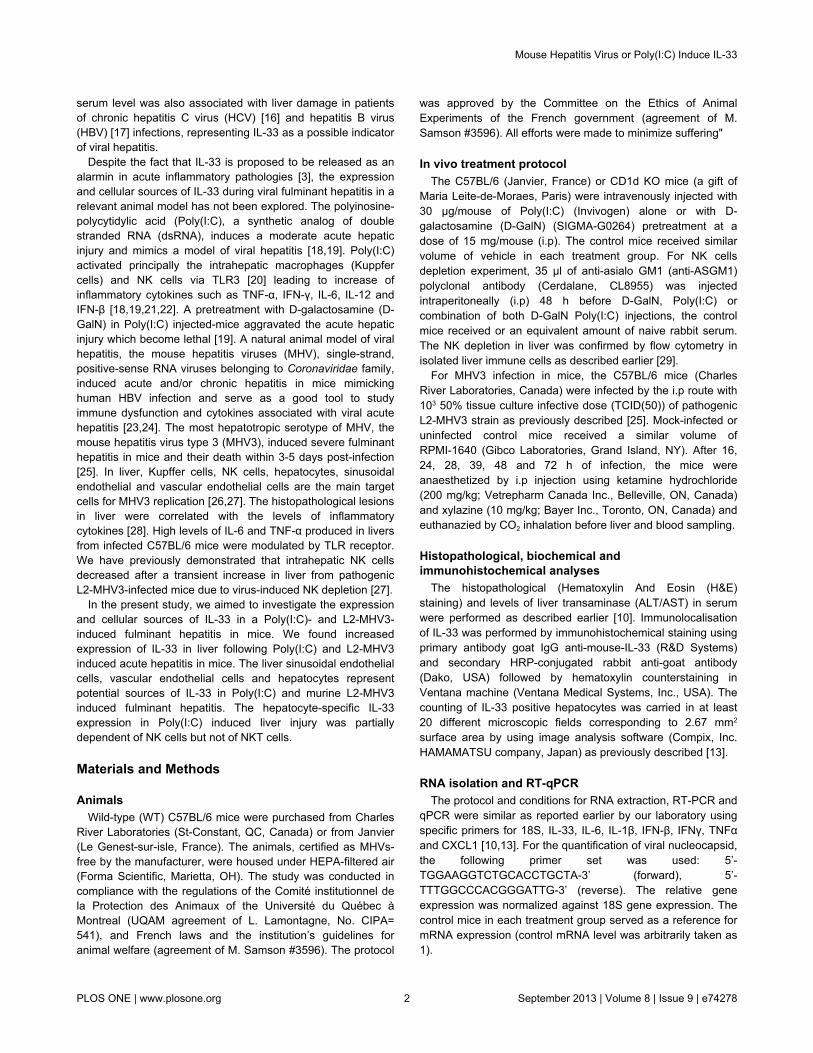
NK cells pre-depletion led to severe D-GalN Poly(I:C)induced acute liver injury, increased pro-inflammatorycytokines and down-regulated IL-33 expression inhepatocytes
NK cells have shown to be crucially important in Poly(I:C)induced liver injury [22]. Here, we studied the effect ofdepletion of NK cells by anti-ASGM1 antibody on D-GalNPoly(I:C) induced acute hepatitis and expression of IL-33. Asexpected, the anti-ASGM1 pre-treatment efficiently depletedNK cells in liver of mice compared to vehicle control orPoly(I:C) treated mice (Figure 3A). However, NK cells depletionled to enhanced liver injury (as evaluated by serum AST/ALT)in D-GalN Poly(I:C) treated mice than Poly(I:C) alone or vehiclecontrol mice (Figure 3B). A milder increase in serumtransaminases was evident between D-GalN Poly(I:C) and NK-depleted D-GalN Poly(I:C) treated mice (Figure 3B).
The mRNA expression of IL-33 was not varied betweencontrol (non NK depleted) and NK- depleted Poly(I:C) or D-GalN Poly(I:C) treated mice although the expression of IL-33was diminished in NK-depleted D-GalN Poly(I:C) mice (Figure3B). The liver sinusoidal endothelial cells (LSEC) and vascularendothelial cells (VEC) expressed IL-33 in NK depletedPoly(I:C) or D-GalN Poly(I:C) treated mice livers andhepatocyte-specific IL-33 expression was evident only afterPoly(I:C) treatment (Figure 3C) (arrows indicate IL-33 positivehepatocytes). The NK depletion led to decrease in number ofIL-33 expressing hepatocytes in Poly(I:C) treated micecompared to non NK depleted mice (Figure 3D) suggesting apartial NK cells dependent regulation of IL-33 in hepatocytes.
The pro-inflammatory cytokine expression of TNF-α, IL-1βand IL-6 was up-regulated in Poly(I:C) or D-GalN Poly(I:C)treated mice when compared with control mice (p <0.01)(Figure 3E). NK cell depletion did not decrease the expressionlevel of the inflammatory cytokines (Figure 3E). The mRNAexpression of IFN-γ was not significantly varied among thetreated and control groups of mice except in NK-depleted D-GalN Poly(I:C) treated mice (Figure 3E). The expression ofTRAIL was significantly increased in both NK depleted and nondepleted mice following Poly(I:C) or D-GalN Poly(I:C)administration when compared with control mice (p <0.01)) butsignificantly decrease in NK depleted mice after D-GalNPoly(I:C) treatment (p <0.05) when compared with D-GalNPolyIC treated mice (Figure 3E). The expression ofCXCL1 only increased in NK depleted D-GalN Poly(I:C) treatedmice when compared to control mice (p <0.05) (Figure 3E) thatmay correlate with development of inflammatorymicroenvironment in liver and neutrophil migration during liverinjury.
Mouse Hepatitis Virus or Poly(I:C) Induce IL-33
PLOS ONE | www.plosone.org 3 September 2013 | Volume 8 | Issue 9 | e74278

NKT cells deficiency protected mice against Poly(I:C)-induced liver injury but up-regulated hepatocyte-specific IL-33 expression
The role of NKT cells in Poly(I:C)-induced liver injury is notwell known but we have observed a partial decrease in NKTcells percentages in anti-ASGM1-treated mice (Figure 4A). Weaimed to verify the impact of NKT cells deficiency in Poly(I:C)-induced liver injury and IL-33 expression in liver in CD1d KO(NKT KO) mice. WT control and NKT KO mice showedincreased liver injury (2000 to 5000 AST/ALT levels) after D-GalN Poly(I:C) administration with a milder hepatoprotection inNKT KO mice (p <0.05) when compared with WT treated mice(Figure 4A). Liver histology revealed hemorrhagic lesions inliver after D-GalN Poly(I:C) treatment in NKT KO mice withoutremarkable liver injury in Poly(I:C) or vehicle control NKT KO
mice (Figure 4B, upper panel). The hepatocyte-specific IL-33expression was evident in Poly(I:C)-administered NKT KO micebut not in D-GalN Poly(I:C)-treated or control NKT KO mice(Figure 4B, lower panel). The number of IL-33 expressinghepatocytes significantly increased in Poly(I:C)-administeredNKT KO mice compared to WT controls (p <0.05) but not in D-GalN Poly(I:C)-treated or control NKT KO mice (Figure 4C).These results suggested that NKT cells have a protective effecton liver injury in association with increased expression of IL-33in hepatocytes during Poly(I:C)-induced liver injury.
L2-MHV3 induced fulminant hepatitis in mice wasassociated with increased expression of IL-33 in liver. Thepathogenic strain of mouse hepatitis virus (L2-MHV3) inducesfulminant hepatitis in C57BL/6 mice [23] and mimics a model offulminant viral hepatitis (HBV) in human. We investigated the
Figure 1. Liver injury and IL-33 expression in D-GalN, Poly(I:C), D-GalN Poly(I:C) treated mice. (A) Levels of serum AST/ALT(IU/L) and relative fold change in mRNA expression of IL-33 in WT mice treated with Poly(I:C) (30 µg/mouse i.v.) and/or D-GalNPoly(I:C) at 8h of post injection. (B) Sections of mice liver following PBS, D-GalN, Poly(I:C) and D-GalN Poly(I:C) treatment werestained with H&E for histopathology (arrows indicating hemorrhagic lesions in liver) and for immunolocalisation of IL-33 by usingprimary antibody goat IgG anti-mouse-IL-33 and secondary HRP-conjugated rabbit anti-goat antibody with hematoxylincounterstaining (black arrows and red arrows indicating IL-33 positive hepatocytes and vascular/sinusoidal endothelial cells,respectively). Scale bar was 200 µm. (C) Comparison of number of IL-33 expressing hepatocytes in PBS and Poly(I:C) treated miceat 8h.doi: 10.1371/journal.pone.0074278.g001
Mouse Hepatitis Virus or Poly(I:C) Induce IL-33
PLOS ONE | www.plosone.org 4 September 2013 | Volume 8 | Issue 9 | e74278

expression and cellular sources of IL-33 in L2-MHV3 inducedacute hepatitis in C57BL/6 mice. The kinetics of L2-MHV3infection in mice exhibited increase in serum AST/ALT levelsfollowing 16, 24, 48 and 72h of viral infection with severe andpeak liver injury at 72h (p <0.001) (Figure 5A). Accordingly, theliver mRNA expressions of IFN-β and nucleocapsid of MHV3that served as markers of viral infection, increased significantlyat 16, 24, 48 and 72h of post infection with peak at 72h (Figure5B). Interestingly, the mRNA expression of IL-33 wassignificantly up-regulated following L2-MHV3 infection reachingmaximum at 72h post infection (p <0.001) (Figure 5B).
The histology of liver tissues showed important perivascularand parenchymal zone of liver injury at 72h of L2-MHV3infection compared to vehicle control mice liver and noappreciable liver injury at 16, 24 and 48h time points (Figure5C). The immunostaining of IL-33 in livers of L2-MHV3 infectedmice revealed induced expression of IL-33 in liver sinusoidalendothelial cells, vascular endothelial cells and hepatocytes(Figure 5D). The liver sinusoidal endothelial cells and vascularendothelial cells represented inducible expression of IL-33 at16, 24, 28, 32, 48 and 72h of L2-MHV3 infection compared tovehicle control mice (Figure 5D). However, the kinetics of
Figure 2. Cytokine expression of TNF-α, IL-1β, IL-6, TRAIL,CXCL1, and IFN-γ in D-GalN, Poly(I:C), D-GalN Poly(I:C)induced hepatitis mice. Relative fold change in mRNAexpression of TNF-α, IL-1β, IL-6, TRAIL, CXCL1, and IFN-γ inlivers of Poly(I:C) (30 µg/mouse i.v.) and/or D-GalN Poly(I:C)treated mice at 8h of post injection. The PBS-treated miceserved as a reference for mRNA expression.doi: 10.1371/journal.pone.0074278.g002
hepatocyte-specific IL-33 expression was specifically found at24, 28 and 32h of L2-MHV3 induced liver injury. In accordance,the number of IL-33 expressing hepatocytes following L2-MHV3 hepatitis increased significantly at 24, 28 and 32h ofinfection (Figure 5E). Our data showed that IL-33 expression isup-regulated in liver sinusoidal and vascular endothelial cellsand hepatocytes during L2-MHV3-induced fulminant hepatitis.
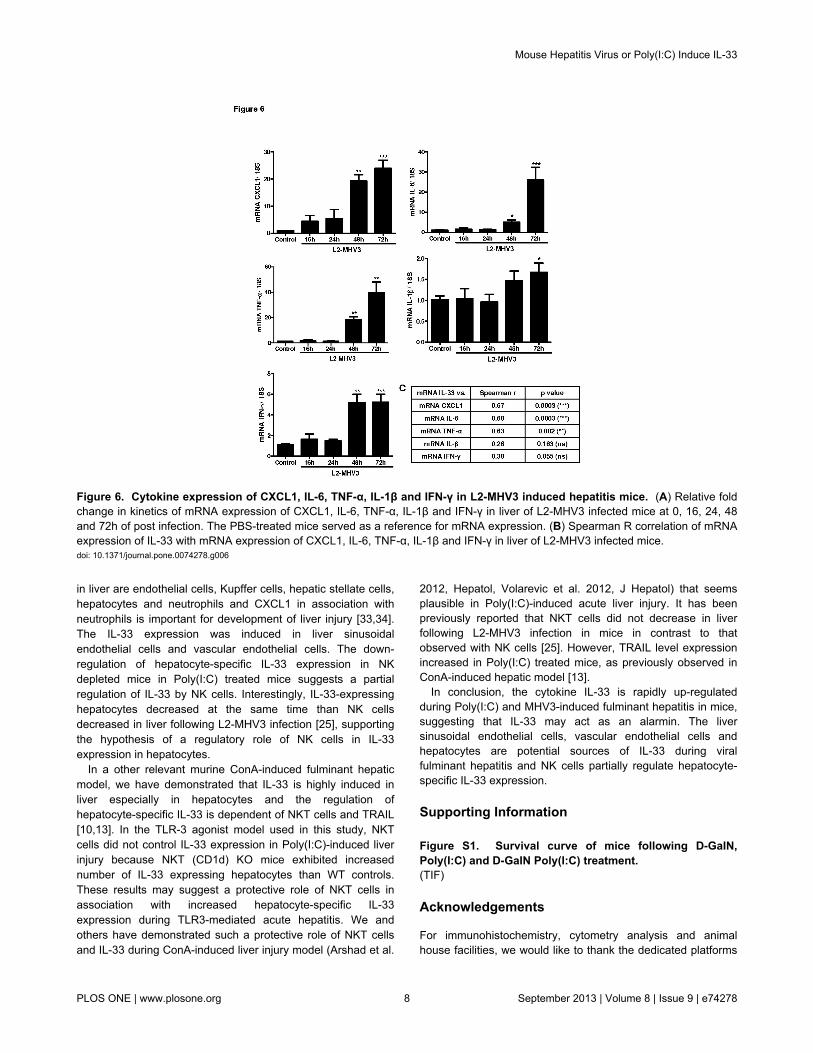
MHV3 infection in mice up-regulated pro-inflammatorycytokine expression in liver
The inflammatory cytokines play an important role indevelopment of fulminant hepatitis. Therefore, we studied theexpression of pro-inflammatory cytokine expression in liverafter L2-MHV3 infection. The kinetics of TNF-α, CXCL1, IFN-γand IL-6 showed a similar time dependent increasing trend withpeak expression at 72h of L2-MHV3 infection (Figure 6A). ThemRNA expression of IL-1β was not greatly increased followingL2-MHV3 hepatitis compared to control mice (Figure 6A). Asignificant correlation between mRNA expression of IL-33 andTNF-α, CXCL1 and IL-6 was evident but not with IL-1β or IFN-γ(Figure 6B). Hence, the elevated pro-inflammatory cytokinemicro-environment is important for development of L2-MHV3-induced acute hepatitis in mice.
Discussion
The over-expression of IL-33 and ST2 is associated withacute and chronic liver diseases in mice and human. IL-33 andsST2 have shown to be up-regulated in acute on chronic andchronic hepatic failure [15] and in chronic HBV and HCVinfections in human [14,16,17]. The cellular sources of IL-33 inviral fulminant hepatitis are not well known. Accordingly, themurine fulminant hepatic models of TLR3 agonist, Poly(I:C),and pathogenic mouse hepatitis virus (L2-MHV3) are relevantacute viral hepatic models in human. Thus, we aimed to knowthe expression and regulation of IL-33 in Poly(I:C) and L2-MHV3 induced hepatitis in mice. The Poly(I:C) administration inmice induced moderate hepatic injury while co-administrationof D-GalN and Poly(I:C) led to very severe fulminant hepatitisin mice. The liver injury induced by D-GalN Poly(I:C) treatmentwas associated with hemorrhagic lesions in liver and elevatedpro-inflammatory cytokines as reported earlier [19,21,22]. Theunderlying mechanism of Poly(I:C)-induced liver injury ismediated by activation of Kuppfer cells and NK cells in a TLR3dependent pathway [20] in association with increasedinflammatory cytokines. Inducible expression of IL-33 wasfound in liver sinusoidal endothelial cells and vascularendothelial cells following Poly(I:C) and D-GalN Poly(I:C)treatment. However, hepatocyte-specific IL-33 expression wasonly evident in Poly(I:C) treated mice with increased number ofIL-33 expressing hepatocytes compared to control mice. It maybe plausible that hepatocyte specific inhibition of transcriptionby D-GalN prevented hepatocyte-specific IL-33 expression attranscript and protein level following D-GalN Poly(I:C)treatment. However, innate immune stimulation by the TLR3agonist alone can induce IL-33 expression in liver especially inhepatocytes. The regulation of IL-33 by TLR viral and bacterialligands have been demonstrated in human corneal epithelial
Mouse Hepatitis Virus or Poly(I:C) Induce IL-33
PLOS ONE | www.plosone.org 5 September 2013 | Volume 8 | Issue 9 | e74278
cells and fibroblasts [30,31] as well as in murine macrophages[32]. The IL-33 was up-regulated by Poly(I:C) stimulation inmurine macrophages and its transcriptional regulation wasdependent of two transcription factors, IFN regulatory factor-3(IRF-3) and CREB [32]. Here, we add TLR3 mediatedexpression of IL-33 in liver sinusoidal endothelial cells, vascularendothelial cells and hepatocytes in acute hepatitis in mice in apathophysiological context. In addition, TLR3 expression isalso rapidly increased in liver from L2-MHV3 infected mice(results not shown).
While in a viral murine model, IL-33 has been shown to beexpressed by radio-resistant cells of the spleenic T cell zone inlymphocytic choriomeningitis virus (LCMV) infection [7], theabove data prompted us to compare the IL-33 expression in anatural viral infection in liver in using the serotype 3 of mousehepatitis viral infection MHV model [23,25]. We demonstrated
the cellular expression of IL-33 in sinusoidal and vascularendothelial cells and hepatocytes at various times of thefulminant viral hepatitis induced by L2-MHV3. The L2-MHV3-induced liver injury was associated with significant increases inserum transaminases ALAT/ASAT, viral nucleocapsid, IFN-βand pro-inflammatory cytokine and chimiokine expression. Theincreased CXCL1 during MHV3 induced acute hepatitis maylead to chemotaxis/infiltration of neutrophils as an earlyresponse to liver infection and development of inflammatorymicroenvironment. The mechanism of MHV3-induced liverinjury have shown to be dependent on activation of target cellsof virus like Kupffer cells, NK cells, hepatocytes, sinusoidalendothelial and vascular endothelial cells [26,27]. Here wehave shown that mRNA expression of IL-33 was over-expressed after L2-MHV3-induced hepatitis in mice. Thetranscript level of IL-33 was highly increased in L2-MHV3
Figure 3. Liver injury and cytokine expression of TNF-α, IL-1β, IL-6, TRAIL, CXCL1, IFN-γ and IL-33 in non NK depleted andNK depleted D-GalN, Poly(I:C), D-GalN Poly(I:C) treated mice. (A) Pre-depletion (48h before) of NK cells by anti-ASGM1antibody in mice and confirmation of NK cells percentage by flow cytometry analysis (CD3-FITC and NK1.1-PE markers) in Vehicle(control), Poly(I:C)-, NK-depleted poly(I-C)-, and NK-depleted D-GalN Poly(I:C)-treated mice (B) Levels of serum AST/ALT (IU/L)and relative fold change in mRNA expression of IL-33 in NK depleted or not with Poly(I:C) (30 µg/mouse i.v.) or D-GalN Poly(I:C)-treated mice at 8h of post injection. (C) Immunostaining of IL-33 in livers of NK depleted or not with Poly(I:C) (30 µg/mouse i.v.) orD-GalN Poly(I:C) treated mice at 8h of post injection. (D) Comparison of number of IL-33 expressing hepatocytes in PBS or Vehicle,Poly(I:C)-treated and NK-depleted Poly(I:C)-treated mice. (E) Relative fold change in mRNA expression of TNF-α, IL-1β, IL-6,TRAIL, CXCL1, and IFN-γ in livers of NK depleted or not with Poly(I:C) (30 µg/mouse i.v.) and/or D-GalN Poly(I:C) treated mice(C57Bl/6) at 8h of post injection.doi: 10.1371/journal.pone.0074278.g003
Mouse Hepatitis Virus or Poly(I:C) Induce IL-33
PLOS ONE | www.plosone.org 6 September 2013 | Volume 8 | Issue 9 | e74278

induced hepatitis than Poly(I:C) treated mice demonstrating adifference between TLR-3 agonist and natural virus infection inliver. TLR3 expression is also rapidly increased in liver from L2-MHV3 infected mice (results not shown), suggesting thatanother factor may be also involved in the increase of IL-33.The hepatocyte-specific expression of IL-33 in hepatocyteswas associated with beginning of L2-MHV3 induced liver injury(24, 28 and 32h) and the inducible expression of IL-33 in liversinusoidal endothelial cells and vascular endothelial cells wassustained during the whole infection period (16 to 72h).
We next studied the role of NK and NKT cells in Poly(I:C)induced IL-33 expression in liver. The depletion of NK cells byanti-AGSM1 antibody in mice did not inhibit increased liverinjury in D-GalN sensitized Poly(I:C) treated mice and had anyeffect in Poly(I:C) alone administration. These results are
Figure 4. Liver injury and IL-33 expression in WT and NKT(CD1d) KO mice following Poly(I:C) and D-GalN Poly(I:C)treatment. (A) Levels of serum AST/ALT (IU/L) in WT andCD1d KO mice following PBS, Poly(I:C) (30 µg/mouse i.v.)and/or D-GalN Poly(I:C) treatment at 8h of post injection. (B)Liver histology (H and E) and immunostaining of IL-33 in liversof CD1d KO mice treated with PBS, Poly(I:C) and/or D-GalNPoly(I:C). (C) Comparison of number of IL-33 expressinghepatocytes in WT and CD1d KO mice following PBS, Poly(I:C)and/or D-GalN Poly(I:C) treatment at 8h of post injection.doi: 10.1371/journal.pone.0074278.g004
contrary to earlier findings which showed that pre-depletion ofNK cells protected mice against D-GalN Poly(I:C) induced liverinjury [22]. The difference seemed to be related with kineticsand dose of D-GalN Poly(I:C) used in these studies. Thesignificant increase in CXCL1 expression in NK-depleted micemay explain the interplay of immune cells migration in liver i.e.depletion of one immune cell population relatively compensatethe other immune cell population. The major sources of CXCL1
Figure 5. Liver injury and IL-33 expression in L2-MHV3induced fulminant hepatitis in mice. (A) Levels of serumAST/ALT (IU/L) in mice infected with L2-MHV3 (103 TCID(50))or vehicle at 0, 16, 24, 48 and 72h of post infection. (B)Relative fold change in kinetics of mRNA expression of IFN-β,nucleocapsid of MHV3 and IL-33 in livers of L2-MHV3 inducedhepatitis. (C) Sections of mice liver following vehicle or L2-MHV3 infection (16, 24, 48 and 72h) were stained with H&E forhistopathology (arrows indicating zone of liver injury). (D)Immunolocalisation of IL-33 in livers of L2-MHV3 fulminanthepatic tissues by using primary antibody goat IgG anti-mouse-IL-33 and secondary HRP-conjugated rabbit anti-goat antibodywith hematoxylin counterstaining (black arrows and red arrowsindicating IL-33 positive hepatocytes and vascular/sinusoidalendothelial cells, respectively)). Scale bar was 50 µm. (E)Comparison of number of IL-33 expressing hepatocytes invehicle and L2-MHV3 fulminant hepatic tissues (16, 24 and32h).doi: 10.1371/journal.pone.0074278.g005
Mouse Hepatitis Virus or Poly(I:C) Induce IL-33
PLOS ONE | www.plosone.org 7 September 2013 | Volume 8 | Issue 9 | e74278
in liver are endothelial cells, Kupffer cells, hepatic stellate cells,hepatocytes and neutrophils and CXCL1 in association withneutrophils is important for development of liver injury [33,34].The IL-33 expression was induced in liver sinusoidalendothelial cells and vascular endothelial cells. The down-regulation of hepatocyte-specific IL-33 expression in NKdepleted mice in Poly(I:C) treated mice suggests a partialregulation of IL-33 by NK cells. Interestingly, IL-33-expressinghepatocytes decreased at the same time than NK cellsdecreased in liver following L2-MHV3 infection [25], supportingthe hypothesis of a regulatory role of NK cells in IL-33expression in hepatocytes.
In a other relevant murine ConA-induced fulminant hepaticmodel, we have demonstrated that IL-33 is highly induced inliver especially in hepatocytes and the regulation ofhepatocyte-specific IL-33 is dependent of NKT cells and TRAIL[10,13]. In the TLR-3 agonist model used in this study, NKTcells did not control IL-33 expression in Poly(I:C)-induced liverinjury because NKT (CD1d) KO mice exhibited increasednumber of IL-33 expressing hepatocytes than WT controls.These results may suggest a protective role of NKT cells inassociation with increased hepatocyte-specific IL-33expression during TLR3-mediated acute hepatitis. We andothers have demonstrated such a protective role of NKT cellsand IL-33 during ConA-induced liver injury model (Arshad et al.
2012, Hepatol, Volarevic et al. 2012, J Hepatol) that seemsplausible in Poly(I:C)-induced acute liver injury. It has beenpreviously reported that NKT cells did not decrease in liverfollowing L2-MHV3 infection in mice in contrast to thatobserved with NK cells [25]. However, TRAIL level expressionincreased in Poly(I:C) treated mice, as previously observed inConA-induced hepatic model [13].
In conclusion, the cytokine IL-33 is rapidly up-regulatedduring Poly(I:C) and MHV3-induced fulminant hepatitis in mice,suggesting that IL-33 may act as an alarmin. The liversinusoidal endothelial cells, vascular endothelial cells andhepatocytes are potential sources of IL-33 during viralfulminant hepatitis and NK cells partially regulate hepatocyte-specific IL-33 expression.
Supporting Information
Figure S1. Survival curve of mice following D-GaIN,Poly(I:C) and D-GaIN Poly(I:C) treatment.(TIF)
Acknowledgements
For immunohistochemistry, cytometry analysis and animalhouse facilities, we would like to thank the dedicated platforms
Figure 6. Cytokine expression of CXCL1, IL-6, TNF-α, IL-1β and IFN-γ in L2-MHV3 induced hepatitis mice. (A) Relative foldchange in kinetics of mRNA expression of CXCL1, IL-6, TNF-α, IL-1β and IFN-γ in liver of L2-MHV3 infected mice at 0, 16, 24, 48and 72h of post infection. The PBS-treated mice served as a reference for mRNA expression. (B) Spearman R correlation of mRNAexpression of IL-33 with mRNA expression of CXCL1, IL-6, TNF-α, IL-1β and IFN-γ in liver of L2-MHV3 infected mice.doi: 10.1371/journal.pone.0074278.g006
Mouse Hepatitis Virus or Poly(I:C) Induce IL-33
PLOS ONE | www.plosone.org 8 September 2013 | Volume 8 | Issue 9 | e74278

(i.e. H2P2, cytometry plateform and animal house platforms) ofSFR BIOSIT, University of Rennes 1, France. We also thankVanessa Thouault for technical assistance.
Author Contributions
Conceived and designed the experiments: MIA CPP LL MS.Performed the experiments: MIA SPD CPP AL MR CLC CB
MS VG. Analyzed the data: MIA CPP LM MS. Contributedreagents/materials/analysis tools: MIA SPD CPP AL MR CLCVG CB MS. Wrote the manuscript: MIA CPP LM MS.
References
1. Schmitz J, Owyang A, Oldham E, Song Y, Murphy E et al. (2005) IL-33,an interleukin-1-like cytokine that signals via the IL-1 receptor-relatedprotein ST2 and induces T helper type 2-associated cytokines.Immunity 23: 479-490. doi:10.1016/j.immuni.2005.09.015. PubMed:16286016.
2. Ali S, Huber M, Kollewe C, Bischoff SC, Falk W et al. (2007) IL-1receptor accessory protein is essential for IL-33-induced activation of Tlymphocytes and mast cells. Proc Natl Acad Sci U S A 104:18660-18665. doi:10.1073/pnas.0705939104. PubMed: 18003919.
3. Liew FY, Pitman NI, McInnes IB (2010) Disease-associated functions ofIL-33: the new kid in the IL-1 family. Nat Rev Immunol 10: 103-110. doi:10.1038/nri2692. PubMed: 20081870.
4. Liew FY (2012) IL-33: a Janus cytokine. Ann Rheum Dis 71 Suppl 2:i101-i104 doi:10.1136/annrheumdis-2011-200589. PubMed: 22460136.
5. Le Goffic R, Arshad MI, Rauch M, L’Helgoualc’h A, Delmas B et al.(2011) Infection with influenza virus induces IL-33 in murine lungs. AmJ Respir Cell Mol Biol 45: 1125-1132. doi:10.1165/rcmb.2010-0516OC.PubMed: 21642589.
6. Chang YJ, Kim HY, Albacker LA, Baumgarth N, McKenzie AN et al.(2011) Innate lymphoid cells mediate influenza-induced airway hyper-reactivity independently of adaptive immunity. Nat Immunol 12:631-638. doi:10.1038/ni.2045. PubMed: 21623379.
7. Bonilla WV, Fröhlich A, Senn K, Kallert S, Fernandez M et al. (2012)The alarmin interleukin-33 drives protective antiviral CD8(+) T cellresponses. Science 335: 984-989. doi:10.1126/science.1215418.PubMed: 22323740.
8. Becerra A, Warke RV, de Bosch N, Rothman AL, Bosch I (2008)Elevated levels of soluble ST2 protein in dengue virus infected patients.Cytokine 41: 114-120. doi:10.1016/j.cyto.2007.11.001. PubMed:18226917.
9. Miyagaki T, Sugaya M, Yokobayashi H, Kato T, Ohmatsu H et al.(2011) High levels of soluble ST2 and low levels of IL-33 in sera ofpatients with HIV infection. J Invest Dermatol 131: 794-796. doi:10.1038/jid.2010.366. PubMed: 21150924.
10. Arshad MI, Rauch M, L’Helgoualc’h A, Julia V, Leite-de-Moraes MC etal. (2011) NKT cells are required to induce high IL-33 expression inhepatocytes during ConA-induced acute hepatitis. Eur J Immunol 41:2341-2348. doi:10.1002/eji.201041332. PubMed: 21557213.
11. Arshad MI, Piquet-Pellorce C, Samson M (2012) IL-33 and HMGB1alarmins: sensors of cellular death and their involvement in liverpathology. Liver Int, 32: 1200–10. PubMed: 22530772.
12. Volarevic V, Mitrovic M, Milovanovic M, Zelen I, Nikolic I et al. (2012)Protective role of IL-33/ST2 axis in Con A-induced hepatitis. J Hepatol56: 26-33. doi:10.1016/S0168-8278(12)60072-5. PubMed: 21703183.
13. Arshad MI, Piquet-Pellorce C, L’Helgoualc’h A, Rauch M, Patrat-DelonS et al. (2012) Tumor Necrosis Factor Related Apoptosis InducingLigand (TRAIL), but Not FasL and Tumor Necrosis Factor alpha(TNFa), Regulates Interleukin (IL)-33 Expression in MurineHepatocytes During Acute Hepatitis. Hepatology 56: 2353-2362. doi:10.1002/hep.25893. PubMed: 22961755.
14. Marvie P, Lisbonne M, L’Helgoualc’h A, Rauch M, Turlin B et al. (2010)Interleukin-33 overexpression is associated with liver fibrosis in miceand humans. J Cell Mol Med 14: 1726-1739. PubMed: 19508382.
15. Roth GA, Zimmermann M, Lubsczyk BA, Pilz J, Faybik P et al. (2010)Up-regulation of interleukin 33 and soluble ST2 serum levels in liverfailure. J Surg Res 163: e79-e83. doi:10.1016/j.jss.2010.04.004.PubMed: 20638676.
16. Wang J, Zhao P, Guo H, Sun X, Jiang Z et al. (2012) Serum IL-33levels are associated with liver damage in patients with chronichepatitis C. Mediat Inflamm, 2012: 2012: 819636. PubMed: 22315510
17. Wang J, Cai Y, Ji H, Feng J, Ayana DA et al. (2012) Serum IL-33Levels Are Associated with Liver Damage in Patients with ChronicHepatitis B. J Interferon Cytokine Res 32: 248-253. doi:10.1089/jir.2011.0109. PubMed: 22304300.
18. Alexopoulou L, Holt AC, Medzhitov R, Flavell RA (2001) Recognition ofdouble-stranded RNA and activation of NF-kappaB by Toll-like receptor3. Nature 413: 732-738. doi:10.1038/35099560. PubMed: 11607032.
19. Dejager L, Libert C (2008) Tumor necrosis factor alpha mediates thelethal hepatotoxic effects of poly(I:C) in D-galactosamine-sensitizedmice. Cytokine 42: 55-61. doi:10.1016/j.cyto.2008.01.014. PubMed:18331798.
20. Cavanaugh PF Jr., Ho YK, Bardos TJ (1996) The activation of murinemacrophages and natural killer cells by the partially thiolated doublestranded RNA poly(I)-mercapto poly(C). Res Commun Mol PatholPharmacol 91: 131-147. PubMed: 8832906.
21. Schwabe RF, Seki E, Brenner DA (2006) Toll-like receptor signaling inthe liver. Gastroenterology 130: 1886-1900. doi:10.1053/j.gastro.2006.01.038. PubMed: 16697751.
22. Hou X, Zhou R, Wei H, Sun R, Tian Z (2009) NKG2D-retinoic acid earlyinducible-1 recognition between natural killer cells and Kupffer cells in anovel murine natural killer cell-dependent fulminant hepatitis.Hepatology 49: 940-949. doi:10.1002/hep.22725. PubMed: 19177594.
23. Lamontagne L, Descoteaux JP, Jolicoeur P (1989) Mouse hepatitisvirus 3 replication in T and B lymphocytes correlate with viralpathogenicity. J Immunol 142: 4458-4465. PubMed: 2542412.
24. Aparicio JL, Peña C, Retegui LA (2011) Autoimmune hepatitis-likedisease in C57BL/6 mice infected with mouse hepatitis virus A59. IntImmunopharmacol 11: 1591-1598. doi:10.1016/j.intimp.2011.05.020.PubMed: 21635973.
25. Jacques A, Bleau C, Martin JP, Lamontagne L (2008) Intrahepaticendothelial and Kupffer cells involved in immunosuppressive cytokinesand natural killer (NK)/NK T cell disorders in viral acute hepatitis. ClinExp Immunol 152: 298-310. doi:10.1111/j.1365-2249.2008.03628.x.PubMed: 18336588.
26. Godfraind C, Langreth SG, Cardellichio CB, Knobler R, Coutelier JP etal. (1995) Tissue and cellular distribution of an adhesion molecule inthe carcinoembryonic antigen family that serves as a receptor formouse hepatitis virus. Lab Invest 73: 615-627. PubMed: 7474935.
27. Lehoux M, Jacques A, Lusignan S, Lamontagne L (2004) Murine viralhepatitis involves NK cell depletion associated with virus-inducedapoptosis. Clin Exp Immunol 137: 41-51. doi:10.1111/j.1365-2249.2004.02501.x. PubMed: 15196242.
28. Jacques A, Bleau C, Turbide C, Beauchemin N, Lamontagne L (2009)Macrophage interleukin-6 and tumour necrosis factor-alpha are inducedby coronavirus fixation to Toll-like receptor 2/heparan sulphatereceptors but not carcinoembryonic cell adhesion antigen 1a.Immunology 128: e181-e192. doi:10.1111/j.1365-2567.2008.02946.x.PubMed: 19740307.
29. Lisbonne M, L’Helgoualc’h A, Nauwelaers G, Turlin B, Lucas C et al.(2011) Invariant natural killer T-cell-deficient mice display increasedCCl(4) -induced hepatitis associated with CXCL1 over-expression andneutrophil infiltration. Eur J Immunol 41: 1720-1732. doi:10.1002/eji.201041006. PubMed: 21469102.
30. Zhang L, Lu R, Zhao G, Pflugfelder SC, Li DQ (2011) TLR-mediatedinduction of pro-allergic cytokine IL-33 in ocular mucosal epithelium. IntJ Biochem Cell Biol 43: 1383-1391. doi:10.1016/j.biocel.2011.06.003.PubMed: 21684348.
31. Sponheim J, Pollheimer J, Olsen T, Balogh J, Hammarström C et al.(2010) Inflammatory bowel disease-associated interleukin-33 ispreferentially expressed in ulceration-associated myofibroblasts. Am JPathol 177: 2804-2815. doi:10.2353/ajpath.2010.100378. PubMed:21037074.
32. Polumuri SK, Jayakar GG, Shirey KA, Roberts ZJ, Perkins DJ et al.(2012) Transcriptional Regulation of Murine IL-33 by TLR and Non-TLRAgonists. J Immunol, 189: 50–60. PubMed: 22634618.
33. Stefanovic L, Brenner DA, Stefanovic B (2005) Direct hepatotoxic effectof KC chemokine in the liver without infiltration of neutrophils. Exp BiolMed (Maywood) 230: 573-586. PubMed: 16118408.
Mouse Hepatitis Virus or Poly(I:C) Induce IL-33
PLOS ONE | www.plosone.org 9 September 2013 | Volume 8 | Issue 9 | e74278

34. Patrick AL, Rullo J, Beaudin S, Liaw P, Fox-Robichaud AE (2007)Hepatic leukocyte recruitment in response to time-limited expression of
TNF-alpha and IL-1beta. Am J Physiol Gastrointest Liver Physiol 293:G663-G672. doi:10.1152/ajpgi.00070.2007. PubMed: 17656447.
Mouse Hepatitis Virus or Poly(I:C) Induce IL-33
PLOS ONE | www.plosone.org 10 September 2013 | Volume 8 | Issue 9 | e74278