iis oollaat tiio onn ioff nneeww … · modo controllato e accelerato, ciò che in natura assicura...
TRANSCRIPT

IISSOOLLAATTIIOONN OOFF NNEEWW TTHHEERRMMOOPPHHIILLEESS
FFRROOMM CCOOMMPPOOSSTT AANNDD TTHHEEIIRR
SSEELLEECCTTIIOONN FFOORR TTHHEE CCOONNVVEERRSSIIOONN OOFF
LLIIGGNNOOCCEELLLLUULLOOSSIICC BBIIOOMMAASSSS
Alessia Gioiello
Dottorato in Scienze Biotecnologiche – XXIX ciclo
Università di Napoli Federico II


Dottorato in Scienze Biotecnologiche – XXIX ciclo
Università di Napoli Federico II
IISSOOLLAATTIIOONN OOFF NNEEWW TTHHEERRMMOOPPHHIILLEESS
FFRROOMM CCOOMMPPOOSSTT AANNDD TTHHEEIIRR
SSEELLEECCTTIIOONN FFOORR TTHHEE CCOONNVVEERRSSIIOONN OOFF
LLIIGGNNOOCCEELLLLUULLOOSSIICC BBIIOOMMAASSSS
Alessia Gioiello
Dottorando: Alessia Gioiello
Relatore: Prof. Giovanni Sannia
Co-Relatore: Dott.ssa Annarita Poli
Coordinatore: Prof. Giovanni Sannia

Index Pag.
Abstract 1
Riassunto 2
Chapter 1 Introduction 8
1.0 Composting Process 9
1.1 Thermophilic microorganisms
11
1.2
Composting sites
12
1.3 Cellulase and Xylanase activities
14
1.4 Pectinase, Inulinase, Gellan-lyase, Pullulanase and Lipase activities
16
1.5 Lignocellulosic Biomass 18
1.6 Biorefinery Approach 19
1.7 State of the art and Future prospect
20
Chapter 2 Materials and Methods 21
2.0 Sampling 22
2.1 Culture-media 22
2.2 Isolation of strains after enrichment (A)
23
2.3 Isolation of strains on selective media (B)
24
2.4 Physiological tests of new isolates
24
2.5 Phenotypic characterization 25
2.5.1 Catalase test 25
2.5.2 Oxidase test 25
2.5.3 Tests alternative to Gram staining
25
2.5.4 Indole Test 26
2.5.5 Tyrosine degradation test 26
2.5.6 Urease Test 26
2.5.7 Hippurate hydrolysis test 26

2.5.8 Gelatin hydrolysis test 26
2.5.9 Voges-Proskauer Test 27
2.5.10 Nitrate and Nitrite reduction 27
2.5.11 Phenylalanine decomposition test
27
2.5.12 Protease test on agar plates 27
2.5.13 Antibiotics sensitivity 27
2.6 Study of exopolysaccharide production of strain N.8
28
2.7 Genotypic characterization 28
2.7.1 DNA extraction 28
2.7.2 Analysis of 16S rRNA gene sequence
29
2.7.3 DNA-DNA hybridization 29
2.7.4 Evaluation of DNA G+C content (%mol)
30
2.7.5 Phylogenetic tree 30
2.8 Chemotaxonomic study 30
2.8.1 Lipid analysis of Aeribacillus strains N.8 and N.6B
30
2.9 Screening of enzymatic activities on agar plates
31
2.10 Fractionation of cell components for CESCO strains
31
2.11 Determination of protein concentration
32
2.12 Xylanase activity assay 32
2.13 Reducing Sugars Assay 32
2.14 Screening of cellulase activity
33
2.15 Enzymatic assay of cellulase activity
33
2.16 Enzymatic assay of β-xylosidase, arabino-furanosidase and cellobio-hydrolase activity
33
2.17 Determination of protein molecular mass: Electrophoresis and
34
zymogram
2.18 Study of cellulase activity in strain N.3TH2
34
2.18.1 Temperature and pH Curves 34
2.18.2 Effect of different carbon sources on Cellulase production
34
2.18.3 Thermostability 35
2.18.4 Substrate specificity of Cellulase Activity
35
2.18.5 Cellulase activity at different incubation times
35
2.18.6 Analysis of enzymatic hydrolysis products
35
2.18.7 Ethanol assay 35
2.18.8 Effect of Organic Solvents on Cellulase Activity
36
2.18.9 Effect of Metal Ions and Enzyme Inhibitors on Cellulase Activity
36
2.19
Glucosidase and cellobio-hydrolase assay
36
2.20 Screening of other enzymatic activities at the “Bulgarian Academy of Sciences” (Sofia, BG)
36
2.20.1 Colorimetric test of pectinase on agar plates
37
2.20.2 Lipase qualitative test 37
2.20.3 Colorimetric test of Gellan-Lyase activity
37
2.20.4 Pectinase Activity assay 37
2.20.5 Inulinase Activity Assay 38
2.20.6 Pullulanase Activity Assay 38
2.20.7 Gellan-Lyase Activity Assay 39
2.20.8 Qualitative test of enzymatic activity using Azurine cross-linked substrates (AZCL)
39
Chapter 3 Results 40
3.0 Description of strains 41

isolated from CESCO
3.1 Description of strains isolated from DISSPA
45
3.2 Phylogenetic analysis of isolates
46
3.3 DNA-DNA hybridization analysis
48
3.4 Biochemical characterization of isolates
49
3.5 Chemotaxonomic study of Aeribacillus strains N.8 and N.6B
57
3.6 Chemotaxonomic study of Geobacillus strains N.3BX and N.3BC
61
3.7 Study of Xylanase activity of Geobacillus strains N.3BX and N.3BC
63
3.8 β-xylosidase and arabinofuranosidase activity of Geobacillus strains N.3BX and N.3BC
66
3.9 Cellulase activity of strain N.3TH2
71
3.10 Study of Extracellular Cellulase activity of strain N.3TH2
73
3.10.1 Effect of different carbon sources on Cellulase production of strain N.3TH2
73
3.10.2 Substrate specificity of Cellulase Activity
74
3.10.3 Thermostability 75
3.10.4 Cellulase activity at different times
75
3.10.5 Analysis of cellulase hydrolysis products
76
3.10.6 Effect of organic solvents 77
3.10.7 Effect of different metal ions and other agents on the enzyme activities
77
3.10.8 Ethanol production 78

3.11 Cellulase Activity of strain CV2-1
79
3.12 Thermobifida fusca 80
3.13 Screening of other enzymatic activities at the “Bulgarian Academy of Sciences” (Sofia, BG)
80
3.13.1 Qualitative test of enzymatic activity using Azurine cross-linked substrates (AZCL)
83
Chapter 4 Conclusion and Discussion 85
Acknowledgements 93
References List 94
Seminars and Courses 100
Activity-Conference Partecipations-Oral Comunications-Awards
100
Publications on scientific journals
101
Appendix 102
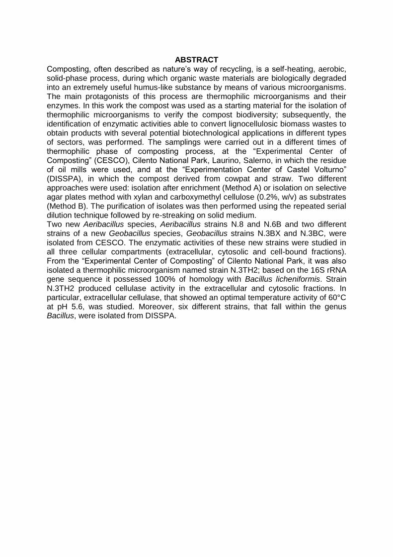
ABSTRACT
Composting, often described as nature‟s way of recycling, is a self-heating, aerobic, solid-phase process, during which organic waste materials are biologically degraded into an extremely useful humus-like substance by means of various microorganisms. The main protagonists of this process are thermophilic microorganisms and their enzymes. In this work the compost was used as a starting material for the isolation of thermophilic microorganisms to verify the compost biodiversity; subsequently, the identification of enzymatic activities able to convert lignocellulosic biomass wastes to obtain products with several potential biotechnological applications in different types of sectors, was performed. The samplings were carried out in a different times of thermophilic phase of composting process, at the “Experimental Center of Composting” (CESCO), Cilento National Park, Laurino, Salerno, in which the residue of oil mills were used, and at the “Experimentation Center of Castel Volturno” (DISSPA), in which the compost derived from cowpat and straw. Two different approaches were used: isolation after enrichment (Method A) or isolation on selective agar plates method with xylan and carboxymethyl cellulose (0.2%, w/v) as substrates (Method B). The purification of isolates was then performed using the repeated serial dilution technique followed by re-streaking on solid medium. Two new Aeribacillus species, Aeribacillus strains N.8 and N.6B and two different strains of a new Geobacillus species, Geobacillus strains N.3BX and N.3BC, were isolated from CESCO. The enzymatic activities of these new strains were studied in all three cellular compartments (extracellular, cytosolic and cell-bound fractions). From the “Experimental Center of Composting” of Cilento National Park, it was also isolated a thermophilic microorganism named strain N.3TH2; based on the 16S rRNA gene sequence it possessed 100% of homology with Bacillus licheniformis. Strain N.3TH2 produced cellulase activity in the extracellular and cytosolic fractions. In particular, extracellular cellulase, that showed an optimal temperature activity of 60°C at pH 5.6, was studied. Moreover, six different strains, that fall within the genus Bacillus, were isolated from DISSPA.

2
RIASSUNTO Il compost, rappresenta il prodotto finale di un processo, noto come compostaggio, in cui la materia organica di scarto è biologicamente degradata da diverse classi di microrganismi, tra cui batteri, funghi e attinomiceti. Il compostaggio riproduce in modo controllato e accelerato, ciò che in natura assicura il riciclaggio dei nutrienti con la formazione di un prodotto finale che risulta essere ricco di nutrienti ed igienicamente sicuro. Generalmente, i microrganismi mesofili, la cui temperatura ottimale di crescita varia dai 25 ai 45°C, iniziano il processo di compostaggio; utilizzando le fonti di carbonio presenti nella materia organica, producono CO2, rilasciandola nell‟ambiente. Per effetto dell‟aumento del metabolismo di questi microrganismi, la temperatura all‟interno dei cumuli di compost aumenta, dando spazio all‟azione dei microrganismi termofili, che agiscono a temperature superiori ai 50°C: i termofili degradano la sostanza organica più facilmente digeribile, consumando ossigeno e producendo anidride carbonica ed energia. In circa 72 ore le temperature aumentano da 50 a 70°C: questa rappresenta la fase “attiva” del compostaggio, che procede fin quando c‟è degradazione dei nutrienti da parte dei microrganismi presenti. Per diminuzione dell‟attività metabolica di questi microrganismi, la temperatura diminuisce fino ai 37°C e i mesofili ricolonizzano i cumuli di compost, entrando nella fase di “curing”. In questa fase, operata principalmente da funghi e attinomiceti, c‟è la degradazione della sostanza organica più complessa, con la produzione delle sostanze umiche. A tal punto, animali di piccole dimensioni, colonizzano il compost e, sminuzzando e rimescolando i prodotti organici ed inorganici, danno origine al compost maturo (Chen et al., 2011). I microrganismi termofili rappresentano i protagonisti della fase attiva del compostaggio, che avviene alle alte temperature e con un‟elevata richiesta di ossigeno. In particolare, in questo progetto di ricerca il compost ha rappresentato il punto di partenza per l‟isolamento di microrganismi termofili, studiandone in tal modo la biodiversità del compost stesso e focalizzando poi l‟attenzione sulle attività enzimatiche da essi prodotte, principalmente attività cellulolitiche ed emicellulolitiche, per la conversione di biomasse lignocellulosiche. Infatti, gli enzimi dei termofili, i termozimi, trovano numerose applicazioni in diversi processi biotecnologici e industriali, come nelle industrie alimentari, farmaceutiche, nei processi di sbiancamento della carta, nella produzione di biocarburanti e nella conversione di biomasse. I vantaggi di operare alle elevate temperature sono notevoli: al loro funzionamento è abbinato un aumento della velocità di reazione, alta solubilità dei substrati e una riduzione del rischio di contaminazione da parte di altri microrganismi. Inoltre, l'aumento di temperatura ha una notevole influenza sulla biodisponibilità e la solubilità dei composti organici ed è accompagnato da una diminuzione della viscosità e da un aumento del coefficiente di diffusione di composti organici (Bruins et al., 2001). Il campionamento del compost è stato effettuato in due siti di compostaggio, in diverse fasi del processo. In particolare, un primo campionamento è stato eseguito presso il “Centro Sperimentale di Compostaggio”, (CESCO), nel Parco Nazionale del Cilento, Laurino, Salerno, dove sono utilizzati residui derivanti dai frantoi oleari. In questo sito di compostaggio, il materiale di partenza è sottoposto a una fase di pre-trattamento, in cui vengono aggiunti sfalci di potatura e/o lana, al fine di ottenere un compost di qualità. I campioni sono stati raccolti in diversi stadi della fase attiva del processo di compostaggio e sono stati nominati come segue: -CC-3 (Strutturante legno vergine, T: 66.6°C);

3
-CC-5 (Bioreattore 15 giorni di incubazione, T: 66.4°C); -CC-6 (Ammendante compost misto finale, T: 62°C); -CC-8 (Curing 1 sotto telo, 30 giorni, T: 51.04°C); -CC-10 (Curing 2, 15 giorni, 69.84°C). Il secondo campionamento è stato effettuato presso il “Centro di Sperimentazione di Castel Volturno”, DISSPA, Università degli Studi di Napoli Federico II, in cui il compost deriva da sterco di mucca e legno. In tal caso sono stati effettuati due differenti prelievi: il primo (1CV) al 15° giorno del processo di compostaggio, quando la temperatura era di 43.6°C, il secondo (2CV) al 13° giorno del processo alla temperatura di 58.9°C. Con lo scopo di isolare microrganismi termofili dai campionamenti effettuati, sono stati utilizzati due differenti approcci: isolamento dopo arricchimento in mezzo liquido (Metodo A); oppure l‟isolamento è avvenuto su terreni selettivi con xilano e carbossimetil cellulosa (CMC) (0.2%), come substrati, per isolare direttamente le colonie con attività xilanasica e cellulasica, rispettivamente (Metodo B). In entrambi i casi colonie pure sono state ottenute mediante la tecnica delle diluizioni seriali seguita da successivi passaggi in piastra (Romano et al., 2004). In particolare dal “Centro Sperimentale di Compostaggio” (CESCO), sono stati isolati tre differenti ceppi nominati N.3TH1, N.8 e N.6B dopo arricchimento in mezzo liquido (Metodo A) dai campioni CC-3 (strutturante legno vergine), CC-8 (Curing 1 sotto telo, 30 giorni) e CC-6 (ammendante compostato misto finale), rispettivamente. Inoltre, dal campionamento CC-3 sono stati isolati altri 3 ceppi, nominati N.3TH2, N.3BX e N.3BC, su piastre contenenti xilano o CMC, come substrati (Metodo B). In particolare, il ceppo N.3TH2 produceva attività cellulasica, mentre i ceppi N.3BX e N.3BC mostravano attività xilanasica. Dal campionamento effettuato presso il “Centro di Sperimentazione di Castel Volturno” (DISSPA), sono stati isolati sei differenti ceppi, dal punto di vista morfologico, in seguito a selezione su piastra con xilano e carbossimetil cellulosa (0.2%, w/v), come substrati (Metodo B). In particolare, dal campione compost 1CV sono stati isolati i ceppi CV1-1 e CV1-2 che mostravano attività xilanasica, mentre dal campione compost 2CV sono stati isolati i ceppi CV2-1, CV2-2, CV2-3 e CV2-4, i quali producevano attività cellulasica, ma non xilanasica. Di ciascun ceppo isolato sono state individuate le condizioni di temperatura, pH e salinità ottimali per la loro crescita. In particolare, essi crescevano a temperature comprese tra i 50 e 60°C, ad eccezione dei microrganismi N.3BX e N.3BC, il cui optimum di temperatura era di 70°C. I ceppi mostravano una crescita ottimale a valori di pH compresi tra 7.0 e 9.0 e ad una concentrazione salina a valori di NaCl tra il 5 e il 7% (p/v). Gli isolati dal “Centro Sperimentale di Compostaggio” (CESCO), sono stati poi studiati dal punto di vista genetico; in particolare essi sono stati identificati attraverso l‟utilizzo dell‟EzTaxon-e server (http://www.ezbiocloud.net/eztaxon; Kim et al., 2012) sulla base delle sequenze del gene che codificava il 16S rRNA ottenuto dal servizio di sequenziamento “BMR Genomics Service”. In particolare, i ceppi N.3TH1, N.6B e N.8 sono risultati essere appartenenti al genere Aeribacillus e mostravano la più alta percentuale di omologia con Aeribacillus pallidus H12T DSM 3670 (99.8 %). Il genere Aeribacillus è stato proposto da Minãna-Galbis et al., (2010) quando Geobacillus pallidus (Scholz et al., 1988, Banat et al., 2004) è stato riclassificato nel nuovo genere come Aeribacillus pallidus.
4
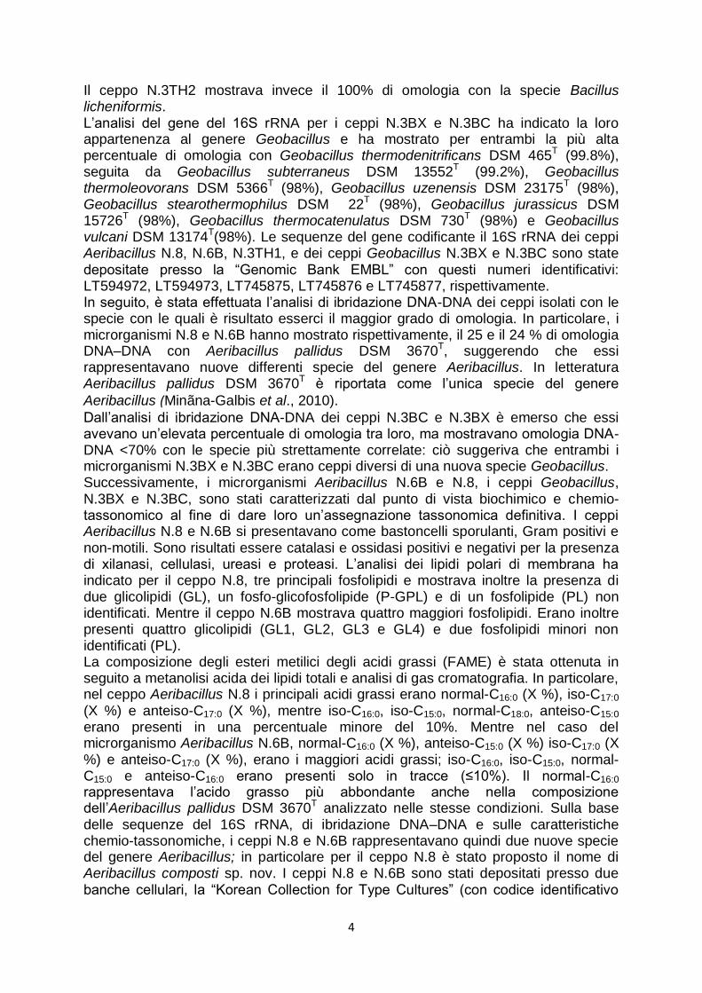
Il ceppo N.3TH2 mostrava invece il 100% di omologia con la specie Bacillus licheniformis. L‟analisi del gene del 16S rRNA per i ceppi N.3BX e N.3BC ha indicato la loro appartenenza al genere Geobacillus e ha mostrato per entrambi la più alta percentuale di omologia con Geobacillus thermodenitrificans DSM 465T (99.8%), seguita da Geobacillus subterraneus DSM 13552T (99.2%), Geobacillus thermoleovorans DSM 5366T (98%), Geobacillus uzenensis DSM 23175T (98%), Geobacillus stearothermophilus DSM 22T (98%), Geobacillus jurassicus DSM 15726T (98%), Geobacillus thermocatenulatus DSM 730T (98%) e Geobacillus vulcani DSM 13174T(98%). Le sequenze del gene codificante il 16S rRNA dei ceppi Aeribacillus N.8, N.6B, N.3TH1, e dei ceppi Geobacillus N.3BX e N.3BC sono state depositate presso la “Genomic Bank EMBL” con questi numeri identificativi: LT594972, LT594973, LT745875, LT745876 e LT745877, rispettivamente. In seguito, è stata effettuata l‟analisi di ibridazione DNA-DNA dei ceppi isolati con le specie con le quali è risultato esserci il maggior grado di omologia. In particolare, i microrganismi N.8 e N.6B hanno mostrato rispettivamente, il 25 e il 24 % di omologia DNA–DNA con Aeribacillus pallidus DSM 3670T, suggerendo che essi rappresentavano nuove differenti specie del genere Aeribacillus. In letteratura Aeribacillus pallidus DSM 3670T è riportata come l‟unica specie del genere
Aeribacillus (Minãna-Galbis et al., 2010).
Dall‟analisi di ibridazione DNA-DNA dei ceppi N.3BC e N.3BX è emerso che essi avevano un‟elevata percentuale di omologia tra loro, ma mostravano omologia DNA-DNA <70% con le specie più strettamente correlate: ciò suggeriva che entrambi i microrganismi N.3BX e N.3BC erano ceppi diversi di una nuova specie Geobacillus. Successivamente, i microrganismi Aeribacillus N.6B e N.8, i ceppi Geobacillus, N.3BX e N.3BC, sono stati caratterizzati dal punto di vista biochimico e chemio-tassonomico al fine di dare loro un‟assegnazione tassonomica definitiva. I ceppi Aeribacillus N.8 e N.6B si presentavano come bastoncelli sporulanti, Gram positivi e non-motili. Sono risultati essere catalasi e ossidasi positivi e negativi per la presenza di xilanasi, cellulasi, ureasi e proteasi. L‟analisi dei lipidi polari di membrana ha indicato per il ceppo N.8, tre principali fosfolipidi e mostrava inoltre la presenza di due glicolipidi (GL), un fosfo-glicofosfolipide (P-GPL) e di un fosfolipide (PL) non identificati. Mentre il ceppo N.6B mostrava quattro maggiori fosfolipidi. Erano inoltre presenti quattro glicolipidi (GL1, GL2, GL3 e GL4) e due fosfolipidi minori non identificati (PL). La composizione degli esteri metilici degli acidi grassi (FAME) è stata ottenuta in seguito a metanolisi acida dei lipidi totali e analisi di gas cromatografia. In particolare, nel ceppo Aeribacillus N.8 i principali acidi grassi erano normal-C16:0 (X %), iso-C17:0 (X %) e anteiso-C17:0 (X %), mentre iso-C16:0, iso-C15:0, normal-C18:0, anteiso-C15:0 erano presenti in una percentuale minore del 10%. Mentre nel caso del microrganismo Aeribacillus N.6B, normal-C16:0 (X %), anteiso-C15:0 (X %) iso-C17:0 (X %) e anteiso-C17:0 (X %), erano i maggiori acidi grassi; iso-C16:0, iso-C15:0, normal-C15:0 e anteiso-C16:0 erano presenti solo in tracce (≤10%). Il normal-C16:0 rappresentava l‟acido grasso più abbondante anche nella composizione dell‟Aeribacillus pallidus DSM 3670T analizzato nelle stesse condizioni. Sulla base delle sequenze del 16S rRNA, di ibridazione DNA–DNA e sulle caratteristiche chemio-tassonomiche, i ceppi N.8 e N.6B rappresentavano quindi due nuove specie del genere Aeribacillus; in particolare per il ceppo N.8 è stato proposto il nome di Aeribacillus composti sp. nov. I ceppi N.8 e N.6B sono stati depositati presso due banche cellulari, la “Korean Collection for Type Cultures” (con codice identificativo

5
KCTC 33824 e KCTC 33821, rispettivamente) e la “Japan Collection of Microorganisms” (con codice identificativo JCM 31580 e JCM 31579, rispettivamente). I ceppi Geobacillus N.3BX e N.3BC si presentavano come bastoncelli, non motili, Gram-positivi e formanti spore. Essi risultavano essere positivi alla presenza di catalasi e ossidasi, ma negativi alla presenza di ureasi, proteasi e alla formazione di indolo. Entrambi i ceppi erano capaci di idrolizzare lo xilano e l‟ippurato. L‟analisi dei lipidi polari di membrana ha indicato in entrambi i ceppi due principali lipidi polari:, descritti anche nelle specie tassonomicamente correlate come il G. subterraneus DSM 13552T e G. vulcani DSM 13174T. L‟analisi su TLC dei lipidi polari dei ceppi N.3BX e N.3BC, mostrava inoltre la presenza di un glicolipide (GL) e due fosfolipidi (PL) non identificati. L‟analisi spettroscopica dei lipidi neutri ha permesso di identificare il menachinone MK-7 come chinone respiratorio predominante in entrambi i ceppi confermando l‟MK-7 come marker tassonomico per le specie Geobacillus (Nazina et al., 2001). La composizione degli esteri metilici degli acidi grassi (FAME) è risultata essere per il ceppo N.3BX: iso-C17:0 (X %), iso-C15:0 (X %), anteiso-C17:0 (X%) e iso-C16:0 (X %) come maggiori acidi grassi e tracce di normal-C16:0 (X %), anteiso-C15:0 (X %), iso-C18:0 (X %), normal-C15:0 (X %), normal-C17:0 (X %) e normal-C18:0 (X %). Nel ceppo N.3BC i principali esteri metilici degli acidi grassi erano iso-C17:0 (X %), iso-C15:0 (X %), anteiso-C17:0 (X %) e iso-C16:0 (X %), mentre normal-C16:0 (X %), anteiso-C15:0 (X %), normal-C15:0 (X %), normal-C17:0 (X %), iso-C18:0 (X %) e normal-C18:0 (X %) erano presenti in tracce. In entrambi, erano presenti tracce di normal-C17:0, iso-C18:0 e normal-C18:0 che non erano presenti nella composizione degli acidi grassi della specie più strettamente correlata, Geobacillus thermodenitrificans DSM 465T analizzato nelle stesse condizioni. Effettuata l‟analisi genetica e la caratterizzazione biochimica e chemio-tassonomica dei nuovi isolati, l‟attenzione è stata posta sull‟individuazione di attività enzimatiche di interesse biotecnologico prodotte dai nuovi ceppi batterici, per la conversione di biomasse lignocellulosiche. In particolare, sono state studiate le attività enzimatiche nelle frazioni extracellulari e intracellulari prodotte dai ceppi Geobacillus N.3BX e N.3BC e dal ceppo Bacillus licheniformis N.3TH2. I ceppi N.3BX e N.3BC producevano attività xilanasica, citosolica ed extracellulare, con una temperatura ottimale tra 60 e 70°C, con un optimum a 70°C, con l‟eccezione della xilanasi extracellulare del ceppo N.3BC la cui temperatura ottimale era di 60°C, in un ampio range di pH a valori tra 4.0 e 9.0, con un optimum a pH 8.0. Nel ceppo N.3BC è stata individuata attività β-xilosidasica nel citosol e nella membrana, mentre il ceppo N.3BX produceva solo una β-xilosidasi citosolica. Le β-xilosidasi di entrambi i ceppi avevano una temperatura ottimale di 60°C in condizioni standard. I ceppi N.3BX e N.3BC producevano inoltre attività arabinofuranosidasica in tutti i compartimenti cellulari: in ogni caso la temperatura ottimale era di 70°C. La maggiore attività arabinofuranosidasica è stata riscontrata nella frazione extracellulare del ceppo N.3BC. I pesi molecolari degli enzimi sono stati determinati mediante elettroforesi su gel di poliacrilammide (SDS-PAGE) e zimogram. Il ceppo N.3TH2 mostrava invece attività cellulasica extracellulare e citosolica, con una temperatura ottimale di 60 e 70°C, rispettivamente; entrambe le cellulasi risultavano attive in un ampio intervallo di pH, con un optimum a valori intorno a 5.0-5.6. In particolar modo, è stata studiata l‟attività cellulasica extracellulare, in seguito a precipitazione con 80% ammonio solfato. La cellulasi extracellulare, mostrava un peso molecolare di circa 37 kDa e, risultava essere stabile per 10 minuti di incubazione, dimezzando l‟attività dopo un‟ora a 60°C. Tra i diversi substrati di

6
crescita testati (CMC, glucosio, cellobiosio), in presenza di glucosio registrava la maggiore produzione di cellulasi, mostrando un‟attività relativa (207%) circa il doppio rispetto alla crescita del microrganismo avvenuta in presenza di CMC (100%), a 60°C per 1h di incubazione. Il monitoraggio dell‟attività a diversi tempi di incubazione mostrava un incremento dai 30 minuti fino a circa 3h di incubazione, registrando poi un plateau fino alle 24h. La produzione di glucosio andava dai 0.85 mg/ml a 30 minuti di incubazione a 1.99 mg/ml dopo 3 ore di incubazione. I prodotti d‟idrolisi analizzati su TLC a diversi tempi d‟incubazione, erano cellobiosio e cellotriosio, facendo supporre che l‟enzima sia una “endocellulasi”. La cellulasi extracellulare del ceppo N.3TH2 ha mostrato un‟alta resistenza ai solventi organici, aumentando in alcuni casi l‟attività, come in presenza di benzene, toluene, n-decano, esa-decano, n-esano. Inoltre, è risultata essere stabile in presenza di diversi cationi e detergenti testati, essa potrebbe quindi rappresentare una candidata ideale per applicazioni industriali, in particolare nella formulazione dei detergenti (Ladeira et al., 2015). Per quanto riguarda i microrganismi isolati dai campionamenti effettuati presso il “Centro di Sperimentazione di Castel Volturno” (DISSPA), dall‟analisi del gene che codifica il 16S rRNA è emersa la loro appartenenza al genere Bacillus. In particolare i ceppi CV1-1 e CV1-2 hanno mostrato una percentuale di omologia pari al 100% con Bacillus thermodenitrificans, mentre i ceppi CV2-1, CV2-2, CV2-3 e CV2-4 avevano una percentuale di omologia pari al 100% con Bacillus licheniformis. Tutti i ceppi isolati si presentavano come piccoli bastoncelli quando cresciuti nel mezzo TSB alle loro temperature ottimali, in particolare, i ceppi CV2-1, CV2-2, CV2-3 e CV2-4 dopo 24 ore di incubazione mostravano un film cellulare sulla superficie del mezzo di coltura. Questi microrganismi producevano diverse attività enzimatiche, tra cui cellulasi, xilanasi, amilasi e proteasi. Infine, i ceppi Aeribacillus N.6B e N.8, isolati dai campionamenti CC-6 e CC-8, rispettivamente, del “Centro Sperimentale di Compostaggio” (CESCO) e i ceppi Bacillus CV2-1 e CV2-3, isolati dal campionamento 2CV del “Centro di Sperimentazione di Castel Volturno” (DISSPA), sono stati selezionati per effettuare lo studio di varie attività enzimatiche presso la “Bulgarian Academy of Sciences”, the Stephan Angeloff, Istituto di Microbiologia, Sofia (BG). In particolare, sono state utilizzate le frazioni extracellulari e gli omogenati cellulari di ciascun ceppo ottenuti in seguito ad una crescita di 24 ore nelle loro condizioni ottimali. Da questo studio, è emerso che tutti i microrganismi producevano attività gellan-liasica, sia nella frazione intracellulare che extracellulare, ad eccezione del ceppo N.8, il quale mostrava questa attività solo nella frazione intracellulare. Il ceppo N.6B mostrava inoltre attività pectinasica e inulinasica nella frazione intracellulare. I ceppi CV2-1 e CV2-3, mostravano oltre all‟attività gellan-liasica, anche attività pectinasica in entrambe le frazioni e producevano lipasi extracellulare solo se cresciuti in presenza di (1%, m/v) Tween 80 nel mezzo colturale, come substrato induttore. Inoltre, solo il ceppo CV2-3 mostrava attività inulinasica nella frazione intracellulare. I ceppi CV2-1 e CV2-3 risultavano essere positivi all‟idrolisi del collagene, in particolare l'attività collagenasica era maggiormente presente nella frazione extracellulare del ceppo CV2-1. In questo progetto di ricerca, il compost ha quindi rappresentato il punto di partenza per l‟isolamento di microrganismi termofili, in particolare sono stati isolati due nuovi ceppi appartenenti al genere Aeribacillus (N.8 e N.6B) e due nuovi differenti ceppi del genere Geobacillus (N.3BX e N.3BC). Mentre, alcuni dei microrganismi isolati avevano una percentuale di omologia pari al 100% con microrganismi già noti, come nel caso del ceppo N.3TH2 Bacillus licheniformis. Sono state successivamente
7
studiate le attività enzimatiche prodotte dai microrganismi isolati, in particolare le attività cellulolitiche ed emicellulotiche. Gli enzimi prodotti dai termofili isolati, in particolare xilanasi e cellulasi, potrebbero essere, infatti, utilizzati nella conversione di biomasse lignocellulosiche al fine di ottenere prodotti con potenziali applicazioni biotecnologiche. Difatti, dalla conversione delle biomasse lignocellulosiche è possibile ottenere monosaccaridi, ovvero zuccheri fermentabili per la produzione di biocarburanti ad esempio, e oligosaccaridi di cui potrebbero esserne studiate le proprietà fisiche, chimiche e biologiche dato le loro diverse applicazioni in campo biotecnologico.

8
Chapter 1
INTRODUCTION

9
1.0 Composting process Composting, often described as nature‟s way of recycling, is a self-heating, aerobic, solid-phase process, during which organic waste materials (for example food waste, manure, leaves, grass trimmings, paper, and coffee grounds, etc.) are biologically degraded into an extremely useful humus-like substance by means of various microorganisms including bacteria, fungi and actinomycetes. The product resulting from
this process is defined "compost" (from Latin compositum, consisting of more than one
substance) that stabilizes biologically numerous type of organic waste by converting them
into a final product rich in humus. The compost rich in nutrients and hygienically safe, is achieved reproducing in a controlled and accelerated way the processes that in nature
ensure the recycling of nutrients (Ecochem, An Earth Friendly Company). Human control of the biological decomposition process is what differentiates composting from the natural decomposition of organic matter, in fact regulating and optimizing conditions ensures a faster process and the generation of a quality end product. Once optimal physical conditions are established, microbes colonize the organic materials and initiate the composting process. Many of the microbes involved in decompositions are present in the wastes themselves. Soil microbes (such as bacteria, actinomycetes, fungi and protozoa) are introduced when the wastes are mixed with soil or inoculated with finished compost. Microorganisms use carbon compounds present in the organic materials as an energy source, transforming them into carbon dioxide (CO2), and releasing into the environment. As carbon compound is lost from the compost pile, the compost becomes more condensed and air spaces within the pile become smaller and the oxygen remaining in the pile is quickly consumed by the resident microorganisms (Chen et al., 2011). The composting process is carried out by different classes of microbes, such as mesophiles and thermophiles. Generally, mesophilic microorganisms, which function best between 30 and 50°C, initiate the compost process (Ecochem, An Earth Friendly
Company). As microbial activity increases soon after compost piles are formed, temperatures and density within the piles also increase and thermophilic microorganisms take over at temperatures above 50°C. The temperature in the compost pile typically increases rapidly from 50 to 70°C within 24 to 72 hours of pile formation, and can stay there for several days depending on feedstocks properties, pile size and environmental conditions. This represents the “active phase” of composting, during which decomposition is the most rapid. It continues until the materials containing nutrient and energy within the piles have been transformed. As microbial activity decrease, the pile compost temperature gradually declines approximately 37°C. Mesophilic microorganisms recolonize the pile, and the compost enters in the “curing phase”. The oxygen consumption during curing declines and organic materials continue to decompose and are converted to biologically stable humic substances that represent the mature or finished compost (Fig. 1). Potentially toxic organic acids and resistant compounds are also stabilized during curing. A long curing phase is needed if the compost is unfinished or immature and this is possible if the compost pile contained too little oxygen or either too little or too much moisture (Chen et al., 2011).

10
Figure 1. Temperature changes in the composting process (Adapted by Chen et al., 2011).
Therefore, the composting is a process, lasting from to 12/45 days, that evolves in three phases:
the first one, called thermophilic or bio-oxidation in which there is the degradation of the organic substance easier to degrade (such as sugars) by aerobic microorganisms that using O2, produce CO2 and energy. In this way occurs an increase of temperature up to 60°C determining the action of thermophilic microorganisms;
in the second one, the biological transformation of the most resistant organic
substances takes place. In particular, the most resistant polymers are degraded mainly by fungi and actinomycete activities, and only barely by bacteria. In this step the synthesis of humic substances starts by resulting in the typical smell of fresh soil;
finally in the last phase, there is the maturation of the compost itself. Several
animals of small size colonize the compost by contributing to the shredding and mixing of the organic and inorganic compounds.
The high temperatures of the first phase (between 50 and 70°C) result in faster breakdown of organic materials, kill pathogens and destroy weed seeds. However, excessively high temperatures (<70-75°C) can inhibit microbial activity. In particular, the main protagonists of the first phase, which occurs at high temperatures and with a high oxygen demand, are thermophilic microorganisms.

11
1.1 Thermophilic microorganisms Thermophiles are able to live and proliferate in environments with extreme physical (temperature, pressure, radiation) and geochemical parameters (salinity, pH, redox potential) (Dalmaso et al., 2015; Finore et al., 2016, Lama et al., 2012; Nicolaus et al., 2010). In particular thermophilic microorganisms are isolated in environments characterized by high temperatures and they grow more rapidly above 40°C (Stetter, 1999) with an optimum temperature between 60 and 80°C (Gul-Guven et al., 2008; Poli et al., 2006a, 2009, 2012). They are able to live, withstand and operate at high temperatures thanks to the production of biomolecules and particular structures, such as the structure of plasma membrane. A characteristic of thermophiles regards their phylogenetic position, which would suggest that they form part of the oldest life forms. By using 16S rDNA sequence comparison, an archaeal phylogenetic tree has been proposed, with a tripartite division of the living world consisting of the domains Eucarya, Bacteria, and Archaea (Andrade et al., 1999). In particular, thermophilic microorganisms belong both to the Archaea and Bacteria Domains (Fig. 2).
Figure 2. A new view of phylogenetic tree of life obtained by using ribosomial protein
sequences from each organism (Hug et al., 2016).
12
Thermophiles are found in various geothermally heated regions of the Earth, such as hot springs like those in Yellowstone National Park, in submarine volcanic areas, such as solfatara fields and in deep sea hydrothermal vents, as well as decaying
plant matter, such as compost (Stetter, 1999). Thermophilic microorganisms were also isolated in the thermophilic phase of composting belonging to the genera Thermus, Geobacillus and Bacillus (Blanc et al., 1999): Thermus thermophilus (Lyon et al., 2000), aerobic, Gram negative, with an optimum growth temperature of 70°C and characterized by an evident xylanolytic activity; Geobacillus toebii sp. nov. (Sung et al., 2002), aerobic, Gram positive staining with an optimum temperature of 60°C and characterized by a thermostable D-amino acid aminotransferases (Lee et al., 2006); Planifilum composti sp. nov. (Han et al., 2013), aerobic, Gram positive staining, isolated from compost in Korea with an optimum growth temperature of 55°C and Thermus composti sp. nov. (Vajna et al., 2012), aerobic, Gram negative staining, isolated from the thermophilic phase of the composting process for oyster mushroom substrate preparation, with an optimum temperature of 65–75°C. Further, two thermophilic microorganisms have been isolated and characterized from compost: Geobacillus toebii subsp. decanicus subsp. nov. (Poli et al., 2006b) Gram positive staining, with an optimum growth temperature of 65°C and Geobacillus galactosidasius (Poli et al., 2011), aerobic, Gram positive staining , with optimum growth temperature of 70°C and had α-galactosidase and α-glucosidase activities. They were isolated from “Pomigliano Ambiente” s.p.a. (Pomigliano, Naples, Italy) and from the “Experimental System of Composting” (Teora, Avellino, Italy), respectively, in which the compost derives from green waste and organic fraction of solid urban waste. 1.2 Composting sites In this research project the sampling was performed at two composting sites; the first one in the province of Salerno, “Experimental Center of Composting” (CESCO) in the Cilento National Park, Loc. Iscariello Laurino (SA), in which are used residues deriving from oil mils. Olive mill waste water, a by-product in olive oil manufacturing, results rich in biophenols, such as tyrosol and hydroxytyrosol, that are finding practical applications in the food, pharmaceutical, cosmetic and nutraceutical industries (Delisi et al., 2016). In particular, in this site recycling of waste water mills occurs through to the system developed by the project TIRSAV PLUS (INNOVATIVE TECHNOLOGIES FOR RECICLYNG OF OLIVE RESIDUES AND WASTE WATER OIL MILLS). The system methodological-productive of use of wastewater mills developed by TIRSAV PLUS is based on a composting process that is within same process is the vegetation water that olive-residue oil to produce quality compost (Fig. 3).

13
Figure 3. Example plan of technology system of composting at “Experimental Center of
Composting”(CESCO) in the Cilento National Park, Loc. Iscariello Laurino (SA).
To obtain these results, after a first pretreatment step, in which are added structuring/soil such as mowings of pruning and/or wool, the mixture obtained can follow two alternative lines of maturation:
- PCS (Passive Composting Simplified) that consists in a pretreatment phase, low maturation and secondary treatment. After the pretreatment the organic material is insert into plastic container in presence of air (Bins/Big bags) (Fig. 4A) (80 days); during this phase the aerobic bacteria convert the organic substance until a totally maturation (15-16 weeks). Following a refinement phase in order to reduce the volume of the organic substance (until 50%). Finally, the temperature arrives over to 55°C (more than 10 days) for the sanitation.
- ACC (Active Composting Composite) that has in common with PCS line the pretreatment phase, followed by active maturation and refinement. A mixture after pretreatment is inserted in the “Biocontainer” (Fig. 4B) in which a process of accelerated bio-oxidation (14-21 days) in presence of oxygen occurs. During this step, there is a microbial activity that causes an increase of temperature until 65°C (3 days). Subsequently the biocontainer are downloaded on reinforced concrete (30 days). When the maturation is completed, the temperature goes down to 25-35°C. The mature compost starts to refinement by rotary riddle and it is ready for marketing.
Figure 4. A) Bins/Big bags and and B) Bioreactor at “Experimental Center of Composting”(CESCO) in the Cilento National Park, Loc. Iscariello Laurino (SA).
14
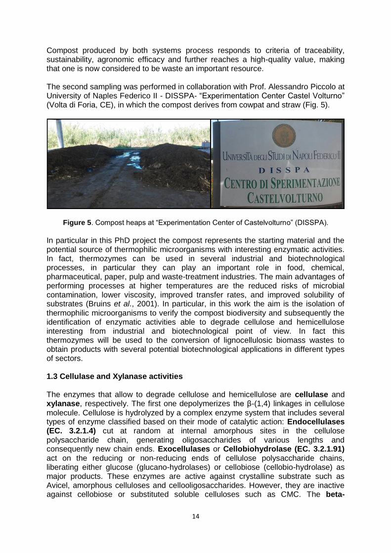
Compost produced by both systems process responds to criteria of traceability, sustainability, agronomic efficacy and further reaches a high-quality value, making that one is now considered to be waste an important resource. The second sampling was performed in collaboration with Prof. Alessandro Piccolo at University of Naples Federico II - DISSPA- “Experimentation Center Castel Volturno” (Volta di Foria, CE), in which the compost derives from cowpat and straw (Fig. 5).
Figure 5. Compost heaps at “Experimentation Center of Castelvolturno” (DISSPA).
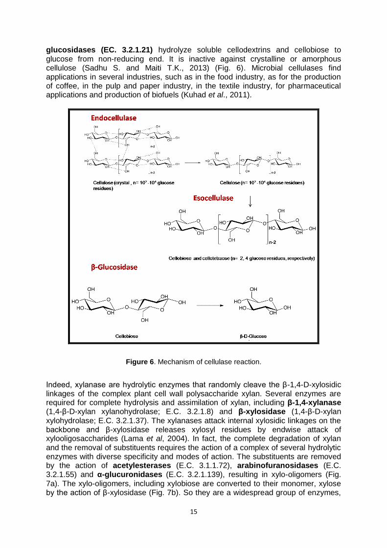
In particular in this PhD project the compost represents the starting material and the potential source of thermophilic microorganisms with interesting enzymatic activities. In fact, thermozymes can be used in several industrial and biotechnological processes, in particular they can play an important role in food, chemical, pharmaceutical, paper, pulp and waste-treatment industries. The main advantages of performing processes at higher temperatures are the reduced risks of microbial contamination, lower viscosity, improved transfer rates, and improved solubility of substrates (Bruins et al., 2001). In particular, in this work the aim is the isolation of thermophilic microorganisms to verify the compost biodiversity and subsequently the identification of enzymatic activities able to degrade cellulose and hemicellulose interesting from industrial and biotechnological point of view. In fact this thermozymes will be used to the conversion of lignocellulosic biomass wastes to obtain products with several potential biotechnological applications in different types of sectors. 1.3 Cellulase and Xylanase activities The enzymes that allow to degrade cellulose and hemicellulose are cellulase and xylanase, respectively. The first one depolymerizes the β-(1,4) linkages in cellulose molecule. Cellulose is hydrolyzed by a complex enzyme system that includes several types of enzyme classified based on their mode of catalytic action: Endocellulases (EC. 3.2.1.4) cut at random at internal amorphous sites in the cellulose polysaccharide chain, generating oligosaccharides of various lengths and consequently new chain ends. Exocellulases or Cellobiohydrolase (EC. 3.2.1.91) act on the reducing or non-reducing ends of cellulose polysaccharide chains, liberating either glucose (glucano-hydrolases) or cellobiose (cellobio-hydrolase) as major products. These enzymes are active against crystalline substrate such as Avicel, amorphous celluloses and cellooligosaccharides. However, they are inactive against cellobiose or substituted soluble celluloses such as CMC. The beta-
15
glucosidases (EC. 3.2.1.21) hydrolyze soluble cellodextrins and cellobiose to glucose from non-reducing end. It is inactive against crystalline or amorphous cellulose (Sadhu S. and Maiti T.K., 2013) (Fig. 6). Microbial cellulases find applications in several industries, such as in the food industry, as for the production of coffee, in the pulp and paper industry, in the textile industry, for pharmaceutical applications and production of biofuels (Kuhad et al., 2011).
Figure 6. Mechanism of cellulase reaction.
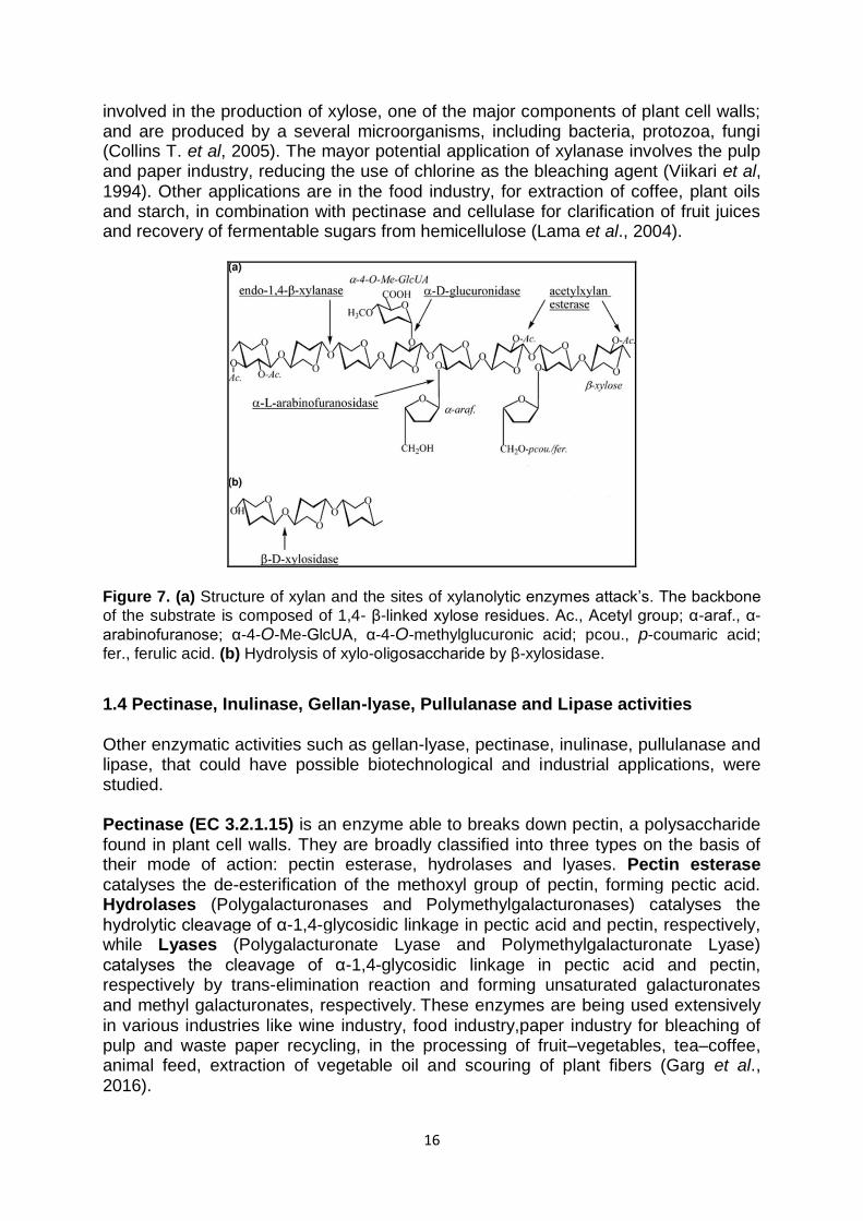
Indeed, xylanase are hydrolytic enzymes that randomly cleave the β-1,4-D-xylosidic linkages of the complex plant cell wall polysaccharide xylan. Several enzymes are required for complete hydrolysis and assimilation of xylan, including β-1,4-xylanase (1,4-β-D-xylan xylanohydrolase; E.C. 3.2.1.8) and β-xylosidase (1,4-β-D-xylan xylohydrolase; E.C. 3.2.1.37). The xylanases attack internal xylosidic linkages on the backbone and β-xylosidase releases xylosyl residues by endwise attack of xylooligosaccharides (Lama et al, 2004). In fact, the complete degradation of xylan and the removal of substituents requires the action of a complex of several hydrolytic enzymes with diverse specificity and modes of action. The substituents are removed by the action of acetylesterases (E.C. 3.1.1.72), arabinofuranosidases (E.C. 3.2.1.55) and α-glucuronidases (E.C. 3.2.1.139), resulting in xylo-oligomers (Fig. 7a). The xylo-oligomers, including xylobiose are converted to their monomer, xylose by the action of β-xylosidase (Fig. 7b). So they are a widespread group of enzymes,
16
involved in the production of xylose, one of the major components of plant cell walls; and are produced by a several microorganisms, including bacteria, protozoa, fungi (Collins T. et al, 2005). The mayor potential application of xylanase involves the pulp and paper industry, reducing the use of chlorine as the bleaching agent (Viikari et al, 1994). Other applications are in the food industry, for extraction of coffee, plant oils and starch, in combination with pectinase and cellulase for clarification of fruit juices and recovery of fermentable sugars from hemicellulose (Lama et al., 2004).
Figure 7. (a) Structure of xylan and the sites of xylanolytic enzymes attack‟s. The backbone
of the substrate is composed of 1,4- β-linked xylose residues. Ac., Acetyl group; α-araf., α-
arabinofuranose; α-4-O-Me-GlcUA, α-4-O-methylglucuronic acid; pcou., p-coumaric acid;
fer., ferulic acid. (b) Hydrolysis of xylo-oligosaccharide by β-xylosidase.
1.4 Pectinase, Inulinase, Gellan-lyase, Pullulanase and Lipase activities Other enzymatic activities such as gellan-lyase, pectinase, inulinase, pullulanase and lipase, that could have possible biotechnological and industrial applications, were studied. Pectinase (EC 3.2.1.15) is an enzyme able to breaks down pectin, a polysaccharide found in plant cell walls. They are broadly classified into three types on the basis of their mode of action: pectin esterase, hydrolases and lyases. Pectin esterase catalyses the de-esterification of the methoxyl group of pectin, forming pectic acid. Hydrolases (Polygalacturonases and Polymethylgalacturonases) catalyses the hydrolytic cleavage of α-1,4-glycosidic linkage in pectic acid and pectin, respectively, while Lyases (Polygalacturonate Lyase and Polymethylgalacturonate Lyase) catalyses the cleavage of α-1,4-glycosidic linkage in pectic acid and pectin, respectively by trans-elimination reaction and forming unsaturated galacturonates and methyl galacturonates, respectively. These enzymes are being used extensively in various industries like wine industry, food industry,paper industry for bleaching of pulp and waste paper recycling, in the processing of fruit–vegetables, tea–coffee, animal feed, extraction of vegetable oil and scouring of plant fibers (Garg et al., 2016).

17
Inulinase (EC 3.2.1.7) hydrolyze inulin, a plant reserve polysaccharide, into fructose and fructooligosaccharides which are widely used as food additives. Inulin represents a polyfructan consisting of linear chains β -(2,1)-linked fructose residues attached to a terminal sucrose molecule and having fructose as its major composition, it is suitable to be used as a substrate for fructo-oligosaccharides and high fructose syrup production for food, drink and pharmaceutical industries (Nirobol et al., 2012). Moreover, fructose is also used as a substrate for bioethanol fermentation as renewable energy source by Saccharomyces cerevisiae and Zymomonas mobilis (Flemming and Grootwassink, 1979). Gellan lyase (EC 4.2.2.25) is a type of enzyme able to degrade gellan, exopolysaccharide consisting of a linear repeating tetrasaccharide composed of D-glucose (Glc), D-glucuronic acid (GlcA), and L-rhamnose (Rha). Native gellan is so highly viscous that it is difficult to use in food and biopolymer-based industries. So the low-viscosity and low-molecular-mass gellan produced with gellan lyase could be represents a substrate with novel physiological and food technology functions (Hashimoto et al., 1996). Only one thermostable gellan lyase is known and it is produced by thermophilic strain isolated from Bulgarian hot spring (Derekova et al., 2006). Pullulanase (EC 3.2.2.41) is a specific kind of glucanase, an amylolytic extracellular
enzyme, that have specific action on α-1,6 linkages in pullulan, a linear α-glucan consisting essentially of maltotriosyl units connected by 1,6-α-bonds. Enzymes hydrolysing pullulan are classified into groups based on the substrate specificity and reaction products: Pullulanases type I, which are able to hydrolyse
efficiently the α-(1,6) glucosidic bonds in pullulan and branched polysaccharides and type II, also called amylopullulanases, are prominent in starch processing industry
due to the specific debranching capacity of hydrolysing either α-(1,6) or α-(1,4) glucosidic linkages. This enzyme debranch pullulan and gives maltotriose as final product and it also attacks α-(1,4) bonds in starch, amylose, and amylopectin.
Pullulanase is used in various industrial applications, such as in the saccharification of starch, production of High-maltose and fructose corn syrup and as effective additives in dish washing and laundry detergents (Hii et al., 2012). Lipases (EC 3.1.1.3) are a class of enzymes which catalyse the hydrolysis of long chain triglycerides and represent a subclass of esterases. Lipases constitute the most important group of biocatalysts for biotechnological and industrial applications such as in the detergent, food, flavour industry, biocatalytic resolution of pharmaceuticals, esters and amino acid derivatives, making of fine chemicals, agrochemicals, use as biosensor, bioremediation and cosmetics and perfumery (Hasan et al., 2006).

18
1.5 Lignocellulosic Biomass Lignocellulosic biomass include agricultural wastes, forestry residues, grasses and woody materials have great potential for bio-fuel production. They are mostly composed of three major units: cellulose, hemicellulose and lignin. Generally, most of the lignocellulosic biomass is comprised of about 40–50% cellulose, 25–30% hemicellulose, and 15–25% lignin with small amounts of other components, such as proteins, oils and ash (Isigkor and Becer, 2015) (Fig. 8).
Figure 8. Lignocellulosic biomass composition.
Cellulose represents one of the most abundant organic polymer found in nature and consisting of linear chains of 100-1400 of β-(1,4)-linked D-glucose units. It is a major structural component of plant cell walls, which is responsible for mechanical strength: cellulose molecules have a tendency to form hydrogen bonds between hydroxyl groups on the glucose from one chain with oxygen atoms on the same or on a neighbor chain, increasing the rigidity of cellulose and make highly insoluble and highly resistant to most organic solvents. Hemicellulose is the second most abundant heterogeneous polymers and it is often constituted by repeated polymers of pentoses and hexoses, such as xylose, arabinose, galactose and mannose, in different proportions depending on the tipe of biomass, but generally xylose is in most cases the sugar monomer present in the largest amount. Xylopyranose is the backbone of the polymer and connected with β-1,4 linkages. While cellulose is crystalline, strong, and resistant to hydrolysis, hemicellulose has a random, amorphous structure with little strength. Lignin is generally the most complex and

19
smallest fraction. It has a long-chain, heterogeneous polymer composed largely of phenyl-propane units most commonly linked by ether bonds. Lignin acts like a glue by filling the gap between and around the cellulose and hemicellulose complexion with the polymers (Anwar et al., 2014). 1.6 Biorefinery approach A valorization of enzymes produced by thermophiles lies in a “biorefinery” approach: indeed in the biorefinery, the wastes are not considered as material to be disposed of but can be used as a source of renewable energy and value added chemicals (Kamm and Kamm, 2004). In this perspective, the enzymes produced by the new thermophilic microorganisms will be used to degrade lignocellulosic biomass obtaining monosaccharides and oligosaccharides. The first one, since they are fermentable sugars, could be used for the production of biofuels, while the oligosaccharides could be investigated for their chemical and physical properties and biological activity and subsequently employed for potential biotechnological applications. Moreover, by using enzyme cocktails, efforts will be done to improve the conversion of lignocellulosic biomass into mono-and oligosaccharides. In fact, the hydrolysis products represent renewable source of value added compounds that are the basis for the production of a wide spectrum of chemicals that in turn can be used for several purposes in the production of drugs, food additives and biomaterials (Fig. 9).
Figure 9. Example of use of thermophilic enzymes in the conversion of lignocellulosic biomass. Adapted from Strassberger et al., 2014.

20
1.7 State of the art and Future perspective
The increased attention to environmental problems closely related to human health, including the aim to reduce the rising amount of waste in developed societies and the use of nonrenewable materials, have encouraged the use of compost (Grigatti et al. 2007). Today, the use of composting to turn organic wastes into a valuable resources is expanding rapidly in many countries; in fact many types of organic waste can be turned into compost contributing to the sustainable use of resources. Therefore, the compost production doesn‟t satisfy the growing demand, both for lack of facilities, and for the difficulty of producing high quality compost. The use of high quality compost allows a return of organic matter to the soil by restoring fertility at medium and long term that can‟t be obtained in any other way. The characteristics of the waste influence the performance of the composting process, in fact organic wastes are produced from many sources such as agricultural waste, market waste, kitchen waste, urban solid food wastes and municipal solid waste (Kadir et al., 2016). Generally, on the basis of the organic wastes used, the final compost could have many different useful applications, such as soil amendment, fertilization, restoration, landfill covering, landscape gardening and plant disease suppression (García de la Fuente et al., 2006). In particular, compost is usually used as fertilizer in place of chemical ones for the high content of nutrients (nitrogen, phosphorus, calcium and potassium) (Becker et al., 2010), but on the other hand the potential use of compost as source of thermophilic bacteria is also known. Recently, several thermophilic bacteria present in the compost were isolated and characterized such as, Thermus thermophilus (Lyon et al., 2000), Geobacillus toebii sp. nov. (Sung et al., 2002), Planifilum composti sp. nov. (Han et al., 2013), Geobacillus thermodenitrificans and Aneurinibacillus thermoaerophilus (Charbonneau et al., 2012). Moreover, thermophilic bacteria isolated from compost able to convert lignocellulosic biomass to obtain mono and oligosaccharides, have been reported; in particular, they belonged to Bacillus species, such as B. amyloliquefaciens and B. licheniformis, isolated from raw composting materials. The study of cellulolytic activity of these microorganisms was performed highlighting their potential for cellulose conversion and subsequently, for second generation bioethanol production that represents one of the best alternatives to the fossil fuels (Amore et al., 2012). In this project, following the isolation of thermophilic bacteria from compost, it is expected the use of the enzymes deriving from new microorganisms isolates in the bioconversion reactions for the degradation of lignocellulosic biomass. In particular, it would be also possible concomitant use of more enzymes deriving from different microorganisms isolated in order to increase mono and oligosaccharide yields.

21
Chapter 2
MATERIALS AND METHODS
22
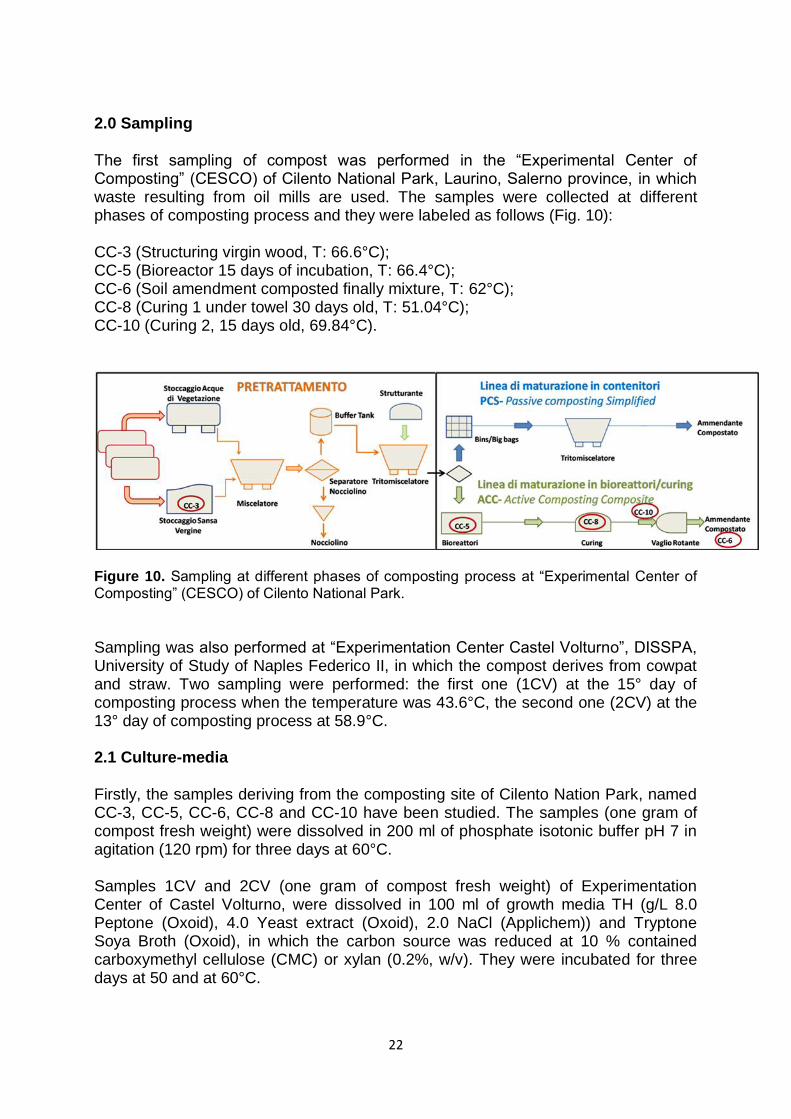
2.0 Sampling The first sampling of compost was performed in the “Experimental Center of Composting” (CESCO) of Cilento National Park, Laurino, Salerno province, in which waste resulting from oil mills are used. The samples were collected at different phases of composting process and they were labeled as follows (Fig. 10): CC-3 (Structuring virgin wood, T: 66.6°C); CC-5 (Bioreactor 15 days of incubation, T: 66.4°C); CC-6 (Soil amendment composted finally mixture, T: 62°C); CC-8 (Curing 1 under towel 30 days old, T: 51.04°C); CC-10 (Curing 2, 15 days old, 69.84°C).
Figure 10. Sampling at different phases of composting process at “Experimental Center of Composting” (CESCO) of Cilento National Park.
Sampling was also performed at “Experimentation Center Castel Volturno”, DISSPA, University of Study of Naples Federico II, in which the compost derives from cowpat and straw. Two sampling were performed: the first one (1CV) at the 15° day of composting process when the temperature was 43.6°C, the second one (2CV) at the 13° day of composting process at 58.9°C. 2.1 Culture-media Firstly, the samples deriving from the composting site of Cilento Nation Park, named CC-3, CC-5, CC-6, CC-8 and CC-10 have been studied. The samples (one gram of compost fresh weight) were dissolved in 200 ml of phosphate isotonic buffer pH 7 in agitation (120 rpm) for three days at 60°C. Samples 1CV and 2CV (one gram of compost fresh weight) of Experimentation Center of Castel Volturno, were dissolved in 100 ml of growth media TH (g/L 8.0 Peptone (Oxoid), 4.0 Yeast extract (Oxoid), 2.0 NaCl (Applichem)) and Tryptone Soya Broth (Oxoid), in which the carbon source was reduced at 10 % contained carboxymethyl cellulose (CMC) or xylan (0.2%, w/v). They were incubated for three days at 50 and at 60°C.

23
10 ml of the suspensions of both composting sites, obtained for all samples, that represent the “First enrichment”, have been used as inocula in flasks with 100 ml of different media and were incubated for 48 hours at 60 and 70°C. The following media were used: TH (g/L): 8.0 Peptone (Oxoid), 4.0 Yeast extract (Oxoid), 2.0 NaCl (Applichem); after melting of chemicals, the pH value was adjusted to 7.2 with NaOH 1 M. Bactomarine (g/L): 5.0 Peptone (Oxoid), 1.0 Yeast extract (Oxoid), 19.45 NaCl (Applichem), 8.8 MgCl2 (Applichem), 3.24 Na2SO4 (Applichem), 1.8 CaCl2 (J.T.Baker), 0.5 ml/L Ferric citrate (10gr/L) (Carlo Erba), 10 ml/L Solution A, 10 ml/L Solution B, 10 ml/L Solution C, 10 ml/L Solution D. Solution A: 55.0 g/L KCl (J.T.Baker); Solution B: 3.4 g/L SrCl2 (J.T. Baker), 2.2 g/L H3BO3 (Merk), 8.0 g/L KBr (Sigma); Solution C: NaHCO3 (Applichem) 16.0 g/L; Solution D: 0.16 g/L NH3NO3 (Sigma), NaHPO4 0.8 g/L (Applichem). Initial pH was adjusted to 7.2 with NaOH 1 M. YN (g/L) 6.0 Yeast extract, 6.0 NaCl at pH 5.6; M162 (g/L): 4.0 NaCl (Applichem), 0.6 Yeast extract (Oxoid), 0.53 NH4Cl (J.T.Baker), Solution A, 60 ml/L, Solution B, 20 ml/L, Solution C, 100 ml/L.
Solution A: 35.58 g/L Na₂HPO₄ x 2H₂O (Applichem); Solution B: 27.19 g/L KH₂PO₄ (Carlo Erba); Solution C: 1.0 g/L Acetic Acid nitrile (Applichem), 5.0 ml/L Nitsch‟S trace elements (Nitsch&Nitsch, 1956), 0.01 M Ferric citrate (Carlo Erba), 0.4 g/L
CaSO₂ x 6H₂O (Carlo Erba), 2.0 g/L MgCl₂ x 6H₂O (Applichem); TSB (Oxoid) (g/L): Pancreatic digest of casein 17.0, Papaic digest of soybean meal 3.0, NaCl 5.0.; Di-basic potassium phosphate 2.5, glucose 2.5; Nutrient Broth (Oxoid) (g/L): “Lab-Lemco” Powder 1.0, Yeast extract 2.0, Peptone 5.0, NaCl 5.0. The solid media were prepared with agar (Oxoid) (2% w/v). In order to select microorganisms with specific enzymatic activity, xylan (Birchwood) or carboxymethyl cellulose (CMC) (Oxoid), were added in the preparation of solid growth media. All growth media were sterilized at 121°C for 20 min. The occurred growth was monitored by spectrophotometric lecture to λ 540 nm (BECKMAN COULTER DV 730 Life Science UV/Vis). 2.2 Isolation of strains after enrichment (A) The occurred growth in liquid media for 48 hours of incubation at 60 and 70°C, was used as inoculum for the same solid medium obtained by adding agar 2.0 % (w/v), for 48 hours at 60 and 70°C. Bacterial strains that grew vigorously were selected and transferred into the corresponding liquid media. They were purified using the repeated serial dilution technique followed by re-streaking on solid medium (Romano et al., 2004). The purity of isolates was examined based on cell shape under a microscope and colony homogeneity on the plates. Cellular morphology and motility were determined by phase-contrast microscopy (Zeiss) and colony morphology was determined with a Leica M8 stereomicroscope using cultures grown on agar plates

24
for 24 h at the optimal temperature. In particular, the strains named N.3TH1, N.8 and N.6B were isolated from the sample compost CC-3, CC-6B and CC-8 of “Experimental Center of Composting” (CESCO) (Table 1). 2.3 Isolation of strains on selective agar plates (B) The occurred growth in liquid media for 48 hours of incubation at 60 and 70°C, was used as inoculum for the same solid medium with xylan or CMC (0.2%, w/v), as substrates, for 48 hours at 60 and 70°C. Bacterial strains that grew vigorously and showed the presence of xylanase or cellulase when tested on agar plates with xylan or CMC (0.2%, w/v), were selected and transferred into the corresponding liquid media. They were purified using the repeated serial dilution technique followed by re-streaking on solid medium (Romano et al., 2004). The purity of isolates was examined based on cell shape under a microscope and colony homogeneity on the plates. Cellular morphology and motility were determined by phase-contrast microscopy (Zeiss) and colony morphology was determined with a Leica M8 stereomicroscope using cultures grown on agar plates for 24 h at the optimal temperature. On the basis of enzymatic activities the strains named N.3TH2, N.3BX and N.3BC were isolated from the sample compost CC3 (Experimental Center of Composting, CESCO), the strains named CV1-1 and CV1-2 were isolated from the sample compost 1CV (Experimentation Center of Castel Volturno) and the strains named CV2-1, C2-2, CV2-3 and CV2-4 were isolated from the sample compost 2CV (Experimentation Center of Castel Volturno) (Table 1).
Table 1. Strains isolated using Method A or B and their corresponding composting site of
isolation.
Strains Isolation Method Compost sample
N.3TH1 A CC-3 N.3TH2 B CC-3
N.8 A CC-8 N.6B A CC-6
N.3BX A CC-3 N.3BC A CC-3
CV1-1 B 1CV CV1-2 B 1CV
CV2-1 B 2CV CV2-2 B 2CV
CV2-3 B 2CV CV2-4 B 2CV
2.4 Physiological tests of new isolates In order to determine the optimal conditions of growth of the new isolates, several studies were performed. In particular, the temperature range for growth was determined by incubating the isolates in the temperature interval of 45-80°C. The pH range was, indeed, determined verifying the growth of each microorganism using optimal growth medium buffered for pH values ranging from 5 to 10. Salinity tests were carried out varying the concentration of NaCl in the growth medium from 0 to 20 %. For the strains N.8 and N.6B the ionic and osmotic sensitivity and the optimal pH value for growth were also studied using the BIOLOG Phenotype MicroArrayTM
25
plates PM9 and PM10, respectively, prepared as recommended by the manufacturer‟s instructions. Carbon and nitrogen sources usage of strains N.8 and N.6B was evaluated using the BIOLOG Phenotype MicroArrayTM plates PM1, PM2a and PM3 (Biolog, Inc., Hayward, California, USA). The cellular growth was monitored by spectrophotometric lecture to λ 540 nm. 2.5 Phenotypic characterization The strains isolated from sample compost of “Experimental Center of Composting” (CESCO), named N.6B, N.8, N.3BX and N.3BC were selected to perform several biochemical and phenotypic tests to give them a definitive taxonomic assignment. 2.5.1 Catalase test 5 ml of liquid bacterial growth after an incubation of 24 h at optimal growth conditions, were mixed with 3% (w/v) hydrogen peroxide solution. The rapid elaboration of oxygen bubbles occurs in the presence of catalase. 2.5.2 Oxidase test The presence of oxidase enzyme was assayed with Kovacs Oxidase Reagent (1% tetra-methyl-p-phenylenediaminedihydrochloride, in water). The test was performed using a filter paper in a Petri dish and adding 3 drops of freshly prepared oxidase reagent. Using a sterile glass rod, a colony of tested microorganisms collected from a culture plate was smeared on the filter paper containing Kovacs Reagent. Oxidase positive microorganisms gave blue color within 5-10 seconds, and in oxidase negative organisms, color did not change. 2.5.3 Tests alternative to Gram staining KOH string test In alternative to Gram stain, a rapid non staining method was used. With a sterile loop, a visible amount of bacterial growth was transferred from an agar plate to the glass slide containing a drop of 3% (w/v) KOH. If the bacteria-KOH suspension became markedly viscid or gels within 5 to 60 s, the isolate was Gram negative. Bactident Aminopeptidase In order to determine the presence of the L-alanine aminopeptidase a growth of 24 h on agar plate was suspended in 2 ml of distilled water in glass test tube. The aminopeptidase test strip (MikrobiologieBactident aminopeptidase, Merck, Germany) was inserted into the test tube containing the bacteria suspension and incubated at 37°C for 10 minutes. A clear yellow coloration of the bacteria suspension could be seen after only 10 minutes in the case of most aminopeptidase-positive microorganisms.

26
2.5.4 Indole Test In order to determine the ability of the organism to convert tryptophan into the indole, the growth of bacteria was perfomed in “Tryptophan broth” ((w/v) 10 g/L Peptone, 1% (w/v) tryptophan and with an addition of NaCl optimal for each strain). After 24 or 48 hours of incubation 5 drops of Kovacs reagent (150 ml Butanol, 50 ml hydrochloric acid and 10 g of 4 (p)-dimethylaminobenzaldehyde were added. The 4 (p)-dimethylaminobenzaldehyde reacts with indole present in the medium to form a red- rose dye, so a positive result is shown by the presence of a red or red-violet color in the surface alcohol layer of the broth, while a negative result appears yellow. 2.5.5 Tyrosine degradation test In order to evaluate the ability of the strains to degrade tyrosine, the growth was performed at the standard conditions in agar media with 5 g/L (w/v) of tyrosine. A blank was represented by the medium without inoculum. After 1, 3, 5 and 10 days of incubation the dark medium staining indicated that hydrolysis of tyrosine occurred and so the positivity reaction. The blank showed no staining. 2.5.6 Urease Test In order to evaluate the presence of enzyme urease, the growth of bacteria was performed in “Urea Broth”, constitute so follows (g/L) (w/v): 9.1 KH2PO4, 9.5 Na2HPO4, 0.1 yeast extract, 0.01 phenol red. The inoculum of strains in the standard culture medium represented the blank. Urea is broken down by urease into carbon dioxide and ammonia; ammonia turns the medium alkaline and phenol red changes from yellow to red/pink as the pH increases. After 24 or 48 hours of incubation at optimal temperature for each strain, a color change of medium indicated the urea digestion. 2.5.7 Hippurate hydrolysis test The presence of hippuricase was evaluated performing the growth of each strain in standard media with 10 g/L (w/v) sodium hippurate. The inoculum of strains in the standard culture medium without sodium hippurate represented the blank. After 5 and 10 days of incubation 1.5 ml of 50% H2SO4 were added. The formation of insoluble crystals (for the release of benzoic acid) indicated that the hydrolysis occurred (positivity reaction). 2.5.8 Gelatin hydrolysis test In order to detect the ability to produce gelatinase that liquefy gelatin, the bacteria were inoculated on the tube containing nutrient gelatin (120 g/L, w/v) medium and incubated at their optimal temperature. After incubation the tubes were placed at 4°C for 15-30 minutes (until control is gelled) to check for gelatin liquefaction. In fact gelatin normally liquefies at 28°C and above; to confirm that liquefaction was due to gelatinase activity, the tubes were placed at 4°C. The liquefaction of gelatin indicates that hydrolysis occurred suggesting the presence of the gelatinase enzyme.

27
2.5.9 Voges-Proskauer Test Voges-Proskauer is a test used to detect acetoin in a bacterial broth culture. The test was performed after 1 and 3 days of incubation of each strain at their optimal temperature in the Voges-Proskauer broth (g/L) (100.0 NaCl, 7.0 pepton, 5.0 glucose, 5.0 KH2PO4) by adding α-naphthol (2.2 g α-naphtol in 10.5 ml of EtOH.
Slowly were added 6.5 ml of H₂SO₄, 40.5 ml EtOH and 4 ml of H₂O) and potassium hydroxide. A cherry red color indicates a positive result, while a yellow-brown color indicates a negative result. 2.5.10 Nitrate and Nitrite reduction For nitrate and nitrite reduction, optimal medium of each strain containing 0.1 % (w/v) KNO3 (Media A) or 0.001% (w/v) NaNO2 (Media B) were used, respectively. After 1 and 3 days of incubation at respective optimal growth temperature, 1 ml of Reactive A (0.8% (w/v) sulfanilic acid in 5N acetic acid 30%) and 1 ml of Reactive B (0.6% (w/v) α-naphthylamine in 5N acetic acid 30%), were added. If the microorganism reduced nitrate to nitrite there was a formation of nitrous acid, that reacting with sulfanilic acid produced diazotized sulfanilic acid. This latter reacted with α-naphthylamine to form a red-colored compound. A color change to red in the Media A indicated a positive nitrate reduction test, while if no red color forms, there was no nitrate to reduce. While, in the Media B the color change to yellow indicated a positive nitrite reduction test, while if no yellow color forms, there was no nitrite to reduce.
2.5.11 Phenylalanine decomposition test Phenylalanine decomposition was tested by flooding cultures with 10% (w/v) FeCl3 solution on solid optimal medium containing 0.2 % (w/v) phenylalanine after a growth of 24 hours at respective optimal growth temperature. The development of green colour on the plate indicated the presence of phenylpyruvic acid deriving from the decomposition of phenylalanine (positivity reaction). 2.5.12 Protease test on agar plates The presence of protease activity of isolates was assayed on agar plates in which on TSB solid medium was stratified a milk solution prepared as follow: 7% (w/v) skim milk powder (Oxoid) with 3% (w/v) agar. TSB was sterilized at 121°C for 20 min, while 7% (w/v) milk powder solution was sterilized at 115°C for 10 min. The presence of a clear halo around the colonies indicates milk hydrolysis occurred and the presence of protease. 2.5.13 Antibiotics sensitivity In order to evaluate the sensitivity of isolates to antibiotics, the growths of the strains N.3TH1, N.8, N.6B, N.3BX and N.3BC was performed on agar plate in presence of Sensi-disks (6mm; Oxoid). If bacteria were sensitive to the antibiotic, a clear ring, or zone of inhibition, appeared around the disks indicating poor growth.

28
2.6 Study of exopolysaccharide production of strain N.8 In order to test the ability of the strain N.8 to produce exopolysaccharide, maltose, mannose and sucrose were used as sole carbon source; 1% (w/v) of each substrate was added in a medium containing % (w/v): 0.89 Na2HPO4; 0.63 KH2PO4; 0.5 NaCl; 0.02 MgSO4; 0.01 CaCl; 0.04 yeast extract and 0.0001 thiamine. One milliliter of trace element solution containing (mg/L): 440 ZnSO4·7H2O; 2300 FeSO4·7H2O; 50 CuSO4·5H2O; 50 CoSO4·5H2O, was added to 1 L medium (Radchenkova et al., 2013). After 48 hours of incubation at 60°C the cells were removed by centrifugation at 10,000 rpm for 40 minutes in the late stationary phase and the supernatants were precipitated by an equal volume of cold ethanol added dropwise with stirring in ice bath, held at -20 °C overnight and then centrifuged for 40 min at 10,000 rpm, 4 °C. The pellets were dissolved in hot water, dialyzed against distilled water and then dried. The samples were tested for the total carbohydrate content by using the method of Dubois et al. (1956), with glucose as a standard. 2.7 Genotypic characterization The strains isolated from sample compost of “Experimental Center of Composting” (CESCO), named N.3TH1, N.3TH2, N.3BX, N.3BC, N.6B, and N.8 and six strains from sample compost of “Experimentation Center of Castel Volturno”, named CV1-1, CV1-2, CV2-1, CV2-2, CV2-3 and CV2-4, were selected and have been studied from genetic point of view. 2.7.1 DNA extraction DNA was extracted and purified from bacterial cell culture (about 250 mg of dry cells for each strain) using the Genomic-DNA-Buffer Set and the Genomic-tip-100/G columns (QiagenSpA, Milano, Italy), according to the manufacturer‟s instructions with minor modifications. DNA was dissolved in TE buffer (10 mM Tris pH 8, 1 mm EDTA) and serial diluted to obtain a working solution (WS) of 50 µg/ml, as evaluated by UV-absorbance using a BioPhotometer (Eppendorf, Germany). WS DNA concentration was confirmed by fluorimetric measurements using the Quant-iT DNA assay Kit (Invitrogen, Milano, Italy); DNA size was estimated by 0.8% DNA-grade agarose (Bio-Rad, Segrate-Milano, Italy) electrophoresis using kDNA as molecular weight marker (DNAs size > 32 kDa). Working solutions were diluted to a final concentration of 1 ng/ml in 0.1 x Saline Sodium Citrate (SSC) containing 2.5 ng/ml herring sperm DNA. DNA was denatured by 10 min at 100°C followed by quick immersion in water-ice bath. The strains N.3TH2, CV2-1, CV2-2, CV2-3 and CV2-4, after incubation of 24 hours at 50°C showed a thick layer of biofilm on the surface of the growth liquid media TSB. Before of DNA extraction, the biofilm was subjected to a specific treatment, in which it was washed 3 times with 0.15 M PBS pH 7.2 and then centrifugated at 4,000xg for 40 minutes at 4°C. Pellet was treated with 0.1 M NaOH for 4h, at 37°C, 120 rpm. After centrifugation at 20,000xg for 20 min at 4°C, the pellet obtained was used for DNA extraction.

29
2.7.2 Analysis of 16S rRNA gene sequence The almost complete 16S rRNA gene sequence was determined by direct sequencing of PCR-amplified 16S rRNA product. One colony of each isolate, after 24h of incubation, was dissolved in 500 µl of distilled water and boiled for 5 minutes. Then the boiled colonies were shipped for sequencing of 16S rRNA gene to the BMR Genomics-Service (Padova, Italy). The 16S rRNA gene sequences of the strains were compared with closely related sequences of reference organisms from the FASTA network service. 2.7.3 DNA-DNA hybridization For DNA-DNA hybridization analysis an amount of 50–80 ng/dot of DNA from each strain to screening, were blotted in quadruplicate on nylon membrane positively charged (Roche, Germany) by using a Dot-blot apparatus (Bio-Rad) connected to a soft vacuum. Dots were washed twice by 0.1 x SSC. A standard curve from 20 to 120 ng DNA/dot from the strain to the probe was included in the analysis to estimate the linearity response of assay. The DNA was cross-linked to nylon by 3 min UV exposure and by 1 h backing under-vacuum at 120°C. Membranes were frozen at –20°C until analysis. A measure of 1 µg of DNA from the strain to probe, sheared by ultrasonic treatment (Branson mod B-12, Gene´ve, Switzerlandat), was digoxigenin-dUTP labeled over-night in a 20 µl reaction mixture using the hexanucleotide random priming procedure (Dig DNA Labeling kit, Roche) according to manufacturer‟s instructions. Membranes were pre-hybridized for 3 h at 41°C in DIG Easy-Hyb solution (Roche) and hybridized over-night at 41°C, using a rollertube hybridization incubator (GFL, Germany), in DIG EasyHyb solution containing 20 pg/ml of Dig-labeled probe, heat-denatured as above described or by 10 min at 68°C in DIG Easy-Hyb solution. Stringency washes were: twice for 5 min at room temperature in 2 x SSC solution containing 0.1 x SDS, twice for 15 min at 68°C in 0.1 x SSC solution containing 0.1 x SDS. Immune-detection was performed using the anti-Digoxigenin-AP antibody (anti-digoxigenin FAB fragment conjugated to alkaline-phosfatase) the CDP-Star chemiluminescent substrate and the DIG Wash and Block buffer set Kit, all reagents and relative instructions were from Roche. Chemiluminescence was quantified in condition of time-exposure linearity by using a VersaDOC 4000 apparatus (Bio-Rad) equipped by the Quantityone software version 4.6. The DNA–DNA value percentage was calculated according to Jahnke (1994) by putting as 100% the media of the chemilumiscence values (adjusted volume intensity x mm2) from the homologous DNA dots, taking in account the linear response of the DNA standard curve. The media standard deviation of replicate samples did not exceeded 5%. Cross-experiments (probe A vs. B, probe B vs. A) showed variation coefficients in the homology values within 10%. The following species have been used for comparison in the DNA-DNA hybridization analysis with strains N.3TH1, N.6B, N.8, N.3BX and N.3BC: Aeribacillus pallidus DSM3670T, Geobacillus thermoleovorans DSM 5366T, Geobacillus stearothermophilus DSM 22T, Geobacillus uzenensis DSM 23175T, Geobacillus jurassicus DSM 15726T, Geobacillus subterraneus DSM 13552T, Geobacillus thermodenitrificans DSM 465T, Geobacillus thermocatenulatus DSM 730T and Geobacillus vulcani DSM 13174T that were obtained from the Deutsche Sammlung

30
von Mikroorganismen und Zellkulturen, Brunschweig, Germany (DSMZ) and were grown according to the DSMZ catalogue and to the author‟s collection. 2.7.4 Evaluation of DNA G+C content (%mol) The G+C mol% content was evaluated by modifying the procedure originally described by Gonzalez and SaizJimenez (2002). Briefly, DNA samples were subjected to thermal denaturation in a reaction mixture containing a final volume of 25 µl: 10 mM Tris pH 8, 1 mM EDTA, 20 mM NaCl, 19 of the fluorescent DNA-intercalating dye EVA-green (Biotium) and 200 ng of DNA, by using an iQ5 (Bio-Rad) PCR-Real-Time apparatus. Determinations were performed in quadruplicate in a 96-well plate sealed by an optical tape (BioRad). Well factors were obtained from a replicate plate containing the mixture without DNA, while the experimental plate was inserted in the iQ5 apparatus during a hold step at 37°C. The incubation at 37°C was resumed for further 20 min, followed by a melting protocol from 50 to 100°C in step of 0.2°C, dwell time 15 s and acquisition of fluorescence data for each step. The fluorescence data were exported to the „„Melting Profiler‟‟ version 0.7 software (Bio-Rad) for the evaluation of Tm. Bacterial DNA samples of known G+C content, ranging from 30 to 60%, were included for a standard curve G+C versus Tm design (linear correlation coefficient 0.999%) and the G+C content of the unknowns was extrapolated from the curve (Romano et al., 2007). 2.7.5 Phylogenetic tree A phylogenetic tree of strains was constructed by using the software package MEGA version 5 (Tamura et al., 2011) after multiple alignment of the data by CLUSTAL_X (Thompson et al., 1997). Distances (distance options according to the Kimura two-parameter model; Kimura, 1980) and clustering were based on the neighbour-joining and maximum likelihood methods (Saitou and Nei, 1987; Tamura et al., 2011). 2.8 Chemotaxonomic study The strains isolated from sample compost of “Experimental Center of Composting” (CESCO), named N.6B and N.8 were selected and have been studied from chemotaxonomic point of view. 2.8.1 Lipid analysis of Aeribacillus strain N.8 and N.6B N.8 and N.6B lipid extracts were obtained by freeze-dried cells harvested at stationary growth phase after a growth conducted at temperature of 60°C and 50°C, respectively, for 24 hours in TSB medium. Aeribacillus pallidus DSM 3670T (Minãna-Galbis et al., 2010) was used for chemotaxonomic comparison. Quinones were extracted from freeze-dried cells with n-hexane and were purified by thin layer chromatography (TLC) on silica gel (0.25 mm, F254, Merck) eluted with n-hexane/ethylacetate (96:4, by vol.). The purified UV-bands from TLC were then analysed by LC/MS on a reverse-phase RP-18 Lichrospher column eluted with n-hexane/ethylacetate (99:1, by vol.) with a flow rate of 1.0 ml min-1 and identified by ESI/MS and 1H-NMR spectra (Nicolaus et al., 2001). NMR spectra, recorded at the NMR Service of Institute of Biomolecular Chemistry of CNR (Pozzuoli, Italy), were
31
acquired on a Bruker DPX-300 operating at 300 MHz, using a dual probe. The residual cellular pellet, after n-hexane extraction, was subject to a following extraction with CHCl3/MeOH/H2O (65:25:4, by vol.) for polar lipids recovery. The polar lipid extracts were analysed by TLC on silica gel (0.25 mm, F254, Merck) eluted in the first dimension with CHCl3/MeOH/H2O (65:25:4, by vol.) and in the second dimension with CHCl3/MeOH/Acetic acid/H2O (80:12:15:4, by vol.). All polar lipids were detected by spraying the plates with 0.1 % (w/v) Ce(SO4)2 in 1 M H2SO4 or with 3 % (w/v) methanolic solution of molybdophosphoric acid followed by heating at 100 °C for 5 min. Phospholipids and aminolipids were detected by spraying TLC with the Dittmer- Lester and the ninhydrin reagents, respectively, and glycolipids were visualized with α-naphthol (Nicolaus et al., 2001).
Cells of Aeribacillus pallidus DSM 3670T (Minãna-Galbis et al., 2010) and cells of microorganisms N.6B and N.8 used for fatty acid methyl esters (FAMEs) analysis, were grown in TSB medium at temperature of 50°C for the firsts two strains and 60°C for the strain N.8 and harvested at stationary growth phase. Fatty acid methyl esters (FAMEs) were obtained from complex lipids by acid methanolysis and analysed using a Hewlett Packard 5890A gas chromatograph fitted with a FID detector, as previously reported (Nicolaus et al., 2001).
2.9 Screening of enzymatic activities on agar plates The presence of enzymatic activities in the isolates that are able to hydrolyze xylan and carboxymethyl cellulose, was assayed on agar plates containing xylan (0.2%, w/v) or CMC (0.2%, w/v), respectively. After an incubation of 24h at appropriate growth temperatures, 10 ml of Congo Red solution (Sigma) (0.2%, w/v) followed by a solution of NaCl 1M, were added to the plates. The presence of the clear halo around the colonies indicated that the hydrolysis of specific substrates occurred suggesting the presence of enzymatic activities. The presence of amylase in the isolates was assayed on agar plates containing starch (0.2%, w/v). After incubation of 24h at appropriate growth temperatures, 10 ml
of Lugol‟s reagent (10.0 g KI (Sigma), 5.0 g I₂ Crystals (Riedel-deHaёn) in 100 ml of distillated water), diluted 1:5 with water, were added to a plate. The presence of the clear halo around the colonies indicated the presence of amylase. 2.10 Fractionation of cell components for CESCO strains The isolates were grown at their optimal conditions for 24 h. Culture broths were centrifuged at 10,000 rpm for 40 min at 4 °C and cell-free supernatant were
concentrated with 80% (NH₄)₂SO₄ and then dialyzed against 50 mM TRIS-HCl pH 7. The protein pellets were washed with their respective buffer and centrifugated at 6,000 rpm for 20 minutes, for two times. Cells were suspended in their respective buffer and treated with lysozyme (3 mg of lysozyme /g of wet cell) at 37°C for 1 hour. If necessary, the cells were completely lysed with ultrasonic treatment (Heat Systems Instrument) for 15 minutes. Cell extracts were separated by centrifugation at 6,000 rpm for 40 min into cytosolic fractions (supernatants) and cell-wall fractions (pellets). Then, the cell-wall fractions were suspended, treated with 0.5% Triton X-100 for 30 min at 4°C under easy agitation, and centrifuged at 15,000 rpm for 40 min (cell-bound fractions).

32
The three cellular compartments (extracellular pp(NH₄)₂SO₄, cytosolic fractions and cell-bound fractions) were assayed for protein content and for several enzymatic activities. 2.11 Determination of protein concentration Protein concentration was determined by a Bradford colorimetric assay method using Bio-Rad reagent. The mix contained 50 µl of sample, 1150 µl of distillated water and 300 µl of reagent; the blank contained only water and reagent. The quantity of protein was determined by spectrophotometric lecture to λ 595 nm after 10 minutes at room temperature. It was utilized a calibration curve with known quantities of bovine serum albumin (BSA), as standard. 2.12 Xylanase activity assay
The xylanase activity was assayed using the supernatant pp(NH₄)₂SO₄, cytosolic and cell-bound fractions, deriving from a growth of 24 hours in a medium containing xylan (0.2%). The assay was prepared as follows: Blank of xylan: 175 µl of 50 mM TRIS-HCl, pH 7 + 175 µl of 2% (w/v) xylan; Blank of sample: 117 μg of protein sample in 350 µl (final volume) of 50 mM TRIS-HCl, pH 7; Mix of reaction: 117 μg of protein sample + 175 µl of 2% (w/v) xylan in 350 µl (final volume) of 50 mM TRIS-HCl, pH 7. For the strains isolated from “Experimentation Center of Castel Volturno”, the cell-free supernatants deriving from a microbial growth conducted in the presence of xylan (0.2%) for 24 hours, were used. 175 µl of these samples were assayed in a final volume of 350 µl. The reaction mixtures were incubated in a range temperature ranging from 45 to 80°C for 1 hour. After, the amount of reducing sugars released was measured by using 3,5-dinitrosalicylic acid (DNS method). One unit of xylanase activity was expressed as 1μmol of reducing sugars (xylose equivalent) released in 1 hour under the above conditions. The xylanase activity was indicated as Relative Activity (%) in 1 hour of incubation. 2.13 Reducing Sugars Assay The contents of reducing sugars were measured by 3,5-dinitrosalicylic acid assay through Bernfeld method, using a calibration curve of xylose. The reagent was constituted (w/v, %): NaOH (1.6); 3,5-dinitrosalicylic acid (DNS) (1); Potassium sodium tartrate (30). The reagent was added to sample in the ratio 1:1 and incubated for 5 minutes at 100°C. The reactions were stopped on ice and diluted with two volumes of distilled water and spectrophotometric lectures were carried out at wavelength of λ546 nm.
33
2.14 Screening of cellulase activity
50 μl of supernatants pp(NH₄)₂SO₄, the cell wall-membrane and cytosolic fraction of each sample, deriving from a cell growth of 24 hours, were loaded on agar (1%) plate containing CMC (0.5% w/v) as substrate in 50 mM TRIS-HCl pH 7 and incubated at respective temperatures. After 24 hours of incubation 10 ml of Congo Red solution (Sigma) (0.2%, w/v) followed by a solution of 1M NaCl were added to the plate. The presence of high zone of clearance around the hypothetical enzymatic solution inoculated indicates hydrolysis occurred and subsequently, the presence of cellulase activity. 2.15 Enzymatic assay of cellulase activity
Cellulase activity was assayed on the supernatants pp(NH₄)₂SO₄, cytosolic fractions and the cell bound fractions deriving from a microbial growth conducted in optimal condition for 24 hours in a medium containing carboxymethyl cellulose (0.2%). The assay was prepared as follows: Blank of CMC: 175 µl of 50 mM TRIS-HCl, pH 7 + 175 µl of 1% (w/v) CMC; Blank of sample: 117 µg of protein sample in 350 µl (final volume) of 50 mM TRIS-HCl, pH 7; Mix of reaction: 117 μg of protein sample + 175 µl of 1% (w/v) CMC in 350 µl (final volume) of 50 mM TRIS-HCl, pH 7. For the strains isolated from “Experimentation Center of Castel Volturno”, the cell-free supernatants deriving from a microbial growth conducted in the presence of carboxymethyl cellulose (0.2%) for 24 hours, were used. 175 µl of these samples were assayed in a final volume of 350 µl. The reactions were incubated at a temperature range from 45 to 80°C for 1 hour. After, it was measured the amount of reducing sugars released by using 3,5-dinitrosalicylic acid (DNS method). One unit of cellulase activity was expressed as 1μmol of reducing sugars (glucose equivalent) released in 1 hour under the above conditions. The cellulase activity was expressed as Relative Activity (%) in 1 hour of incubation. 2.16 Enzymatic assay of β-xylosidase, arabino-furanosidase and cellobio-hydrolase activity The β-xylosidase, arabino-furanosidase and cellobio-hydrolase activities were
assayed using the supernatants pp(NH₄)₂SO₄, the cytosolic and cell bound fractions deriving from growths of 24 hours conducted at optimal growth conditions in the respective culture medium. The assays were conducted using the appropriate quantity of protein sample in 50 mM TRIS-HCl pH 7 with 100 μl p-Nitro-Phenyl β-D-xylopiranoside, p-Nitro-Phenyl α-L-arabinofuranoside and 4-NitroPhenyl β-D-cellobioside as substrates, respectively, in a final volume of 1 ml. The reactions were incubated at a temperature range of 50-80°C for 20 minutes, following by the addition
of 1 ml of 1M Na₂CO₃ and 3 ml of distillated water. The spectrophotometric lectures
were carried out at wavelength of 420 nm. One unity of β-xylosidase, arabino-
furanosidase and cellobio-hydrolase represents the amount of enzyme able to release 1 µmol of p-Nitrophenol at 60 and 70°C in 20 minutes of incubation.

34
2.17 Determination of protein molecular mass: Electrophoresis and zymogram The molecular masses of the enzymes (xylanase, cellulase, β-xylosidase and arabino-furanosidase) were determined by sodium dodecyl sulfate polyacrylamide gel electrophoresis (SDS-PAGE) using Mini Protean II apparatus (Bio-Rad). The electrophoresis were carried out as described by Laemmli (1970) using 10% acrylamide in the resolving gel, 5% acrylamide in the stacking gel and TRIS-glycine buffer, pH 8.3 containing SDS 1 g/L as running buffer. Markers for molecular mass determination were employed (LMW-Low Molecular Weight, 97–14.4 kDa). The electrophoretic run (1h-1,30h) was performed at room temperature to a constant voltage (150 V). Protein bands were stained with 0.1% Coomassie brilliant blue R-250 and destained with a mixture of distilled water/methanol/acetic acid (50:40:10 by vol.). For zymogram analysis, gels were incubated with 2% xylan or with 1% CMC for xylanase and cellulase activity, respectively, at room temperature for 30 minutes on shaker. After opportune rinses with respective buffers, the gels were incubated in their buffers, at appropriate temperatures for 30 minutes. Following incubation with 0.1% Congo red for 30 minutes at room temperature on shaker and washing with 1M NaCl for 15 minutes for two times until the bands of hydrolysis were visible. For zymogram analysis of β-xylosidase and arabinofuranosidase, 1% agarose gels in 50 mM phosphate pH 7.0 were prepared with 50 µg/ml 1-4-methylumbelliferyl-7-β-D-xylopyranoside (Sigma) and 50 µg/ml 4-methylumbelliferyl-α-arabinofuranoside (Sigma) as substrates, respectively. The protein samples were not heated prior to electrophoresis. The zymogram gels were incubated at 70°C for 15 minutes and then the presence of fluorescent bands were visualized under UV light at λ365 nm. 2.18 STUDY OF CELLULASE ACTIVITY IN STRAIN N.3TH2 2.18.1 Temperature and pH Curves In order to identify the optimal temperature and pH of cellulase produced by the isolate N.3TH2, the activity was measured at temperatures ranging from 40 to 80°C in 50 mM TRIS-HCl pH 7.0. Optimal pH was measured in universal buffer at pH values ranging from 3.0 to 10.0 at the optimal temperature. 2.18.2 Effect of different carbon sources on Cellulase production In order to identify the optimal conditions for the production of cellulase activity for the strain N.3TH2, the growth of microorganism was effectuated in standard medium TSB and in TSB with CMC (w/v) (0.2%), or Glucose (w/v) (1%) or Cellobiose (w/v) (1%) as substrates. The microbial growth after 24 h at 50°C was harvested (10,000 rpm for 40‟).
The cell-free supernatants were concentrated with 80% (NH₄)₂SO₄ and then dialyzed
against 50 mM acetate buffer pH 5.6. The extracellulars pp(NH₄)₂SO₄ were assayed for protein content and cellulase activity in the conditions above described. The cellulase activity recovered from TSB complex medium, enriched with CMC, was considered to be the reference value (100%).

35
2.18.3 Thermostability
The thermostability of cellulase was determined using the supernatant pp(NH₄)₂SO₄ after a growth in TSB with glucose (1%) as substrate for 24 h at 50°C. 100 µl of sample in a volume of 175 µl 50 mM acetate pH 5.6 were pre-incubated a 60°C for 10‟, 30‟, 1h and 2h. After the step of pre-incubation, the samples (175 µl) were assayed for cellulase activity with 1% CMC in a final volume of 350 µl for 1 hour at 60°C. A control was performed using a protein sample without pre-incubation. After, it was measured the amount of reducing sugars released by using 3,5-dinitrosalicylic acid (DNS method). 2.18.4 Substrate specificity of Cellulase Activity In order to study the substrate specificity for cellulase activity for the strain N.3TH2, the supernatant pp(NH4)2SO4 deriving from a growth of 24 hours in TSB medium with glucose 1% (w/v), was assayed for cellulase activity using 1% (w/v) of CMC, Cellobiose (SIGMA), β-glucan (SIGMA), Avicel (SIGMA), Laminarine (SIGMA), Lichenan (Megazyme) or Cellulose (SIGMA), as substrates. 70 µl of sample in 50 mM Acetate buffer pH 5.6 were assayed in the presence of each substrate in a final volume of 350 µl for 1 hour at 60°C. After, it was measured the amount of reducing sugars released by using 3,5-dinitrosalicylic acid (DNS method). The activity of the enzyme using CMC as substrate was considered to be the reference value (100%). 2.18.5 Cellulase activity at different incubation times The cellulase activity of strain N.3TH2 was monitoring at different times using the
supernatant pp(NH₄)₂SO₄ after a growth in TSB with glucose (1%) as substrate for 24 h a 50°C. The reaction mix was prepared with 100 µl sample in a volume of 175 µl in 50 mM acetate pH 5.6 and 175 µl of CMC 1.4% and incubated at 60°C for 30 minutes, 1h, 2h, 3h, 5h and 24 hours. After, it was measured the amount of reducing sugars released by using 3,5-dinitrosalicylic acid (DNS method). 2.18.6 Analysis of enzymatic hydrolysis products The hydrolysis of mono- and oligosaccharides from 30 minutes to 24 hours was monitored by thin layer chromatography (TLC) on 0.2-mm silica-gel (type 60; Merck,
Germany) developed with CH₃CH₂CH₂CH₂OH (Sigma Aldrich)/CH₃COOH (Carlo Erba)/ H2O (6/2/2; v/v/v). Spots were detected by a specific reagent for carbohydrates, α-naphtol. The standards used were glucose, cellobiose (C2), cellotriose (C3), cellotetraose (C4), cellopentose (C5) and celloesose (C6).
2.18.7 Ethanol assay
The extracellular fraction of strain N.3TH2 from growth of 24 h at 50°C in medium standard with glucose (1%, w/v) or CMC (0.2%, w/v) as substrates, were assayed for ethanol production according to kit Megazyme K-ETOH.

36
2.18.8 Effect of Organic Solvents on Cellulase Activity
To determine the influence of organic solvents on the extracellular cellulase of N.3TH2, the activity was assayed in standard conditions by incorporating one of the following solvents at 15% (v/v) or 25% (v/v): pyridine (Carlo Erba), ethanol (VMR CHEMICALS), benzene (Riedel-deHaën), toluene (Lab-Scan), exane (BHD Laboratory Reagents) n-decane (BHD Laboratory Reagents), xylene (Lab-Scan), esa-decane (AldrienChem). The activity of the enzyme in the standard assay conditions was considered to be the reference value (100%).
2.18.9 Effect of Metal Ions and Enzyme Inhibitors on Cellulase Activity
To determine the influence of metal ions, the cellulase activity was assayed in standard conditions in the presence of various metal ions. All metals were used in the chloride form except for Zn2+ and Cu2+, which were assayed in sulphate form. In particular the following metal ions, at a final concentration of 10 mM, were used: HgCl2 (SIGMA-ALDRICH), FeCl3 (SIGMA-ALDRICH), MnCl2 (SIGMA-ALDRICH), MgCl2 (SIGMA-ALDRICH), CaCl2 (JT BAKER), BoCl2 x 2H2O (JT BAKER), NaCl (APLLICHEM), CuSO4 x 5H2O (SIGMA-ALDRICH), ZnSO4x7H2O (SIGMA-
ALDRICH). EDTA (APPLICHEM), β-Mercaptoethanol (SIGMA-ALDRICH), Urea
(SIGMA-ALDRICH), Triton (SIGMA-ALDRICH), dithiothreitol (DTT) (SIGMA-ALDRICH), N-Bromosuccinimide (SIGMA ALDRICH) and 4(hydroxymercuri)-benzoic acid (SIGMA-ALDRICH) were used as enzyme inhibitors with a final concentration of 10 mM, except for Triton with a final concentration of 0.1%. The activity of the enzyme in the standard assay conditions was considered to be the reference value (100%).
2.19 Glucosidase and cellobio-hydrolase assay The Glucosidase and cellobiohydrolase activities of strain N.3TH2 were assayed
using the supernatants pp(NH₄)₂SO₄ deriving from cell growths of 24 hours conducted at optimal growth conditions. The assays were conducted using 100 μl of sample in 50 mM acetate buffer pH 5.6 with 100 μl p-Nitro-Phenyl α or β-D-glucopiranoside or 4-NitroPhenyl β-D-cellobioside as substrates, in a final volume of 1 ml. The reactions were incubated at 60°C for 1 hour, following by the addition of 1
ml of 1M Na₂CO₃ and 3 ml of distillated water. The spectrophotometric lectures were carried out at wavelength of 420 nm. One unity of glucosidase and cellobio-hydrolase represents the amount of enzyme able to release 1 µmol of p-Nitrophenol at 60°C for 1 hour of incubation. 2.20 Screening of other enzymatic activities at the “Bulgarian Academy of Sciences” (Sofia, BG) The strains N.6B and N.8, isolated from the compost sample CC-6 and CC-8, respectively, of “Experimental Center of Composting” (CESCO) and the strains CV2-1 and CV2-3, from the compost sample 2CV of Experimentation Center of Castel Volturno, were selected to perform a screening of several enzymatic activities at the “Bulgarian Academy of Sciences” The Stephan Angeloff Institute of Microbiology, Sofia (BG). The microbial growth of each strain after 24 h of incubation in the optimal conditions was centrifugated at 10,000 rpm for 40‟. The cell-free supernatants were harvested

37
and the cells were resuspended for two times with 50 mM TRIS-HCl pH 7, treated with lysozyme (3 mg of lysozyme/g of wet cell) and DNAsi (50 µl/g of wet cell) at 37°C for 1 hour (cell homogenates). The supernatants and cell homogenates of each strain were assayed for protein content and for gellan-lyase, pectinase, inulinase, pullulanase and lipase activity. 2.20.1 Colorimetric test of pectinase on agar plates The presence of pectinase activity was assayed on agar plates containing polygalacturonic acid (0.2%, w/v) (ICN Biomedicals Inc) as substrate. 100 µl of supernatant deriving from a growth of 24 hours in TSB medium with and without polygalacturonic acid (0.2%, w/v) as substrate and 100 µl of cell homogenate from a growth of 24 hours in TSB medium, were used as inocula in agar plates containing polygalacturonic acid (0.2%, w/v). After incubation of 24h at appropriate growth temperatures, the plates were stained with 10 ml of Ruthenium red solution (0.05%, w/v) for 20 minutes followed by washes with distillated water for 30 minutes. The presence of the clear halo around the colonies indicated that the hydrolysis of specific substrates occurred suggesting the presence of enzymatic activities.
2.20.2 Lipase qualitative test
The presence of lipase activity was assayed on agar plates containing CaCl2 x 2H2O (0.02%) and Tween 80 (1%, w/v) (FLUKA). 100 µl of supernatant, deriving from a growth of 24 hours in medium with and without Tween 80 (0.05%, w/v) as substrate, and 100 µl of cell homogenate deriving from a growth of 24 hours in TSB medium were used as inocula in agar plates containing Tween 80 (1%, w/v). After 24 hours of incubation at the appropriate temperature the presence of the clear halo around the colonies indicated that the hydrolysis of specific substrate occurred suggesting the presence of enzymatic activity.
2.20.3 Colorimetric test of Gellan-Lyase activity
The presence of Gellan-lyase activity was assayed on hungry-agar plates containing (w/v) Na-azide (0.01%), agar (1.5%) and gelzan (0.2%) (SIGMA) in H2O. 100 µl of supernatant from a growth of 24 hours in TSB medium with or without gelzan 0.2% and 100 µl of intracellular fraction deriving from a growth of 24 hours in TSB medium at optimal growth temperature were used as inocula in agar plates containing gelzan (0.2%, w/v). Staining with Congo red (0.2%) was used as a qualitative method for proving gellan lyase activity. The colonies having gellan lyase activity showed a yellowish halo around the growth. 2.20.4 Pectinase Activity assay The pectinase activity was assayed using the supernatant deriving from a growth of 24 hours in TSB medium with and without polygalacturonic acid (0.2%, w/v) (SIGMA-ALDRICH) as substrate and the cell homogenate from a growth of 24 hours in TSB medium at optimal growth temperature of each strain.

38
The assay was prepared as follows: Sample Control: 50 µl of sample + 50 µl of H2O;
Substrate Control: 50 µl of 1% (w/v) polygalacturonic acid in 50 mM TRIS-HCl pH 7 + 50 µl of H2O;
Mix Reaction (in duplicate): 50 µl of 1% (w/v) polygalacturonic acid 50 in mM TRIS-HCl pH 7 + 50 µl of sample;
Reaction Control: 100 µl of H2O.
The reaction mixtures were incubated at 60°C for 30 minutes on shaker (120 rpm). Then reaction stopped by adding 100 μl of 3,5-Dinitrosalicylic acid (DNS) then was placed in boiling water for 10 min. The absorbance was measured at λ540 nm. One unit of pectinase activity was expressed as 1 μmol of reducing sugars (glucose equivalent) released in 30 minutes under the above conditions. The pectinase activity was indicated as Relative Activity (%) in 30 minutes of incubation. 2.20.5 Inulinase Activity Assay The inulinase activity was assayed using the supernatant deriving from a growth of 24 hours in TSB medium with and without inulin (0.2%, w/v) (ICN Biomedicals Inc) as substrate and the cell homogenate from a growth of 24 hours in TSB medium at optimal growth temperature of each strain. The assay was prepared as follows: Sample Control: 50 µl of sample + 50 µl of H2O;
Substrate Control: 50 µl of 1% (w/v) inulin in 50 mM TRIS-HCl pH 7 + 50 µl of H2O;
Mix Reaction (in duplicate): 50 µl of 1% (w/v) inulin in 50 mM TRIS-HCl pH 7 + 50 µl of sample;
Reaction Control: 100 µl of H2O.
The reaction mixtures were incubated at 60°C for 30 minutes on shaker (120 rpm). Then reaction stopped by adding 100 μl of 3,5-Dinitrosalicylic acid (DNS) then was placed in boiling water for 10 min. The absorbance was measured at λ540 nm. The inulinase activity was expressed as Relative Activity (%) in 30 minutes of incubation. 2.20.6 Pullulanase Activity Assay The pullulanase activity was assayed using the supernatant deriving from a growth of 24 hours in TSB medium with and without pullulan (0.2%, w/v) (ICN Biomedicals Inc) as substrate and the cell homogenate from a growth of 24 hours in TSB medium at optimal growth temperature of each strain. The assay was prepared as follows: Sample Control: 50 µl of sample + 50 µl of H2O;
Substrate Control: 50 µl of 1% (w/v) pullulan in 50 mM TRIS-HCl pH 7 + 50 µl of H2O;
39
Mix Reaction (in duplicate): 50 µl of 1% (w/v) pullulan in 50 mM TRIS-HCl pH 7 + 50 µl of sample;
Reaction Control: 100 µl of H2O.
The reaction mixtures were incubated at 60°C for 30 minutes on shaker (120 rpm). Then reaction stopped by adding 100 μl of 3,5-Dinitrosalicylic acid (DNS) then was placed in boiling water for 10 min. The absorbance was measured at λ540 nm. The pullulanase activity was expressed as Relative Activity (%) in 30 minutes of incubation. 2.20.7 Gellan-Lyase Activity Assay The gellan lyase activity was assayed using the supernatant deriving from a growth of 24 hours in TSB medium with and without gelzan (0.2%, w/v) (ICN Biomedicals Inc) as substrate and the cell homogenate from a growth of 24 hours in TSB medium at optimal growth temperature of each strain. The assay was prepared as follows: Sample Control: 50 µl of sample + 50 µl of H2O;
Substrate Control: 50 µl of 1% (w/v) gelzan in 50 mM TRIS-HCl pH 7 + 50 µl of H2O;
Mix Reaction (in duplicate): 50 µl of 1% (w/v) gelzan in 50 mM TRIS-HCl pH 7 + 50 µl of sample;
Reaction Control: 100 µl of H2O.
The reaction mixtures were incubated at 60°C for 30 minutes on shaker (120 rpm). Then reaction stopped by adding 100 μl of 3, 5-Dinitrosalicylic acid (DNS) then was placed in boiling water for 10 min. The absorbance was measured at λ540 nm. The gellan-lyase activity was indicated as Relative Activity (%) in 30 minutes of incubation. 2.20.8 Qualitative test of enzymatic activity using Azurine cross-linked substrates (AZCL) In order to test the ability of strains N.6B, N.8, CV2-1 and CV2-3 to degrade different substrates, 100 µl of extracellular and cell homogenates deriving from a growth of 24 hours in TSB medium in the optimal conditions, were inoculated on “hungry-agar” plates constituted of (w/v) 1.5% agar, 0.01% Na-azide and 0.05% of the following AZCL substrates (MEGAZYME): Arabinoxylan, Galactomannan, Curdlan, Arabinan,
Dextran, Xyloglucan, Amylose, Cellulose, Galactan, β-Glucan, Chitosan, Pullulan,
Collagen and Xylan. The plates were incubated for 72 hours at 50°C, with exception of samples of strain N.8 at 60°C.

40
Chapter 3
RESULTS

41
3.0 Description of strains isolated from CESCO From the “Experimental Center of Composting” (CESCO) of Cilento National Park, in which the compost derived from residue of oil mills, six different strains, from morphological point of view, were isolated from composting phase named CC-3, CC-6B and CC-8. The composting suspensions in isotonic buffer, obtained after three days of incubation at temperature of 60°C under agitation (120 rpm), were used to inoculate different growth media. After 48 h at temperatures of 60 and 70°C, the microbial growth was used as inoculum for the corresponding solid growth medium. Two different methods of isolation were used; in particular, strains named N.3TH1, N.8 and N.6B were isolated after growth in liquid media and then were purified using the repeated serial dilution technique followed by re-streaking of isolate colonies on solid medium. They were isolated from sample compost CC-3 (structuring virgin wood), CC-8 (Curing 1 under towel 30 days old) and CC-6 (Soil amendment composted finally mixture), respectively. Furthermore, from the sample compost CC-3 were also isolated other three strains, named N.3TH2, N.3BX and N.3BC, through the selective agar plates method with xylan and Carboxymethyl cellulose (0.2%, w/v) as substrates, and then were purified using the repeated serial dilution technique followed by re-streaking of isolate colonies on solid medium (Table 2).
Table 2. Strains isolated from “Experimental Center of Composting” (CESCO).
Strains Isolation Method
Enzymatic activities
Compost sample
N.3TH1 A n.a. CC-3 N.3TH2 B Cellulase CC-3
N.8 A n.a. CC-8 N.6B A n.a. CC-6
N.3BX B Xylanase CC-3 N.3BC B Xylanase CC-3
Isolation by using enrichment liquid media (A) and using selective agar plates (B) methods. n.a: no enzymatic activities.
In particular, the strain N.3TH2 showed cellulase activity, while the strains N.3BX and N.3BC produced xylanase activity (Fig. 11).
Figure 11. Example of colorimetric enzymatic assay on agar plate. A) Presence of xylanase
and B) cellulase activity of strains isolated from “Experimental Center of Composting” (CESCO).

42
The cells of all strains were rods and aerobe. Cells of strains N.3TH1 and N.6B formed white circular colonies and smooth margins on solid medium TSB after 24 hours of incubation. Colonies of strain N.8 on solid medium TSB were cream, smooth, circulars with shiny surface after 24 h. While, the cells of the isolates N.3BC and N.3BX formed cream circular colonies with irregular margins on solid medium TH. Moreover, cells of strain N.3TH2 were motile rods, but formed white biofilm on solid medium TSB after 24 hours of incubation (Table 3).
Table 3. Cell morphology of strains isolated from “Experimental Center of Composting” (CESCO).
The optimal growth temperature was 60°C for the strains N.3TH1 and N.8 in medium TH and TSB, respectively, and of 50°C for the strains N.6B and N.3TH2 in medium TSB; while for the strains N.3BX and N.3BC, the optimal growth temperature was 70°C in TH media (Figure 12 A and B).
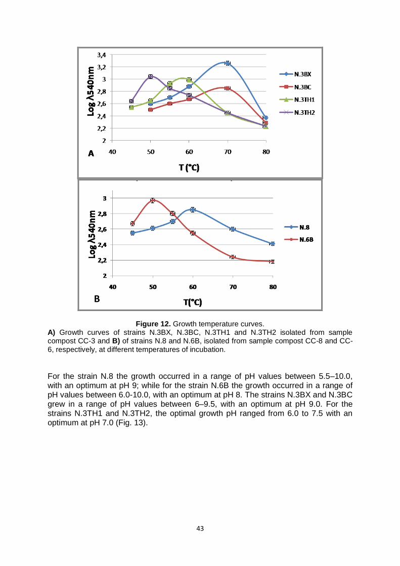
43
Figure 12. Growth temperature curves. A) Growth curves of strains N.3BX, N.3BC, N.3TH1 and N.3TH2 isolated from sample compost CC-3 and B) of strains N.8 and N.6B, isolated from sample compost CC-8 and CC-6, respectively, at different temperatures of incubation.
For the strain N.8 the growth occurred in a range of pH values between 5.5–10.0, with an optimum at pH 9; while for the strain N.6B the growth occurred in a range of pH values between 6.0-10.0, with an optimum at pH 8. The strains N.3BX and N.3BC grew in a range of pH values between 6–9.5, with an optimum at pH 9.0. For the strains N.3TH1 and N.3TH2, the optimal growth pH ranged from 6.0 to 7.5 with an optimum at pH 7.0 (Fig. 13).
B
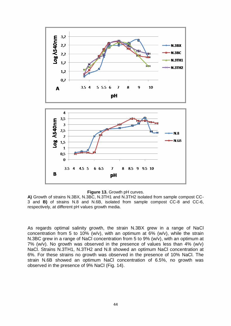
44
Figure 13. Growth pH curves. A) Growth of strains N.3BX, N.3BC, N.3TH1 and N.3TH2 isolated from sample compost CC-3 and B) of strains N.8 and N.6B, isolated from sample compost CC-8 and CC-6, respectively, at different pH values growth media. As regards optimal salinity growth, the strain N.3BX grew in a range of NaCl concentration from 5 to 10% (w/v), with an optimum at 6% (w/v), while the strain N.3BC grew in a range of NaCl concentration from 5 to 9% (w/v), with an optimum at 7% (w/v). No growth was observed in the presence of values less than 4% (w/v) NaCl. Strains N.3TH1, N.3TH2 and N.8 showed an optimum NaCl concentration at 6%. For these strains no growth was observed in the presence of 10% NaCl. The strain N.6B showed an optimum NaCl concentration of 6.5%, no growth was observed in the presence of 9% NaCl (Fig. 14).

45
Figure 14. Growth NaCl (%) curves. A) Growth curves of strains N.3BX, N.3BC, N.3TH1 and N.3TH2 isolated from sample compost CC-3 and B) of strains N.8 and N.6B, isolated from sample compost CC-8 and CC-
6, respectively, at different NaCl concentrations in the growth media. 3.1 Description of strains isolated from DISSPA Six different strains, from morphological point of view, were isolated from compost collected from the “Experimentation Center of Castel Volturno”, in which the compost derives from cowpat and straw. The strains have been isolated after a preliminary screening of specific enzymatic activities on agar plates: the isolation was performed in presence of CMC and xylan (0.2 %, w/v) as substrates, in order to select, directly, the colonies with cellulase and xylanase activities, respectively. In particular, from the sample compost 1CV were isolated the strains CV1-1 and CV1-2, while from the sample compost 2CV were isolated the strains named CV2-1, CV2-2, CV2-3 and CV2-4. The optimal growth temperature was of 50°C for all microorganisms, with the exception of strain CV1-2 that grew at 60°C. The growth culture medium for all strains was TSB. In particular, the isolates CV2-1, CV2-2,

46
CV2-3 and CV2-4 produced cellulase activity, in fact after 24 hours of incubation on solid medium there were evident zone of hydrolysis around the colonies that indicated the digestion of cellulose. These strains were found to be negative for xylan hydrolysis, while strains CV1-1 and CV1-2 were able to degrade xylan, but they weren‟t negative for cellulose degradation (Table 4).
Table 4. Strains isolated from “Experimentation Center of Castel Volturno”.
Strains Isolation Method
Enzymatic activities
Compost sample
CV1-1 B Xylanase 1CV
CV1-2 B Xylanase 1CV CV2-1 B Cellulase 2CV
CV2-2 B Cellulase 2CV CV2-3 B Cellulase 2CV
CV2-4 B Cellulase 2CV B: Isolation by using selective agar plates methods.
All strains were also positive to starch hydrolysis. In figure 15 the clear halo around the colonies after 24 hours of incubation indicated the presence of amylase activity (Fig. 15).
Figure 15. Example of colorimetric amylase assay on agar plate. The halo showed the presence of extracellular amylase in the strains isolated from “Experimentation Center of Castel Volturno” (DISSPA).
3.2 Phylogenetic analysis of isolates The 16S rRNA gene sequences obtained from BMR Genomics Service of strains N.8, N.6B, N.3TH1, N.3BX and N.3BC were deposited to the Genomic Bank EMBL under the following accession numbers LT594972, LT594973, LT745875, LT745876 and LT745877, respectively. The isolates were identified using the EzTaxon-e server (http://www.ezbiocloud.net/eztaxon; Kim et al., 2012) on the basis of 16S rRNA gene sequence. Strains N.3TH1, N.6B and N.8 belonged to Aeribacillus genus and were most closely related to Aeribacillus pallidus strain H12T DSM 3670 (99.8 %). Indeed, strains N.3BX and N.3BC belonged to the Geobacillus genus and showed 100% of identity between them. In particular, they had high similarity to Geobacillus thermodenitrificans DSM 465T (99.8%), Geobacillus subterraneus DSM13552T

47
(99.2%), Geobacillus thermoleovorans DSM 5366T (98%), Geobacillus uzenensis DSM 23175T (98%), Geobacillus stearothermophilus DSM 22T (98%), Geobacillus jurassicus DSM 15726T (98%), Geobacillus thermocatenulatus DSM 730T (98%) and Geobacillus vulcani DSM 13174T (98%). Strain N.3TH2 belonged to Bacillus genus, in particular showed 100% of homology with Bacillus licheniformis. Moreover, from the analysis of 16S rRNA gene sequence, was emerged that the strains isolated from “Experimentation Center of Castel Volturno” belonged to Bacillus genus, in particular strains CV1-1 and CV1-2 showed 100% of homology with Bacillus thermodenitrificans, while strains CV2-1, CV2-2, CV2-3 showed 100% of homology CV2-4 with Bacillus licheniformis. The phylogenetic tree of strains N.8 and N.6B reconstructed using the neighbour-joining method showed that strains N.8 and N.6B were a member of the genus Aeribacillus, while strains N.3BX and N.3BC were members of the genus Geobacillus (Fig. 16).
Figure 16. Neighbour-joining phylogenetic tree based on 16S rRNA gene sequences, showing the relationship between strains N.3TH1, N.8, N.6B, N.3BX and N.3BC and related taxa. Aneuribacillus thermoaerophilus DSM 10154T (X94196) was used as outgroup.
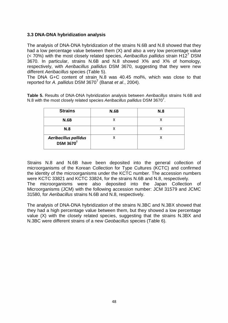
48
3.3 DNA-DNA hybridization analysis The analysis of DNA-DNA hybridization of the strains N.6B and N.8 showed that they had a low percentage value between them (X) and also a very low percentage value (< 70%) with the most closely related species, Aeribacillus pallidus strain H12T DSM 3670. In particular, strains N.6B and N.8 showed X% and X% of homology, respectively, with Aeribacillus pallidus DSM 3670, suggesting that they were new different Aeribacillus species (Table 5). The DNA G+C content of strain N.8 was 40.45 mol%, which was close to that reported for A. pallidus DSM 3670T (Banat et al., 2004).
Table 5. Results of DNA-DNA hybridization analysis between Aeribacillus strains N.6B and N.8 with the most closely related species Aeribacillus pallidus DSM 3670T.
Strains N.6B N.8
N.6B X X
N.8 X X
Aeribacillus pallidus DSM 3670T
X X
Strains N.8 and N.6B have been deposited into the general collection of microorganisms of the Korean Collection for Type Cultures (KCTC) and confirmed the identity of the microorganisms under the KCTC number. The accession numbers were KCTC 33821 and KCTC 33824, for the strains N.6B and N.8, respectively. The microorganisms were also deposited into the Japan Collection of Microorganisms (JCM) with the following accession number: JCM 31579 and JCMC 31580, for Aeribacillus strains N.6B and N.8, respectively. The analysis of DNA-DNA hybridization of the strains N.3BC and N.3BX showed that they had a high percentage value between them, but they showed a low percentage value (X) with the closely related species, suggesting that the strains N.3BX and N.3BC were different strains of a new Geobacillus species (Table 6).

49
Table 6. Results of DNA-DNA hybridization analysis between Geobacillus strains N.3BX and N.3BC with the most closely related species.
Strains N.3BX N.3BC
N.3BX X X
N.3BC X X
G. stearothermophilus
X X
G. thermoleovorans
X X
G. jurassicus X X
G. uzenensis X X
G. subterraneus X n.d.
G. thermodenitrificans
X n.d.
G. thermocatenulatus
X n.d.
G. vulcani X n.d.
Geobacillus thermodenitrificans DSM 465T, Geobacillus subterraneus DSM 13552T, Geobacillus thermoleovorans DSM 5366T, Geobacillus uzenensis DSM 23175T, Geobacillus stearothermophilus DSM 22T, Geobacillus jurassicus DSM 15726T, Geobacillus thermocatenulatus DSM 730T and Geobacillus vulcani DSM 13174T. n.d.: not determined
3.4 Biochemical characterization of isolates The strains isolated from sample compost of “Experimental Center of Composting” (CESCO), named N.6B, N.8, N.3BX and N.3BC were selected to perform several biochemical and phenotypic tests to give them a definitive taxonomic assignment. Some tests were performed also for strains CV1-1, CV1-2, CV2-1, CV2-2, CV2-3 and CV2-4 isolated from “Experimentation Center of Castel Volturno” (DISSPA). Cells of strains N.8 and N.6B were Gram-positive stain rods (1.5 µm), non-motile and endospore-forming. They were catalase and oxidase positive but negative for urease and indole formation. Strain N.8 was able to hydrolyse L-tyrosine, hippurate and xylan but not starch, CMC, casein, gelatine and phenylalanine. Strain N.6B was able to hydrolyse hippurate, but not L-tyrosine, xylan, starch, CMC, casein, gelatine and phenylalanine. They did not reduce nitrate and nitrite and they were found positive for aminopeptidase test and negative in the Voges–Proskauer reaction. The comparison of the physiological, biochemical and molecular properties of strains N.8 and N.6B with their nearest phylogenetic neighbour A. pallidus DSM 3670T was reported in Table 7.
50
Table 7. Characteristics of strains N.8 and N.6B and the closely related species, Aeribacillus pallidus DSM 3670T.
X Aeribacillus strains N.8 and N.6B were susceptible to (amount per disc): streptomycin (25 µg), gentamicin (30 µg), bacitracin (10 U), novobiocin (30 µg), penicillin (10 U), ampicillin (25 µg), erythromycin (30 µg), tetracycline (30 µg), neomycin (30 µg), fusidic acid (10 µg), lincomycin (15 µg), vancomycin (30 µg), chloramphenicol (10 µg) and kanamycin (30 µg). In the Table 8 and 9 are reported the information regarding carbon and nitrogen sources utilization evaluated by using BIOLOG Phenotype MicroArrayTM plates PM1 and PM2a, respectively (Biolog, Inc., Hayward, California, USA).
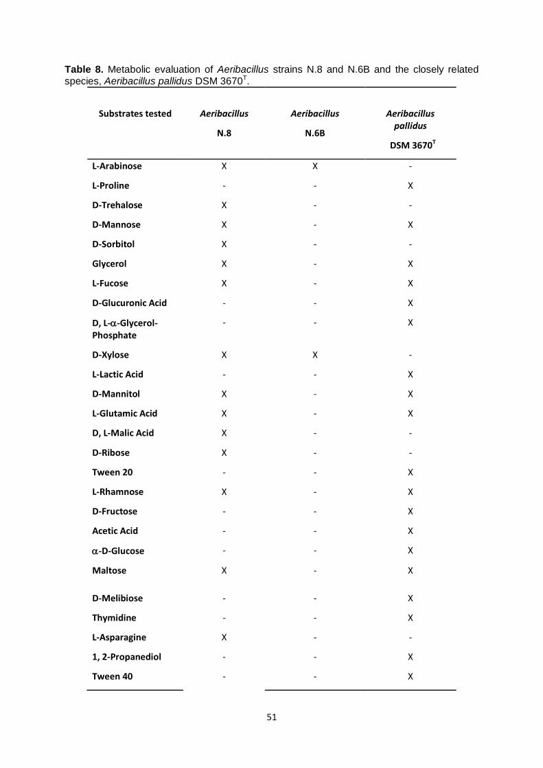
51
Table 8. Metabolic evaluation of Aeribacillus strains N.8 and N.6B and the closely related species, Aeribacillus pallidus DSM 3670T.
Substrates tested
Aeribacillus
N.8
Aeribacillus
N.6B
Aeribacillus pallidus
DSM 3670T
L-Arabinose X X -
L-Proline - - X
D-Trehalose X - -
D-Mannose X - X
D-Sorbitol X - -
Glycerol X - X
L-Fucose X - X
D-Glucuronic Acid - - X
D, L--Glycerol-Phosphate
- - X
D-Xylose X X -
L-Lactic Acid - - X
D-Mannitol X - X
L-Glutamic Acid X - X
D, L-Malic Acid X - -
D-Ribose X - -
Tween 20 - - X
L-Rhamnose X - X
D-Fructose - - X
Acetic Acid - - X
-D-Glucose - - X
Maltose X - X
D-Melibiose - - X
Thymidine - - X
L-Asparagine X - -
1, 2-Propanediol - - X
Tween 40 - - X
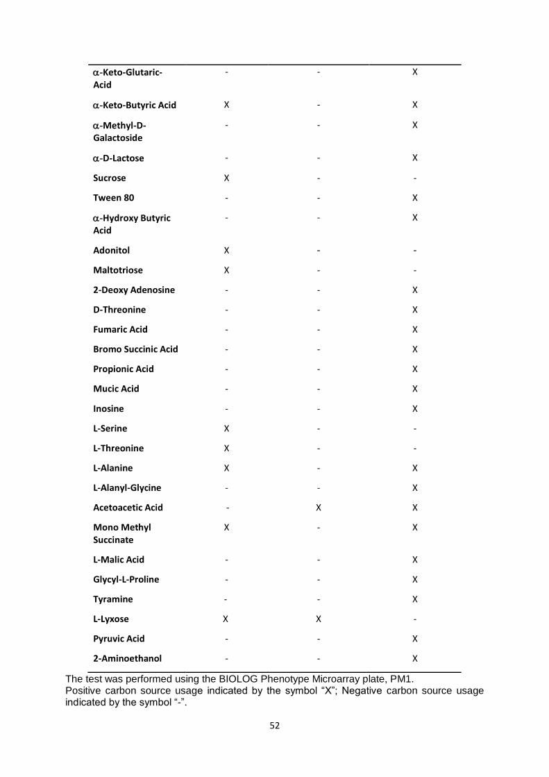
52
-Keto-Glutaric-Acid
- - X
-Keto-Butyric Acid X - X
-Methyl-D-Galactoside
- - X
-D-Lactose - - X
Sucrose X - -
Tween 80 - - X
-Hydroxy Butyric Acid
- - X
Adonitol X - -
Maltotriose X - -
2-Deoxy Adenosine - - X
D-Threonine - - X
Fumaric Acid - - X
Bromo Succinic Acid - - X
Propionic Acid - - X
Mucic Acid - - X
Inosine - - X
L-Serine X - -
L-Threonine X - -
L-Alanine X - X
L-Alanyl-Glycine - - X
Acetoacetic Acid - X X
Mono Methyl Succinate
X - X
L-Malic Acid - - X
Glycyl-L-Proline - - X
Tyramine - - X
L-Lyxose X X -
Pyruvic Acid - - X
2-Aminoethanol - - X
The test was performed using the BIOLOG Phenotype Microarray plate, PM1. Positive carbon source usage indicated by the symbol “X”; Negative carbon source usage indicated by the symbol “-”.
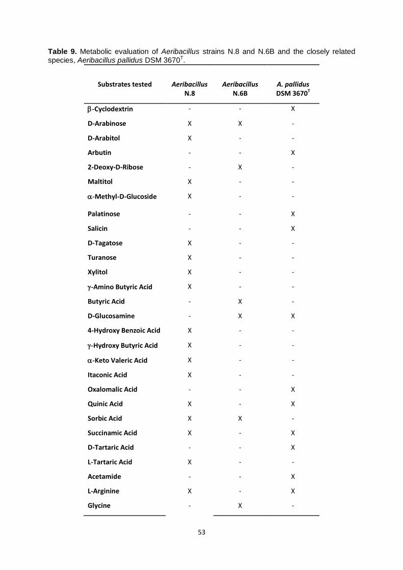
53
Table 9. Metabolic evaluation of Aeribacillus strains N.8 and N.6B and the closely related species, Aeribacillus pallidus DSM 3670T.
Substrates tested
Aeribacillus N.8
Aeribacillus N.6B
A. pallidus DSM 3670T
-Cyclodextrin - - X
D-Arabinose X X -
D-Arabitol X - -
Arbutin - - X
2-Deoxy-D-Ribose - X -
Maltitol X - -
-Methyl-D-Glucoside X - -
Palatinose - - X
Salicin - - X
D-Tagatose X - -
Turanose X - -
Xylitol X - -
-Amino Butyric Acid X - -
Butyric Acid - X -
D-Glucosamine - X X
4-Hydroxy Benzoic Acid X - -
-Hydroxy Butyric Acid X - -
-Keto Valeric Acid X - -
Itaconic Acid X - -
Oxalomalic Acid - - X
Quinic Acid X - X
Sorbic Acid X X -
Succinamic Acid X - X
D-Tartaric Acid - - X
L-Tartaric Acid X - -
Acetamide - - X
L-Arginine X - X
Glycine - X -
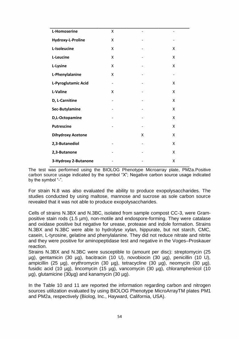
54
L-Homoserine X - -
Hydroxy-L-Proline X - -
L-Isoleucine X - X
L-Leucine X - X
L-Lysine X - X
L-Phenylalanine X - -
L-Pyroglutamic Acid - - X
L-Valine X - X
D, L-Carnitine - - X
Sec-Butylamine - - X
D,L-Octopamine - - X
Putrescine - - X
Dihydroxy Acetone X X
2,3-Butanediol - - X
2,3-Butanone - - X
3-Hydroxy 2-Butanone - - X
The test was performed using the BIOLOG Phenotype Microarray plate, PM2a.Positive carbon source usage indicated by the symbol “X”; Negative carbon source usage indicated by the symbol “-”. For strain N.8 was also evaluated the ability to produce exopolysaccharides. The studies conducted by using maltose, mannose and sucrose as sole carbon source revealed that it was not able to produce exopolysaccharides. Cells of strains N.3BX and N.3BC, isolated from sample compost CC-3, were Gram-positive stain rods (1.5 µm), non-motile and endospore-forming. They were catalase and oxidase positive but negative for urease, protease and indole formation. Strains N.3BX and N.3BC were able to hydrolyse xylan, hippurate, but not starch, CMC, casein, L-tyrosine, gelatine and phenylalanine. They did not reduce nitrate and nitrite and they were positive for aminopeptidase test and negative in the Voges–Proskauer reaction. Strains N.3BX and N.3BC were susceptible to (amount per disc): streptomycin (25 µg), gentamicin (30 µg), bacitracin (10 U), novobiocin (30 µg), penicillin (10 U), ampicillin (25 µg), erythromycin (30 µg), tetracycline (30 µg), neomycin (30 µg), fusidic acid (10 µg), lincomycin (15 µg), vancomycin (30 µg), chloramphenicol (10 µg), glutamicine (30µg) and kanamycin (30 µg). In the Table 10 and 11 are reported the information regarding carbon and nitrogen sources utilization evaluated by using BIOLOG Phenotype MicroArrayTM plates PM1 and PM2a, respectively (Biolog, Inc., Hayward, California, USA).

55
Table 10. Metabolic evaluation of Geobacillus strains N.3BX and N.3BC.
Substrates tested
Geobacillus N.3BX
Geobacillus N.3BC
L-Arabinose X -
L-Seryne X X
L-Threonine X X
L-Fucose - X
L-Phenylalanine X X
L-Valine X -
L-Lyxose X X
D-Ribose X X
The test was performed using the BIOLOG Phenotype Microarray plate, PM1. Positive carbon source usage indicated by the symbol “X”; Negative carbon source usage indicated by the symbol “-”.
Table 11. Metabolic evaluation of Geobacillus strains N.3BX and N.3BC.
Substrates tested
Geobacillus N.3BX
Geobacillus N.3BC
D-Arabinose X X
Glycine X X
L-Histidine X X
L-Homoserine X -
Hydroxy-L-Proline X -
L-Isoleucine X X
L-Leucine X X
L-Lysine - X
L-Valine X X
Putrescine X X
Dhydroxy Acetone X X
The test was performed using the BIOLOG Phenotype Microarray plate, PM2. Positive carbon source usage indicated by the symbol “X”; Negative carbon source usage indicated by the symbol “-”.
56
The comparison of the physiological, biochemical and molecular properties of strains N.3BX and N.3BC with their closely related Geobacillus species was reported in Table 12.
Table 12. Characteristics of strains N.3BX and N.3BC and the closely related species.
X
Geobacillus subterraneus DSM 13552T, Geobacillus thermoleovorans DSM 5366T, Geobacillus stearothermophilus DSM 22T, Geobacillus uzenensis DSM 23175T, Geobacillus jurassicus DSM 15726T, Geobacillus thermodenitrificans DSM 465T and Geobacillus vulcani DSM 13174T. n.d.: not determined.
Strains CV1-1, CV1-2, CV2-1, CV2-2, CV2-3 and CV2-4, isolated from sample compost 1CV and 2CV of “Experimentation Center of Castel Volturno” were aerobe and appeared as small motile rods when grown on solid media TSB at their optimal temperature. In particular, strains CV2-1, CV2-2, CV2-3 and CV2-4 after a growth of 24 hour in the standard growth conditions, showed a biofilm formation on the surface of liquid culture medium that observed at microscope, appeared as cells-extracellular matrix aggregates. The isolates CV2-1, CV2-2, CV2-3 and CV2-4 were positive for protease (Fig. 17), oxidase, and catalase; all of them were Gram-positive stain.
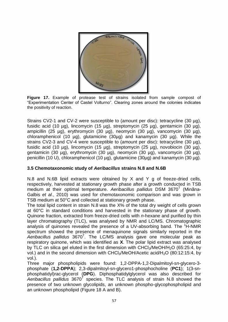
57
Figure 17. Example of protease test of strains isolated from sample compost of “Experimentation Center of Castel Volturno”. Clearing zones around the colonies indicates the positivity of reaction.
Strains CV2-1 and CV-2 were susceptible to (amount per disc): tetracycline (30 µg), fusidic acid (10 µg), lincomycin (15 µg), streptomycin (25 µg), gentamicin (30 µg), ampicillin (25 µg), erythromycin (30 µg), neomycin (30 µg), vancomycin (30 µg), chloramphenicol (10 µg), glutamicine (30µg) and kanamycin (30 µg). While the strains CV2-3 and CV-4 were susceptible to (amount per disc): tetracycline (30 µg), fusidic acid (10 µg), lincomycin (15 µg), streptomycin (25 µg), novobiocin (30 µg), gentamicin (30 µg), erythromycin (30 µg), neomycin (30 µg), vancomycin (30 µg), penicillin (10 U), chloramphenicol (10 µg), glutamicine (30µg) and kanamycin (30 µg). 3.5 Chemotaxonomic study of Aeribacillus strains N.8 and N.6B N.8 and N.6B lipid extracts were obtained by X and Y g of freeze-dried cells, respectively, harvested at stationary growth phase after a growth conducted in TSB medium at their optimal temperature. Aeribacillus pallidus DSM 3670T (Minãna-Galbis et al., 2010) was used for chemotaxonomic comparison and was grown in TSB medium at 50°C and collected at stationary growth phase. The total lipid content in strain N.8 was the X% of the total dry weight of cells grown at 60°C in standard conditions and harvested in the stationary phase of growth. Quinone fraction, extracted from freeze-dried cells with n-hexane and purified by thin layer chromatography (TLC), was analysed by NMR and LC/MS. Chromatographic analysis of quinones revealed the presence of a UV-absorbing band. The 1H-NMR spectrum showed the presence of menaquinone signals similarly reported in the Aeribacillus pallidus 3670T. The LC/MS analysis gave one molecular peak as respiratory quinone, which was identified as X. The polar lipid extract was analysed by TLC on silica gel eluted in the first dimension with CHCl3/MeOH/H2O (65:25:4, by vol.) and in the second dimension with CHCl3/MeOH/Acetic acid/H2O (80:12:15:4, by vol.). Three major phospholipids were found: 1,2-DPPA-1,2-Dipalmitoyl-sn-glycero-3-phosphate (1,2-DPPA); 2,3-dipalmitoyl-sn-glycero1-phosphocholine (PC1); 1(3-sn-phosphatidyl)rac-glycerol (DPG). Diphosphatidylglycerol was also described for Aeribacillus pallidus 3670
T species. The TLC analysis of strain N.8
showed the
presence of two unknown glycolipids, an unknown phospho-glycophospholipid and an unknown phospholipid (Figure 18 A and B).

58
X
Figure 18. Thin Layer Chromatography (TLC) showing the polar lipids of strain N.8. A) TLC showing the phospholipids of strain N.8 detected by spraying TLC with the Dittmer- Lester reagent (Nicolaus et al., 2001). First dimension: CHCl3/MeOH/H2O (65:25:4, by vol.); second dimension: CHCl3/MeOH/Acetic acid/H2O (80:12:15:4, by vol). (B) TLC showing the
glycolipids of strain N.8. First dimension: CHCl3/MeOH/H2O (65:25:4, by vol.); second dimension: CHCl3/MeOH/Acetic acid/H2O (80:12:15:4, by vol.). Glycolipids were detected by spraying with α-naphthol reagent (Nicolaus et al., 2001). P-GPL: unknown phospho-
glycophospholipid; GL: unknown glycolipid.
The total lipid content in strain N.6B was the X% of the total dry weight of cells grown at 50°C in standard conditions and harvested in the stationary phase of growth. Chromatographic analysis of quinones revealed the presence of a UV-absorbing band. The 1H-NMR spectrum showed the presence of menaquinone signals similarly reported in the Aeribacillus pallidus 3670T. The LC/MS analysis gave two molecular peaks as respiratory quinone, which was identified as X and X. Four major phospholipids were found. The TLC analysis of strain N.6B showed the presence of two major unknown glycolipids (GL1 and GL2), two minor unknown glycolipids (GL3 and GL4) and two unknown phospholipids (PL) (Fig. 19 A and B).
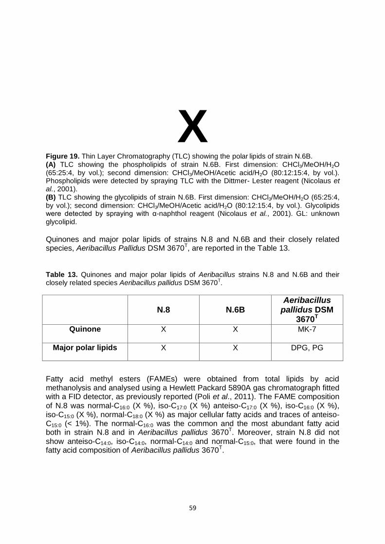
59
X
Figure 19. Thin Layer Chromatography (TLC) showing the polar lipids of strain N.6B. (A) TLC showing the phospholipids of strain N.6B. First dimension: CHCl3/MeOH/H2O (65:25:4, by vol.); second dimension: CHCl3/MeOH/Acetic acid/H2O (80:12:15:4, by vol.). Phospholipids were detected by spraying TLC with the Dittmer- Lester reagent (Nicolaus et al., 2001). (B) TLC showing the glycolipids of strain N.6B. First dimension: CHCl3/MeOH/H2O (65:25:4, by vol.); second dimension: CHCl3/MeOH/Acetic acid/H2O (80:12:15:4, by vol.). Glycolipids were detected by spraying with α-naphthol reagent (Nicolaus et al., 2001). GL: unknown glycolipid.
Quinones and major polar lipids of strains N.8 and N.6B and their closely related species, Aeribacillus Pallidus DSM 3670T, are reported in the Table 13. Table 13. Quinones and major polar lipids of Aeribacillus strains N.8 and N.6B and their closely related species Aeribacillus pallidus DSM 3670T.
N.8
N.6B
Aeribacillus pallidus DSM
3670T Quinone X X MK-7
Major polar lipids X X DPG, PG
Fatty acid methyl esters (FAMEs) were obtained from total lipids by acid methanolysis and analysed using a Hewlett Packard 5890A gas chromatograph fitted with a FID detector, as previously reported (Poli et al., 2011). The FAME composition of N.8 was normal-C16:0 (X %), iso-C17:0 (X %) anteiso-C17:0 (X %), iso-C16:0 (X %), iso-C15:0 (X %), normal-C18:0 (X %) as major cellular fatty acids and traces of anteiso-C15:0 (< 1%). The normal-C16:0 was the common and the most abundant fatty acid both in strain N.8 and in Aeribacillus pallidus 3670T. Moreover, strain N.8 did not show anteiso-C14:0, iso-C14:0, normal-C14:0 and normal-C15:0, that were found in the fatty acid composition of Aeribacillus pallidus 3670T.

60
The FAME composition of N.6B was normal-C16:0 (X %), anteiso-C15:0 (X %) iso-C17:0 (X %), anteiso-C17:0 (X %), iso-C16:0 (X %), iso-C15:0 (X %), normal-C15:0 (X %) as major cellular fatty acids and traces of anteiso-C16:0 (X %). The normal-C16:0 was the common and the most abundant fatty acid both in strain N.8 and in Aeribacillus pallidus 3670T. Moreover, strain N.6B showed anteiso-C15:0, that was found in traces in the fatty acid composition of strains N.8 and Aeribacillus pallidus 3670T.
The cellular fatty acids of strains N.8, N.6B and Aeribacillus pallidus 3670T (Minãna-Galbis et al., 2010) are compared in the Table 14.
Table 14. Cellular fatty acid composition of strains N.8 and N.6B and their closely related species of the genus Aeribacillus.
X
*Values are relative percentages of the total fatty acids. -: Not detected.
61
3.6 Chemotaxonomic study of Geobacillus strains N.3BX and N.3BC N.3BX and N.3BC lipid extracts were obtained by X and Y g of freeze-dried cells, respectively, harvested at stationary growth phase after a growth conducted in TH medium at their optimal temperature. Geobacillus thermodenitrificans DSM 465T, Geobacillus subterraneus DSM 13552T, Geobacillus jurassicus DSM 15726T, Geobacillus thermocatenulatus DSM 730T and Geobacillus vulcani DSM 13174T were used for chemotaxonomic comparison and were grown in their standard conditions and collected at stationary growth phase. The total lipid content in strains N.3BX and N.3BC was the X% and Y%, respectively, of the total dry weight of cells grown at 70°C in standard conditions and harvested in the stationary phase of growth. Quinone fractions, extracted from freeze-dried cells with n-hexane and purified by thin layer chromatography (TLC), were analysed by NMR and LC/MS. Chromatographic analysis of quinones revealed the presence of a UV-absorbing band. The 1H-NMR spectrum showed the presence of menaquinone signals similarly reported in the others Geobacillus species. The LC/MS analysis gave one molecular peak as respiratory quinone, which was identified as X. The polar lipid extracts were analysed by TLC on silica gel eluted in the first dimension with CHCl3/MeOH/H2O (65:25:4, by vol.) and in the second dimension with CHCl3/MeOH/Acetic acid/H2O (80:12:15:4, by vol.). In both strains two major phospholipids were found. The TLC analysis of strains N.3BX and N.3BC showed the presence of one unknown glycolipids and two unknown phospholipids (Figure 20 A and B).
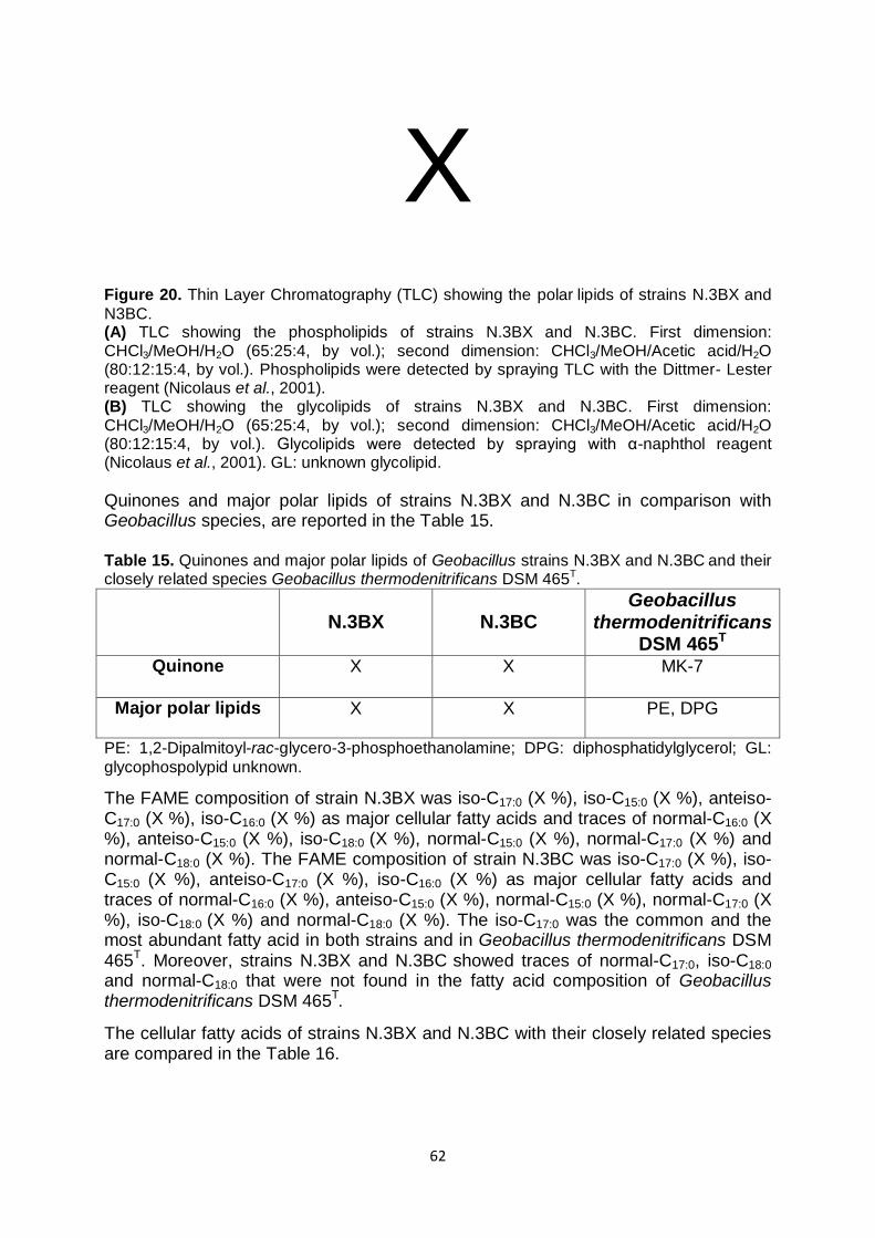
62
X
Figure 20. Thin Layer Chromatography (TLC) showing the polar lipids of strains N.3BX and
N3BC. (A) TLC showing the phospholipids of strains N.3BX and N.3BC. First dimension: CHCl3/MeOH/H2O (65:25:4, by vol.); second dimension: CHCl3/MeOH/Acetic acid/H2O (80:12:15:4, by vol.). Phospholipids were detected by spraying TLC with the Dittmer- Lester reagent (Nicolaus et al., 2001). (B) TLC showing the glycolipids of strains N.3BX and N.3BC. First dimension: CHCl3/MeOH/H2O (65:25:4, by vol.); second dimension: CHCl3/MeOH/Acetic acid/H2O (80:12:15:4, by vol.). Glycolipids were detected by spraying with α-naphthol reagent (Nicolaus et al., 2001). GL: unknown glycolipid.
Quinones and major polar lipids of strains N.3BX and N.3BC in comparison with Geobacillus species, are reported in the Table 15. Table 15. Quinones and major polar lipids of Geobacillus strains N.3BX and N.3BC and their closely related species Geobacillus thermodenitrificans DSM 465T.
N.3BX
N.3BC
Geobacillus thermodenitrificans
DSM 465T Quinone X X MK-7
Major polar lipids X X PE, DPG
PE: 1,2-Dipalmitoyl-rac-glycero-3-phosphoethanolamine; DPG: diphosphatidylglycerol; GL:
glycophospolypid unknown.
The FAME composition of strain N.3BX was iso-C17:0 (X %), iso-C15:0 (X %), anteiso-C17:0 (X %), iso-C16:0 (X %) as major cellular fatty acids and traces of normal-C16:0 (X %), anteiso-C15:0 (X %), iso-C18:0 (X %), normal-C15:0 (X %), normal-C17:0 (X %) and normal-C18:0 (X %). The FAME composition of strain N.3BC was iso-C17:0 (X %), iso-C15:0 (X %), anteiso-C17:0 (X %), iso-C16:0 (X %) as major cellular fatty acids and traces of normal-C16:0 (X %), anteiso-C15:0 (X %), normal-C15:0 (X %), normal-C17:0 (X %), iso-C18:0 (X %) and normal-C18:0 (X %). The iso-C17:0 was the common and the most abundant fatty acid in both strains and in Geobacillus thermodenitrificans DSM 465T. Moreover, strains N.3BX and N.3BC showed traces of normal-C17:0, iso-C18:0
and normal-C18:0 that were not found in the fatty acid composition of Geobacillus thermodenitrificans DSM 465T.
The cellular fatty acids of strains N.3BX and N.3BC with their closely related species are compared in the Table 16.

63
Table 16. Cellular fatty acid composition of strains N.3BX and N.3BC with their closely related species Geobacillus thermodenitrificans DSM 465T.
Fatty acid
N.3BX*
N.3BC*
Geobacillus thermodenitrificans
DSM 465T*
iso-C15:0 X Y 21.55
anteiso-C15:0 X Y 2.02
n-C15:0 X Y 1.69
iso-C16:0 X Y 14.64
n-C16:0 X Y 7.27
iso-C17:0 X Y 39.52
anteiso-C17:0 X Y 13.31
n-C17:0 X Y -
iso-C18:0 X Y -
n-C18:0 X Y -
*Values are relative percentages of the total fatty acids. -: Not detected.
3.7 Study of Xylanase activity of Geobacillus strains N.3BX and N.3BC Strains N.3BX and N.3BC showed an extracellular and cytosolic xylanase activity, measured at different temperatures using soluble xylan as substrate. In particular, extracellular and cytosolic xylanase of strain N.3BC showed to be active in a range temperature between 50 and 80°C with an optimum at X°C at pH X after 1 hour of incubation. The relative activities at 50 and 80°C for extracellular xylanase, were about 54% and 44%, respectively, and 68% and 46%, respectively, for cytosolic enzyme, respect to the activity at 70°C (Fig. 21A). While, for the strain N.3BX the optimal temperature activity for extracellular and cytosolic xylanase was 60 and 70°C, respectively, at pH 7 after 1 hour of incubation. In particular, the extracellular enzyme was active even at 70°C, keeping at that temperature, still the 60% as residual activity. The cytosolic xylanase was active in a temperature range between 50 and 80°C, showing a relative activity of about 70% and 40%, respectively, compared to the activity at 70°C in the same standard conditions (Fig. 21B).
64
X
Figure 21. Temperature curve of extracellular and cytosolic xylanase of microorganisms A) N.3BC and B) N.3BX. The values, after 1 hour of incubation, were indicated as Relative Activity (%) and they were subtracted from the contribution by the single sample and substrate at the time zero.
Extracellular xylanase of strains N.3BC and N.3BX showed to be active in a wide range of pH between 4.0 and 9.0, with an optimum at pH 8.0. The relative activities at pH 4.0 and 9.0 were 65% and 90%, respectively, for xylanase of N.3BC and 50% and 65%, respectively, for xylanase of N.3BX, compared to the activity at pH 8.0. (Fig. 22 A and B).

65
X
Figure 22. pH curve of extracellular xylanase of microorganisms A) N.3BC and B) N.3BX.
Buffer solution: Acetate pH 4 (A4), Acetate pH 5 (A5), Acetate pH 5.6 (A5.6), Phosphate pH 6 (P6); Phosphate pH 6.5 (P6.5), Phosphate pH 7 (P7), Phosphate pH 8 (P8), Glycine pH 9 (G9). The values after 1 hour of incubation were expressed as Relative Activity (%) and they were subtracted from the contribution by the single sample and substrate at the time zero.
The molecular masses of xylanase of strains N.3BX and N.3BC were estimated by SDS-PAGE and zymogram gels. In particular, the molecular masses were 100 KDa for extracellular and cytosolic xylanase of strain N.3BX and 50 KDa for extracellular xylanase of N.3BC. For cytosolic xylanase of strain N.3BC were not visible bands (Fig. 23).
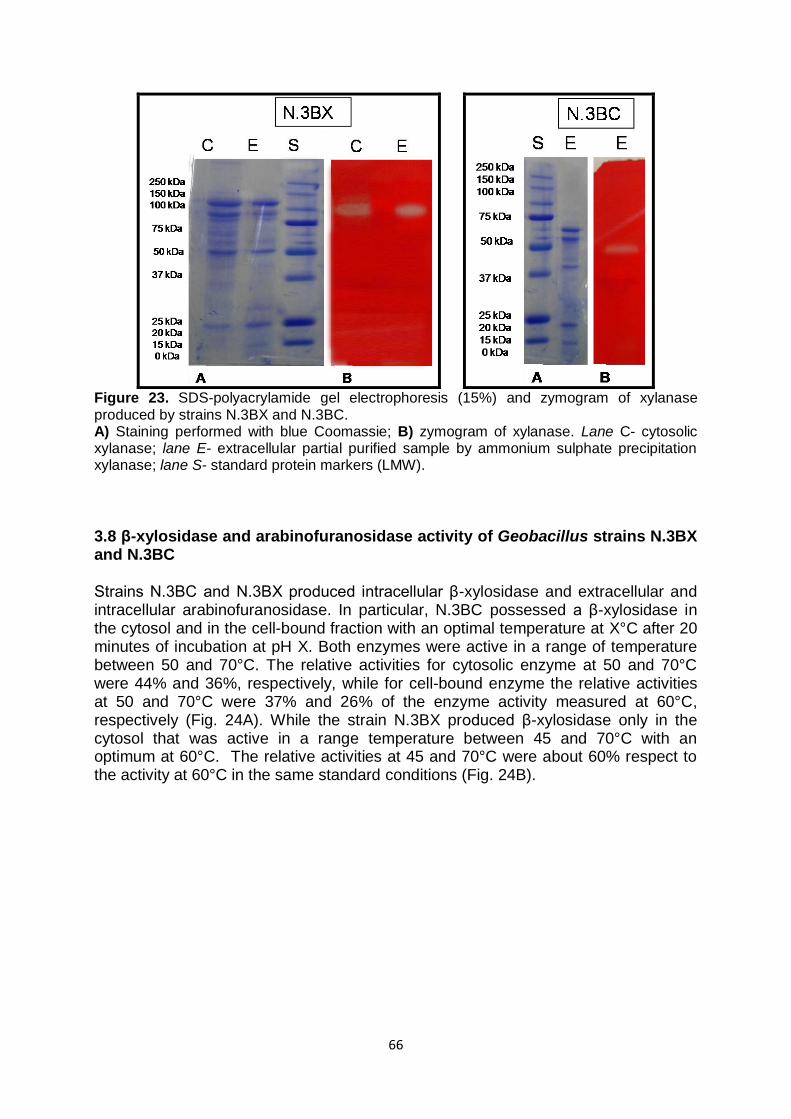
66
Figure 23. SDS-polyacrylamide gel electrophoresis (15%) and zymogram of xylanase produced by strains N.3BX and N.3BC. A) Staining performed with blue Coomassie; B) zymogram of xylanase. Lane C- cytosolic xylanase; lane E- extracellular partial purified sample by ammonium sulphate precipitation xylanase; lane S- standard protein markers (LMW).
3.8 β-xylosidase and arabinofuranosidase activity of Geobacillus strains N.3BX and N.3BC Strains N.3BC and N.3BX produced intracellular β-xylosidase and extracellular and intracellular arabinofuranosidase. In particular, N.3BC possessed a β-xylosidase in the cytosol and in the cell-bound fraction with an optimal temperature at X°C after 20 minutes of incubation at pH X. Both enzymes were active in a range of temperature between 50 and 70°C. The relative activities for cytosolic enzyme at 50 and 70°C were 44% and 36%, respectively, while for cell-bound enzyme the relative activities at 50 and 70°C were 37% and 26% of the enzyme activity measured at 60°C, respectively (Fig. 24A). While the strain N.3BX produced β-xylosidase only in the cytosol that was active in a range temperature between 45 and 70°C with an optimum at 60°C. The relative activities at 45 and 70°C were about 60% respect to the activity at 60°C in the same standard conditions (Fig. 24B).

67
X
Figure 24. Temperature curve of β-xylosidase of microorganisms A) N.3BC and B) N.3BX.
The values after 20 minutes of incubation were expressed as Relative Activity (%) and they were subtracted from the contribution by the single sample and substrate at the time zero.
Molecular masses of β-xylosidase of strains N.3BX and N.3BC were estimate by SDS-PAGE and 1% agarose gels. In particular, for cytosolic and cell-bound β-xylosidase of strain N.3BC, were visible two fluorescent bands of 30 and 60 KDa, while agarose gel of N.3BX cytosolic β-xylosidase showed two fluorescent bands, the
first one at about 100 KDa, the second one of 70 KDa, under UV light at 365 nm (Fig. 25).

68
Figure 25. SDS-polyacrylamide gel electrophoresis (15%) and 1% agarose gel of the β-
xylosidase produced by strains N.3BC and N.3BX.
A) Staining performed with blue Coomassie; B) 1% agarose gel in 50 mM phosphate pH 7.0 prepared with 50 µg/ml 1-4-methylumbelliferyl-7-β-D-xylopyranoside. The presence of fluorescent bands were visualized under UV light at λ365 nm. Lane C-
cytosolic β-xylosidase; lane M- cell-bound β-xylosidase; Lane S- standard protein markers
(LMW).
As regards the arabinofuranosidase activity, the microorganism N.3BC showed a strong extracellular arabinofuranosidase, active from 60 to 80°C with an optimal temperature (100% of activity) at 70°C at pH 7 after 20 minutes of incubation. The relative activities at 40 and 60°C were 40% and 46% of the enzymatic activity measured at 70°C, respectively. Strain N.3BC also produced arabinofuranosidase activity in the cytosolic and cell-bound fractions, active both in a range temperature between 50 and 80°C, with an optimum at 70°C at pH 7 after 20 minutes of incubation (Fig. 26A). Arabinofuranosidase activity was also produced by strain N.3BX in all three cellular compartments. The optimal temperature of activity was in all compartments of X°C at pH X after 20 minutes of incubation. In particular, in the cell bound fraction the arabinofuranosidase was active in a wide range of temperature (50-80°C) respect to the extracellular and cytosolic fractions. The relative activities at 50 and 80°C were 49% and 83% respect to the activity at 70°C (Fig. 26B).

69
X
Figure 26. Temperature curve of arabinofuranosidase of microorganisms A) N.3BC and B)
N.3BX. The values after 20 minutes of incubation were expressed as Relative Activity (%) and they were subtracted from the contribution by the single sample and substrate at the time zero. Molecular masses of arabinofuranosidase activities of strains N.3BX and N.3BC were estimate by SDS-PAGE and 1% agarose gels. In particular, 1% agarose gels of arabinofuranosidase of strain N.3BC showed that the molecular masses of extracellular and cell-bound enzymes were 150 KDa, while as regard the cytosolic enzyme were visible two clear fluorescent bands of 75 and 15 KDa under UV light at
365 nm. While, 1% agarose gel of arabinofuranosidase produced by strain N.3BX showed one clear fluorescent band of about 200 kDa relative to extracellular, cell-bound and cytosolic fractions (Fig. 27).

70
Figure 27. SDS-polyacrylamide gel electrophoresis (15%) of the arabinofuranosidase produced by strains N.3BC and N.3BX. A) Staining performed with blue Coomassie; B) 1% agarose gel in 50 mM phosphate pH 7.0 prepared with 50 µg/ml 4-methylumbelliferyl-α-arabinofuranoside. The presence of fluorescent bands were visualized under UV light at λ365 nm. Lane C- cytosolic arabinofuranosidase; lane M- cell-bound arabinofuranosidase; lane E- extracellular partial purified sample by ammonium sulphate precipitation arabinofuranosidase; Lane S- standard protein markers (LMW).

71
3.9 Cellulase activity of strain N.3TH2 Strain N.3TH2 isolated from composting site of Cilento National Park at compost sample CC-3, produced extracellular and weak cytosolic cellulase, measured at different temperatures using soluble carboxymethyl cellulose as substrate. In particular, extracellular enzyme was found to be active in a wide temperature range between 40 and 70°C, with an optimum at 60°C; the relative activities were 52% and 72% at 40 and 70°C, respectively, compared to the activity at the optimal temperature in the same standard conditions. While cytosolic cellulase showed an optimal temperature at 70°C and was found to be active in a range temperature between 60 and 80°C, with relative activity of 36% and 43%, respectively, compared to the activity at 70°C (Fig. 28A and B).
Figure 28. Temperature curve of cellulase activity produced by strain N.3TH2.
A) Extracellular pp(NH₄)₂SO₄ and B) cytosolic cellulase of strain N.3TH2. The values after 1
hour of incubation were expressed as Relative Activity (%) and they are subtracted from the contribution by the single sample and substrate at the time zero.

72
Extracellular cellulase was active in a wide pH range between 3.5 and 9.0, with an optimum at pH 5.6. The relative activities at pH values of 3.5 and 9.0 were about 66% and 40% compared to the activity at 60°C (100% of activity) (Fig. 29A), while cytosolic cellulase was found to be active at pH range between 3.0 and 8.0, with relative activity of about 40% and 70%, respectively, compared to the activity at 70°C (Fig. 29B).
Figure 29. pH curve of cellulase produced by strain N.3TH2.
A) Extracellular pp(NH₄)₂SO₄ and B) cytosolic cellulase of strain N.3TH2 at 60°C and 70°C,
respectively, for 1 hour of incubation. Buffer solution: Glycine pH 3 (G3), Acetate pH 4 (A4), Acetate pH 4.6 (A4.6), Acetate pH 5 (A5), Acetate pH 5.6 (A5.6), Phosphate pH 6 (P6); Phosphate pH 6.5 (P6.5), Phosphate pH 7 (P7), TRIS-HCl pH 7 (T7), Phosphate pH 8 (P8), Glycine pH 9 (G9) and pH 10 (G10). The values after 1 hour of incubation were expressed as Relative Activity (%) and they were subtracted from the contribution by the single sample and substrate at the time zero.
3.10 Study of Extracellular Cellulase activity of strain N.3TH2
B
A

73
Strain N.3TH2 produced extracellular and cytosolic cellulase; in particular, several tests were performed in order to establish the optimal conditions for the expression of extracellular cellulase. SDS-PAGE analysis and zymogram showed a single band with a molecular mass of about 37 KDa (Fig. 30).
Figure 30. SDS-polyacrylamide gel electrophoresis (15%) of the extracellular cellulase produced by strain N.3TH2. Staining was performed with blue Coomassie. Lane S- standard protein markers (LMW)1,; lane E- extracellular cellulase N.3TH2 partial purified sample by ammonium sulphate precipitation; lane Z- zymogram of extracellular cellulase partial purified.
3.10.1 Effect of different carbon sources on Cellulase production of strain N.3TH2 The activity of extracellular cellulase was assayed under standard conditions, using
the precipitate pp(NH₄)₂SO₄ after a growth of 24h in TSB medium standard and in
presence of CMC (0.2%, w/v), glucose or cellobiose (1%). The activity in presence of CMC as substrate of growth, was considered the reference value (100% of Relative Activity); in particular the maximum production of cellulase activity, evaluated by DNS method, was obtained using glucose as substrate of growth, showing 207% of relative activity compared to the activity registered in the presence of CMC as substrate of growth, at 60°C for 1 hour of incubation (Fig. 31).
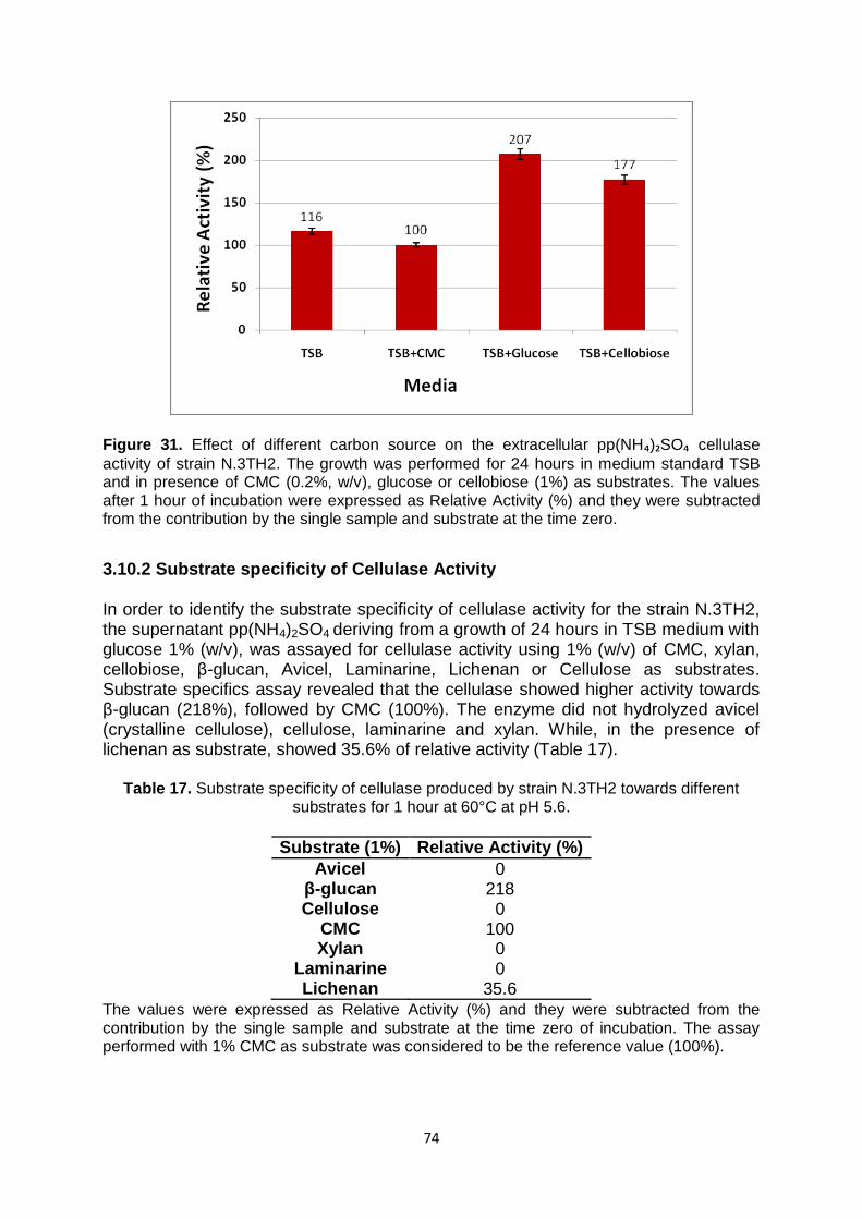
74
Figure 31. Effect of different carbon source on the extracellular pp(NH₄)₂SO₄ cellulase
activity of strain N.3TH2. The growth was performed for 24 hours in medium standard TSB and in presence of CMC (0.2%, w/v), glucose or cellobiose (1%) as substrates. The values after 1 hour of incubation were expressed as Relative Activity (%) and they were subtracted from the contribution by the single sample and substrate at the time zero.
3.10.2 Substrate specificity of Cellulase Activity In order to identify the substrate specificity of cellulase activity for the strain N.3TH2, the supernatant pp(NH4)2SO4 deriving from a growth of 24 hours in TSB medium with glucose 1% (w/v), was assayed for cellulase activity using 1% (w/v) of CMC, xylan, cellobiose, β-glucan, Avicel, Laminarine, Lichenan or Cellulose as substrates. Substrate specifics assay revealed that the cellulase showed higher activity towards β-glucan (218%), followed by CMC (100%). The enzyme did not hydrolyzed avicel (crystalline cellulose), cellulose, laminarine and xylan. While, in the presence of lichenan as substrate, showed 35.6% of relative activity (Table 17).
Table 17. Substrate specificity of cellulase produced by strain N.3TH2 towards different substrates for 1 hour at 60°C at pH 5.6.
Substrate (1%) Relative Activity (%)
Avicel 0 β-glucan 218 Cellulose 0
CMC 100 Xylan 0
Laminarine 0 Lichenan 35.6
The values were expressed as Relative Activity (%) and they were subtracted from the contribution by the single sample and substrate at the time zero of incubation. The assay performed with 1% CMC as substrate was considered to be the reference value (100%).

75
3.10.3 Thermostability In order to identify the thermostability of cellulase extracellular of strain N.3TH2 the sample was pre-incubated at 10‟, 30‟, 1 and 2 hours before to performing the enzymatic assay in standard conditions; the control was represented by the sample without pre-incubation. The results, as reported in the figure 32, showed that enzyme was stable for 10 minutes at 60°C and the half-life of cellulase activity was 1 hour of incubation (47%), while it retained 17.8 % of initial activity at 60°C after 2 hours of incubation.
Figure 32. Thermostability of extracellular cellulase N.3TH2. The values were reported as
Relative Activity (%) after 1 hour of incubation at 60°C at pH 5.6 and they were subtracted from the contribution by the single sample and substrate at the time zero of incubation. The control was represented by the enzyme activity tested in the standard assay conditions.
3.10.4 Cellulase activity at different times Extracellular cellulase N.3TH2 activity was monitored at different time intervals from 30 minutes up to 24 hours. There was an increasing of enzymatic activity from 30 minutes to 3 hours of incubation, in particular it was registered an increase from 0.85 mg/ml of glucose at 30 minutes to 1.99 mg/ml of glucose after 3 hours of incubation. As showed from 3 to 24 hours of incubation it was reached a plateau with production of about glucose 2.0 mg/ml (Fig. 33).

76
Figure 33. Extracellular cellulase N.3TH2 activity at different incubation times (0.5, 1, 2, 3, 5 and 24 hours) at 60°C at pH 5.6. The values were expressed as mg/ml of glucose and they were subtracted from the contribution by the single sample and substrate at the time zero of incubation.
3.10.5 Analysis of cellulase hydrolysis products Hydrolysis products of extracellular cellulase N.3TH2 were analyzed at different times of incubation (30 minutes, 1h, 2h, 3h, 5h and 24h), by thin layer cromatography (TLC) with the use of appropriate reference standards. As reported in the figure 34, the major end products detected at different times were cellobiose (C2) and cellotriose (C3) with increase of their production after 3 hours of incubation at 60°C. Therefore, these end products suggested that the enzyme was an “endocellulase”.
Figure 34. Thin layer chromatography analysis of hydrolysis products released by pp(NH4)2SO4 extracellular cellulase of strain N.3TH2 at different times of incubation at 60°C at pH 5.6 with 1% CMC. The solvent system used was buthanol/acetic acid/water (60:20:20
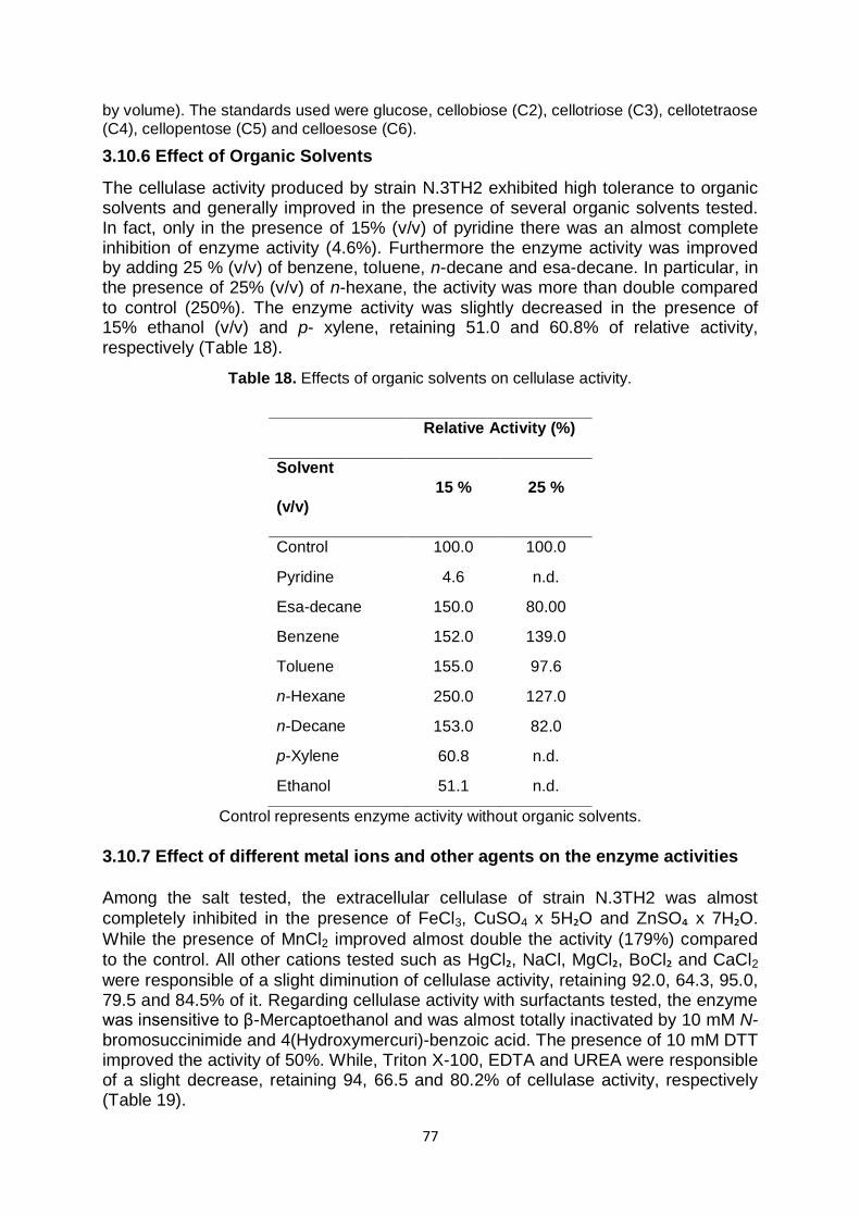
77
by volume). The standards used were glucose, cellobiose (C2), cellotriose (C3), cellotetraose (C4), cellopentose (C5) and celloesose (C6).
3.10.6 Effect of Organic Solvents
The cellulase activity produced by strain N.3TH2 exhibited high tolerance to organic solvents and generally improved in the presence of several organic solvents tested. In fact, only in the presence of 15% (v/v) of pyridine there was an almost complete inhibition of enzyme activity (4.6%). Furthermore the enzyme activity was improved by adding 25 % (v/v) of benzene, toluene, n-decane and esa-decane. In particular, in the presence of 25% (v/v) of n-hexane, the activity was more than double compared to control (250%). The enzyme activity was slightly decreased in the presence of 15% ethanol (v/v) and p- xylene, retaining 51.0 and 60.8% of relative activity, respectively (Table 18).
Table 18. Effects of organic solvents on cellulase activity.
Relative Activity (%)
Solvent
(v/v)
15 % 25 %
Control 100.0 100.0
Pyridine 4.6 n.d.
Esa-decane 150.0 80.00
Benzene 152.0 139.0
Toluene 155.0 97.6
n-Hexane 250.0 127.0
n-Decane 153.0 82.0
p-Xylene 60.8 n.d.
Ethanol 51.1 n.d.
Control represents enzyme activity without organic solvents.
3.10.7 Effect of different metal ions and other agents on the enzyme activities
Among the salt tested, the extracellular cellulase of strain N.3TH2 was almost
completely inhibited in the presence of FeCl3, CuSO4 x 5H₂O and ZnSO₄ x 7H₂O.
While the presence of MnCl2 improved almost double the activity (179%) compared
to the control. All other cations tested such as HgCl₂, NaCl, MgCl₂, BoCl₂ and CaCl2
were responsible of a slight diminution of cellulase activity, retaining 92.0, 64.3, 95.0, 79.5 and 84.5% of it. Regarding cellulase activity with surfactants tested, the enzyme was insensitive to β-Mercaptoethanol and was almost totally inactivated by 10 mM N-bromosuccinimide and 4(Hydroxymercuri)-benzoic acid. The presence of 10 mM DTT improved the activity of 50%. While, Triton X-100, EDTA and UREA were responsible of a slight decrease, retaining 94, 66.5 and 80.2% of cellulase activity, respectively (Table 19).
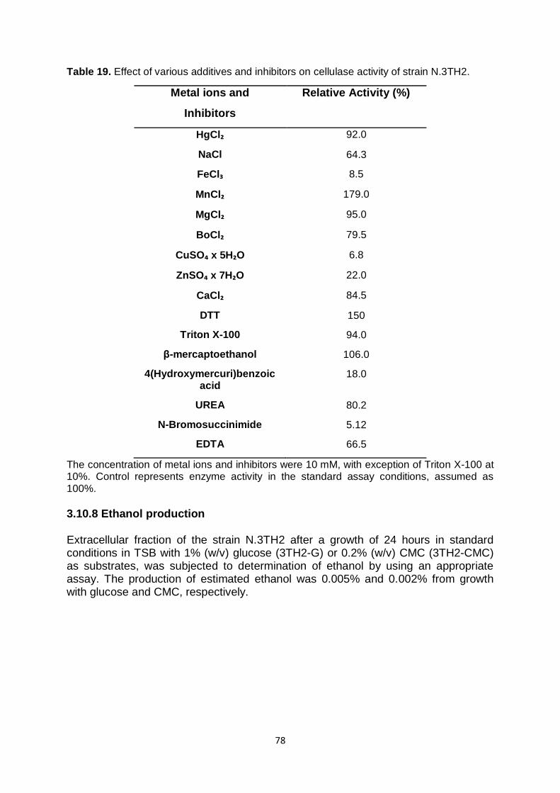
78
Table 19. Effect of various additives and inhibitors on cellulase activity of strain N.3TH2.
Metal ions and
Inhibitors
Relative Activity (%)
HgCl₂ 92.0
NaCl 64.3
FeCl₃ 8.5
MnCl₂ 179.0
MgCl₂ 95.0
BoCl₂ 79.5
CuSO₄ x 5H₂O 6.8
ZnSO₄ x 7H₂O 22.0
CaCl₂ 84.5
DTT 150
Triton X-100 94.0
β-mercaptoethanol 106.0
4(Hydroxymercuri)benzoic acid
18.0
UREA 80.2
N-Bromosuccinimide 5.12
EDTA 66.5
The concentration of metal ions and inhibitors were 10 mM, with exception of Triton X-100 at 10%. Control represents enzyme activity in the standard assay conditions, assumed as 100%. 3.10.8 Ethanol production Extracellular fraction of the strain N.3TH2 after a growth of 24 hours in standard conditions in TSB with 1% (w/v) glucose (3TH2-G) or 0.2% (w/v) CMC (3TH2-CMC) as substrates, was subjected to determination of ethanol by using an appropriate assay. The production of estimated ethanol was 0.005% and 0.002% from growth with glucose and CMC, respectively.
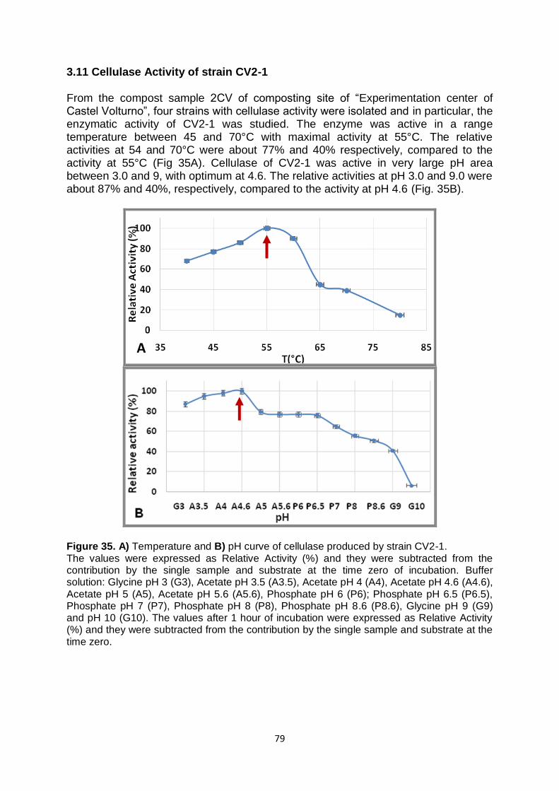
79
3.11 Cellulase Activity of strain CV2-1 From the compost sample 2CV of composting site of “Experimentation center of Castel Volturno”, four strains with cellulase activity were isolated and in particular, the enzymatic activity of CV2-1 was studied. The enzyme was active in a range temperature between 45 and 70°C with maximal activity at 55°C. The relative activities at 54 and 70°C were about 77% and 40% respectively, compared to the activity at 55°C (Fig 35A). Cellulase of CV2-1 was active in very large pH area between 3.0 and 9, with optimum at 4.6. The relative activities at pH 3.0 and 9.0 were about 87% and 40%, respectively, compared to the activity at pH 4.6 (Fig. 35B).
Figure 35. A) Temperature and B) pH curve of cellulase produced by strain CV2-1. The values were expressed as Relative Activity (%) and they were subtracted from the contribution by the single sample and substrate at the time zero of incubation. Buffer solution: Glycine pH 3 (G3), Acetate pH 3.5 (A3.5), Acetate pH 4 (A4), Acetate pH 4.6 (A4.6), Acetate pH 5 (A5), Acetate pH 5.6 (A5.6), Phosphate pH 6 (P6); Phosphate pH 6.5 (P6.5), Phosphate pH 7 (P7), Phosphate pH 8 (P8), Phosphate pH 8.6 (P8.6), Glycine pH 9 (G9) and pH 10 (G10). The values after 1 hour of incubation were expressed as Relative Activity (%) and they were subtracted from the contribution by the single sample and substrate at the time zero.
A
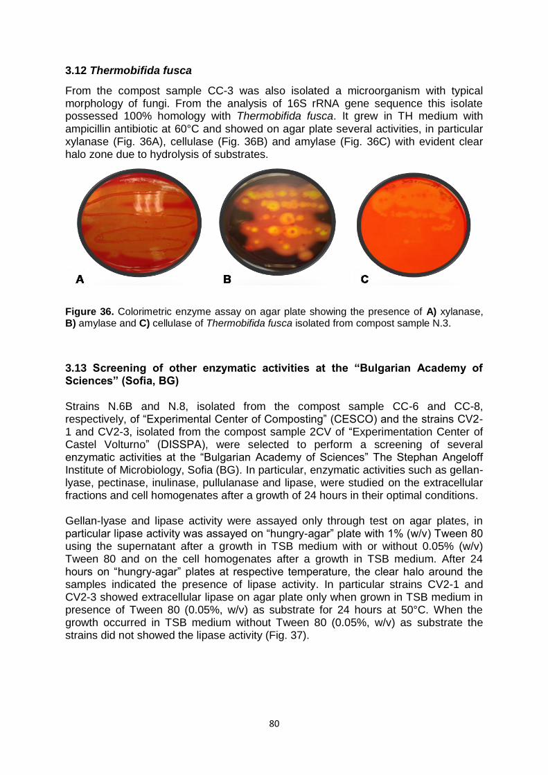
80
3.12 Thermobifida fusca
From the compost sample CC-3 was also isolated a microorganism with typical morphology of fungi. From the analysis of 16S rRNA gene sequence this isolate possessed 100% homology with Thermobifida fusca. It grew in TH medium with ampicillin antibiotic at 60°C and showed on agar plate several activities, in particular xylanase (Fig. 36A), cellulase (Fig. 36B) and amylase (Fig. 36C) with evident clear halo zone due to hydrolysis of substrates.
Figure 36. Colorimetric enzyme assay on agar plate showing the presence of A) xylanase, B) amylase and C) cellulase of Thermobifida fusca isolated from compost sample N.3.
3.13 Screening of other enzymatic activities at the “Bulgarian Academy of Sciences” (Sofia, BG) Strains N.6B and N.8, isolated from the compost sample CC-6 and CC-8, respectively, of “Experimental Center of Composting” (CESCO) and the strains CV2-1 and CV2-3, isolated from the compost sample 2CV of “Experimentation Center of Castel Volturno” (DISSPA), were selected to perform a screening of several enzymatic activities at the “Bulgarian Academy of Sciences” The Stephan Angeloff Institute of Microbiology, Sofia (BG). In particular, enzymatic activities such as gellan-lyase, pectinase, inulinase, pullulanase and lipase, were studied on the extracellular fractions and cell homogenates after a growth of 24 hours in their optimal conditions. Gellan-lyase and lipase activity were assayed only through test on agar plates, in particular lipase activity was assayed on “hungry-agar” plate with 1% (w/v) Tween 80 using the supernatant after a growth in TSB medium with or without 0.05% (w/v) Tween 80 and on the cell homogenates after a growth in TSB medium. After 24 hours on “hungry-agar” plates at respective temperature, the clear halo around the samples indicated the presence of lipase activity. In particular strains CV2-1 and CV2-3 showed extracellular lipase on agar plate only when grown in TSB medium in presence of Tween 80 (0.05%, w/v) as substrate for 24 hours at 50°C. When the growth occurred in TSB medium without Tween 80 (0.05%, w/v) as substrate the strains did not showed the lipase activity (Fig. 37).
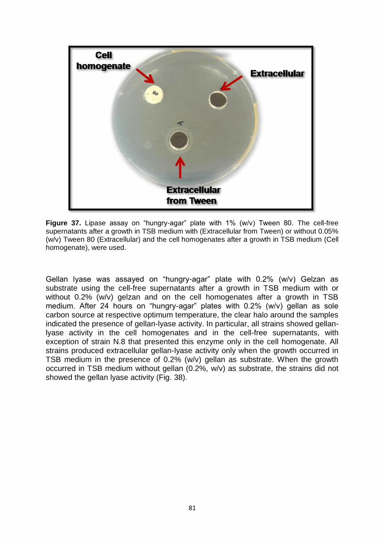
81
Figure 37. Lipase assay on “hungry-agar” plate with 1% (w/v) Tween 80. The cell-free supernatants after a growth in TSB medium with (Extracellular from Tween) or without 0.05% (w/v) Tween 80 (Extracellular) and the cell homogenates after a growth in TSB medium (Cell homogenate), were used.
Gellan lyase was assayed on “hungry-agar” plate with 0.2% (w/v) Gelzan as substrate using the cell-free supernatants after a growth in TSB medium with or without 0.2% (w/v) gelzan and on the cell homogenates after a growth in TSB medium. After 24 hours on “hungry-agar” plates with 0.2% (w/v) gellan as sole carbon source at respective optimum temperature, the clear halo around the samples indicated the presence of gellan-lyase activity. In particular, all strains showed gellan-lyase activity in the cell homogenates and in the cell-free supernatants, with exception of strain N.8 that presented this enzyme only in the cell homogenate. All strains produced extracellular gellan-lyase activity only when the growth occurred in TSB medium in the presence of 0.2% (w/v) gellan as substrate. When the growth occurred in TSB medium without gellan (0.2%, w/v) as substrate, the strains did not showed the gellan lyase activity (Fig. 38).

82
Figure 38. Extracellular gellan-lyase activity produced by strains N.8, N.6B, CV2-1 and CV2-
3. After a growth on “hungry-agar” plate with 0.2% (w/v) a solution of 0.2% (w/v) Congo Red followed by a solution of NaCl 1M, were added to the plates. The presence of the clear halo around the colonies indicated that the hydrolysis of gelzan occurred (positivity reaction).
Strains N.6B, CV2-1 and CV2-3 showed pectinase activity in the cell homogenates, measured with soluble polygalacturonic acid as substrate. The major activity was present in the cell homogenates of strain CV2-3 at 60°C and pH 7. The relative pectinase activity of strains N.6B and CV2-3 were 64% and 86%, respectively, compared to the pectinase of strain CV2-3 (Tab. 20).
Table 20. Comparison of pectinase activity produced in the cell homogenates of strains N.6B, N.8, CV2-1 and CV2-3.
Strains Pectinase
(Relative Activity %)
N.6B 64
N.8 -
CV2-1 68
CV2-3 100
The values were expressed as Relative Activity (%) and they were subtracted from the contribution by the single sample and substrate at the time zero of incubation. The Relative activity of strain CV2-3 was assumed as 100%.
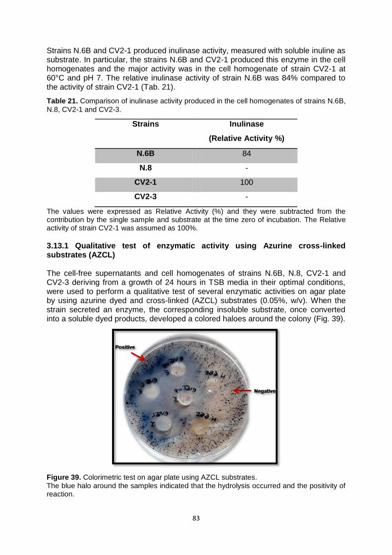
83
Strains N.6B and CV2-1 produced inulinase activity, measured with soluble inuline as substrate. In particular, the strains N.6B and CV2-1 produced this enzyme in the cell homogenates and the major activity was in the cell homogenate of strain CV2-1 at 60°C and pH 7. The relative inulinase activity of strain N.6B was 84% compared to the activity of strain CV2-1 (Tab. 21).
Table 21. Comparison of inulinase activity produced in the cell homogenates of strains N.6B,
N.8, CV2-1 and CV2-3.
Strains Inulinase
(Relative Activity %)
N.6B 84
N.8 -
CV2-1 100
CV2-3 -
The values were expressed as Relative Activity (%) and they were subtracted from the contribution by the single sample and substrate at the time zero of incubation. The Relative activity of strain CV2-1 was assumed as 100%. 3.13.1 Qualitative test of enzymatic activity using Azurine cross-linked substrates (AZCL) The cell-free supernatants and cell homogenates of strains N.6B, N.8, CV2-1 and CV2-3 deriving from a growth of 24 hours in TSB media in their optimal conditions, were used to perform a qualitative test of several enzymatic activities on agar plate by using azurine dyed and cross-linked (AZCL) substrates (0.05%, w/v). When the strain secreted an enzyme, the corresponding insoluble substrate, once converted into a soluble dyed products, developed a colored haloes around the colony (Fig. 39).
Figure 39. Colorimetric test on agar plate using AZCL substrates. The blue halo around the samples indicated that the hydrolysis occurred and the positivity of reaction.

84
In particular, the strain N.6B showed extracellular hydrolysis of galactomannan, while in the extracellular and intracellular compartment showed hydrolysis of xyloglucan and β-glucan. It was found to be negative to hydrolysis of the following substrates: amilose, arabinoxyan, curdulan, arabinan, dextran, cellulose, galactan, chitosan, pullulan and collagen. The strain CV2-3 produced extracellular cellulase, while the strain CV2-1 also in the intracellular compartment. The strains CV2-1 and CV2-3, both in the supernatants and in the cell homogenates, showed enzymatic hydrolysis of arabinan, xyloglucan, amilose and galactan, but they were not able to hydrolyse arabinoxylan, galactomannan, curdulan, dextran, chitosan, pullulan and xylan. Moreover, both strains produced collagenasic extracellular activity, more evident in the strain CV2-1. The strain N.8 it was found to be negative for each AZCL substrates tested (Table 22).
Table 22. Summary of enzymatic activities on agar plate using AZCL-substrates of strains
N.6B, N.8, CV2-1 and CV2-3.
AZCL SUBSTRATES
N.6B
N.8
CV2-1
CV2-3
Cell Homogenate
Extrac. Fraction
Cell Homogenate
Extrac. Fraction
Cell Homogenate
Extrac. Fraction
Cell Homogenate
Extrac. Fraction
Arabinoxylan - - - - - - - -
Galactomannan - +++ - - - - - -
Curdlan - - - - - - - -
Arabinan - - - - + +++ +++ +++
Dextran - - - - - - - -
Xyloglucan +++ ++ - - ++ +++ ++ +
Amylose - - - - ++ +++ ++ ++
Cellulose - - - - + + - -
Galactan - - - - ++ ++ ++ ++
β-Glucan ++ ++ - - - ++ ++ ++
Chitosan - - - - - - - -
Pullulan - - - - - - - -
Collagen - - - - - +++ + ++
Xylan - - - - - - - -
-: negative, +: weak, ++ positive, +++: very positive; Extrac.: Extracellular sample.

85
Chapter 4:
CONCLUSION AND DISCUSSION

86
In this research project the compost was used as a starting material for the isolation of thermophilic microorganisms; subsequently, enzymatic activities able to convert lignocellulosic biomass wastes to obtain products with several potential biotechnological applications in different types of sectors, were investigated. In particular, from composting site “Experimental Center of Composting” (CESCO), which waste resulting from oil mills were used, strains named N.3TH1, N.8 and N.6B were isolated, from sample compost CC-3 (structuring virgin wood), CC-8 (Curing 1 under towel 30 days old) and CC-6 (Soil amendment composted finally mixture), respectively. Furthermore, from the sample compost CC-3 were also isolated other three strains, producing cellulolytic or hemicellulolytic enzymes, named N.3TH2, N.3BX and N.3BC. While, from compost collected at the “Experimentation Center of Castel Volturno” (DISSPA), University of Study of Naples Federico II, in which the compost derives from cowpat and straw, six different strains from morphological point of view, were isolated. In particular, from the sample compost 1CV, strains named CV1-1 and CV1-2, that showed xylanase activity on agar plate, were isolated; while from the sample compost 2CV, strains named CV2-1, CV2-2, CV2-3 and CV2-4, that showed cellulase activity, were isolated. Thermophilic strains isolated from “Experimental Center of Composting” (CESCO) and from “Experimentation Center of Castel Volturno” (DISSPA), are reported in the Table 23. Table 23. Optimal growth conditions of strains isolated from “Experimental Center of Composting” (CESCO) and from “Experimentation Center of Castel Volturno” (DISSPA).
Strains
Origin of strains
Growth Medium
Optimal Growth
Temperature (°C)
Optimal
Growth pH
Optimal Growth Salinity (NaCl %,
w/v)
N.3TH1 CESCO TH X X 6.0
N.3TH2 CESCO TSB X X 6.0
N.8 CESCO TSB X X 6.0
N.6B CESCO TSB X X 6.5
N.3BX CESCO TH X X 6.0
N.3BC CESCO TH X X 7.0
CV1-1 DISSPA TSB 50 7.0 5.0
CV1-2 DISSPA TSB 60 7.0 5.0
CV2-1 DISSPA TSB 50 7.0 5.0
CV2-2 DISSPA TSB 50 7.0 5.0
CV2-3 DISSPA TSB 50 7.0 5.0
CV2-4 DISSPA TSB 50 7.0 5.0
87
Among the strains isolated, four of them were a new thermophilic species. In particular, strains N.8 and N.6B represented two novel species of Aeribacillus genus; for strain N.8 the name Aeribacillus composti sp. nov. was proposed. Strains N.8 and N.6B, based on the 16S rRNA gene sequence homology, were most closely related to Aeribacillus pallidus strain H12T DSM 3670 (99.8 %). In literature, it has been reported that Aeribacillus pallidus DSM 3670T was the sole species of Aeribacillus genus (Minãna-Galbis et al., 2010). The genus Aeribacillus was first proposed by Minãna-Galbis et al., (2010) when Geobacillus pallidus (Scholz et al, 1988, Banat et al., 2004) was reclassified in a new genus, as Aeribacillus pallidus. This genus belongs to the Firmicutes phylum and it is most closely related to the genera Geobacillus and Anoxybacillus (Minãna-Galbis et al., 2010). Cells of strains Aeribacillus N.8 and N.6B, were Gram positive rods (1.5 µm) and endospore-forming, similar to cells of Aeribacillus pallidus. Aeribacillus strains N.8 and N.6B were found to be negative for xylanase activity, while Aeribacillus pallidus showed xylanase activity. The analysis of cellular fatty acids, that represents an important chemotaxonomic marker, showed various differences between the new Aeribacillus strains N.8 and N.6B and their closely related species Aeribacillus pallidus DSM 3670T (Tab. 24). Therefore, based on the 16S rRNA gene sequences, DNA–DNA hybridization and chemotaxonomic characteristics, strains N.8 and N.6B represented a new different species of Aeribacillus genus. From the sample compost CC-3 of “Experimental Center of Composting” (CESCO) were also isolated two different strains of the same a new Geobacillus species, named N.3BX and N.3BC. Based on 16S rRNA gene sequence homology, they showed high similarity to Geobacillus thermodenitrificans DSM465T (99.8%), Geobacillus subterraneus DSM13552T (99.2%), Geobacillus thermoleovorans DSM 5366T (98%), Geobacillus uzenensis DSM 23175T (98%), Geobacillus stearothermophilus DSM 22T (98%), Geobacillus jurassicus DSM 15726T (98%), Geobacillus thermocatenulatus DSM 730T (98%) and Geobacillus vulcani DSM 13174T (98%). The analysis of DNA-DNA hybridization of the strains N.3BC and N.3BX showed that they had a high percentage value between them, but they showed a very low percentage value (< 70%) with the closely related species, suggesting that the strains N.3BX and N.3BC were different strains of a new Geobacillus species. The genus Geobacillus comprises a group of Gram-positive
thermophilic bacteria, originally classified as group of Bacillus sp. (Nazina et al., 2001). Cells of Geobacillus strains N.3BX and N.3BC, were Gram positive rods (1.5 µm), non-motile and endospore-forming as described for Geobacillus genus. They were catalase negative and oxidase positive at difference of G. vulcani, G. stearothermophilus and G. thermoleovorans that were found to be negative for both enzymes. From cellular fatty acids composition, an important chemotaxonomic marker, were emerged various differences between the new Geobacillus strains N.3BX and N.3BC and their closely related Geobacillus species compared (Tab. 24). Therefore, based on the 16S rRNA gene sequences homology, DNA–DNA hybridization and chemotaxonomic characteristics, strains N.3BX and N.3BC represented new different thermophilic strains of a new species of the genus Geobacillus.

88
Table 24. Neutral, polar lipids and FAME composition.
X
GL: unknown glycolipid, PL: unknown phospholipid, GPL: unknown glycophospholipid, G-GPL: unknown Phospho-glycophospholipid.
Moreover, Geobacillus strains N.3BX and N.3BC produced hemicellulolytic enzymes, in particular they showed extracellular and cytosolic xylanase with additional hemicellulases including β-xylosidase and arabinofuranosidase. Molecular masses of xylanase, β-xylosidase and arabinofuranosidase produced by strains N.3BX and N.3BC, are summarized in the table 25.

89
Table 25. Molecular mass estimated by SDS-PAGE and zymogram.
N.3BX N.3BC
Xylanase Extracellular <100 kDa
Cytosol
<100 kDa
Extracellular
50 KDa
Cytosol
n. d.
β-
xylosidase
*Cytosol
1st band about 70 kDa;
2nd band 100-150 kDa
*Cytosol 1st band 30 kDa; 2nd band 60 kDa
*Cell-bound
1st band 30 kDa;
2nd band 60 kDa
Arabinofu
ranosidase
*Extracellular
200 kDa
*Cytosol 200 kDa
*Cell-
bound
200
kDa
*Extracellular
150 kDa
*Cytosol 1st band 15 kDa; 2nd band 75 kDa
*Cell-bound
150 kDa
*samples not boiled
There are several reports of xylanase producing bacteria including Geobacillus
genus, but only a few of them had both xylanase and β-xylosidase, like G. stearothermophilus and G. thermantarcticus (Lama et al., 2004). In particular,
xylanase and β-xylosidase produced by Geobacillus strains N.3BX and N.3BC
showed interesting properties for biotechnological applications, such as optimal temperature and pH activity and the absence of cellulase activity; the lack of cellulase activity represents a fundamental prerequisite in the bleaching paper by using xylanase (Viikari et al, 1994). Geobacillus strains N.3BX and N.3BC also produced intracellular and extracellular arabinofuranosidase; in literature, it has been described thermophilic arabinofuranosidase activity produced by a Gram-positive soil bacterium, Geobacillus stearothermophilus T6, which had an extensive system for the utilization of plant cell-wall polysaccharides, including xylan, arabinan and galactan (Lansky et al, 2014). Moreover, it has been reported a thermophilic strain, Geobacillus sp. DC3, capable of producing hemicellulolytic enzymes such as in the case of Geobacillus strains N.3BX and N.3BC. In particular, Geobacillus sp. DC3 produced endoxylanase (39.5 U/mg protein), β-xylosidase (0.209 U/mg of protein) and arabinofuranosidase (0.230 U/mg of protein) after the bacterium was grown in 1% (w/v) xylan for 24 h. After 24 hours of growth in media with 0.2% (w/v) xylan as substrate, Geobacillus strains N.3BX and N.3BC produced a lower extracellular xylanase with 2.7 and 3.3 U/mg of protein, respectively, but expressed a high level of cytosolic β-xylosidase (251 and 2.807 U/mg of protein for strains N.3BX and N.3BC, respectively) and the cell-free supernatant arabinofuranosidase (6.698 and 1.130 U/mg of protein for strains N.3BX and N.3BC, respectively) compared to Geobacillus sp. DC3 (Bergdale et al., 2014).

90
Furthermore, from the sample compost CC-3 of the “Experimental Center of Composting” (CESCO) was also isolated a thermophilic strain producing an extracellular and cytosolic cellulase activity named N.3TH2 that showed 100% of homology with Bacillus licheniformis. In particular, the properties of extracellular cellulase, that represented an endocellulase, was studied, supporting its potential application for industrial hydrolysis of renewable biomass such as lignocelluloses. In literature have been reported several strains of Bacillus as cellulolytic microorganisms isolated from compost; among them Bacillus amyloliquefaciens B31C produced extracellular proteins exhibiting higher cellulase activity levels than the other strains (Amore et al., 2012). Moreover, three cellulolytic strains of Bacillus subtilis, SL9-9, C5-16 and S52-2, isolated from soil, have been reported. Among them, in the cell-free supernatant, strains SL9-9 and C5-16 showed considerable cellulase activity, reaching their maxima after 72 h of incubation, with production of 0.9 and 0.8 U/mL, respectively (Kim et al., 2012), while the strain Bacillus licheniformis N.3TH2 showed higher activity after 24 hours of incubation with production of 3.7 U/ml. Furthermore, the activity of cellulase produced by strain N.3TH2 exhibited high tolerance to organic solvents. The organic solvent stable cellulases were found to be quite attractive in recent days due their industrial applications (Shafiei et al., 2011). In particular, the cellulase activity of strain N.3TH2 improved in presence of several organic solvents tested, such as benzene, toluene, n-decane, esa-decane, n-hexane. The increase of enzyme activity by organic solvents might be due to residues of carried-over non polar hydrophobic solvents providing an interface, keeping the enzyme in an open conformation which resulting stimulated activation (Zaks and Klibanov, 1988). The extracellular cellulase of strain N.3TH2 was stable in presence of several cations tested. In particular, the presence of MnCl2 improved almost double the activity (179%) compared to the control. Similar results were reported for cellulase produced by Bacillus halodurans CAS-1, in which the presence of MnCl2 enhanced the activity (Annamalai et al., 2013). Regarding cellulase activity with surfactants tested, the enzyme was insensitive to β-Mercaptoethanol and was almost totally inactivated by 10 mM N-bromosuccinimide and 4(Hydroxymercuri)benzoic acid. The presence of 10 mM DTT improved the activity of 50%. While, Triton X-100, EDTA and UREA were responsible of a slight diminution. The stability of this enzyme in presence of the most surfactants and detergents tested, could be useful in detergent industry (Ladeira et al., 2015). From the “Experimentation Center of Castel Volturno” were isolated six strains belonged to Bacillus genus. They were aerobe and appeared as small motile rods when grown on solid media TSB at their optimal temperature. These strains produced several interesting enzymatic activities, such as cellulase, xylanase, protease and amylase that could have a possible applications in industrial practice (Table 26).
Table 26. Enzymatic activities produced by strains isolated from DISSPA.
CV1-1 CV1-2 CV2-1 CV2-2 CV2-3 CV2-4
Xylanase ++ ++ - - - - Cellulase - - +++ +++ ++ ++
Amylase + + +++ +++ ++ ++ Protease n.d. n.d. + + + +

91
Moreover, the strains Aeribacillus N.6B and N.8, isolated from the compost sample CC-6 and CC-8, respectively, of “Experimental Center of Composting” (CESCO) and the strains Bacillus CV2-1 and CV2-3, isolated from the compost sample 2CV of “Experimentation Center of Castel Volturno” (DISSPA), were selected to perform a screening of several enzymatic activities at the “Bulgarian Academy of Sciences” The Stephan Angeloff Institute of Microbiology, Sofia (BG). The strains produced, in different way, several enzymatic activities, such as lipase, pectinase and inulinase; in particular, among the activities tested all strains showed gelzan-lyase activity. In literature it has been reported only one thermostable gellan-lyase and it was produced by Geobacillus stearothermophilus 98, thermophilic strain isolated from Bulgarian hot spring (Derekova et al., 2006). Moreover, strains CV2-1 and CV2-3 also produced collagenase activity; in particular bacterial collagenolytic proteases have many interesting and useful qualities. They have been reported other two microorganisms isolated from soil, Bacillus licheniformis and Bacillus amyloliquefaciens, producers collagenase activity (Israel-Roming et al., 2015) (Tab. 27). Table 27. Screening of enzymatic activities of strains N.6B, N.8, CV2-1 and CV2-3
performed at the Bulgarian Academy of Sciences” The Stephan Angeloff Institute of Microbiology, Sofia (BG).
ACTIVITY N.6B N.8 CV2-1 CV2-3
Cell
Homogenate
Extrac.
Fraction
Cell
Homogenate
Extrac.
Fraction
Cell
Homogenate
Extrac.
Fraction
Cell
Homogenate
Extrac.
Fraction
Inulinase + - - - - - + -
Lypase - - - - - + - +
Pectinase + - - - + + ++ +
Pullulanase - - - - - - - -
Gelzan-
Lyase + + + - ++ + ++ +
Collagenase - - - - - +++ + ++
The growth was performed in the standard conditions of microorganisms for 24 hours.

92
In the present PhD research project were isolated from compost new thermophilic microorganisms belonged to Aeribacillus species (N.8 and N.6B) and Geobacillus species (N.3BX and N.3BC). Subsequently, the enzymatic activities of the isolates were studied in all three cellular compartments, in particular cellulolytic or hemicellulolytic enzymes were found. In future studies, strategies could be developed and realized in order to use these thermophilic enzymes for the conversion of lignocellulosic biomass to obtain products with several potential biotechnological applications in different types of sectors. In fact, from the conversion of lignocellulosic biomass it is possible to obtain fermentable sugars, that could be used for the production of biofuels, and oligosaccharides that could be investigated for their chemical and physical properties and biological activity and subsequently employed for potential biotechnological applications (Fig. 40). Furthermore, the completion of taxonomic study of isolates from both composting sites, is in progress.
Figure 40. Conventional and proposed use of compost.

93
Acknowledgements
This work was partially supported by a project founded in the frame of Operative National Programme Research and Competitiveness – BioPoliS PON03PE_00107_1 „Development of green technologies for production of BIOchemicals and their use in preparation and industrial application of POLImeric materials from agricultural biomasses cultivated in a sustainable way in Campania region‟. I thank Dr. Antonio Feola for sampling in the Composting Experimental Center (CESCO) in Cilento National Park and Dr. Piccolo for sampling in the Experimentation Center of Castelvolturno (DISSPA).
Personal Acknowledgements
I primi ringraziamenti vanno alla Dott.ssa Barbara Nicolaus, che mi ha permesso di svolgere questo lavoro di dottorato nel suo gruppo e che ha rappresentato per me una guida fondamentale e al mio Tutor, il Prof. Giovanni Sannia, resosi sempre disponibile.
Ringrazio il mio Co-tutor, la Dott.ssa Annarita Poli che è stata il mio punto di riferimento durante questi tre anni, dal punto di vista scientifico, ma soprattutto umano. La ringrazio per avermi supportato ed avermi guidato fino alla fine di questo percorso.
Questo lavoro è stato possibile grazie ai suggerimenti e al supporto della Dott.ssa Lama e grazie al contributo del tecnico Edoardo Pagnotta.
Ringrazio inoltre i tecnici Ida Romano e Valeria Calandrelli, disponibili in qualsiasi momento ne avessi bisogno.
Ringraziamenti fondamentali vanno ai miei “compagni di viaggio”, la Dott.ssa Ilaria Finore e il tecnico Luigi Leone, presenti in qualsiasi momento e al Dr. Roberto Abbamondi.
Ringrazio le mie amiche e colleghe Valentina, Michela, Federica, Egle con le quali ho condiviso parte di questo percorso.
Infine, ma non meno importanti, i ringraziamenti vanno alla mia famiglia, a mio marito, ai miei genitori e ai miei nonni, senza i quali non sarei arrivata fino a qui.
I must thank especially Prof. Margarita Kamburova and her research group. During my staying at Bulgarian Academy of Sciences, Institute of Microbiology (Sofia, BG), she has been warm and enthusiastic about my presence.
94
References List
http://www.fao.org/docrep/007/y5104e/y5104e05.htm
https://www.cals.uidaho.edu/edcomm/pdf/CIS/CIS1179.pdf
Amore A., Pepe O., Ventorino V., Birolo L., Giangrande C., Faraco V., 2013, Industrial waste based compost as a source of novel cellulolytic strains and enzymes. FEMS Microbiol Lett, 339, 93–101;
Andrade M.M.C.; Pereira Jr, Antranikian G., 1999, Extremely thermophilic microorganisms and their polymer-hidrolytic enzymes. Rev Microbiol, 30, 287-298.
Annamalai N., Rajeswari M.V., Elayaraja S., Balasubramanian T., 2013, Thermostable, haloalkaline cellulase from Bacillus halodurans CAS 1 by conversion of lignocellulosic wastes. Carbohydr Polym, 94 409–415;
Anwar Z., Muhammad G., Muhammad I., 2014, Agro-industrial lignocellulosic biomass a key to unlock the future bio-energy: A brief review Journal of Radiation Research and Applied Sciences. J Radiat Res Appl Sci, 7, 163-173;
Banat, I.M., Marchant, R., Rahman, T.J., 2004, Geobacillus debilis sp. nov., a novel obligately thermophilic bacterium isolated from a cool soil environment, and reassignment of Bacillus pallidus to Geobacillus pallidus comb. nov. Int J Syst Evol Microbiol, 54, 2197–2201;
Becker S.J., Ebrahimzadeh A., Plaza Herrada B.M., Lao M.T., 2010, Characterization of Compost Based on Crop Residues: Changes in Some Chemical and Physical Properties of the Soil after Applying the Compost as Organic Commun Soil Sci Plant Anal,41, 696–708;
Bergdale, T.E., Hughes, S.R., Bang, S.S., 2014, Thermostable hemicellulases of a bacterium, Geobacillus sp. DC3, isolated from the former Homestake gold mine in Lead, South Dakota, Appl Biochem Biotechnol, 172- 3488;
Blanc, M., Marilley, L., Beffa, T., Aragno, M., 1999, Thermophilic bacterial communities in hot composts as revealed by most probable number counts and molecular (16S rDNA) methods. FEMS Microbiol Ecol, 28, 141.149;
Bruins M.E., Janssen A.E., Boom R.M., 2001, Thermozymes and their applications: a
review of recent literature and patents. Appl Biochem Biotechnol, 90,155-86;
Charbonneau D.M., Meddeb-Mouelhi F., Boissinot M., Sirois M., Beauregard M., 2012, Identification of Thermophilic Bacterial Strains Producing Thermotolerant Hydrolytic Enzymes from Manure Compost. Indian J Microbiol, 52(1), 41–47;
Chen L., de Haro Marti M., Moore A., Falen C., 2011, The composting process. Dairy compost production and use in Idaho, CIS 1179;
Collins T., Gerday C., Feller G., 2005, Xylanases, xylanase families and extremophilic xylanases. FEMS Microbiol Rev, 29, 3-23;
Dalmaso G.Z.L., Ferreira D., Vermelh A.B., 2015, Marine Extremophiles: A Source of Hydrolases for Biotechnological Applications. Mar Drugs. 134, 1925-1965;

95
De Bertoldi, M., Vallini, G., Pera. A., 1983. The biology of composting, a review. Waste Manage Res. 1, 167–176;
Delisi R., Saiano F., Pagliaro M., Ciriminna R., 2016, Quick assessment of the economic value of olive mill waste water. Chem Cent J, 10,63;
Derekova A., Sjøholm C., Mandeva R., Michailova L., Kambourova M., 2006,
Biosynthesis of a thermostable gellan lyase by newly isolated and characterized strain of Geobacillus stearothermophilus 98. Extremophiles, 4, 321-6;
Dubois, M., Gilles, K.A., Hamilton, J.K., Rebers, P.A., Smith, F. 1956. Colorimetric methods for determination of sugars and related substances. Anal Chem, 28, 350–356;
Delisi R., Saiano F., Pagliaro M., Ciriminna R., 2016, Quick assessment of the economic value of olive mill waste water. Chem Cent J, 10, 63;
Ecochem, An Earth Friendly Company, http://www.ecochem.com/t_compost_faq2.html;
Finore I., Lama L., Poli A., Di Donato P., Nicolaus B., 2015, Biotechnology implications of extremophiles as life pioneers and wellspring of valuable biomolecules in Microbial Factories: Biodiversity, Biofuels, Biopolymers, Bioactive molecules and Waste treatment. Kalia V.V. editor, Vol 2, pp.193-216;
Finore I., Orlando P., Di Donato P., Leone L., Nicolaus B., Poli A., 2016, Nesterenkonia aurantiaca, sp. nov., an alkaliphilic actinobacterium isolated from Cape King (Antarctica). Int J Syst Evol Microbiol, 66, 1554–1560;
Flemming S.E., Grootwassink J.W., 1979, Preparation of High-Fructose Syrup from the Tubers of Jerusalem artichoke (Helianthus tuberosus L.). Crit Rev Food Sci Nutr, 12, 1- 28;
Frock A.D., Kelly R.M., 2012. Extreme thermophiles: moving beyond single-enzyme biocatalysis. Curr Op Chem Eng, 1, 1-10;
García de la Fuente, R., Carrión C., Botella S., Fornes F., Noguera V., Abad M., 2006, Biological oxidation of elemental sulphur added to three composts from different feedstocks to reduce their pH for horticultural purposes. Biores Technol, 98, 3561–3569;
Garg G., Singh A., Kaur A., Singh R., Kaur J., Mahajan R. 2016 Microbial pectinases: an ecofriendly tool of nature for industries. Biotechnol, 6,1- 47;
Gonzalez J.M., Saiz-Jimenez C.A., 2002, Fluorimetric method for the estimation of G+C mol% content in microorganisms by thermal denaturation temperature, Environ Microbiol, 4(11), 770-3;
96
Gul-Guven R., Guven K., Poli A., Nicolaus B., 2008, Anoxybacillus kamchatkensis subsp. asaccharedens subsp. nov., a thermophilic bacterium isolated from a hot spring in Batman. JGAM, 54(6), 327-334;
Grigatti, M., Giorgioni M. E., Cavani L., Ciavatta C., 2007, Vector analysis in the study of the nutritional status of Philodendron cultivated in compost-based media. Scientia Horticulturae, 112, 448–455;
Han S.I., Lee J.C., Lee H.J., Whang K.S., 2013, Planifilum composti sp. nov., a thermophile isolated from compost. Int J Syst Evol Microbiol, 63, 4557-61;
Hasan F., Aamer A.S., Hameed A., 2006, Industrial applications of microbial lipases. Enzyme Microb Technol, 39, 2, 235-251;
Hashimoto W., Inose T., Nakajima H., Sato N., Kimura S., Murata K., 1996, Purification and Characterization of Microbial Gellan Lyase. Appl Environ Microbiol, 62, 1475–1477;
Hii S.L., Tan J.S., Ling T.C., Ariff A.B., 2012, Pullulanase: role in starch hydrolysis
and potential industrial applications. Enzyme Res, 921-362;
Hug L.A, Baker B.J., Anantharaman K., Brown C.T, Probst A.J, Castelle C.J, Butterfield C.N., Hernsdorf W.A., Amano Y., Ise K., Suzuki Y., Dudek N., Relman D.A, Kari M. Finstad K.M., Amundson R., Banfield BCT and J., 2016, A new view of the tree of life. Nat Microbiol, 1, 16048;
Isigkor F.H., Becer C.R., 2015, Lignocellulosic biomass: a sustainable platform for the production of bio-based chemicals abd polymers. Polym Chem, 6, 4497-4559;
Israel-Roming, F., Lutaa G., Balana D., Gherghinaa E., Petruta Corneaa Calina, Mateia F., 2015, Time and Temperature Stability of Collagenase Produced by Bacillus licheniformis, Science Direct, 6, 579 – 584;
Kadir A.A., Azhari, N.W., Jamaludin S.N., 2016, An Overview of Organic Waste in Composting, EDP Sciences, 47, 05025;
Kamm B., Kamm M., 2004, Principles of biorefineries. Appl and Microbiol Biotech, 64, 137;
Kim, O.S., Cho, Y.J., Lee, K., Yoon, S.H., Kim, M., Na, H., Park, S.C., Jeon, Y.S., Lee, J.H. & other authors, 2012, Introducing EzTaxon-e: a prokaryotic 16S rRNA gene sequence database with phylotypes that represent uncultured species. Int J Syst Evol Microbiol, 62, 716–721; Kim, Y.K., Lee S.C., Cho, Y.Y., Oh, H.J., Ko, Y.H. Isolation of Cellulolytic Bacillus subtilis Strains from Agricultural Environments. ISRN Microbiology, 2, 2012:650563; Kimura, M., 1980, A simple method for estimating evolutionary rates of base substitutions through comparative studies of nucleotide sequences. J Mol Evol 16, 111–120;

97
Kuhad R.C., Gupta R., Singh A. 2011, Microbial Cellulases and Their Industrial Applications. Enzyme Res, 10 pp, 280696;
Ladeira S.A., Cruz, E., Delatorre, A.B., Barbosa, J.B, Leal Martins, M.L., 2015, Cellulase production by thermophilic Bacillus sp. SMIA-2 and its detergent compatibility. Electron J Biotechnol, 18, 110–115;
Lama L., Calandrelli V., Gambacorta A., Nicolaus B., 2004, Purification and characterization of thermostable xylanase and β-xylosidase by the thermophilic bacterium Bacillus thermantarcticus. Res Microbiol, 155, 283-289;
Lama L., Poli A., Nicolaus B., 2012, Geobacillus thermantarcticus as source of biotechnological thermozymes and exopolysaccharides: a review. Curr Trends Microbiol, Vol. 8, 1-12;
Lansky S., Salama R., Dann R., Shner I, Manjasetty BA., Belrhali H., Shoham Y., Shoham G., 2014, Cloning, purification and preliminary crystallographic analysis of Ara127N, a GH127 β-L-arabinofuranosidase from Geobacillus stearothermophilus T6. Acta Crystallogr F Struct Biol Commun, 70(8)1038–1045;
Laowklom N., Chantanaphan R., Pinphanichakarn P., 2012, Production, Purification and Characterization of Inulinase from a Newly Isolated Streptomyces sp. CP01. Nat Resour J, 3, 137-144;
Lee S.G., Hong S.P., Song J.J., Kim S.J., Kwak M.S., Sung M.H., 2006, Functional and structural characterization of thermostable D-amino acid aminotransferases from Geobacillus spp.”; Appl Env Microbiol, 72(2), 1588-94;
Lyon P.F., Beffa T., Blanc M., Auling G., Aragno M., 2000, Isolation and Characterization of Highly Thermophilic Xylanolytic Thermus thermophilus strains from Hot Composts. Can J Microbiol, 46, 1029-1035; Minãna-Galbis, D., Pinzòn, D. L., Lorén, J. G., Manresa, A´., Oliart-Ros, R. M., 2010, Reclassification of Geobacillus pallidus (Scholz et al.1988) Banat et al. 2004 as Aeribacillus pallidus gen. nov., comb. nov. Int J Syst Evol Microbiol 60, 1600–1604; Nazina T.N., Tourova T.P., Poltaraus A.B, Novikova, E.V., Grigoryan A.A., Ivanova A.E., Lysenko A.M., Petrunyaka V.V., Osipov G.A., Belyaev S.S., Ivanov M.V., 2001, Taxonomic study of aerobic thermophilic bacilli: descriptions of Geobacillus subterraneus gen. nov., sp. nov. and Geobacillus uzenensis sp. nov. from petroleum reservoirs and transfer of Bacillus stearothermophilus, Bacillus thermocatenulatus, Bacillus thermoleovorans, Bacillus kaustophilus, Bacillus thermoglucosidasius and Bacillus thermodenitrificans to Geobacillus as the new combinations G. stearothermophilus, G. thermocatenulatus, G. thermoleovorans, G. kaustophilus, G. thermoglucosidasius and G. thermodenitrificans, Int J Syst Evol Microbiol, 51, 433–446; Nicolaus, B., Manca, M.C., Lama, L., Esposito, E., Gambacorta, A., 2001, Lipid modulation by environmental stresses in two models of extremophiles isolated from Antarctica. Polar Biol, 24, 1–8;

98
Nicolaus B., Kambourova M., Oner E.T., 2010, Exopolysaccharides from extremophiles: from fundamentals to biotechnology. Environ Technol, 10, 1145-58;
Nirobol L., Rungtrakarn C., Pairoh P., 2012, Production, Purification and Characterization of Inulinase from a Newly Isolated Streptomyces sp. CP01. Nat Resour J, 3, 137-144;
Nitsch J.P. and Nitsch C., 1956, Auxin-dependent growth of excised Helianthus tuberosus tissues. Am J Bot, 43, 839-851;
Poli A., Guven K., Romano I., Pirinccioglu H., Guven R.G., Euzeby JPM, Matpan F., Acer O., Orlando P., Nicolaus B., 2012, Geobacillus subterraneus subsp. aromaticivorans subsp. nov., a novel thermophilic and alkaliphilic bacterium isolated from a hot spring in Sırnak, Turkey. JGAM, 58, 437-446;
Poli A., Esposito E., Lama L., Orlando P., Nicolaus G., de Appolonia F., Gambacorta A. and Nicolaus B., 2006a, Anoxybacillus amylolyticus sp. nov., a thermophilic amylase producing bacterium isolated from Mount Rittmann (Antarctica). Syst Appl Microbiol, 29, 300-307;
Poli A., Laezza G., Gul-Guven R., Orlando P., Nicolaus B., 2011, Geobacillus galactosidasius sp. nov., a new thermophilic galactosidase-producing bacterium isolated from compost. Syst Appl Microb, 34, 419-423;
Poli A., Romano I., Caliendo G., Nicolaus G., Orlando P., de Falco A., Lama L., Gambacorta A., Nicolaus B., 2006b, Geobacillus toebii subsp. decanicus subsp. nov., a hydrocarbon-degrading, heavy metal resistant bacterium from hot compost. J Gen App Microb, 52, 223-234;
Poli A., Romano I., Cordella P., Orlando P., Nicolaus B., Ceschi Berrini C., 2009, Anoxybacillus thermarum sp. nov., a novel thermophilic bacterium isolated from thermal mud in Euganean hot springs, Abano Terme, Italy. Extremophiles, 13, 867-874;
Radchenkova N., Vassilev S., Panchev I., Anzelmo G., Tomova I., Nicolaus B., Kuncheva M., Petrov K., Kambourova M. et al., 2013, Production and Properties of Two Novel Exopolysaccharides Synthesized by a Thermophilic Bacterium Aeribacillus Pallidus 418, Appl Biochem Biotechnol, 171 (1), 31-43;
Romano I., Lama L., Orlando P., Nicolaus B., Giordano A., Gambacorta A., 2007, Halomonas sinaiensis sp. nov., a novel halophilic bacterium isolated from a salt lake inside Ras Muhammad Park, Egypt. Extremophiles, 11, 789–796;
Romano I., Lama L., Schiano Moriello V., Poli A., Gambacorta A. and Nicolaus B., 2004, Isolation of a new thermohalophilic Thermus thermophilus strain from hot spring, able to grow on renewable sources of polysaccharides. Biotech Lett, 26, 45-49;

99
Romano I., Poli A., Lama L., Gambacorta A., Nicolaus B., 2005, Geobacillus thermoleovorans subsp. stromboliensis subsp. nov., isolated from the geothermal volcanic environment. J Gen Appl Microbiol, 51, 183-189;
Saitou, N. & Nei, M., 1987, The neighbor-joining method: a new method for reconstructing phylogenetic trees. Mol Biol Evol, 4, 406–425;
Sadhu S. and Maiti T.K., 2013, Cellulase Production by Bacteria: A Review. Br Mcrobiol Res J, 3(3), 235-258;
Shafiei, M., Ziaee, A. A., & Amoozegar, M. A., 2011, Purification and characterization of an organic-solvent-tolerant halophilic-amylase from the moderately halophilic Nesterenkonia sp. strain F. J Ind Microbiol Biotech, 38, 275–281;
Spanò A., Gugliandolo C., Lentinia V., Maugeri T.L, Anzelmo G., Poli A., Nicolaus B., 2013, A novel EPS-producing strain of Bacillus licheniformis isolated from a shallow vent off Panarea Island (Italy). Curr Microbiol, 67, 21-29;
Stetter K.O., 1999, Minireview, Extremophiles and their adaptation to hot environments. FEBS Lett, 542, 22-25;
Strassberger Z., Tanase S., Rothenberg G., 2014, The pros and cons of lignin
valorisation in an integrated biorefinery. RSC Adv, 4, 25310-2531;
Sung M.H., Kim H., Bae J.W., Rhee S.K., Jeon C.O., Kim K., Kim J.J., Hong S.P., Lee S.G., Yoon J.H., Park Y.H and Baek D.H., 2002, Geobacillus toebii sp. nov., a novel thermophilic bacterium isolated from hay compost. Int J Syst Evol Microbiol, 52, 2251–2255;
Tamura, K., Peterson, D., Peterson, N., Stecher, G., Nei M., Kumar, S., 2011, MEGA5: molecular evolutionary genetics analysis using maximum likelihood, evolutionary distance, and maximum parsimony methods. Mol Biol Evol, 28, 2731–2739; Thompson, J.D., Gibson, T.J., Plewniak, F., Jeanmougin, F., Higgins, D. G., 1997, The CLUSTAL_x windows interface: flexible strategies for multiple sequence alignment aided by quality analysis tools. Nucleic Acids Res, 25, 4876–4882; Vajna B., Kanizsai S., Kèki Z., Marialigeti K., Schumann P., Toth E.M., 2012, Thermus composti sp. nov., isolated from oyster mushroom compost. Int J Syst Evol Microbiol, 62, 1486-1490;
Viikari L., Kantelinen Anne, Sundquist Jorma, Linko Matti, 1994, Xylanases in bleaching: From an idea to the industry. FEMS Microbiol, 13, 335-350;
Zaks A., Klibanov AM., 1988, Enzymatic Catalysis in Nonaqueous Solvents. J Biol Chem., 263(7), 3194-3201.

100
SEMINARS AND COURSES
28/4/2014: “Nanomaterials-based biosensing systems for diagnostics and environment monitoring applications”, Prof Arben Merkoci Dipartimento di Scienze Chimiche Università degli Studi di Napoli Federico II;
25/6/2014: “The microorganism Thermus as biological lab model”, Prof. Beringuer Dipartimento di Biologia Università degli Studi di Napoli Federico II;
22-23-24/10/2014: Seminars cycle in “Le tre giornate ICB”-CNR-ICB, Pozzuoli;
31/1/2015: Organizator of conference “BioUniverse”, Sala Rossa Monte Sant‟Angelo, Università degli Studi di Napoli Federico II;
11/2/2015: il “Granfalloon” del Bioetanolo di seconda generazione, Prof. Fierro, Dipartimento di Biologia Università degli Studi di Napoli Federico II;
25/2/2015: “Ruolo della pressione metabolica nel controllo della tolleranza immunologica e dell‟infiammazione cronica, Prof. Luigi Matarese, CNR-ICB Pozzuoli;
19/3/2015: “Pancreatic progenitor cell roadmap”, Geppino Falco, Biogem, Dipartimento di Biologia Università degli Studi di Napoli Federico II- Sala Conferenze CNR-ICB Pozzuoli;
26/3/2015: “Thyroid with fins: developmental and thereotical insights for organogenesis and congenital hypothyroidism”, Paolo Sordino, Sala Conferenze CNR-ICB Pozzuoli;
2/4/2015: “Basi molecolari della patogenesi del melanoma”, Giuseppe Palmieri, CNR-ICB Pozzuoli;
30/4/2015: “Transcription factor control in pluripotent stem cells”, Antonio Simeone, PhD, Sala Conferenze CNR-ICB Pozzuoli;
11/5/2015: “MICROBIOLOGY: QUO VADIS?”, ASM, Università degli Studi di Napoli Federico II;
12/5/2015: il progetto ENERBIOCHEM, un sistema integrato di bioraffineria, Eccellenze Campane, Via Brin 69, Napoli;
22/10/2015: “Identification of potential noveldrugs from un common actinomycetes”, Lùcia Carrano,PhD, (F.I.I.R.V), Sala Conferenze CNR-ICB Pozzuoli;
March-May 2015: Bioinformatic Course, Prof. Eugenio Notomista, Dipartimento di Biologia - VIA CINTIA - COMPLESSO MONTE S. ANGELO;
19/5/2016: “The chemical-transcriptional landscape of small molecole”, Prof. Diego Di Bernardo, Sala Conferenze CNR-ICB Pozzuoli.
ACTIVITY-CONFERENCE PARTECIPATIONS-ORAL COMUNICATIONS-AWARDS
February 2015-May 2016: Support activities for graduating students in master's degree in biological sciences;
11/5/2015: Vinner of inscriptions at the “ASM Society”, ASM, in the context “MICROBIOLOGY: QUO VADIS?”, Università degli Studi di Napoli Federico II;

101
7/4/2016: Poster presentation; Alessia Gioiello, Barbara Nicolaus, Licia Lama, Annarita Poli, “The bioconversion of lignocellulosic wastes by thermophilic microorganisms from compost”; XXI IUPAC CHEMRAWN CONFERENCE- SOLID URBAN WASTE MANAGEMENT, CNR Piazzale Aldo Moro 7, Roma.
4-5/4/2016: Poster presentation; Alessia Gioiello, Barbara Nicolaus, Licia Lama, Annarita Poli, “The bioconversion of lignocellulosic wastes by thermophilic microorganisms from compost”; Advanced Training Course on Emerging Biothecnologies for Sustainable Waste Management and Biorefinery Development, Università degli Studi di Napoli Federico II, Sala Azzurra Monte Sant‟Angelo.
12/7/2016: Partecipation as a teacher to the “Project of Formation Biopolis” PON03PE_00107_1/F- Module A9 Downstream di enzimi. Presentation of seminary entitled “Applicazioni enzimi da estremofili”.
30/11/2016: Oral comunication of seminary titled “Isolation of new thermophilic microorganisms from compost and their selection for the conversion of lignocellulosic biomass” at the “Bulgarian Academy of Sciences”, The Stepahn Angeloff Institute of Microbiology, Sofia, BG.
1/3/2016-28/2/2017: Vinner of Research grant for conducting of activity titled “Isolamento di microrganismi termofili da compost e loro selezione per la conversione di biomasse di scarto lignocellulosiche”, Istituto di Chimica Biomolecolare del CNR di Pozzuoli (NA), under supervision of Dr Annarita Poli.
2/11-2/12/2016: Research acitivity at the “Bulgarian Academy of Sciences”, The Stepahn Angeloff Institute of Microbiology, Sofia, Bulgaria, Prof. Margarita Kambourova.
PUBLICATIONS ON SCIENTIFIC JOURNALS:
Barbara Nicolaus, Annarita Poli, Paola Di Donato, Ida Romano, Giusi Laezza, Alessia Gioiello, Sergio Ulgiati, Florinda Fratianni, Filomena Nazzaro, Pierangelo Orlando and Stefano Dumontet. Pb2+ Effects on Growth, Lipids, and Protein and DNA Profiles of the Thermophilic Bacterium Thermus Thermophilus. Microorganisms (2016), 4, 45;
Alessia Gioiello, Ilaria Finore, Annarita Poli, Barbara Nicolaus. Estremozimi e
biotecnologie del futuro. La Chimica & L'Industria. (Aprile 2017);
Ilaria Finore, Alessia Gioiello, Luigi Leone, Pierangelo Orlando, Ida Romano, Barbara Nicolaus, Annarita Poli. Aeribacillus composti sp. nov., a thermophilic bacillus isolated from olive mill pomace compost. International Journal of Systematic and Evolutionary Microbiology. (ACCEPTED WITH MAJOR REVISIONS);
Annarita Poli, Ilaria Finore, Ida Romano, Alessia Gioiello, Licia Lama and Barbara Nicolaus. Microbial diversity in extreme marine habitats and their biomolecules. Microrganisms. (ACCEPTED WITH MAJOR REVISIONS).

102
APPENDIX
103

104
105

106
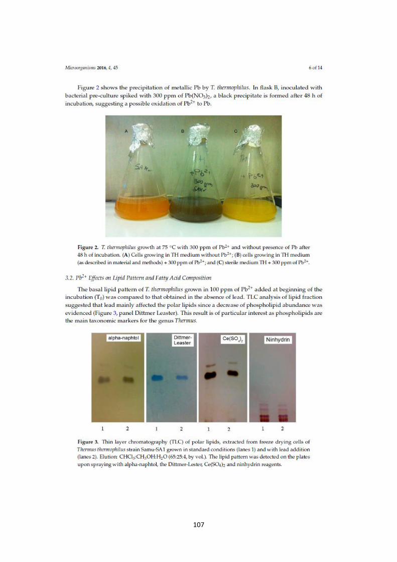
107
108

109

110

111

112

113

114
115
116
This paper is in press and will be published in April 2017 in the journal “La Chimica e l’industria”.
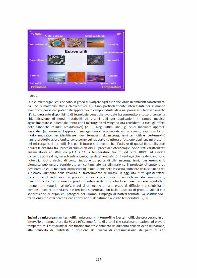
117

118

119
120

121
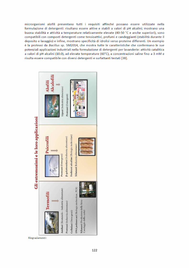
122
123
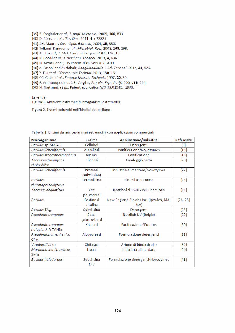
124

125