ii. cardiac glycosides from strophanthus boiviniiii. cardiac glycosides from strophanthus boivinii...
TRANSCRIPT

25
II. Cardiac glycosides from Strophanthus boivinii
2.1 Introduction
In our continuing search for bioactive natural products from Madagascar
rainforests as a part of International Cooperative Biodiversity Group (ICBG) program, we
obtained ethanol extract of the plant Roupellina boivinii, also known as Strophanthus
boivinii.1 This plant extract (MG 2309PE) exhibited antiproliferative activity in the
A2780 ovarian cancer cell line (IC50 = 11 µg/mL) and hence it was selected for further
fractionation to isolate its active components. Bioassay-guided fractionation of this
extract yielded three new cardenolide glycosides, boivinides A, B and F, as well as one
known cytotoxic cardenolide glycoside. The study and structural characterization of these
compounds is reported herein.
2.1.1 Previous studies of Strophanthus genus
Strophanthus boivinii (Fig. 2.1)2 is a
small flowering tree that belongs to the
Apocynaceae family. This family is native
mainly to Africa, extending to South Africa
with a few species in Asia. The family has
424 genera and over 2100 species. The
medicinal plants of this family are known to
contain lignans,3 cytotoxic alkaloids and
cardenolide glycosides.
Figure 2.1 Strophanthus boivinii Source: © TopTropicals.com

26
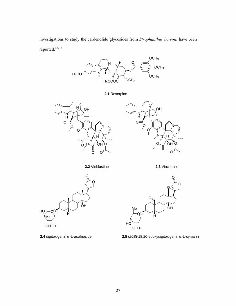
Reserpine (2.1), an indole alkaloid extracted from the roots of Rauwolfia
serpentine, is an antipsychotic and antihypertensive drug.4 The vinca alkaloids,
vincristine (2.2) and vinblastine (2.3), isolated from Catharanthus roseus, are effective
against leukemia.5 The Strophanthus genus within the Apocynaceae family consists of
35-40 species.6 The name strophas anthos (twisted cord flower) is derived from the long
twisted threadlike corolla. The Strophanthus genus includes small shrubs, trees, and vines
which attain an average height of 30 feet.
The genus Strophanthus is most associated with cardenolide glycosides which are
an important class of natural products that can be used as drugs as well as toxins. Since
1500 B. C. the leaves of Strophanthus genus have been used to cure skin ulcerations, to
reduce fever and the decoction of leaves has been a good remedy for gonorrhea.7 As an
example of their use as toxins, Inee, (also known as onaye), obtained from Strophanthus
hispidus8 is used as an arrow poison for hunting in West Africa.
The seeds of Strophanthus gratus have anticoagulant properties and have been
used on wounds and treatment of snake-bites in Africa.3 The seeds and the plant extract of
Strophanthus kombé is beneficial as a cardiac drug or a diuretic.9 These plants are mainly
used in the treatment of congestive heart failure; however, their toxicity limits their
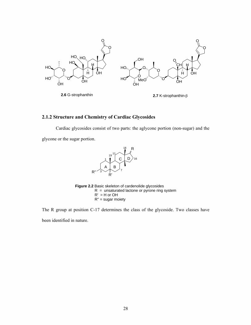
extensive use. Ouabain, a more common name for G-strophanthin (2.6), a poisonous
glycoside obtained from the fruits of Strophanthus gratus,10 is used widely to block the
sodium pump for in vitro studies.11 K-strophanthin (2.7), isolated from Strophanthus
kombé, was also used for the treatment of heart diseases until its adverse side effects were
observed.12 As would be expected, the cytotoxicity and structural characterization of
various cardenolide glycosides have been extensively studied. However, only two
27
investigations to study the cardenolide glycosides from Strophanthus boivinii have been
reported.13, 14
NH
N
H3CO H
H3COOC
O
OCH3
OCH3
OCH3OCH3
H
H O
2.1 Reserpine
OO
ON
OH
N
HH
OH O
OO
O
ON
N
HH
OH O
OO
O
2.2 Vinblastine 2.3 Vincristine
NH
N OH
OO
NH
N OH
O
OOHOMe
OHOH
H
OH
O
2.4 digitoxigenin α-L-acofrioside 2.5 (20S)-18,20-epoxydigitoxigenin α-L-cymarin
O
OO
OCH3
H
OH
O
Me
HO
O
O
28
O
O
OCH
OH
OHO
MeO
O
H
H
2.7 K-strophanthin-β
O
O
O
O
OH
OH
HO
HO
HOOH
O
H
H
2.6 G-strophanthin
HOHO
O
OH
HO
HO
OH
2.1.2 Structure and Chemistry of Cardiac Glycosides
Cardiac glycosides consist of two parts: the aglycone portion (non-sugar) and the
glycone or the sugar portion.
R
R'R''
Figure 2.2 Basic skeleton of cardenolide glycosides R = unsaturated lactone or pyrone ring system R' = H or OH R'' = sugar moiety
1
3 7
19
18
16
11
A B
C D
The R group at position C-17 determines the class of the glycoside. Two classes have
been identified in nature.

29
O
O
Cardenolide:
Figure 2.3 Two classes of cardiac glycosides
O
O
Bufadienolides:
Cardiac glycosides with a cardenolide substituent are generally found in plants,
and cardiac glycosides that contain bufadienolide substituents are obtained from animal
sources. The aglycone consists of a four fused ring system, A, B, C and D. The
stereochemistry of the rings greatly influences the cytotoxicity of these compounds.
Compounds with a trans fusion of A/B and cis fused C/D rings are less active as
compared to the compounds with cis fusions of both A/B and C/D rings. Removal of the
–OH group at the 14β- position does not affect the activity of the compound. The double
bond in the lactone ring plays an important role in the activity of the compound, and
saturation of the ring causes a marked decrease in this activity.15 However, the lactone
ring is not absolutely required, and if it is replaced with a nitrile group, the resulting
compound shows little or no loss of activity. Also, the lactone ring alone does not exhibit
any biological activity. The sugar moiety is not required for the activity of the molecule,
but it is essential for the pharmacokinetics, absorption and half-life of the compound,
etc.15 For example, lanatoside C (2.8) differs from digoxin (2.9) in the sugar residue and
is more lipophilic with greater water solubility and a faster absorption rate than digoxin.

30
O
O
O
OO
O
HOO
O
2.9 Digoxin
O
O
O
OO
O
HOO
O
O
2.8 Lanatoside C
O
O
OH
HO
HO
HOO
OH
OH
HO
HO HO H
OH
H
OHHOHO
Cardiac glycosides are common compounds present in plants. These glycosides
inhibit Na+/K+ ATPase and increase the contraction of the heart muscles. The aglycone
portion of these glycosides is very poisonous, and in low concentrations, exhibits
antiproliferative activity.16 Repke et al. in 1988 established the importance of Na+/K+
ATPase in cancer cell proliferation. Hence, inhibition of Na+/K+ ATPase can play an
important role in apoptosis and cancer cell death.17 However, due to their high levels of
toxicity and adverse side effects, their medicinal use has not been much explored. Further
studies have to be performed to confirm their use as cancer drugs.
2.2 Results and Discussion
Strophanthus boivinni was studied previously by Mr. Eba Adou, a doctoral
student from our research group. The plant extract was subjected to solvent partitioning
between hexane and aqueous MeOH, followed by extraction of the methanol fraction
with dichloromethane. Both methanol and dichloromethane fractions were active and
were filtered through short C18 chromatography columns and separated by HPLC-C18

31
using a MeOH:H2O solvent system. Mr. Adou isolated three known (6.1, 6.2 and 6.4) and
three new cardenolides (6.3, 6.5 and 6.6) from this extract (NO55899). However, the
extract yielded many active fractions, and thus the work on this extract was continued to
further isolate its additional active compounds.
O
O
OOH
H
HO
HO
O
O
6.1 5α− corotoxigenin-β-D-boivinoside
O
O
OOH
H
HO
MeO
O
O
6.2 17α− corotoxigenin-β-D-sarmentoside
O
O
OOH
H
O
MeO
O
6.3 5α, 17α− uzarigenin-3-O-[β-D-glucopyranosyl-
O
HO
HO
OH
OH
1 4)-β-D-sarmentoside
O
O
OOH
H
HO
HO
O
6.4 5α− uzarigenin-3-O-α-L-rhamnoside
OH
IC50 = 0.08 μg/ml IC50 = 2.0 μg/ml
IC50 = 2.0 μg/ml IC50 = 0.08 μg/ml
H H

32
O
O
OOH
H
O
HO
O
O
6.5 5α− uzarigenin-3-O-[β-D-glucosyl-
O
HO
HO
OH
1 4)-β-D-boivinoside
O
O
OOH
H
O
HO
O
O
6.6
O
HO
HO
OH
OH
OH OH
IC50 = 0.2 μg/ml
H
Figure 2.4 Cardenolide glycosides isolated by Mr. Adou from Strophanthus boivinii
2.2.1 Isolation of boivinides A-C and digitoxigenin 3-O-β-D-glucopyranosyl-
(1→ 4)-α-L-acofriopyranoside.
The EtOH extract of the plant, designated MG 2309/11 (3.0 g) was suspended in
aqueous MeOH (MeOH:H2O, 9:1, 100 mL) and extracted with hexanes (3 × 100 mL).
The aqueous layer was then diluted to 50% MeOH (v/v) and extracted with CH2Cl2 (3 ×
180 mL). The aqueous MeOH fraction displayed the highest activity (IC50 = 0.61 µg/mL).
This fraction was chromatographed over a C18 column with 40% MeOH:H2O (0.1%
formic acid) for 40 min followed by 50% MeOH:H2O (0.1% formic acid) for 50 min and
twenty-five subfractions were collected. Fractions 16 and 18 were pure and new
compounds and named boivinide B (2) and boivinide A (1). Fraction IV was loaded on a
C18 column and eluted with 50% MeOH:H2O (0.05% formic acid) for 90 min. Two pure
subfractions were collected; one was a new compound, boivinide F (3) and the other was
a known compound (4), digitoxigenin 3-O-β-D-glucopyranosyl-(1→4)-α-L-
acofriopyranoside.18

33
Scheme 2.1 Fractionation tree for Strophanthus boivinii
MG 2309PEAmt = 3.0 gIC50 =11 μg/mL
Hexane fraction128-2345.4 mgIC50 >20 μg/mL
90% MeOH/water
50%MeOH/H2O fraction128-41.84 gIC50 = 0.61 μg/mL
CH2Cl2 fraction128-3117.9 mgIC50 = 1.5 μg/mL
LIQ-LIQ Partitioning3X DCM
LIQ-LIQ Partioning3X Hexane
131-1 131-2 131-3 131-4 131-5
Yield in mg: 904.9 123.15 282.7 244.8 123.1
IC50 in μg/mL: 3.1 2.0 0.13 0.48 2.8
Open column C18 -30% MeOH:H2O to 100% MeOH
Filtration through C18 open column
HPLC-C18 Isocratic 40% MeOH:H2Owith 0.1% formic acid for 40 min followed by 50% MeOH:H2O with 0.1% formic acid for 50 min
Inactive fractions 16 18 Inactive fractionsYield in mg: 16.7 4.5 5.9 15.3 IC50 inμg/mL: >20 0.46 0.12 >20
HPLC-C18 Isocratic elution with 50% MeOH:H2O with 0.05% formic acid for 40 min
187-1
(B, 2) (A, 1)
189-1
Inactive fractions 9 12 Inactive fractionsYieldin mg: 35.9 3.5 4.9 37.8
IC50 in μg/mL: >20 0.41 0.11 >20
(F, 3)
Filtration through C18 opencolumn
(4)
Loaded ~50 mg
Loaded ~90 mg
Strophanthus boivinii

34
2.2.2 Structure Elucidation of Boivinide A
Boivinide A (compound 1) was obtained as a white amorphous solid. Positive ion
LC-MS gave molecular ion peak at m/z 749.4 [M+K+], consistent with the molecular
composition of C36H54O14. Previous studies on this plant indicated that compound 1
belonged to the class of cardenolide glycosides.
The 1H spectrum of 1 in pyridine-d5 (Fig. 2.5) indicated that the compound was
pure with several oxygenated protons and methyl groups. A peak at δH10.01 (s, H-19)
suggested the presence of an aldehyde proton. The spectrum also showed signals for one
methoxy group at δH 3.62 (s), one methyl at δH 0.93 (s, H-18), and a methyl doublet at
1.63 (d, H-6') indicating the presence of a deoxy sugar moiety in the compound. Two
anomeric proton signals at δH 4.77 (d, H-1') and δH 5.20 (d, H-1'') confirmed the presence
of two sugar moieties in the compound. The 13C spectrum of compound 1 contained 36
signals: one methoxy, two methyls, eleven methylenes, seventeen methines and five
quaternary carbons.
12 10 8 6 4 2 0 PP
Figure 2.5 1H NMR spectrum of compound 1 in pyridine-d5

35
The 1H and 13C NMR signals in pyridine-d5 showed typical signals for an α,β-
unsaturated γ- lactone unit with peaks at δC 176.0 (C-20), δC/δH 74.0 (C-21)/5.03 and 5.27
(br d, J = 18.0 Hz, H-21), δC/δH 118.1 (C-22)/6.10 (br s, H-22), δC 174.8 (C-23). Four spin
systems, A, B, C and D for the aglycone portion were identified by COSY, 1D and 2D
TOCSY spectra. From HSQC and HMBC correlations, the proton-carbon pairs were
connected to each other and 1JCH correlations were determined (Table 1) to obtain a
cardenolide skeleton (2.13).
HMBC correlations from H-17 (δH 2.75, s) to C-12, C-13, C-14, C-15, C-16, C-
20, C-21 and C-22, as well as H-22 (δH 6.12, s) to C-17, indicated the point of
attachment of the lactone ring system to ring D. H-18 (δH 0.93, s) exhibited a very strong
correlation to C-12 (δC 39.9), C-13 (δC 49.9), C-14 (δC 84.1) and C-17 (δC 50.1). These
correlations led to the construction of Fragment 1 and confirmed the C/D ring fusion at C-
13 and C-14.
O
O
OH
H
HH
HHC
Figure 2.6 Key HMBC correlations of fragment 1
12
9
11
23
The fusion of the A/B rings was confirmed by the correlation of H-19 (δH 10.01,
s) to C-1 (δC 31.7), C-5 (δC 43.2), C-9 (δC 48.8); H-5 (δH 1.19, m) to C-1, C-3 (δC 76.9),
C-4 (δC 36.4), C-6 (δC 29.0), C-7 (δC 27.4), C-9 and C-10 (δC 55.5). Similarly, the B/C
ring fusion was confirmed by correlation of H-8 (δH 1.81) to C-6, C-7, C-11(δC 22.4), C-

36
13, and C-14 and of H-9 (1.20, m) to C-1, C-5, C-7, C-8 (δC 43.1), C-10, C-11, C-12 and
C-19 (δC 209.1).
H
H
H
O HH
H
HA
Figure 2.7 Key HMBC correlations of A/B and B/C rings of compound 1
2
3
6
198
A strong HMBC correlation from H-3 to C-1' confirmed that the first sugar was
connected to the aglycone at position 3 on A ring.
O
OMeOOH
H
H
Figure 2.8 Key HMBC correlation for connecting the sugar moiety to the aglycone of compound 1
A33'
6'
In the second sugar, H-1'' (δH 5.20) correlated to C-4' of the first sugar. Similarly,
a correlation of H-4' to C-1'' was observed, suggesting a 1→4 attachment of the two
sugars.
O
OOH
Figure 2.9 Key HMBC correlation for attachmentof sugar II to sugar I of compound 1
O
OMeO
HOHO
HO
OH
H H
A1'
1''
4''
6''
3'

37
The 1H NMR spectrum of compound 1 was very complex, and extensive overlap
of peaks especially between 2.0 and 4.0 ppm, made analysis of the spectrum very
difficult. However, an HSQC experiment in pyridine-d5 was extremely useful for
detecting single bond C-H correlations, especially for assembling the sugar portion, and
made the 1JCH correlations very clear. H-5 (δH 1.19), H-9 (δH 1.20), H-11 (δH 1.22) and H-
12 (δH 1.23) were well resolved and correlated to C-5 (δC 43.2), C-9 (δC 48.9), C-11 (δC
22.3) and C-12 (δC 39.5) respectively. Similarly, H-4' (δH 4.25), H-3'' (δH 4.23) and H-4''
(δH 4.18) correlated to C-4' (δC 76.4), C-3'' (δH 78.5) and C-4'' (δC 72.2) respectively.
The stereochemistry of compound 1 was determined from 1D and 2D ROESY
spectra. The ROESY correlations of H-19 (δH 10.01, s) to Hβ-1 (δH 2.39 m), Hβ-2 (δH
2.20, m), Hβ-4 (δH 2.04, m), Hβ-11 (δH 1.22, m) and H-5 (δH 1.19, m) to Hα-1 (δH 0.90,
m), H-3 (δH 3.97, br s), Hα-6 (δH 1.42, m), H-9 (δH 1.20, m) indicated a trans fusion of the
A and B rings. Correlations of H-8 (δH 1.81) to H-19 (δH 10.01, s) and Hβ-11 (δH 1.22, m)
as well as correlations of H-9 (δH 1.20, m) to H-5 (δH 1.19) and H-18 (δH 0.93, s)
suggested a trans fusion of the B/C rings of the aglycone.
CHO
H
HH
H
H
O
O
CH3
OHH
HH
H
H HH
Figure 2.10 Key ROESY correlations of the aglyconeportion of compound 1
H
The ROESY spectrum also indicated crosspeaks from H-17 (δH 2.75, s) to H2-21
(5.03, m 5.27, d, J = 18.0 Hz), H-22 ((δH 6.12, s), and H2-16 (δH 1.96, m 2.06, m), as well
38
as H-18 (δH 0.93, s) to H-21 and H-22, but not H-17, hence the lactone ring was assigned
the β configuration.
Figure 2.11 ROESY correlations of the D ring and the lactone unit of compound 1
O
O
CH3
OHH
H
H
HH
H
HH
The 1H and 13C chemical shifts for the sugar moieties were assigned as follows:
δC/δH 102.8 (C-1')/4.77 (br d, J = 7.2 Hz) and δC/δH 105.7 (C-1'')/5.20 (br d, J = 8.0 Hz)
indicated the sugars had β-linkages; δC/δH 85.7 (C-3')/3.58 ( dd, J = 10.0, 12.4 Hz), δC/δH
78.6 (C-3'')/4.23 (br s); H-4' appears as a multiplet at δH 4.25 (C-4' δC 76.4); H-5' is a
multiplet at δH 3.77 (C-5' δC 79.0); H-6' is a prominent broad doublet at δH 1.63 (J = 6.8
Hz, C-6' δC 18.1), H-6'' is a methylene with δH 4.38 (m), 4.59 (m) and a 3'-methoxy group
appeared as a singlet at δH 3.62. The glycone portion of the compound was connected
together from COSY, TOCSY and HMBC spectra. The stereochemistry at C-3 position
was confirmed by ROESY correlation of H-3 (δH 3.97) to H-1' (δH 4.77), suggesting a cis
relationship between H-1' and H-3. (2.12)
3 5
6' 19
1'
HH
HO
OHO
H
CH3
H
OHOHO
OH
OH
MeO
Figure 2.12 Key ROESY correlations ofsugar I and the aglycone of compound 1
HH
HH
CHO

39
The two sugars were attached by a 4'→1'' linkage. The H-4' and H-1'' protons
were oriented cis to each other. The chemical shifts of the sugar portion matched the
literature values for β-D-glucopyranosyl-β-D-digitaloside19 very closely. The 1H and 13C
NMR shifts of the shifts of the aglycone portion were similar to those of corotoxigenin.20
This assignment of the sugar and the aglycone moieties in boivinide A led to its structural
assignment as 5α-corotoxigenin-β-D-glucopyranosyl-(1→4)-β-D-digitaloside. This
compound has been isolated and identified for the first time. The final structure of
boivinide A is as shown in Figure 2.13.
OHO
H
CH3OHO
HOOH
OH
MeOH
HH
O
O
OOH
OO
MeOOH
H
OHO
OHHO
O
HO
1 HMBC Correlations
O
O
OOH
OO
MeOOH
H
OHO
OHHO O
HO
CHO
H
HH
H
OH
O
O
CH3
OHH
HH
H
H HH
ROESY Correlations
1
35
8
12
16
21
23
1'3'
6'1''
5''
3'' 19H
H
Figure 2.13 Final structure and key HMBC and ROESY correlations of compound 1
40
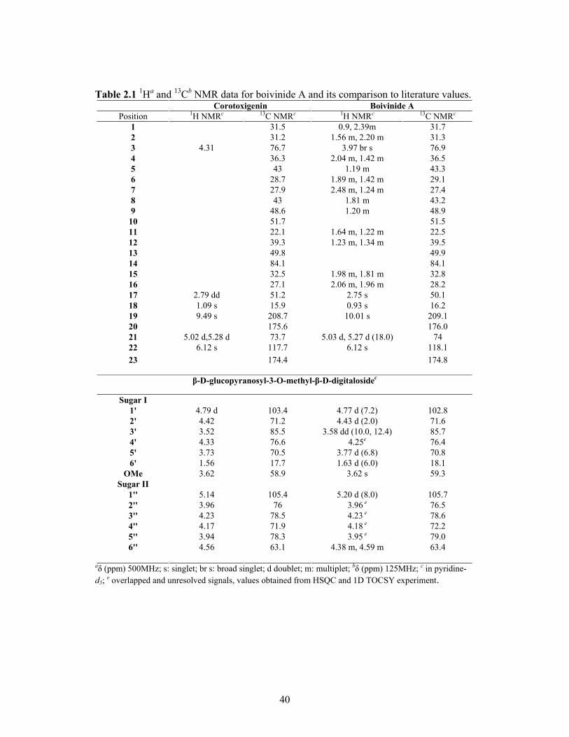
Table 2.1 1Ha and 13Cb NMR data for boivinide A and its comparison to literature values. Corotoxigenin Boivinide A
Position 1H NMRc 13C NMRc 1H NMRc 13C NMRc 1 31.5 0.9, 2.39m 31.7 2 31.2 1.56 m, 2.20 m 31.3 3 4.31 76.7 3.97 br s 76.9 4 36.3 2.04 m, 1.42 m 36.5 5 43 1.19 m 43.3 6 28.7 1.89 m, 1.42 m 29.1 7 27.9 2.48 m, 1.24 m 27.4 8 43 1.81 m 43.2 9 48.6 1.20 m 48.9
10 51.7 51.5 11 22.1 1.64 m, 1.22 m 22.5 12 39.3 1.23 m, 1.34 m 39.5 13 49.8 49.9 14 84.1 84.1 15 32.5 1.98 m, 1.81 m 32.8 16 27.1 2.06 m, 1.96 m 28.2 17 2.79 dd 51.2 2.75 s 50.1 18 1.09 s 15.9 0.93 s 16.2 19 9.49 s 208.7 10.01 s 209.1 20 175.6 176.0 21 5.02 d,5.28 d 73.7 5.03 d, 5.27 d (18.0) 74 22 6.12 s 117.7 6.12 s 118.1 23 174.4 174.8
β-D-glucopyranosyl-3-O-methyl-β-D-digitalosidec
Sugar I 1' 4.79 d 103.4 4.77 d (7.2) 102.8 2' 4.42 71.2 4.43 d (2.0) 71.6 3' 3.52 85.5 3.58 dd (10.0, 12.4) 85.7 4' 4.33 76.6 4.25e 76.4 5' 3.73 70.5 3.77 d (6.8) 70.8 6' 1.56 17.7 1.63 d (6.0) 18.1
OMe 3.62 58.9 3.62 s 59.3 Sugar II
1'' 5.14 105.4 5.20 d (8.0) 105.7 2'' 3.96 76 3.96 e 76.5 3'' 4.23 78.5 4.23 e 78.6 4'' 4.17 71.9 4.18 e 72.2 5'' 3.94 78.3 3.95 e 79.0 6'' 4.56 63.1 4.38 m, 4.59 m 63.4
aδ (ppm) 500MHz; s: singlet; br s: broad singlet; d doublet; m: multiplet; bδ (ppm) 125MHz; c in pyridine-d5; e overlapped and unresolved signals, values obtained from HSQC and 1D TOCSY experiment.

41
2.2.3 Structure Elucidation of Boivinide B
Compound 2 (boivinide B) was also obtained as a white amorphous solid. Positive
ion LC-MS gave a molecular ion peak at m/z 695.4 [M+H]-, consistent with the molecular
formula of C36H54O13. The 1H NMR spectrum was characteristic of a cardenolide
glycoside.
The 1H spectrum of compound 2 in pyridine-d5 (Fig. 2.14) showed the presence
of thirteen oxygenated carbons, one less than boivinide A. The 1H NMR spectrum
showed a signal at δH10.05 (s, H-19) for an aldehyde proton, and signals for one methoxy
at δH 3.62 (s), one methyl group at δH 0.92 (s, H-18), and a methyl doublet at 1.71 (d, H-
6'). Two anomeric proton signals were seen at δH 5.48 (d, H-1') and δH 5.04 (d, H-1''),
indicating the presence of two sugar moieties in the compound.
Figure 2.14 1H NMR spectrum of compound 2 in pyridine-d5
10 9 8 7 6 5 4 3 2 1 PPM

42
The 13C spectrum of compound 2 contained 36 signals: one methoxy, two
methyls, twelve methylenes, sixteen methines and five quaternary carbons, consistent
with a cardenolide framework. H-18 (δH 0.92, s) exhibited correlations to C-12 (δC 39.7),
C-13 (δC 50.2), C-14 (δC 84.5), C-17 (δC 52.2). H-3 (δH 3.92, s) correlated to C-2 (δC
31.6), C-4 (δC 36.6), and C-1'(δC 96.3). The 1H and 13C NMR signals in pyridine-d5
showed typical signals for an α,β-unsaturated γ-lactone unit. The aldehyde proton at C-19
(δC 209.3) position suggested that the aglycone in compound 2 was corotoxigenin,20 and
this was further confirmed by the comparison of the 1H and 13C NMR shifts of the
aglycone of compound 2 with those of compound 1. Further analysis of COSY, 1D and
2D TOCSY confirmed that aglycone portion was the same as that of boivinide A (1).
The ROESY correlations of H-19 (δH 10.05, s) to Hβ-1 (δH 2.40, m), Hβ-2 (δH
2.38, m), Hβ-4 (δH 1.86, m), Hβ-11 (δH 1.62, m) and H-5 (δH 1.20, m) to Hα-1 (δH 0.83,
m), H-3 (δH 3.92, br s), Hα-6 (δH 1.40, m) and H-9 (δH 2.75, m) indicated a trans
orientation of H-5 (δH 1.20, m) and H-19 (δH 10.05, s) to each other. The stereochemistry
at this position was further confirmed by ROESY crosspeaks from H-5 (δH 1.20, m) to H-
3 (δH 3.92, m) and H-3 to H-1' (δH 5.48, d, J = 9.6 Hz). Trans fusion of the B/C rings and
cis fusion of the C/D rings further confirmed that the aglycone of compound 2 was
corotoxigenin.20
43
O
H
OH
O
OH
H
H
H
H
H
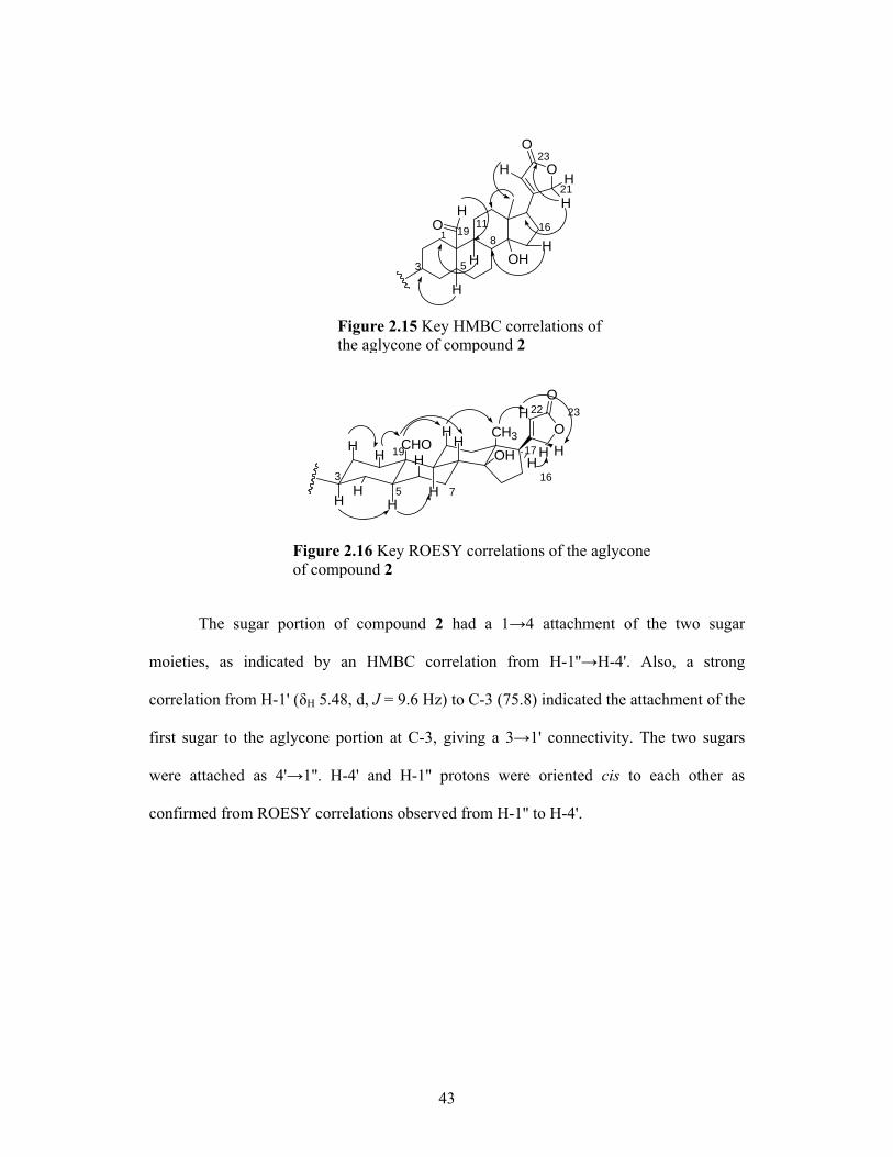
Figure 2.15 Key HMBC correlations of the aglycone of compound 2
1
3 5
1911
23
21
168
3
19
16
17
2322
5 7
CHO
H
HH
H
H
O
O
CH3
OHH
HH
H
H HH
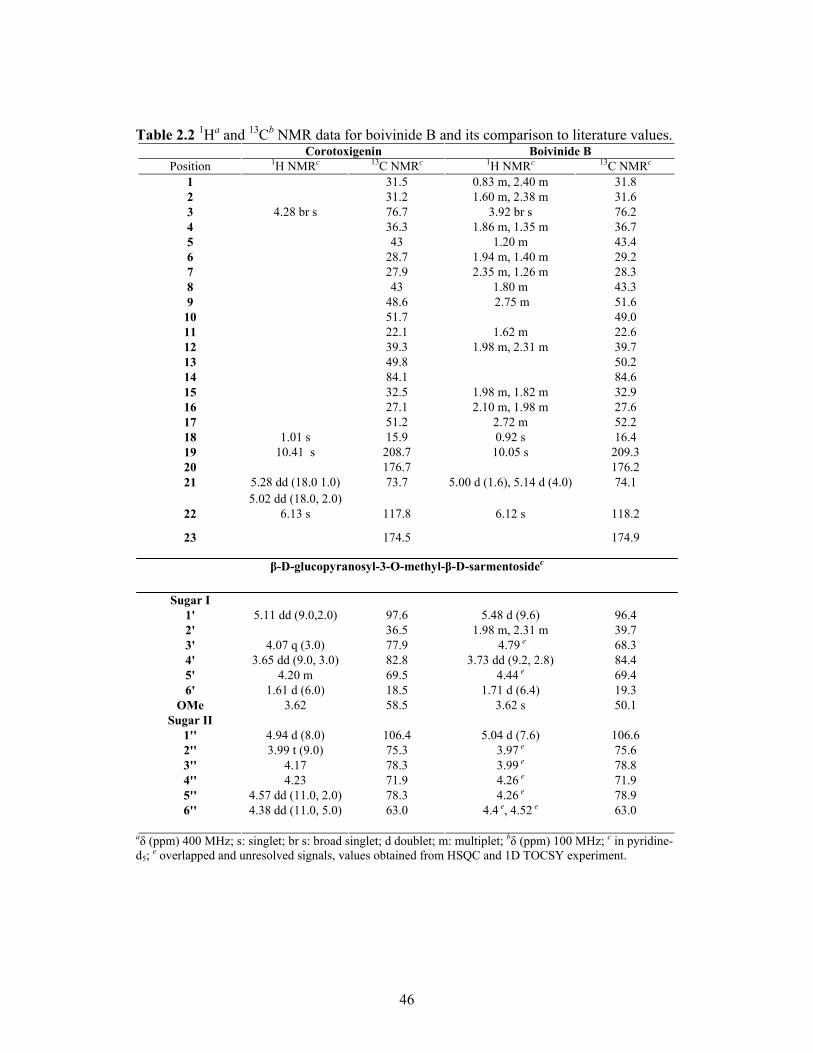
Figure 2.16 Key ROESY correlations of the aglyconeof compound 2
H
The sugar portion of compound 2 had a 1→4 attachment of the two sugar
moieties, as indicated by an HMBC correlation from H-1''→H-4'. Also, a strong
correlation from H-1' (δH 5.48, d, J = 9.6 Hz) to C-3 (75.8) indicated the attachment of the
first sugar to the aglycone portion at C-3, giving a 3→1' connectivity. The two sugars
were attached as 4'→1''. H-4' and H-1'' protons were oriented cis to each other as
confirmed from ROESY correlations observed from H-1'' to H-4'.

44
1'1''3''
6'
3 5
6''
Figure 2.17 Key ROESY correlations of thesugar portion of compound 2
CHO
H
HH
H
OHO
OMe
CH3
H
O
H
HOHO
OH
OH
O
HHH
HH
The HMBC correlations for the sugar portion showed that the sugar moieties of 2
were similar to those of 1, except for the absence of one oxygenated carbon. HMBC,
ROESY and 1D TOCSY spectra suggested that the first sugar of 2 was different from that
of 1. The assignments of the sugars in 2 were made as follows: δC/δH 96.3 (C-1')/5.48 (dd,
J = 9.6 Hz), and δC/δH 106.6 (C-1'')/5.04 (br d, J = 7.6 Hz); δC/δH 39.7 (C-2')/1.98 and
2.31; δC/δH 68.2 (C-3')/4.79 (m), δC/δH 78.8 (C-3'')/3.9 (br s); H-4' appeared as a doublet
at δH 3.73 (dd, J = 9.2, 2.8 Hz), (C-4' δC 84.4); H-5'' was a multiplet at δH 4.26 (C-5'' δC
78.8); H-6' was a prominent broad doublet at δH 1.71 (J = 6.4 Hz, C-6' δC 19.3), H-6'' was
a methylene with δH 4.40, 4.52 and the 3'-methoxy group appeared as a singlet at δH 3.62.
O
OO
O
HOHO
HO
OH
MeO
CHO
HH
H
Figure 2.18 HMBC connection of the sugar to the aglycone of compound 2
31
1'
6'
1''
4''
6''
The NMR spectra of the sugar portion matched the spectra of β-D-
glucopyranosyl-(1→4)-β-D-sarmentoside.21, 22 The structure of boivinide B (compound 2)

45
was thus assigned as 5α-corotoxigenin-β-D-glucopyranosyl-(1→4)-β-D-sarmentoside
(2.19). This compound has not been previously observed in nature.
O
O
OOH
OO
MeOH
OHO
OHHO O
HO
2 HMBC Correlations
O
O
OOH
OO
MeOH
OHO
OHHO O
HO
CHO
H
HH
H
OHO
OMe
H
O
O
CH3
OHH
HH
H
H HH
O
H
HOHO
OH
OH
ROESY Correlations
1
3 5 7
8
12
19 16
21
23
1'
6'
3'
1''
6''
4''
O
HHH
HCH3
HH
Figure 2.19 Key HMBC and ROESY correlations of compound 2
46
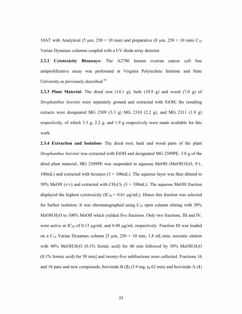
Table 2.2 1Ha and 13Cb NMR data for boivinide B and its comparison to literature values. Corotoxigenin Boivinide B
Position 1H NMRc 13C NMRc 1H NMRc 13C NMRc 1 31.5 0.83 m, 2.40 m 31.8 2 31.2 1.60 m, 2.38 m 31.6 3 4.28 br s 76.7 3.92 br s 76.2 4 36.3 1.86 m, 1.35 m 36.7 5 43 1.20 m 43.4 6 28.7 1.94 m, 1.40 m 29.2 7 27.9 2.35 m, 1.26 m 28.3 8 43 1.80 m 43.3 9 48.6 2.75 m 51.6
10 51.7 49.0 11 22.1 1.62 m 22.6 12 39.3 1.98 m, 2.31 m 39.7 13 49.8 50.2 14 84.1 84.6 15 32.5 1.98 m, 1.82 m 32.9 16 27.1 2.10 m, 1.98 m 27.6 17 51.2 2.72 m 52.2 18 1.01 s 15.9 0.92 s 16.4 19 10.41 s 208.7 10.05 s 209.3 20 176.7 176.2 21 5.28 dd (18.0 1.0) 73.7 5.00 d (1.6), 5.14 d (4.0) 74.1
5.02 dd (18.0, 2.0) 22 6.13 s 117.8 6.12 s 118.2
23 174.5 174.9
β-D-glucopyranosyl-3-O-methyl-β-D-sarmentosidec
Sugar I 1' 5.11 dd (9.0,2.0) 97.6 5.48 d (9.6) 96.4 2' 36.5 1.98 m, 2.31 m 39.7 3' 4.07 q (3.0) 77.9 4.79 e 68.3 4' 3.65 dd (9.0, 3.0) 82.8 3.73 dd (9.2, 2.8) 84.4 5' 4.20 m 69.5 4.44 e 69.4 6' 1.61 d (6.0) 18.5 1.71 d (6.4) 19.3
OMe 3.62 58.5 3.62 s 50.1 Sugar II
1'' 4.94 d (8.0) 106.4 5.04 d (7.6) 106.6 2'' 3.99 t (9.0) 75.3 3.97 e 75.6 3'' 4.17 78.3 3.99 e 78.8 4'' 4.23 71.9 4.26 e 71.9 5'' 4.57 dd (11.0, 2.0) 78.3 4.26 e 78.9 6'' 4.38 dd (11.0, 5.0) 63.0 4.4 e, 4.52 e 63.0
aδ (ppm) 400 MHz; s: singlet; br s: broad singlet; d doublet; m: multiplet; bδ (ppm) 100 MHz; c in pyridine-d5; e overlapped and unresolved signals, values obtained from HSQC and 1D TOCSY experiment.

47
2.2.4 Structure Elucidation of Boivinide F
Compound 3 (boivinide F) was also obtained as a yellow amorphous solid.
Positive ion HRFABMS gave a molecular ion peak at m/z 775.3570 [M+Na]+, consistent
with the molecular formula of C38H56O15 and with the same aglycone portion as
boivinides A and B. The presence of a glucose unit was confirmed by 1D TOCSY, 1D
and 2D ROESY spectra. However, additional peaks appeared at δC 170.0, indicating the
presence of an additional carbonyl group, and a singlet at δH 2.08 (3H, s). Analysis of the
HMBC spectrum led to the conclusion that the first sugar of compound 3 was connected
to the aglycone at C-3. An HMBC correlation from H-1' ((δH 4.82, d, J = 8.4 Hz) to C-3
(δC 77.9) confirmed the 1→3 connectivity of the aglycone and the sugar moieties. The
proton and carbon chemical shifts of the two sugars were assigned as follows: δC/δH 100.6
(C-1')/4.82 (d, J = 8.4 Hz), and δC/δH 105.4 (C-1'')/5.16 (br d, J = 7.6 Hz); δC/δH 72.3 (C-
2')/5.83, indicating that it was highly deshielded; δC/δH 83.4 (C-3')/3.64 ( d, J = 2.8 Hz),
δC/δH 78.7 (C-3'')/4.26 (br s); H-4' appeared as a multiplet at δH 4.46 (C-4' δC 75.1); H-5''
was a multiplet at δH 3.78 (C-5'' δC 71.2); H-6' was a prominent broad doublet at δH 1.60
(J = 6.0 Hz, C-6' δC 18.0), H-6'' was a methylene with δH 4.38, 4.60 and the 3'-methoxy
group appeared as a singlet at δH 3.49.(2.22)
O
OO
OH
OMeO
OHHO
HO
HOH
HH
Figure 2.20 Key HMBC correlations of the two the sugar moieties of compound 3
3
1
1'4'
6'
1''4''
6''
O

48
The acetoxy group (δC 170.0, δC/δH 21.4/2.08) was attached at position C-2' (δC/δH
72.3/5.83) of the first sugar. After comparing the chemical shifts of the first sugar to
literature values, it was concluded that the first sugar is 2-acetyl-3-methyl-β-D-
fucopyranoside.23 The relative stereochemistry of the first sugar was confirmed by
ROESY crosspeaks between H-1' ((δH 4.82, d, J = 8.4 Hz) and H-3 ((δH 3.90, br s); H-3'
(δH 3.62, d, J = 6.4 Hz) and H-5' (δH 3.78, d, J = 2.8 Hz) as well as H-4' (δH 4.47, d, J =
2.0 Hz) and H-1'' (δH 5.16, d, J = 7.6 Hz ) (2.21).
1'
1''3''
6'
3 5
6''
H
HH
H
OHO
H
CH3
H
O
AcO
HOHO
OH
OH
O
H
HHH
H
Figure 2.21 Key ROESY correlations of the two sugar moieties of compound 3
MeO
OH
The comparison of the 1H and 13C NMR data of compound 3 to literature values
led to the conclusion that the aglycone of 3 was corotoxigenin,20 and the sugar moiety
was β-D-glucoypyranosyl-(1→4)-2-acetyl-3-methyl-β-D-fucopyranoside.23 The structure
of boivinide F (compound 3) was thus assigned as 5α-corotoxigenin-β-D-
glucoypyranosyl-(1→4)-2-acetyl-3-methyl-β-D-fucopyranoside (2.22).

49
O
O
O
OO
OH
OH
HO OH
HO
OHMeO
OO
O
3 HMBC Correlations
O
O
OOH
OO
MeOH
OHO
OHHO
O
HOO
1
3 5
8
12
19
18
16
21
23
1'
6'
3'
1''
6''
4''
CHO
H
H H
H
H
O
O
CH3
OHH
HH
H
H HH
ROESY Correlations
OHO
H
CH3O
AcO
HOHO
OH
OH
O
H
HH HMeO
Figure 2.22 Final Structure and key HMBC and ROESY correlations of compound 3

50
Table 2.3 1Ha and 13C bNMR data for boivinide C and its comparison to literature values. Corotoxigenin Boivinide C
Position 1H NMRc 13C NMRc 1H NMRc 13C NMRc 1 31.5 0.98, 2.41m 31.8 2 31.2 1.98 m, 1.45 m 31.6 3 4.28 br s 76.7 3.90 br s 76.2 4 36.3 2.03 m, 1.5 m 36.7 5 43 1.24 m 43.4 6 28.7 1.30 m, 1.30 m 29.2 7 27.9 1.83 m, 1.83 m 28.3 8 43 1.22 m 43.3 9 48.6 2.70 m 51.6
10 51.7 49.0 11 22.1 1.63 m, 1.33 22.6 12 39.3 1.21 m, 1.34 m 39.7 13 49.8 50.2 14 84.1 84.6 15 32.5 1.99 m, 1.82 m 32.9 16 27.1 2.15 m, 1.97 m 27.6 17 51.2 2.75 dd 52.2 18 1.01 s 15.9 0.94 s 16.4 19 10.41 s 208.7 10.04 s 209.3 20 176.7 176.2 21 5.28 dd (18.0 1) 73.7 5.29 d (18.4), 5.01 d (4.0) 74.1
5.02 dd (18.0, 2) 22 6.13 s 117.8 6.14 s 118.2 23 174.5 174.9
β-D-glucopyranosyl-2-O-acteyl-3-O-methyl-β-D-fucopyranoside
Sugar I
1' 4.84 d (8.0) 102.1 4.82 d (8.4) 101.1 2' 5.82 dd (8.0, 10.0) 72.7 5.83 d (7.6) 72.9 3' 3.61 dd (10.2, 3.0) 73.5 3.62 d (6.4) 83.7 4' 4.44 br d (3.0) 75.2 4.47 d (2.0) 75.4 5' 3.73 m 71.8 3.78 br d (2.8) 71.7 6' 1.6 d (6.4) 17.2 1.60 d (6.0) 17.4
OMe 3.45 s 58.5 3.49 s 58.5 C=O 172.2 171.9 Me 2.23 s 21.1 2.08 s 21.2
Sugar II 1'' 5.14 d (7.7) 104.6 5.16 d (7.6) 104.4 2'' 75.9 4.02 e 76.0 3'' 4.25 dd (8.8, 8.8) 77.8 4.26 e 77.9 4'' 4.19 dd (9.4, 8.8) 71.8 4.19 e 71.9 5'' 78.2 3.98 e 78.4 6'' 4.60 d (11.5), 4.84 d (8.0) 63.0 4.38 m, 4.60 m 63.2
aδ (ppm) 500 MHz; s: singlet; br s: broad singlet; d doublet; m: multiplet; bδ (ppm) 125 MHz; c in pyridine-d5; d in methanol-d4; e overlapped and unresolved signals, values obtained from HSQC and 1D TOCSY experiment.

51
2.2.5 The Structure Elucidation of Compound 4
Compound 4 was a yellow solid obtained from the methanol fraction and
HRFABMS gave m/z at 697.3523 [M+H]+, giving a molecular composition of C36H55O13.
The structure was confirmed by examination of 1H and 13C NMR spectra and comparison
of its proton and carbon chemical shifts to literature values.18 Unlike boivinide A-C, the
1H NMR spectrum of compound 4 did not show the presence of an aldehyde proton (Fig.
2.23). Instead, a methyl peak appeared at δH 0.96 ppm. In addition to this, a typical
methyl doublet at δH 1.60 (s, H3-6', J = 6.0) was observed, indicating the presence of a
deoxy sugar in the compound. Two anomeric signals at δC/δH 100.2 (C-1')/ 5.28 (d, J =
2.0 Hz) and δC/δH 104.4 (C-1'')/5.16 (d, J = 7.6 Hz) suggested two sugars in the
compound. The 13C NMR consisted of 36 signals: one methoxy, three methyls, eleven
methylenes, sixteen methines and 5 quaternary carbons. The NMR spectra of the
aglycone of compound 4 matched the literature values for digitoxigenin. The chemical
shifts of compound 4 were similar to that of the known compound, digitoxigenin 3-O-β-
D-glucopyranosyl-(1→4)-α-L-acofriopyranoside.18 However, this compound was isolated
from Strophanthus boivinii for the first time.
6 5 4 3 2 1
Figure 2.23 1H NMR spectrum of compound 4 in methanol-d4

52
O
O
OH
HO
OO
O
OH
HO MeOOH
HO
HO
1
3
8
1916
22
4'1''
Figure 2.24 Digitoxigenin 3-O-β-D-glucopyranosyl-(1 4)-α-L-acofriopyranoside (compound 4)
12

53
Table 2.4 1Ha and 13Cb NMR data for compound 4 and its comparison to literature values. Digitoxigenin Compound 4
Position 1H NMRd 13C NMRc 1H NMd 13C NMc 1 30.1 1.52, 1.42 m 30.6 2 26.9 1.63 m, 1.57 m 27.3 3 4.02 br s 72.4 3.93 br s 72.8 4 31.1 1.46 m, 1.81 m 31.5 5 37.2 1.64 m 37.5 6 27.2 1.28 m, 1.26 m 27.6 7 22.0 1.79 m, 1.27 m 22.4 8 42.0 1.50 m 42.3 9 35.9 1.70 m 36.2
10 35.6 35.9 11 21.6 1.44 m, 1.76 22.0 12 40.0 1.60 m, 1.66 m 40.3 13 50.2 50.1 14 84.7 85.1 15 33.3 1.72 m, 2.16 m 33.6 16 27.4 2.18 m, 1.88 m 27.8 17 2.83 dd (8.5, 6.0) 51.5 2.83 dd 51.9 18 0.88 s 16.3 0.87 s 16.6 19 0.94 s 24.1 0.96 s 24.5 20 175.9 176.5 21 4.92 dd (18.5, 1.5) 73.8 4.88 d (18.4, 4.0), 74.2
5.04 dd (18.5, 1.5) 5.05 d (18.4, 4.0) 22 5.90 br s 117.7 5.85 s 118.1 23 174.5 175.0
β-D-glucopyranosyl-(1 →4)-β-D-acofriopyranoside
Sugar I
1' 5.36 d (2.0) 99.5 5.28 d (2.0) 100.2 2' 4.51 br s 68.5 4.56 br s 72.9 3' 4.04 dd (9.5, 3.0) 82.7 3.92 e 73.3 4' 4.47 t (9.5) 79.6 4.46 e 85.9 5' 68.3 3.78 e 68.8 6' 1.68 d (6.0) 18.7 1.60 d (6.0) 18.9
OMe 3.54 s 56.7 3.34 s 58.5 Sugar II
1'' 5.27 d (8.0) 105.7 5.16 d (7.6) 104.4 2'' 76.1 4.02e 76.0 3'' 78.4 4.26 e 77.9 4'' 72.0 4.19 e 71.9 5'' 78.1 3.98 m 78.4 6'' 4.34 dd (12.0, 4.5), 63.0 4.38 m (12.5, 4.5) 63.2
4.41 dd (12.0, 2.5) 4.60 m (12.0, 2.5)
aδ (ppm) 500MHz; s: singlet; br s: broad singlet; d doublet; m: multiplet; bδ (ppm) 125MHz;c in pyridine-d5 d in methanol-d4; e overlapped and unresolved signals, values obtained from HSQC and 1D TOCSY experiment.

54
2.2.6 Antiproliferative activity of Compounds 1-4
All four compounds were tested for their growth inhibition ability using the
A2780 human ovarian cancer cell line. Compounds 1-3 (boivinide A, B and F) are new
compounds and they exhibited strong growth inhibition. Since 1-3 are new compounds,
no previous activity has been reported for them. However, as they belong to the class of
cardenolide glycosides, they exhibited activities similar to those expected for this group
of compounds. Compound 4 is a known compound, but no biological activity has been
published for it. The activity data for the four compounds, compounds 1-4 is tabulated in
Table 2.5
Table 2.5. Antiproliferative activity of cardenolides against the A2780 human ovarian cancer cell line
Compound IC50 (μg/mL) IC50 ( µM)
1 0.12 0.17 2 0.46 0.66 3 0.41 0.52 4 0.11 0.15
2.3 Experimental Section
2.3.1 General Experimental Procedures- Optical rotations were measured on a Perkin-
Elmer 241 polarimeter. The UV spectra were collected on UV-1210 series and the IR
spectra were measured on a MIDAC M-series FTIR spectrophotometers. 1D and 2D
NMR spectra were obtained on a Varian Inova 400 spectrometer and the chemical shifts
are given in ppm. Mass spectra were obtained on JEOL JMS-HX-110 instrument in a
positive mode and Finnigan LTQ LC/MSn in positive and negative mode ion with sample
elution using MeOH from a C18 column. HPLC was carried out using a Shimadzu LC-
55
10AT with Analytical (5 µm, 250 × 10 mm) and preparative (8 µm, 250 × 10 mm) C18
Varian Dynamax columns coupled with a UV diode array detector.
2.3.2 Cytotoxicity Bioassays- The A2780 human ovarian cancer cell line
antiproliferative assay was performed at Virginia Polytechnic Institute and State
University as previously described.24
2.3.3 Plant Material- The dried root (14.1 g), bark (10.9 g) and wood (7.0 g) of
Strophanthus boivinii were separately ground and extracted with EtOH; the resulting
extracts were designated MG 2309 (3.3 g) MG 2310 (2.2 g), and MG 2311 (1.9 g)
respectively, of which 3.3 g, 2.2 g, and 1.9 g respectively were made available for this
work.
2.3.4 Extraction and Isolation- The dried root, bark and wood parts of the plant
Strophanthus boivinii was extracted with EtOH and designated MG 2309PE. 3.0 g of the
dried plant material, MG 2309PE was suspended in aqueous MeOH (MeOH:H2O, 9:1,
100mL) and extracted with hexanes (3 × 100mL). The aqueous layer was then diluted to
50% MeOH (v/v) and extracted with CH2Cl2 (3 × 180mL). The aqueous MeOH fraction
displayed the highest cytotoxicity (IC50 = 0.61 µg/mL). Hence this fraction was selected
for further isolation. It was chromatographed using C18 open column eluting with 30%
MeOH:H2O to 100% MeOH which yielded five fractions. Only two fractions, III and IV,
were active at IC50 of 0.13 µg/mL and 0.48 µg/mL respectively. Fraction III was loaded
on a C18 Varian Dynamax column [5 µm, 250 × 10 mm, 1.8 mL/min, isocratic elution
with 40% MeOH:H2O (0.1% formic acid) for 40 min followed by 50% MeOH:H2O
(0.1% formic acid) for 50 min] and twenty-five subfractions were collected. Fractions 16
and 18 pure and new compounds, boivinide B (2) (5.9 mg, tR 62 min) and boivinide A (1)

56
(4.5 mg, tR 59 min). Fraction IV was loaded on C18 Varian Dynamax column [8 µm, 250
× 10 mm, 10 mL/min, isocratic elution with 50% MeOH:H2O (0.05% formic acid) for 90
min]. Two pure subfractions were collected and one new compound, boivinide F (3) (3.5
mg, tR 39 min) and one known compound (4) (4.9 mg, tR 49 min) were isolated from this
fraction.
Boivinide A (1): white amorphous solid; +14.0 (c = 0.1, MeOH); UV (MeOH)
λmax (log ε) 217 nm; IR νmax 3409, 2870, 1743, 1616, 1068, 884 cm-1; 1H NMR (400MHz,
pyridine-d5): H2-1:0.90/2.39, H2-2:1.56/2.20, H-3:3.97, H2-4:2.04/1.42, H-5:1.19, H2-
6:1.89/1.42, H2-7:2.48/1.24, H-8:1.81, H-9:1.20, H2-11:1.64/1.22, H2-12:1.23/1.34, H2-
15:1.98/1.81, H-16:2.06/1.96, H-17:2.75, H3-18:0.93, H-19:10.01, H2-21:5.03/5.27, H-
22:6.12, H-1':4.77; H-2':4.43; H-3': 3.58; H-4': 4.25; H-5':3.77; H-6':1.63; OMe:3.62; H-
1'':5.20; H-2'':3.96; H-3'':4.23; H-4'':4.18; H-5'':3.95; H-6'':4.38/4.59. 13C NMR (100
MHz, pyridine-d5) see Table 2.1; LC/MS m/z 749.4 [M+K]+ (calcd. for C36H54O14K+,
749.3)
Boivinide B (2): white amorphous solid; +21.0 (c = 0.1, MeOH); UV (MeOH)
λmax (log ε) 215 nm; IR νmax 3400, 2873, 1739, 1616, 1162, 1071, 1024, 679 cm-1 ; 1H
NMR and 13C NMR (100 MHz, pyridine-d5), see Table 2.2; LC-MS m/z 695.4 [M+H]-
(calcd. for C36H55O13-, 695.4)
Boivinide F (3): yellow amorphous solid; +17.0 (c = 0.1, MeOH); UV (MeOH)
λmax (log ε ) 216 nm; IR νmax 3450, 2871, 1747, 1709, 1372, 1236, 1069, 985, 659 cm-1;
1H NMR and 13C NMR (100 MHz, pyridine-d5), see Table 2.3; HRFABMS m/z 775.3570
(calcd. for C38H56O15Na+, 775.3512)
Compound 4: yellow amorphous solid; +28.0 (c = 0.1, MeOH); UV (MeOH)
25Dα
25Dα
25Dα
25Dα

57
λmax (log ε ) 216 nm; IR νmax 3400, 2927, 1737, 1734, 1593, 1447, 1378, 1351, 1063, 885,
659 cm-1; 1H NMR and 13C NMR (100 MHz, pyridine-d5), see Table 2.4; HRFABMS m/z
697.3523 (calcd. for C36H57O13+, 697.3794)

58
References:
1. http://economy-point.org/s/strophanthus.html
2. Strophanthus boivinni.
http://toptropicals.com/pics/garden/04/3000/2846.jpg as accessed on 08/01/07
3. Cowan, S. S., M.; Abbiw, D. K.; Latif, Z.; Sarker, S. D.; Nash, R. J., Lignans from
Strophanthus gratus. Fitoterapia 2001, 72, 80−82.
4. Lopez-Munoz, F.; Batara, V. S.; Alamo, C.; Cuenca, E. Historical approach to
reserpine discovery and its introduction in psychiatry. Actas Esp. Psiquiatr. 2004,
32, 387−95.
5. Kong, J. M.; Goh, N. K.; Chia, L. S.; Chia, T. F. Recent advances in traditional
plant drugs and orchids. Acta Pharmacologica Sinica 2003, 24, 7−21.
6. Sieber, J. N.; Lee, S. M.; Benson, J. M. Handbook of Natural Toxins. Marcel
Dekker, Amsterdam, 1983, 1, 43−83.
7. Strophanthus gratus. http://www.herbs2000.com/herbs/herbs_strophanthus.htm
8. Definition of inee. In Webster's International Dictionary, 1913.
9. Makarevich, I. F.; Kovganko, N. V.; Gubin, Y. I.; Zhernoklev, K. V.; Slyusarskaya,
T. V.; Yarmolenko, G. N. Strophanthus-Kombe cardenolides: 17-α-strophadogenin.
Chemistry of Natural compounds 1993, 724−729.
10. Furuya, T.; Kawaguchi, K; Hirotani, M. Biotransformation of digitoxigenin by cell
suspension cultures of Strophanthus gratus. Phytochemistry 1988, 27, 2129−2133.
59
11. Gao, J. Y.; Wymore, R. S.; Wang, Y. L.; Gaudette, G. R.; Krukenkamp, I. B.;
Cohen, I. S.; Mathias, R. T. Isoform-specific stimulation of cardiac Na+/K+ pumps
by nanomolar concentrations of glycosides. J. Gen. Physiol. 2002, 119, 297−312.
12. Makarevich, I. F.; Kovalev, S. V. Cardiac glycosides from Strophanthus kombé.
Chemistry of Natural Compounds 2006, 42, 189−193.
13. Shindler, O.; Reinstein, T. Die Glykoside der Samen von Strophanthus boivinni
Baill. Helv. Chim. Acta 1952, 35, 673−687.
14. Russel, J. H. Shindler, O.; Reichstein, T. Die Cardenolide der Blatter von
Roupellina boivinni. Helv. Chim. Acta 1961, 44,163−164.
15. Abang, A. M. The clinical pharmacology of topoisomerase I inhibitors.
Semin.Hematol 1998, 3, 13−21.
16. Haux, J. Digitoxin is a potential anticancer agent for several types of cancer.
Medical Hypotheses 1999, 53, 543−-548.
17. Repke, K. R. H.; Benga, G. H.; Tager, J. H. Biomembranes. Basic and Med. Res.
1988, 161−-173.
18. Endo, H. Warashina, T.; Noro, T.; Castro, V. H.; Mora, G. A.; Poveda, L. J.;
Sanchez, P. E. Cardenolide glycosides from Thevetia ahouai (Linn.) A.DC. Chem.
Pharm. Bull. 1997, 45, 1536−1538.
19. Hanada, R.; Abe, F.; Yamauchi, T. Steroid glycosides from the roots of Nerium
odorum. Phytochemistry 1992, 31, 3183−3187.
20. Abe, F.; Mori, Y.; Yamauchi, T. Cardenolide glycosides from the seeds of Asclepias
curassavica. Chem. Pharm. Bull. 1992, 40, 2917−2920.

60
21. Adou, E. Isolation and characterization of Bioactive compounds from Suriname and
Madagascar flora. PhD. Thesis, Virginia Polytechnic institute and State University,
Blacksburg, 2005.
22 Abe, F. Mori, Y.; Okabe, H.; Yamauchi, T. Strophanthidin glycosides from the roots
of Apocynum venetum var. basikurumon. Chem. Pharm. Bull. 1988, 36,
3811−3815.
23. Khine, M. M.; Franke, K.; Arnold, N.; Porzel, A.; Schmidt, J.; Wessjohann, L. A. A
new cardenolide from the roots of Streptocaulon tomentosum. Fitoterapia 2004, 75,
779−781.
24 Louie, K. G.; Behrens, B. C.; Kinsella, T. J.; Hamilton, T. C.; Grotzinger, K. R.;
McKoy, W. M.; Winker, M. A.; Ozols, R. F. Radiation survival paramenters of anti-
neoplastic drug-sensitive and drug-resistant human ovarian cancer cell-lines and
their modification by buthionine sulfoximine Cancer Res. 1985, 45, 2110−2115.