idua mutational profiling of a cohort of 102 european
TRANSCRIPT
HAL Id: hal-00621299https://hal.archives-ouvertes.fr/hal-00621299
Submitted on 10 Sep 2011
HAL is a multi-disciplinary open accessarchive for the deposit and dissemination of sci-entific research documents, whether they are pub-lished or not. The documents may come fromteaching and research institutions in France orabroad, or from public or private research centers.
L’archive ouverte pluridisciplinaire HAL, estdestinée au dépôt et à la diffusion de documentsscientifiques de niveau recherche, publiés ou non,émanant des établissements d’enseignement et derecherche français ou étrangers, des laboratoirespublics ou privés.
IDUA Mutational Profiling of a Cohort of 102 EuropeanPatients with Mucopolysaccharidosis type I:
Identification and Characterization of 35 Novelalpha-L-iduronidase (IDUA) Alleles
Francesca Bertola, Mirella Filocamo, Giorgio Casati, Matthew Mort, CamilloRosano, Anna Tylki-Szymańska, Beyhan Tuysuz, Orazio Gabrielli, Serena
Grossi, Maurizio Scarpa, et al.
To cite this version:Francesca Bertola, Mirella Filocamo, Giorgio Casati, Matthew Mort, Camillo Rosano, et al.. IDUAMutational Profiling of a Cohort of 102 European Patients with Mucopolysaccharidosis type I: Iden-tification and Characterization of 35 Novel alpha-L-iduronidase (IDUA) Alleles. Human Mutation,Wiley, 2011, 32 (6), �10.1002/humu.21479�. �hal-00621299�
For Peer Review
IDUA Mutational Profiling of a Cohort of 102 European Patients with Mucopolysaccharidosis type I: Identification
and Characterization of 35 Novel alpha-L-iduronidase
(IDUA) Alleles
Journal: Human Mutation
Manuscript ID: humu-2010-0583.R1
Wiley - Manuscript type: Mutation in Brief
Date Submitted by the Author:
22-Jan-2011
Complete List of Authors: Bertola, Francesca; Milano Bicocca University, Consortium for Human Molecular Genetics Filocamo, Mirella; G.Gaslini, Laboratorio Diagnosi Pre-Postnatale Malattie Metaboliche Casati, Giorgio; Milano Bicocca University, Consortium for Human Molecular Genetics Mort, Matthew; Institute of Medical Genetics, School of Medicine, Cardiff University Rosano, Camillo; National Institute for Cancer Research (IST), Bioinformatics and Structural Proteomics Tylki-Szymańska, Anna; The Children’s Memorial Health Institute, Department of Metabolic Diseases Tuysuz, Beyhan; Istanbul University Cerrahpasa, Pediatrics Gabrielli, Orazio; Ospedali Riuniti, Presidio Salesi, Division of Pediatrics Grossi, Serena; IRCCS G. Gaslini, Laboratorio Diagnosi Pre-Postnatale Malattie Metaboliche Scarpa, Maurizio; University Children's Hospital, Department of Pediatrics Parenti, Giancarlo; Federico II University, Department of Pediatrics Antuzzi, Daniela; Catholic University, Department of Pediatric Sciences Dalmau, Jaime; Hospital Infantil La Fe, Division of Metabolism, Di Rocco, Maja; Istituto G. Gaslini, U.O. Pediatria II Dionisi Vici, Carlo; Bambino Gesù Children's Hospital, Division of Metabolism Okur, Ilias; 13Department of Pediatric Nutrition and Metabolism, Department of Pediatric Nutrition and Metabolism Rosell, Jordi; Hospital Son Dureta, Genetics Rovelli, Attilio; University of Milano Bicocca, San Gerardo Hospital, Department of Pediatrics Furlan, Francesca; University of Milano Bicocca, San Gerardo
John Wiley & Sons, Inc.
Human Mutation

For Peer Review
Hospital, Department of Pediatrics Rigoldi, Miriam; University of Milano Bicocca, San Gerardo Hospital, Department of Pediatrics Biondi, Andrea; University of Milano Bicocca, San Gerardo Hospital, Department of Pediatrics Cooper, David; Cardiff University, Institute of Medical Genetics, College of Medicine Parini, Rossella; University of Milano Bicocca, San Gerardo Hospital, Department of Pediatrics
Key Words: IDUA mutations, mucoplysaccharidosis type I, Hurler disease, genotype-phenotype analysis, molecular modelling, MutPred analysis, in vitro splicing analysis
Page 2 of 24
John Wiley & Sons, Inc.
Human Mutation
123456789101112131415161718192021222324252627282930313233343536373839404142434445464748495051525354555657585960
For Peer Review
HUMAN MUTATION
MUTATION IN BRIEF
HUMAN MUTATION Mutation in Brief #____ (2011) Online
© 2011 WILEY-LISS, INC.
Received 18 November 2010; accepted revised manuscript 7 February 2011.
IDUA Mutational Profiling of a Cohort of 102
European Patients with Mucopolysaccharidosis
Type I: Identification and Characterization of 35
Novel αααα-L-iduronidase (IDUA) Alleles
Francesca Bertola1*, Mirella Filocamo2*, Giorgio Casati1, Matthew Mort3, Camillo Rosano4, Anna Tylki-Szymanska5, Beyhan Tüysüz6, Orazio Gabrielli7, Serena Grossi2, Maurizio Scarpa8, Giancarlo Parenti9,10, Daniela Antuzzi11, Jaime Dalmau12, Maja Di Rocco13, Carlo Dionisi Vici14, Ilyas Okur15, Jordi Rosell16, Attilio Rovelli17, Francesca Furlan17, Miriam Rigoldi17, Andrea Biondi17, David N Cooper3, and Rossella Parini17
1Consortium for Human Molecular Genetics, Milano Bicocca University, Monza, Italy; 2S.S.D. Lab. Diagnosi Pre-Postnatale Malattie Metaboliche, IRCCS G. Gaslini, Genova, Italy;3Institute of Medical Genetics, School of Medicine, Cardiff University, Heath Park, Cardiff CF14 4XN, UK; 4Nanobiotecnologie, Istituto Nazionale per la Ricerca sul Cancro, Genova, Italy; 5Department of Metabolic Diseases, The Children's Memorial Health Institute, Warsaw, Poland; 6Division of Genetics, Department of Pediatrics, Istanbul University, Cerrahpasa Faculty of Medicine, Istanbul, Turkey; 7Division of Pediatrics, Polytechnic University of the Marche, Ospedali Riuniti, Presidio Salesi, Ancona, Italy; 8Department of Pediatrics, University Children's Hospital, University of Padua, Italy; 9Telethon Institute of Genetics and Medicine, Napoli, Italy; 10Department of Pediatrics, Federico II University, Napoli, Italy; 11Department of Pediatric Sciences, Catholic University, Roma, Italy; 12Division of Metabolism, Hospital Infantil La Fe, Valencia, España;13S.S. Malattie Rare, U.O. Pediatria II, IRCCS G. Gaslini, Genova, Italy; 14Division of Metabolism, Bambino Gesù Children's Hospital, Roma, Italy;15Department of Pediatric Nutrition and Metabolism, Gazi University Medical School, Ankara, Turkey; 16Department of Genetics, Hospital Universitari Son Dureta, Palma de Mallorca, Spain; 17Department of Pediatrics, University of Milano Bicocca, San Gerardo Hospital, Monza, Italy
*Correspondence to: Dr Francesca Bertola, Consortium for Human Molecular Genetics, Milano Bicocca University, (20052) Monza, Italy. Tel: +39 02 64488055; Fax: +39 02 64488361; E-mail: [email protected] Dr Mirella Filocamo, Lab Diagnosi Pre-Postnatale Malattie Metaboliche, Istituto G. Gaslini, Largo G. Gaslini 5 (16147) Genova, Italy. Tel: +39 010 5636792; Fax: +39 010 383983; E-mail: [email protected] Communicated by Mark H. Paalman
ABSTRACT: Mutational analysis of the IDUA gene was performed in a cohort of 102 European patients with mucopolysaccharidosis type I. A total of 54 distinct mutant IDUA alleles were identified, 34 of which were novel including 12 missense mutations, 2 nonsense mutations, 12 splicing mutations, 5 micro-deletions, 1 micro-duplication 1 translational initiation site mutation, and 1 ‘no-stop’ change (p.X654RextX62). Evidence for the pathological significance of all novel mutations identified was sought by means of a range of methodological approaches, including the assessment of evolutionary conservation, RT-PCR/in vitro splicing analysis, MutPred analysis and visual inspection of the 3D-model of the IDUA protein. Taken together, these data not only demonstrate the remarkable mutational heterogeneity characterizing type 1 mucopolysaccharidosis but also illustrate our increasing ability to make deductions pertaining to the genotype-phenotype relationship in disorders manifesting a high degree of allelic heterogeneity. ©2011 Wiley-Liss, Inc.
KEY WORDS: IDUA, mucoplysaccharidosis type I, Hurler disease, genotype-phenotype analysis, molecular modelling,
MutPred analysis, in vitro splicing analysis
OFFICIAL JOURNAL
www.hgvs.org
Page 3 of 24
John Wiley & Sons, Inc.
Human Mutation
123456789101112131415161718192021222324252627282930313233343536373839404142434445464748495051525354555657585960
For Peer Review
2 Bertola et al.
INTRODUCTION
Mucopolysaccharidosis type I (MPS I) is a rare autosomal recessive disorder resulting from deficiency of the
lysosomal enzyme α-L-iduronidase (IDUA; E.C. 3.2.1.76) which is involved in the degradation of the
glycosaminoglycans (GAGs), namely heparan sulfate and dermatan sulfate. IDUA deficiency leads to the intra-
lysosomal accumulation of undegraded GAG substrates and this coincides with the onset of pathology [Neufeld
and Muenzer, 2001; Clarke, 2007]. Although the MPS-I clinical phenotype represents a continuous spectrum from
the severe to the attenuated forms, three distinct phenotypes have been classically distinguished: 1) severe (Hurler
syndrome, MPS IH; MIM# 607014) when the onset of symptoms is before 12 months of age, with survival no
more than 10 years and mental retardation manifesting before the age of three; 2) intermediate (Hurler/Scheie
syndrome, MPS IH/S; MIM# 607015) when onset of symptoms is between 1 and 6 years, survival is variable and
mental retardation is absent or mild, but never before 3 years of age; and 3) ‘attenuated’ (Scheie syndrome, MPS
IS; MIM# 607016) when symptoms first become apparent after the age of 5, survival is normal and mental
retardation is never present [Neufeld and Muenzer, 2001].
The gene encoding α-L-iduronidase (IDUA; MIM# 252800) maps to chromosome 4p16.3 and contains 14
exons; the cDNA open reading frame (ORF) is ~2 kb in length and encodes a polypeptide of 653 amino acids
(Scott et al., 1990, 1991, 1992). So far, more than 100 different disease-causing IDUA mutations have been
reported (Human Gene Mutation Database; http://www.hgmd.org; Stenson et al., 2009). Several different types of
mutation have been documented: whereas missense mutations may allow for some residual enzyme activity and
are associated with quite a variable clinical phenotype, those mutations that are likely to impact upon RNA
processing (i.e. nonsense, frameshift and splice site mutations) almost invariably result in a more severe phenotype
[Terlato and Cox, 2003]. Non-pathogenic functional polymorphisms may also have a role in modifying the
expression of IDUA mutant alleles, thereby contributing to the phenotypic and clinical heterogeneity characteristic
of MPS I [Scott et al., 1995; Beesley et al, 2001; Matte et al., 2003].
Overall, most mutations are ‘private’, with only four mutations (p.W402X, p.Q70X, p.P533R, p.G51D) being
common in specific populations. The most common IDUA mutation is p.W402X which has a frequency of around
50% in Northern Europe, the United Kingdom, North America [Beesley et al, 2001; Clarke et al., 1994; Scott et
al., 1995] and Spain [Gort et al., 1998], while in Russia, Italy and Brazil its frequency has been estimated to be 4%
[Voskoboeva et al., 1998], 11% [Gatti et al., 1997; Venturi et al., 2002] and 20% [Matte et al., 2000], respectively .
By contrast, the p.Q70X mutation is much more frequent in Scandinavia and Russia (around 50% of alleles) than
in other countries [Bunge et al., 1994; Voskoboeva et al., 1998]. The p.P533R mutation, which probably had a
North African origin, has spread to Mediterranean countries, representing 13% and 10% of IDUA mutant alleles in
Italy and Spain respectively [Alif et al., 2000; Venturi et al., 2002; Voskoboeva et al., 1998]. Finally, the p.G51D
mutation seems to be exclusively Italian with a relative frequency of 13% among IDUA mutant alleles [Venturi et
al., 2002].
We have previously reported IDUA gene mutations from a series of 30 Italian patients [Venturi et al., 2002].
Here we have characterized the underlying IDUA mutations in a group of 102 newly studied European patients,
including 37 Italians, whose condition has been clinically and biochemically diagnosed as MPS I.
MATERIALS AND METHODS
Patients
The present series comprises a total of 102 European patients (pts) affected by MPS I. They are from diverse
ethnic backgrounds: 37 were of Italian origin, 23 Polish, 21 Turkish, 18 Spanish, and 3 patients each from
Hungary, Serbia and Greece, (Table 1). The diagnosis of MPS I was confirmed biochemically in all patients,
demonstrating a defect of IDUA activity either in leukocytes or a fibroblast cell line. As reported in Table 1, the
clinical phenotype, defined as previously reported [Neufeld and Muenzer, 2003], was available for all but three
patients: 59% of patients presented with the severe form of the disease (MPS IH), 22% with the intermediate form
(MPS IH/S) and 15% with the attenuated form (MPS IS); the remaining two patients (2%) manifested a clinical
phenotype ranging between MPS IH-H/S (pt #43) and MPS IH/S-S (pt #92).
Thirty seven Italian cases and one Serbian case were recruited with the help of their attending clinicians. The
remaining 64 patients from different countries were recruited and collected through a collaboration with Genzyme
Corporation (Cambridge, MA, USA) as part of a pan-European project with the aim of performing the molecular
characterization of all MPS I patients in Europe and collecting these data in an MPS I Registry
(https://www.lsdregistry.net/mpsiregistry) [Pastores et al., 2007].
Page 4 of 24
John Wiley & Sons, Inc.
Human Mutation
123456789101112131415161718192021222324252627282930313233343536373839404142434445464748495051525354555657585960
For Peer Review
IDUA Gene Mutations 3
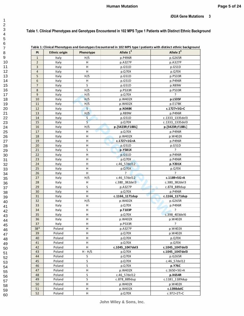
Table 1. Clinical Phenotypes and Genotypes Encountered in 102 MPS Type 1 Patients with Distinct Ethnic Background
Table 1: Clinical Phenotypes and Genotypes Encountered in 102 MPS type I patients with distinct ethnic background
Pt Ethnic origin Phenotype Allele 1§ Allele 2
§
1 Italy H/S p.P496R p.G265R
2 Italy H p.A327P p.A327P
3 Italy H p.G51D p.G51D
4 Italy H p.Q70X p.Q70X
5 Italy H/S p.G51D p.P533R
6 Italy H p.G51D p.P496R
7 Italy S p.G51D p.R89W
8 Italy H/S p.P533R p.P533R
9 Italy H/S p.Q70X ?
10 Italy H/S p.W402X p.L535F
11 Italy H/S p.W402X p.E178K
12 Italy S p.N348K c.1727+5G>C
13 Italy H/S p.R89W p.P496R
14 Italy S p.G51D c.1333_1335del3
15 Italy S p.Q70X c.1333_1335del3
16 Italy H/S p.[S423R;F188L] p.[S423R;F188L]
17 Italy H p.Q70X p.P496R
18 Italy H p.W402X p.W402X
19 Italy H c.1727+1G>A p.P496R
20 Italy H p.G51D p.G51D
21 Italy S p.Y581X ?
22 Italy H p.G51D p.P496R
23 Italy H p.Q70X p.P496R
24 Italy H c.46_57del12 p.Y201X
25 Italy H p.Q70X p.Q70X
26 Italy H ? ?
27 Italy H/S c.46_57del12 c.1189+5G>A
28 Italy H c.380_382del3 c.380_382del3
29 Italy S p.A327P c.878_889dup
30 Italy H p.Q70X p.P496R
31 Italy H c.1166_1171dup c.1166_1171dup
32 Italy H/S p.W402X p.G265R
33 Italy H p.Q70X p.P496R
34 Italy H p.T103P ?
35 Italy H p.Q70X c.398_403del6
36 Italy H p.W402X p.W402X
37 Italy H p.P533R ?
38* Poland H p.A327P p.W402X
39 Poland H p.Q70X p.W402X
40 Poland H p.Q70X p.Q70X
41 Poland H p.Q70X p.Q70X
42 Poland H c.1045_1047del3 c.1045_1047del3
43 Poland H - H/S p.Q70X c.1045_1047del3
44 Poland S p.Q70X p.G265R
45 Poland S p.Q70X c.46_57del12
46 Poland S p.Q70X p.Y76C
47 Poland H p.W402X c.1650+5G>A
48 Poland S c.46_57del12 p.X654R
49 Poland S c.878_889dup c.1181_1189dup
50 Poland H p.W402X p.W402X
51 Poland H p.W402X c.1398delC
52 Poland H p.Q70X c.972+2T>C
Page 5 of 24
John Wiley & Sons, Inc.
Human Mutation
123456789101112131415161718192021222324252627282930313233343536373839404142434445464748495051525354555657585960
For Peer Review
4 Bertola et al.
Table 1 (continued)
53 Poland H p.Q70X p.Q70X
54 Poland H p.Q70X p.Q70X
55 Poland H p.Q70X p.W402X
56 Poland H p.W402X p.W402X
57 Poland H p.Q70X p.Q70X
58 Poland H p.W402X p.W402X
59 Poland S p.G219E p.W306L
69 Poland H/S p.Q70X p.X654R
61 Spain S p.R492P ?
62 Spain H p.Q70X ?
63 Spain H/S p.G51D c.1727+4C>T
64 Spain H/S c.1189+4A>G p.R621X
65 Spain H p.A327P p.P385R
66 Spain H p.W402X p.W402X
67 Spain H p.M1? p.M1?
68 Spain H p.W402X p.W402X
69 Spain NA p.W402X p.L396P
70 Spain NA p.W402X c.1189+4A>G
71 Spain H p.Q70X c.1190-1delG
72 Spain H/S p.P533R p.P533R
73 Spain H c.574delT c.574delT
74 Spain H p.W402X p.W402X
75 Spain H p.W402X p.W402X
76 Spain H p.Q70X p.W402X
77 Spain H p.W402X p.W402X
78 Spain H p.W402X c.385+1G>A
79 Turkey NA c.1727+6T>A ?
80 Turkey H p.P533R p.P533R
81 Turkey H/S c.494-1G>A c.494-1G>A
82 Turkey S p.L490P p.L490P
83 Turkey H/S c.493+1G>A c.493+1G>A
84 Turkey H/S c.46_57del12 c.46_57del12
85^ Turkey H/S ? ?
86 Turkey H c.494-1G>A c.494-1G>A
87^ Turkey H c.956_972+9delinsTA c.956_972+9delinsTA
88^ Turkey H p.W402X p.W402X
89^ Turkey H c.826_828del3 c.826_828del3
90° Turkey H c.494-1G>A c.494-1G>A
91 Turkey H/S c.1727+6T>A p.Q70X
92 Turkey H/S - S c.494-1G>A p.E276K
93 Turkey S p.E276K p.E276K
94 Turkey H/S c.46_57del12 c.46_57del12
95 Turkey H/S p.A436P p.A436P
96 Turkey H p.Q70X p.Q70X
97 Turkey H p.W402X p.W402X
98 Turkey H c.494-1G>A c.494-1G>A
99 Turkey H c.1650+5G>C c.1893delC
100 Hungary H p.Q70X p.W402X
101 Serbian H p.Q70X p.Q70X
102 Greece H/S p.G84R p.E276K
Legend: H=Hurler; S=Scheie; NA=Not available; ?=Unknown; Pt=Patient; Novel mutations are marked in bold. §GenBank-EMBL
accession no. NM_000203.3 (c.DNA considering the A of the ATG translation initiation start site as nucleotide +1) and GenBank-
EMBL accession no. NP_000194 (protein); *Patient not analysed in our Lab; indicates parents’ consanguinity; °Patient’s parents
belonging to the same village; Underlined patients belonging to the same geographical region
Page 6 of 24
John Wiley & Sons, Inc.
Human Mutation
123456789101112131415161718192021222324252627282930313233343536373839404142434445464748495051525354555657585960

For Peer Review
IDUA Gene Mutations 5
Some of the samples from the Italian patients were supplied by the “Cell line and DNA Biobank from patients
affected by Genetic Diseases” in Genova (http://dppm.gaslini.org/biobank/). Following ethical guidelines, all
samples obtained for analysis and storage required prior written informed consent using a form approved by the
Local Ethics Committee.
Cell culture
Fibroblast cells were cultured according to standard procedures. The cell lines were cultured and maintained in
RPMI medium (EuroClone, Gibco, Paisley, UK) containing 15% FCS and penicillin/streptomycin, in a humidified
atmosphere containing 5% CO2 at 37°C.
Enzymatic assay
α-L-iduronidase activity was assayed in homogenates of leukocytes and/or fibroblast cell lines using 4-
methylumbelliferyl-α-L-iduronide (Glycosynth, Cheshire, UK) as a substrate [Stirling et al., 1978]. Total protein
was measured according to the Lowry method [Lowry et al., 1951].
Molecular analysis
Genomic DNA was extracted from circulating lymphocytes and/or cultured fibroblasts using either the Wizard
Genomic DNA Purification kit (Promega, Madison, WI) or a standard phenol-chloroform-based method.
IDUA gene exons, exon–intron boundaries and part of the 5’ untranslated region were PCR amplified using
specific primers designed by reference to the genomic sequence (GenBank-EMBL Accession no. NG_008103.1).
Intronic primers, designed for both PCR and sequence analyses, are listed in Supp. Table S1. PCR amplification
reactions (except for exon 9) were carried out in 25µl vols containing 100ng genomic DNA, buffer 1X with
1.5mM MgCl2, 200µM dNTPs mix, 15pmol each primer, GC-RICH solution 1X and 0.5U proofreading PWO
SuperYield DNA polymerase (Roche, Monza, Italy). Exon 9 PCR-amplification was carried out in 25µl vols
containing 100ng genomic DNA, buffer 1X with 1.5mM MgCl2, 200µM dNTPs mix, 15pmol each primer and
0.5U GoTaq DNA polymerase (Promega, Madison, WI); owing to the use of a no-proofreading DNA polymerase,
this reaction was performed in duplicate. Cycling conditions were: initial denaturation at 96°C for 5 min, 28 cycles
denaturation at 96°C for 1 min, annealing at 63-70°C for 1 min, extension at 72°C for 1 min, followed by final
extension at 72°C for 7 min. Control PCR reactions, in which no template was added, were included during each
set of PCR reactions. PCR products were controlled for contamination by gel electrophoresis and were then
purified using an enzymatic reaction containing 5U exonuclease I (Celbio, Italy) and 1U alkaline phosphatase
(Promega, Madison, WI) under the following conditions: 15 min at 37°C followed by 15 min at 80°C.
Total RNA was prepared from peripheral blood lymphocytes isolated by the Fycoll method or from cultured
fibroblasts using Trizol Reagent (Gibco, Paisley, UK) according to the manufacturer’s instructions. First-strand
cDNAs were synthesized using the High-Capacity cDNA Reverse Transcription Kit (Applied Biosystems, Foster
City, CA) and random hexamer primers. IDUA exonic fragments flanking splicing mutations were amplified and
sequenced using primers and conditions listed in Supp. Table S1.
Sequence analysis of PCR and RT-PCR products was performed in the forward and reverse directions using
BigDye v3.1 terminator technology (Applied Biosystems, Foster City, CA) and then purified with Wizard
MagneSil Sequencing Reaction Clean-Up System (Promega, Madison, WI). Sequencing reactions were carried
out, the products purified according to the manufacturer’s instructions and analyzed on an ABI Prism 3130 Avant
Automatic Sequencer (Applied Biosystems, Foster City, CA). Sequence alterations were confirmed by sequencing
duplicate PCR products.
The question of the pathological authenticity of the novel IDUA sequence alterations detected was addressed by
(i) searching dbSNP (http://www.ncbi.nlm.nih.gov/SNP) for their presence, (ii) screening 100 alleles from healthy
control subjects for each alteration, (iii) modeling the amino acid changes into a homologous three-dimensional
structure of the protein, (iv) employing the MutPred program [Li et al., 2009; Mort et al., 2010] for the missense
mutations, (v) using ESRPred and a neural network for splice site prediction [Krawczak et al., 2007] for the coding
region variants upon splicing, and (vi) using PhyloP method [Pollard et al., 2010] and ClustalW2 program [Larkin
et al., 2007] to measure evolutionary conservation of nucleotides and residues, respectively.
Page 7 of 24
John Wiley & Sons, Inc.
Human Mutation
123456789101112131415161718192021222324252627282930313233343536373839404142434445464748495051525354555657585960

For Peer Review
6 Bertola et al.
Molecular modeling of missense mutations
For modeling of the IDUA missense mutations, an homology model of the IDUA protein was used. This model
was built by Rempel et al. (2005) using the atomic coordinates of β-xylosidase (E.C.3.2.1.37) from
Thermoanaerobacterium saccharolyticum as a template [PDB code 1Y24]. Visual inspection and graphical
representations were then performed using the programs Coot and Chimera, respectively [Emsley and Cowtan,
2004; Pettersen et al., 2004].
Bioinformatic analysis of IDUA variants
Missense variants in the IDUA gene were analysed with a computational model, MutPred [Li et al., 2009, Mort
et al., 2010]. MutPred is designed to assess the likely phenotypic consequences of missense substitutions occurring
within sites of structural and/or functional importance, on the wild-type protein sequence. MutPred can be used to
generate hypotheses as to the underlying molecular mechanism(s) responsible for disease pathogenesis.
The effect of the coding region variants upon splicing [splice site disruption, cryptic splice site activation and
exon skipping via loss of exonic splicing enhancers (ESE) and/or gain of exonic splicing silencers (ESS)] was
ascertained using ESRPred [M. Mort, unpuplished] and a neural network for splice site prediction [Krawczak et
al., 2007].
Evolutionary sequence conservation across an alignment of IDUA orthologues from 44 vertebrate species was
measured using the phyloP method [Pollard et al., 2010]. PhyloP can measure accelerated evolution (more rapid
evolution than expected under neutral drift) as well as evolutionary conservation (slower than expected evolution).
A positive phyloP score represents an evolutionarily conserved nucleotide and a negative phyloP score indicates
that the nucleotide has experienced more rapid evolution than would be expected under neutral drift.
Mutation nomenclature
Mutations are described according to current mutation nomenclature guidelines
(http://www.hgvs.org/mutnomen), ascribing the A of the first ATG translational initiation codon as nucleotide +1
[den Dunnen and Antonarakis, 2000; den Dunnen and Paalman, 2003].
RESULTS AND DISCUSSION
A total of 102 European patients affected by MPS I were recruited by our centre. All patients underwent
molecular characterization and the IDUA genotype was completely ascertained in 93 of the 102 patients.
Mutation detection
All 14 exons, splice junctions and proximal portions of the 5’ and 3’ untranslated regions of the IDUA genes of
the 102 unrelated MPS I patients were investigated by DNA sequence analysis. Putatively pathological IDUA
mutations were identified in 193 of 204 alleles. The genotypes, clinical phenotypes and geographic origins of the
102 patients studied here are listed in Table 1. The characteristics of the 55 distinct IDUA mutations identified are
reported in Table 2. The mutational spectrum comprised 22 missense mutations, 14 splice site alterations, 9 micro-
deletions, 5 nonsense mutations, 3 micro-duplications, 1 translational initiation site mutation and 1 no-stop
mutation. More than 60% (35/55) of these mutant alleles have not been previously reported, attesting to the
extensive allelic heterogeneity of MPS I. The novel lesions resulted from (i) 11 missense mutations (p.Y76C,
p.G84R, p.T103P, p.G219E, p.E276K, p.W306L, p.N384K, p.P385R, p.L396P, p.A436P, p.L535F) as well as two
missense mutations located in cis to each other (p.[F188L;S423R]); (ii) 12 splicing mutations (c.385+1G>A,
c.493+1G>A, c.494-1G>A, c.956_972+9delinsTA, c.1189+4A>G, c.1189+5G>A, c.1190-1delG, c.1650+5G>C,
c.1727+1G>A, c.1727+4C>T, c.1727+5G>C, c.1727+6T>A); (iii) 5 micro-deletions (c.574delT, c.826_828del3,
c.1045_1047del3, c.1398delC and c.1893delC), 1 micro-duplication (c.1166_1171dup), 2 nonsense mutations
(p.201X and p.Y581X), 1 translational initiation site mutation (p.M1?) and 1 ‘no-stop’ mutation
(p.X654RextX62).
Page 8 of 24
John Wiley & Sons, Inc.
Human Mutation
123456789101112131415161718192021222324252627282930313233343536373839404142434445464748495051525354555657585960

For Peer Review
IDUA Gene Mutations 7
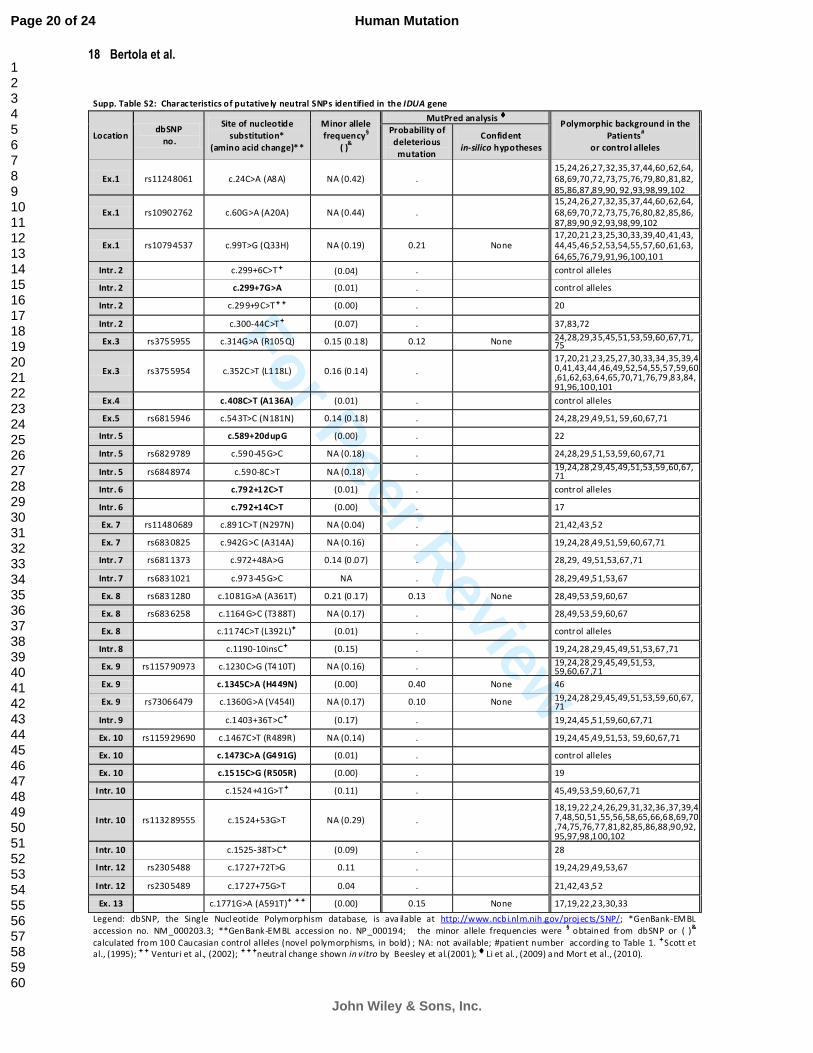
In addition to these putative pathological mutations, a total of 37 putative intragenic IDUA polymorphisms were
identified within either the exons or introns; eight novel SNPs were noted within the coding region (4) and in the
introns (4) (Supp. Table S2). The allele frequencies of the novel variants, derived from 100 normal control alleles,
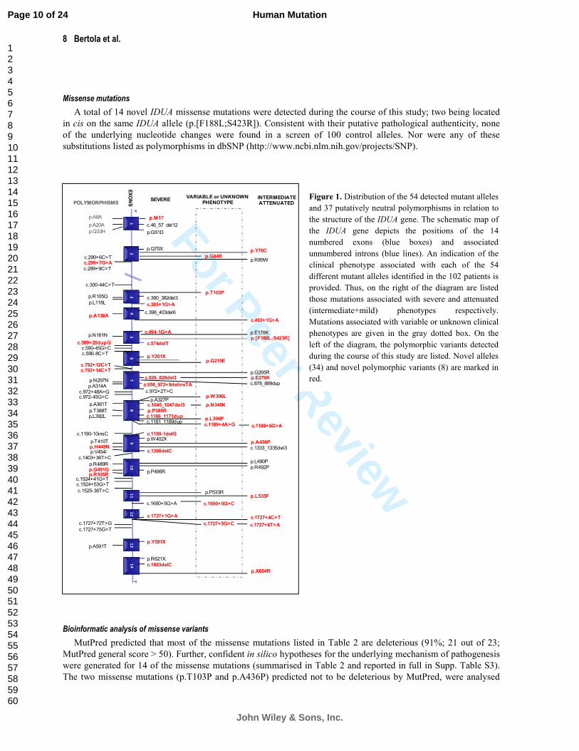
are given in Supp. Table S2. Figure 1 depicts the location of the various mutations and polymorphisms detected in
relation to the structure of the IDUA gene.
Table 2. Characteristics of the IDUA gene mutations identified in the 102 MPS type I patients and MutPred analysis of the missense mutations
Table 2: Characteristics of the IDUA gene mutations identified in the 102 MPS type I patients and MutPred analysis of the missense mutations
Loc. Site of nucleotide
substitution*
P redicted effect on
protein str uc ture **
Type of
mutation
No. of
alleles (%)
Evolutionary
Conser vation (phyloP )
MutPre d analysis of missense mutations #
Re ferences Deleterious
mutation probability
Summary of confident in silico
hypotheses
Ex. 1
c.1A>C p .M1 ? Translational
initiation site 2(0.01) Acc (-0.3) 0.99 Present st udy
§
c.152G>A p.G51 D Missense 10(4.9) Cons (2.7) 0.97 Protein struct ure disruption Bunge et al. (1994 )
c.46_57d el12 p .S16 _A19del In-frame del 8(3.9) . . Bunge et al. (1994 )
Ex. 2
c.208 C>T p.Q70X Nonsense 3 8(18 ) Cons (2.1) . Sco tt e t al. (199 2)
c.227A >G p.Y 76C Missense 1(0.05) Cons (2.7) 0.73 Present st udy
c.2 50G>C p.G84R Missense 1(0.05) Cons (5.5) 0.93 Gain of bi nding site and disruption to
protei n stru cture Present st udy
c.265 C>T p.R89W Missense 2(0.01) Cons (0.3) 0.96 Bunge et al. (1995 )
Ex. 3 c .307A>C p.T10 3P Missense 1(0.05) Cons (0.4) 0.36 Present st udy
c .3 80_382del3 p.L12 7del In-frame del 2(0.01) . . Ventur i et al. (2002)
Intr. 3 c.385+1G>A r.spl? Sp licing 1(0.05) Cons (5.1) . Present st udy
Ex. 4 c .3 98_403del6 p .M 133_G134del In-frame del 1(0.05) . . Ventur i et al. (2002)
Intr. 4 c.493+1G>A r.spl? Sp licing 2(0.01) Cons (6.2) . Present st udy
c.494-1G>A p.R16 6TfsX27 Splicing 9(4.4) Cons (5.7) . P resen t study
Ex. 5
c.532G>A p.E178K Missense 1(0.05) Cons (4.5) 0.97 Gain of functional sites in cluding PTMs Ventur i et al. (2002)
c.[5 62T>C;1269C>A]���� p. [F188L ; S423R]���� Missense 2(0.01) C ons(3.0;1.2) 0.95;0 .92 Gain of methylation at S423 Presen t study
c.574delT p.S192P fsX2 Frameshift 2(0.01) . . P resen t study
Ex. 6 c.6 03C>G p .Y20 1X Nonse nse 1(0.05) Cons (0.7) . P resen t study
c.656G>A p .G2 19E Missense 1(0.05) Cons (5.9) 0.89 Protein struct ure disruption Presen t study
Ex. 7
c.793G>C p.G265R Missense 3(1.4) Cons (6.2) 0.95 Secon dary struct ure disruption , gain of
methylation at G265 Yogalingam et al.(20 04)
c.826G>A p.E27 6K Missense 4(1.9) Cons (6.2) 0.76 Presen t study
c.8 26_828del3 p.E276del In-frame del 2(0.01) . . P resen t study
c.8 78_889du p p .T29 3_Y296dup In-frame dup 2(0.01) . . Bunge et al. (1995)
c .956_972+9delinsTA r .spl? Splicing 2(0.01) . . P resen t study
Intr. 7 c.972+2T>C r.spl? Splicing 1(0.05) Cons (1.6) . Sco tt e t al. (199 3)
Ex. 8
c .917G>T p.W306L Missense 1(0.05) Cons (6.0) 0.82 Loss of catalytic residu e at P309 Presen t study
9 79G>C p .A32 7P Missense 5(2.4) Cons (1.2) 0.84 Protein structure disruption and gain
o f me thylation at K 324 Bunge et al. (1995)
c.1044C>G p.N3 48K Missense 1(0.05) Acc (-0.4) 0.85 Decrease in protein stability and gain
of PTMs Presen t study
c.1045_1047 del3 p .D349del In-frame del 3(1.4) - . P resen t study
c.1154C>G p .P385R Missense 1(0.05) Cons (5.4) 0.82 Protein structure d isruption Presen t study
c.1166_1171 dup p .A38 9_M 390dup In-frame dup 2(0.01) . P resen t study
c.11 87T>C p.L396P Missense 1(0.05) Cons (3.1) 0.83 Decrease in protein stab ility and secondary struct ure disr uption
Presen t study
c.11 81_1189du p p .A3 94_L396dup In-frame dup 1(0.05) . . Bunge et al. (1995)
Intr. 8
c.1189 +4 A>G p.V 371MfsX43 Sp licing 2(0.01) Cons (4.3) . P resen t study
c.1189 +5 G>A p.V 371MfsX43 Sp licing 1(0.05) Cons (2.9) . P resen t study
c.1190 -1delG r .spl? Splicing 1(0.05) - . P resen t study
Ex. 9
c.1205G>A p.W402X Nonsense 3 7(18 ) Cons (1.8) . Sco tt e t al. (199 2)
c.1306G>C p .A436P Missense 2(0.01) Cons (2.8) 0.45 Presen t study
c.13 33_1335del3 p.D445 del In-frame del 2(0.01) . . Bunge et al. (1995)
c.1 398delC p.P467RfsX58 Frameshift 1(0.05) . . P resen t study
Ex. 10
c.1469T>C p.L490P Missense 2(0.01) Cons (0.4) 0.86 Decrease in prot ein stability and
protei n stru cture disru ption Tieu et al. (1995)
c.147 5G>C p.R49 2P Missense 1(0.05) Cons (0.7) 0.83 Loss of binding site Tieu et al. (1995)
c.148 7C>G p.P496R Missense 9(4.4) Cons (4.4) 0.95 Gain o f bin ding site Beesley et al. (2001)
Ex. 11 c.159 8C>G p.P533R Missense 8(3.9) Cons (4.1) 0.94 Gain o f bin ding site Sco tt e t al. (199 2)
c.16 03C>T p.L53 5F Missense 1(0.05) Cons (1.7) 0.59 Presen t study
Intr. 11 c.1650 +5 G>A r.spl? Splicing 1(0.05) Cons (4.7) . Ven turi et al. 2002
c.1 650+5G>C r .spl? Sp licing 1(0.05) Cons (4.7) . Presen t study
Intr. 12
c.1727 +1 G>A r.spl? Sp licing 1(0.05) Cons (3.8) . P resen t study
c.172 7+4 C>T p .L578V fsX14 Sp licing 1(0.05) Cons (0.4) . P resen t study
c.1 727+5G>C Exon 12 skipping Sp licing 1(0.05) Cons (5.4) . P resen t study
c.1727+6T>A Exon 12 skipping Sp licing 2(0.01) Cons (1.1) . P resen t study
Ex. 13 c.1743C>G p .Y58 1X Nonse nse 1(0.05) Acc (-1.1) . P resen t study
Ex. 14
c.1861C>T p.R62 1X Nonsense 1(0.05) Cons (1.5) . Bunge et al. (1994)
c.1 893delC p.F632SfsX105 Frameshift 1(0.05) . P resen t study
c.19 60T>C p.X6 54RextX62 No-stop 2(0.01) Acc (-0 .4) . P resen t study
Legen d: Loc= Location; Ex =ex on; Intr=intron; del=deletion; dup=duplication; Acc = Accelerated; Cons= C onserved; PTMs= Post-translational m od ifications *IDUA gen e
GenBank-EM BL accession no. NM _0 00203.3; * *IDUA gene GenBank-EMBL accessio n no. NP_00019 4; the no vel m utatio ns are given in bold; #Li et al. (2 009) and Mort et
al. (2 010). ����Allele carrying 2 novel mutations in c is; note that the sec ond change occu rs in exon 9 and it was previously report ed as single allele as due to c.1269C>G by
Yogalingam et al. (2004); §mutation pr eviously reported as due to a c.3G>A by Le e-Chen and Wang (1997)
Page 9 of 24
John Wiley & Sons, Inc.
Human Mutation
123456789101112131415161718192021222324252627282930313233343536373839404142434445464748495051525354555657585960
For Peer Review
8 Bertola et al.
Missense mutations
A total of 14 novel IDUA missense mutations were detected during the course of this study; two being located
in cis on the same IDUA allele (p.[F188L;S423R]). Consistent with their putative pathological authenticity, none
of the underlying nucleotide changes were found in a screen of 100 control alleles. Nor were any of these
substitutions listed as polymorphisms in dbSNP (http://www.ncbi.nlm.nih.gov/projects/SNP).
Bioinformatic analysis of missense variants
MutPred predicted that most of the missense mutations listed in Table 2 are deleterious (91%; 21 out of 23;
MutPred general score > 50). Further, confident in silico hypotheses for the underlying mechanism of pathogenesis
were generated for 14 of the missense mutations (summarised in Table 2 and reported in full in Supp. Table S3).
The two missense mutations (p.T103P and p.A436P) predicted not to be deleterious by MutPred, were analysed
Figure 1. Distribution of the 54 detected mutant alleles
and 37 putatively neutral polymorphisms in relation to
the structure of the IDUA gene. The schematic map of
the IDUA gene depicts the positions of the 14
numbered exons (blue boxes) and associated
unnumbered introns (blue lines). An indication of the
clinical phenotype associated with each of the 54
different mutant alleles identified in the 102 patients is
provided. Thus, on the right of the diagram are listed
those mutations associated with severe and attenuated
(intermediate+mild) phenotypes respectively.
Mutations associated with variable or unknown clinical
phenotypes are given in the gray dotted box. On the
left of the diagram, the polymorphic variants detected
during the course of this study are listed. Novel alleles
(34) and novel polymorphic variants (8) are marked in
red.
SEVERE INTERMEDIATE ATTENUATED
p.Q70X
12
34
56
89
10
11
12
13
14
c.380_382del3
p.Y201X
c.826_828del3
p.A327P
p.W402X
c.1650+5G>A
5’
3’
p.E178K
p.G265R
p.R89W
p.P496R
c.878_889dup
p.R621X
p.P385R
p.P533Rp.L535F
c.1727+4C>T
p.N348K
c.46_57 del12
p.G51D
c.1727+1G>A
p.Y581X
p.M1?
p.G84Rp.Y76C
c.385+1G>A
c.493+1G>A
c.494-1G>A
c.574delT
p.E276K
c.956_972+9delinsTA
c.972+2T>C
c.1166_1171dupp.L396P
c.1189+5G>Ac.1189+4A>Gc.1181_1189dup
c.1190-1delG
c.1333_1335del3c.1398delC
p.L490P
p.R492P
c.1727+5G>C c.1727+6T>A
p.X654R
VARIABLE or UNKNOWN PHENOTYPE
EXONSPOLYMORPHISMS
p.A20A
p.A8A
p.Q33H
c.299+9C>T
c.300-44C>T
p.R105Q
p.L118L
c.589+20dupG
p.N181N
c.590-8C>Tc.590-45G>C
c.792+14C>Tc.792+12C>T
p.N297Np.A314A
c.972+48A>Gc.972-45G>C
p.A361T
p.T388T
c.1190-10insC
p.T410Tp.H449Np.V454I
c.1403+36T>C
p.R489Rp.G491Gp.R505R
c.1524+41G>Tc.1524+53G>T
c.1525-38T>C
c.1727+72T>G
c.1727+75G>T
p.A591T
c.1045_1047del3
p.T103P
p.A436P
c.398_403del6
p.[F188L;S423R]
p.G219E
p.W306L
c.1650+5G>C
c.1893delC
c.299+7G>Ac.299+6C>T
p.A136A
p.L392L
7
Page 10 of 24
John Wiley & Sons, Inc.
Human Mutation
123456789101112131415161718192021222324252627282930313233343536373839404142434445464748495051525354555657585960
For Peer Review
IDUA Gene Mutations 9
further (Supp. Table S4). PhyloP scores were used to measure evolutionary conservation at the nucleotide
sequence level across IDUA orthologues derived from 44 vertebrate species. Both mutations (p.T103P and
p.A436P) exhibited positive phyloP scores indicating that these mutations are located at conserved nucleotides. It
should be noted that p.T103P displays only marginal conservation, with a phyloP score of 0.40. Analysis with
SIFT [Ng and Henikoff, 2001] predicted that both mutations would be tolerated in the protein. PolyPhen2
[Adzhubei et al., 2010] however predicted that whilst p.T103P was benign, p.A436P mutation was likely to be
potentially damaging. Neither mutation was predicted to disrupt splicing using a neural network [Krawczak et al.,
2007]. MutPred analysis was also performed on the six polymorphic missense variants identified (listed in Supp.
Table S2) but none of these were predicted to be deleterious (MutPred general scores invariably < 50).
Further evidence for the pathological/functional significance of the missense mutations was then sought from
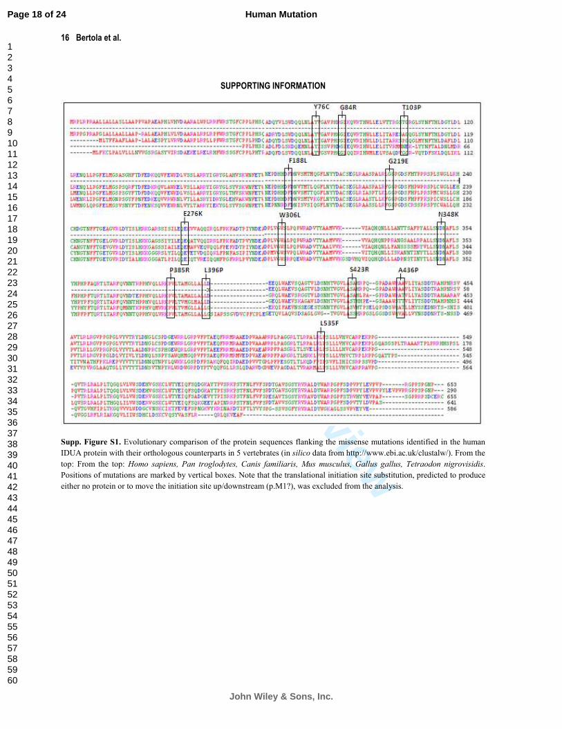
the analysis of the extent of evolutionary conservation of the mutated residues in 6 orthologous (vertebrate) IDUA
proteins from human to Tetraodon nigroviridis. The computational analysis, carried out using ClustalW2
[http://www.ebi.ac.uk/clustalw; Larkin et al., 2007], revealed that among the 5 other vertebrate IDUA protein
sequences examined, Y76, G84, F188, G219, R276, W306, N348, P385, L396, S423, L535 were invariant at these
positions; such conservation over 500 Myrs of evolutionary time is supportive of the functional significance of
these residues and hence the direct involvement of the respective mutations in disease causation. In agreement with
the PolyPhen score results, residue A436 was found to be partially conserved (chimpanzee, dog and chicken) but
T103 was not evolutionarily conserved. (Supp. Figure S1). It may be that the p.A436P mutation is mildly
deleterious, whereas p.T103P may be tolerated and could therefore represent a rare polymorphism.
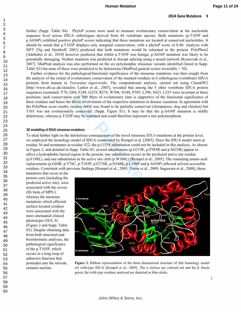
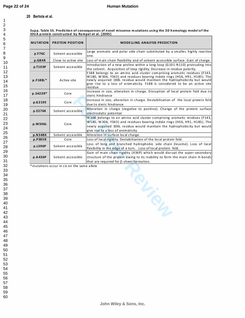
3D modelling of IDUA missense mutations
To shed further light on the deleterious consequences of the novel missense IDUA mutations at the protein level,
we employed the homology model of IDUA constructed by Rempel et al. [2005]. Since the IDUA model starts at
residue 36 and terminates at residue 522, the p.L535F substitution could not be included in this analysis. As shown
in Figure 2, and detailed in Supp. Table S5, several substitutions (p.G219E, p.P385R and p.S423R) appear to
affect a hydrophobic buried region in the protein, one substitution occurs in the predicted active site residue
(p.F188L), and one substitution in the active site cleft (p.W306L) [Rempel et al., 2005]. The remaining amino acid
replacements (p.G84R, p.Y76C, p.T103P, p.E276K, p.N348K, p.L396P and p.A436P) affected solvent accessible
residues. Consistent with previous findings [Rempel et al., 2005; Vazna et al., 2009; Sugawara et al., 2008], those
mutations that occur in the
protein core (including the
predicted active site), were
associated with the severe
(H) form of MPS I,
whereas the missense
mutations which affected
surface-located residues
were associated with the
more attenuated clinical
phenotypes (H/S, S)
(Figure 2 and Supp. Table
S5). Despite obtaining data
from both structural and
bioinformatic analyses, the
pathological significance
of the p.T103P, which
occurs in a long loop of
unknown function that
protrudes into the solvent,
remains unclear.
Figure 2. Ribbon representation of the three dimensional structure of (the homology model
of) wild-type IDUA [Rempel et al., 2005]. The α helices are colored red and the β sheets
green; the wild-type residues analysed are depicted as blue sticks.
Page 11 of 24
John Wiley & Sons, Inc.
Human Mutation
123456789101112131415161718192021222324252627282930313233343536373839404142434445464748495051525354555657585960
For Peer Review
10 Bertola et al.
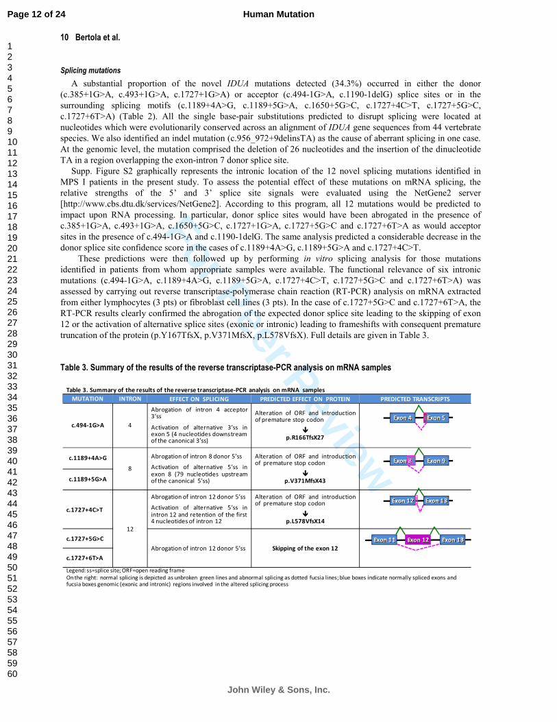
Table 3. Summary of the results of the reverse transcriptase-PCR analysis on mRNA samples
MUTATION INTRON EFFECT ON SPLICING PREDICTED EFFECT ON PROTEIN PREDICTED TRANSCRIPTS
c.494-1G>A 4
Abrogation of intron 4 acceptor3’ss
Activation of alternative 3’ss inexon 5 (4 nucleotides downstreamof the canonical 3’ss)
Alteration of ORF and introductionof premature stop codon
�p.R166TfsX27
c.1189+4A>G
8
Abrogation of intron 8 donor 5’ss
Activation of alternative 5’ss inexon 8 (79 nucleotides upstreamof the canonical 5’ss)
Alteration of ORF and introductionof premature stop codon
�p.V371MfsX43c.1189+5G>A
c.1727+4C>T
12
Abrogation of intron 12 donor 5’ss
Activation of alternative 5’ss inintron 12 and retention of the first4 nucleotides of intron 12
Alteration of ORF and introductionof premature stop codon
�p.L578VfsX14
c.1727+5G>C
Abrogation of intron 12 donor 5’ss Skipping of the exon 12
c.1727+6T>A
Legend: ss=splice site; ORF=open reading frame
On the right: normal splicing is depicted as unbroken green lines and abnormal splicing as dotted fucsia lines; blue boxes indicate normally spliced exons and fucsia boxes genomic (exonic and intronic) regions involved in the altered splicing process
Exon 5Exon 4
Exon 12 Exon 13Exon 11
Exon 8 Exon 9
Exon 12 Exon 13
Splicing mutations
A substantial proportion of the novel IDUA mutations detected (34.3%) occurred in either the donor
(c.385+1G>A, c.493+1G>A, c.1727+1G>A) or acceptor (c.494-1G>A, c.1190-1delG) splice sites or in the
surrounding splicing motifs (c.1189+4A>G, c.1189+5G>A, c.1650+5G>C, c.1727+4C>T, c.1727+5G>C,
c.1727+6T>A) (Table 2). All the single base-pair substitutions predicted to disrupt splicing were located at
nucleotides which were evolutionarily conserved across an alignment of IDUA gene sequences from 44 vertebrate
species. We also identified an indel mutation (c.956_972+9delinsTA) as the cause of aberrant splicing in one case.
At the genomic level, the mutation comprised the deletion of 26 nucleotides and the insertion of the dinucleotide
TA in a region overlapping the exon-intron 7 donor splice site.
Supp. Figure S2 graphically represents the intronic location of the 12 novel splicing mutations identified in
MPS I patients in the present study. To assess the potential effect of these mutations on mRNA splicing, the
relative strengths of the 5’ and 3’ splice site signals were evaluated using the NetGene2 server
[http://www.cbs.dtu.dk/services/NetGene2]. According to this program, all 12 mutations would be predicted to
impact upon RNA processing. In particular, donor splice sites would have been abrogated in the presence of
c.385+1G>A, c.493+1G>A, c.1650+5G>C, c.1727+1G>A, c.1727+5G>C and c.1727+6T>A as would acceptor
sites in the presence of c.494-1G>A and c.1190-1delG. The same analysis predicted a considerable decrease in the
donor splice site confidence score in the cases of c.1189+4A>G, c.1189+5G>A and c.1727+4C>T.
These predictions were then followed up by performing in vitro splicing analysis for those mutations
identified in patients from whom appropriate samples were available. The functional relevance of six intronic
mutations (c.494-1G>A, c.1189+4A>G, c.1189+5G>A, c.1727+4C>T, c.1727+5G>C and c.1727+6T>A) was
assessed by carrying out reverse transcriptase-polymerase chain reaction (RT-PCR) analysis on mRNA extracted
from either lymphocytes (3 pts) or fibroblast cell lines (3 pts). In the case of c.1727+5G>C and c.1727+6T>A, the
RT-PCR results clearly confirmed the abrogation of the expected donor splice site leading to the skipping of exon
12 or the activation of alternative splice sites (exonic or intronic) leading to frameshifts with consequent premature
truncation of the protein (p.Y167TfsX, p.V371MfsX, p.L578VfsX). Full details are given in Table 3.
Table 3. Summary of the results of the reverse transcriptase-PCR analysis on mRNA samples
Page 12 of 24
John Wiley & Sons, Inc.
Human Mutation
123456789101112131415161718192021222324252627282930313233343536373839404142434445464748495051525354555657585960

For Peer Review
IDUA Gene Mutations 11
Genotype-phenotype relationship
Genotypes, clinical phenotypes and geographic origins of the 102 patients studied here are listed in Table 1.
Some 45 (44%) of the patients (pts) were found to be either homozygous for the pan-ethnic IDUA mutations,
p.W402X (12 pts), p.Q70X (9 pts) and p.P533R (3 pts), other known mutations (7 pts) or novel lesions (14 pts).
The remaining 54% of patients were compound heterozygotes (including 7 in whom the second mutant IDUA
allele could not be identified). Finally, the mutant IDUA alleles in two patients (2%), in whom a defect in IDUA
enzyme activity had been documented, remained unidentified.
Overall, we found 68 distinct genotypes including those (7) that were partially characterized. Our results
confirm the high degree of mutational heterogeneity characteristic of MPS I. Extensive allelic heterogeneity often
precludes the recognition of correlations between mutant genotypes and variant clinical phenotypes. This
notwithstanding, in an attempt to obtain new insights into the genotype-phenotype relationship in MPS I, we have
compared the genotypes present in our patient series with those of previously reported patients. Supp. Table S6,
reports the comparable genotypes (13/68) and the respective number of patients per genotype reported in previous
studies on this topic.
Among the analyzed group, 25 patients were either homozygous or compound heterozygous for the two
common deleterious nonsense mutations (p.Q70X and p.W402X); their severe clinical phenotype concurred with
those of 91 previously reported patients with comparable genotypes [Bunge et al., 1994; Gort et al., 1998; Hein et
al., 2003; Li et al., 2002; Matte et al., 2003; Scott et al., 1992; Vazna et al., 2009; Venturi et al., 2002; Voskoboeva
et al. 1998]. In agreement with previously reported genotype-phenotype correlations, a severe phenotype was not
only associated with the p.W402X mutation in compound heterozygosity with both c.1650+5G>A and p.A327P
[Bunge et al., 1994; Venturi et al., 2002; Vazna et al., 2009] but also with homozygosity for p.A327P and p.G51D
[Gatti et al., 1997]. Also consistent with previous data [Beesley et al, 2001; Tieu et al., 1995 ], homozygosity for
p.L490P and compound heterozygosity for p.Q70X and c.1333_1335del3 yielded mild phenotypes in two other
patients (Supp. Table S6). Although for these 38 patients (37%), the predicted genotype-phenotype relationship
may be considered informative, for the remaining group with previously described genotypes, the correlations only
partially concurred (Supp. Table S6). This latter group comprised 6 patients, 3 of whom were homozygous either
for the p.P533R mutation or for the microdeletion c.46_57del12 (2 pts), and another patient who was compound
heterozygous for p.G51D and p.P533R. Comparison between the genotype-phenotype correlation data from our 6
patients and 23 patients from other studies [Alif et al., 1999; Hein et al., 2003; Gatti et al., 1997; Hein et al., 2003,
Laradi et al., 2005; Matte et al., 2003; Scott et al., 1992; Venturi et al., 2002] confirmed the phenotypic variability
of these mutations (p.P533R and c.46_57del12). Similarly, in our patient series (Table 1), these two latter
mutations (p.P533R and c.46_57del12), although present in both the homozygous or compound heterozygous
state, and in a significant number of individuals (viz. pts #5, #8, #37, #72, #80 and pts #24, #27, #45, #48, #84,
#94, respectively), were found to be associated with a wide range of clinical severity.
Additional conclusions were drawn by individually comparing some mutations in our series (Table 1) with
those from previous studies. According to Hein et al. [2003], mildly deleterious consequences could be confirmed
for the mutation p.R89W, contributing to the mild form of MPS I noted in pts #7 and #13, in whom this allele was
in association with the already known severe mutations, p.G51D and p.P496R, respectively. In our own patients,
this latter allele (p.P496R) was invariably found to be associated with the severe form of MPS I (pts #6, #17, #19,
#22, #23, #30, #33); this finding confirms the assessment of its detrimental nature, made in vitro, by Beesley et al.
[2001]. The only patient harbouring p.P496R to exhibit an intermediate clinical form of MPS I (pt #1) was a
compound heterozygote in whom the clinical effect of this detrimental allele was probably attenuated by the
relative benign influence of p.G265R. In support of this latter supposition was the compound heterozygosity
(p.G265R+p.Q70X) observed in the Scheie patient #44 (Table 1). Conversely, conclusions as to the genotype-
phenotype relationship manifested by mutation c.494-1G>A, found in either the homozygous (pts #81, #86, #90,
#98) or compound heterozygous (pt #92) state, were equivocal since this lesion was associated with very different
degrees of clinical severity, even in patients who appeared to share the same genetic background (Table 1). It may
be that other genetic or non-genetic factors can modulate the clinical phenotype in MPS I. Among the possible
genetic factors involved, as previously reported in the same MPS I [Yogalingam et al., 2004] or in other MPSs
such as MPS II [Lualdi et al., 2006], are differentially spliced-RNA transcripts that might result from the splicing
mutation c.494-1G>A, and could potentially be considered as modifiers of the clinical phenotype [Nissim-Rafinia
and Kerem, 2002]. Unfortunately, RNA samples were not available to permit experimental verification.
Page 13 of 24
John Wiley & Sons, Inc.
Human Mutation
123456789101112131415161718192021222324252627282930313233343536373839404142434445464748495051525354555657585960
For Peer Review
12 Bertola et al.
Another factor limiting our ability to define effective genotype-phenotype correlations was probably our
difficulty in attributing one or other clinical phenotype in a given case, bearing in mind that the clinical spectrum
of disease in MPS I is in reality a continuum of phenotypes with gradually changing severity. This is particularly
true for the so called ‘intermediate form’, where it is difficult to be truly objective, especially when the patients are
young at the time of diagnosis and there is no standardized scoring index of severity.
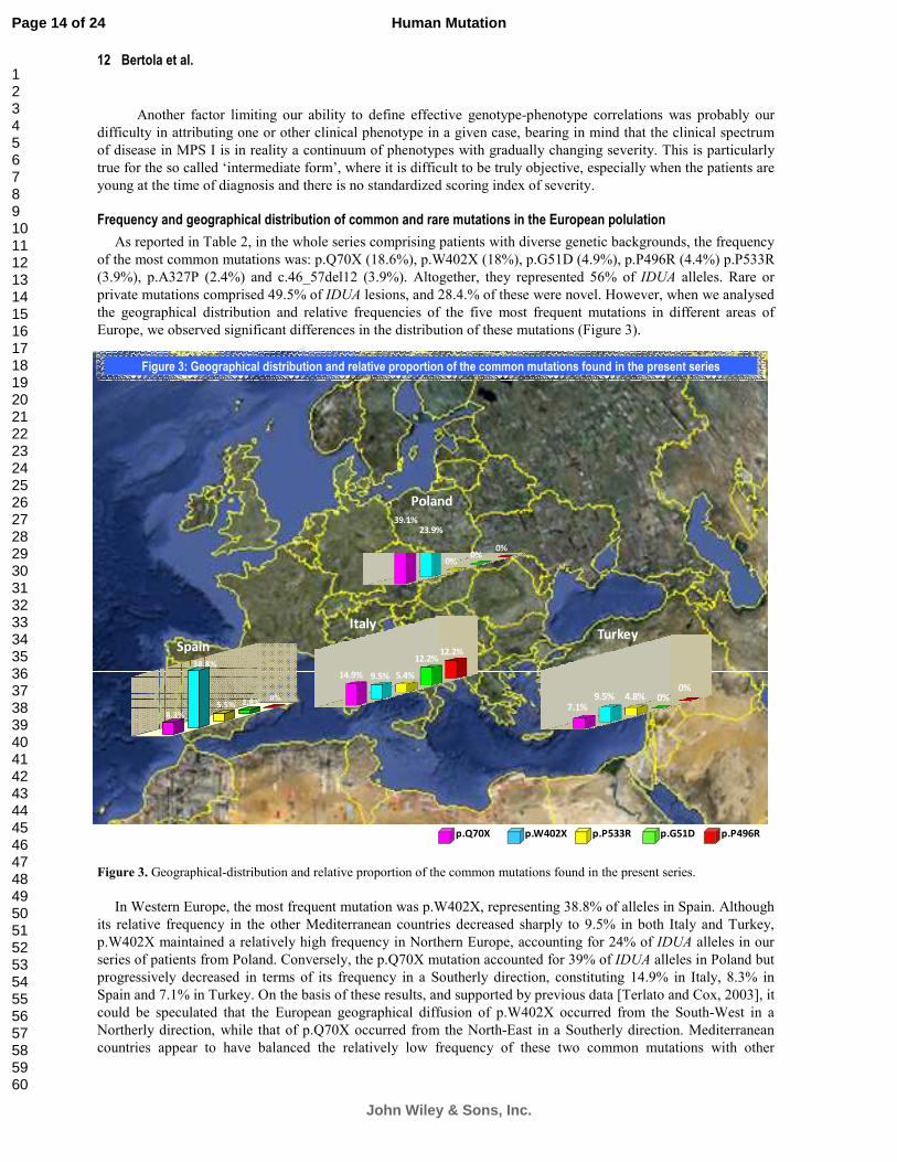
Frequency and geographical distribution of common and rare mutations in the European polulation
As reported in Table 2, in the whole series comprising patients with diverse genetic backgrounds, the frequency
of the most common mutations was: p.Q70X (18.6%), p.W402X (18%), p.G51D (4.9%), p.P496R (4.4%) p.P533R
(3.9%), p.A327P (2.4%) and c.46_57del12 (3.9%). Altogether, they represented 56% of IDUA alleles. Rare or
private mutations comprised 49.5% of IDUA lesions, and 28.4.% of these were novel. However, when we analysed
the geographical distribution and relative frequencies of the five most frequent mutations in different areas of
Europe, we observed significant differences in the distribution of these mutations (Figure 3).
Figure 3. Geographical-distribution and relative proportion of the common mutations found in the present series.
In Western Europe, the most frequent mutation was p.W402X, representing 38.8% of alleles in Spain. Although
its relative frequency in the other Mediterranean countries decreased sharply to 9.5% in both Italy and Turkey,
p.W402X maintained a relatively high frequency in Northern Europe, accounting for 24% of IDUA alleles in our
series of patients from Poland. Conversely, the p.Q70X mutation accounted for 39% of IDUA alleles in Poland but
progressively decreased in terms of its frequency in a Southerly direction, constituting 14.9% in Italy, 8.3% in
Spain and 7.1% in Turkey. On the basis of these results, and supported by previous data [Terlato and Cox, 2003], it
could be speculated that the European geographical diffusion of p.W402X occurred from the South-West in a
Northerly direction, while that of p.Q70X occurred from the North-East in a Southerly direction. Mediterranean
countries appear to have balanced the relatively low frequency of these two common mutations with other
14.9% 9.5% 5.4%
12.2%12.2%
39.1%23.9%
0%0%
0%
7.1%9.5% 4.8% 0%
0%
Spain
Italy
Poland
Turkey
p.Q70X p.W402X p.P533R p.G51D p.P496R
0%2.8%5.5%
38.8%
8.3%
Figure 3: Geographical distribution and relative proportion of the common mutations found in the present series
Page 14 of 24
John Wiley & Sons, Inc.
Human Mutation
123456789101112131415161718192021222324252627282930313233343536373839404142434445464748495051525354555657585960

For Peer Review
IDUA Gene Mutations 13
mutations specific to this geographical region viz. p.P533R, p.P496R and p.G51D, the latter two being specific to
(and recurrent in) the Italian population, and c.494-1G>A, a Turkish-specific mutation (not shown in the figure).
The relatively low frequency of the two usually common mutations (p.Q70X and p.W402X) in Italy and Turkey,
results in an increase in IDUA allelic heterogeneity in these two countries as compared to Northern European
countries. Thus, rare mutations accounted for 50% and 71.4% of alleles in Italy and Turkey, respectively, as
compared to Poland and Spain where they were 37% and 41.6% respectively. The finding that European countries
vary widely in terms of both the distribution and frequency of IDUA gene mutations has important implications for
the selection of screening strategies in individual countries. The existence of dramatic differences in mutational
heterogeneity and mutation prevalence highlights the importance of multi-national screening studies in helping to
elucidate the genotype-phenotype relationship in disorders such as MPS I that are characterized by extensive
allelic heterogeneity.
ACKNOWLEDGMENTS
European patient samples were obtained through a collaboration with Genzyme Corporation, Cambridge, MA,
USA. Some samples were also obtained from the “Cell Line and DNA Biobank from Patients Affected by Genetic
Diseases” (G. Gaslini Institute) – Telethon Genetic Biobank Network (Project No. GTB07001A). We also thank
Prof. Generoso Andria (Napoli), Dr. Rita Barone (Catania), Prof. Cesare Danesino (Pavia), Prof. Paola Di Natale
(Napoli), Dr. Graziella Uziel (Milano), for their contribution of clinical information and patient samples.
REFERENCES
Adzhubei IA, Schmidt S, Peshkin L, Ramensky VE, Gerasimova A, Bork P, Kondrashov AS, Sunyaev SR. 2010. A method and
server for predicting damaging missense mutations. Nat Methods 7:248-249.
Alif N, Hess K, Straczek J, Sebbar S, Belahsen Y, Mouane N, Abkari A, Nabet P, Gelot MA. 2000. Mucopolysaccharidosis
type I in Morocco: clinical features and genetic profile. Arch Pediatr 7:597-604.
Beesley CE, Meaney CA, Greenland GA, Adams V, Vellodi A, Young EP, Winchester BG. 2001. Mutational analysis of 85
mucopolysaccharidosis type I families: frequency of known mutations, identification of 17 novel mutations and in vitro
expression of missense mutations. Hum Genet 109:503-511.
Bunge S, Kleijer WJ, Steglich C, Beck M, Schwinger E, Gal A. 1995. Mucopolysaccharidosis type I: identification of 13 novel
mutations of the alpha-L-iduronidase gene. Hum Mutat 6:91-94.
Bunge S, Kleijer WJ, Steglich C, Beck M, Zuther C, Morris CP, Schwinger E, Hopwood JJ, Scott HS, Gal A. 1994.
Mucopolysaccharidosis type I: identification of 8 novel mutations and determination of the frequency of the two common
alpha-L-iduronidase mutations (W402X and Q70X) among European patients. Hum Mol Genet 3:861-866.
Cartegni L, Chew SL, Krainer AR. 2002. Listening to silence and understanding nonsense: exonic mutations that affect
splicing. Nat Rev Genet 3:285-298.
Clarke LA, Nelson PV, Warrington CL, Morris CP, Hopwood JJ, Scott HS. 1994. Mutation analysis of 19 North American
mucopolysaccharidosis type I patients: identification of two additional frequent mutations. Hum Mutat 3:275–282.
Clarke LA (Updated [September 21, 2007]). Mucopolysaccharidosis Type I. In: GeneReviews at GeneTests: Medical Genetics
Information Resource (database online). Copyright, University of Washington, Seattle. 1993-2007. Available at
http://www.genetests.org
den Dunnen JT, Antonarakis SE. 2000. Mutation nomenclature extensions and suggestions to describe complex mutations: a
discussion. Hum Mutat 15:7-12.
den Dunnen JT, Paalman MH. 2003. Standardizing mutation nomenclature: why bother? Hum Mutat 2:181-182.
Emsley, P. & Cowtan, K. 2004. Coot: model-building tools for molecular graphics. Acta Crystallogr. D 60:2126-2132
Page 15 of 24
John Wiley & Sons, Inc.
Human Mutation
123456789101112131415161718192021222324252627282930313233343536373839404142434445464748495051525354555657585960

For Peer Review
14 Bertola et al.
Gatti R, DiNatale P, Villani GR, Filocamo M, Muller V, Guo XH, Nelson PV, Scott HS, Hopwood JJ. 1997. Mutations among
Italian mucopolysaccharidosis type I patients. J Inherit Metab Dis 20:803-806.
Gort L, Chabas A, Coll MJ. 1998. Analysis of five mutations in 20 mucopolysaccharidosis type 1 patients: high prevalence of
the W402X mutation. Hum Mutat 11:332–333.
Hein LK, Hopwood JJ, Clements PR, Brooks DA. 2003. The alpha-L-iduronidase mutations R89Q and R89W result in an
attenuated mucopolysaccharidosis type I clinical presentation. Biochim Biophys Acta 1639:95–103.
Krawczak M, Thomas NS, Hundrieser B, Mort M, Wittig M, Hampe J, Cooper DN. 2007. Single base-pair substitutions in
exon-intron junctions of human genes: nature, distribution, and consequences for mRNA splicing. Hum Mutat 28:150-158.
Laradi S, Tukel T, Erazo M, Shabbeer J, Chkioua L, Khedhiri S, Ferchichi S, Chaabouni M, Miled A, Desnick RJ. 2005.
Mucopolysaccharidosis I: alpha-L-Iduronidase mutations in three Tunisian families. J Inherit Metab Dis 28:1019-1026.
Larkin M.A., Blackshields G., Brown N.P., Chenna R., McGettigan P.A., McWilliam H., Valentin F., Wallace I.M., Wilm A.,
Lopez R., Thompson J.D., Gibson T.J. and Higgins D.G. 2007. ClustalW and ClustalX version 2. Bioinformatics 23(21):
2947-2948.
Li B, Krishnan VG, Mort ME, Xin F, Kamati KK, Cooper DN, Mooney SD, Radivojac P. 2009. Automated inference of
molecular mechanisms of disease from amino acid substitutions. Bioinformatics 25:2744-2750.
Li P, Wood T, Thompson JN. 2002. Diversity of mutation and distribution of single nucleotide polymorphic alleles in the
human alpha-L-iduronidase (IDUA) gene. Genet Med 4:420-426
Lowry OH, Rosebrough NJ, Farrand AL, Randall RJ. 1951. Protein measurement with the Folin-Phenol reagents. J Biol Chem
193:265-275.
Lualdi S, Pittis MG, Regis S, Parini R, Allegri A, Furlan F, Bembi B, Filocamo M. 2006. Multiple cryptic splice sites can be
activated by IDS point mutations generating misspliced transcripts. J Mol Med 84:692-700.
Matte U, Yogalingam G, Brooks D, Leistner S, Schwartz I, Lima L, Norato DY, Brum JM, Beesley C, Winchester B, Giugliani
R, Hopwood JJ. 2003. Identification and characterization of 13 new mutations in mucopolysaccharidosis type I patients.
Mol Genet Metab 78: 37-43.
Mort M, Evani US, Krishnan VG, Kamati KK, Baenziger PH, Bagchi A, Peters BJ, Sathyesh R, Li B, Sun Y, Xue B, Shah NH,
Kann MG, Cooper DN, Radivojac P, Mooney SD. 2010. In silico functional profiling of human disease-associated and
polymorphic amino acid substitutions. Hum Mutat 31:335-346.
Nalla VJ and Rogan PK. 2005. Automated splice site analysis by information theory. Hum Mut 25:334-342.
Neufeld EF, Muenzer J. 2001 The mucopolysaccharidoses. in Scriver CR, Beaudet AL, Sly WS, Valle D (eds): The metabolic
and molecular bases of inherited disease, 8th Ed McGraw-Hill, , New York pp 3421-3452.
Ng PC, Henikoff S. 2001. Predicting deleterious amino acid substitutions. Genome Res. 11:863-874.
Nissim-Rafinia M, Kerem B. 2002. Splicing regulation as a potential genetic modifier. Trends Genet 18:23–27
Pastores GM, Arn P, Beck M, Clarke JT, Guffon N, Kaplan P, Muenzer J, Norato DY, Shapiro E, Thomas J, Viskochil D,
Wraith JE. 2007. The MPS I registry: design, methodology, and early findings of a global disease registry for monitoring
patients with mucopolysaccharidosis type I. Mol Genet Metab 91:37-47.
Pettersen EF, Goddard, TD, Huang CC, Couch GS, Greenblatt DM, Meng EC, Ferrin TE. 2004. UCSF Chimera - a
visualization system for exploratory research and analysis. J Comput. Chem. 25:1605-1612.
Pollard KS, Hubisz MJ, Rosenbloom KR, Siepel A. 2010. Detection of nonneutral substitution rates on mammalian
phylogenies. Genome Res. 20:110-121.
Rempel BP, Clarke LA, Withers SG. 2005. A homology model for human α-l-iduronidase: Insights into human disease. Mol
Genet Metab 85:28-37
Scott HS, Anson DS, Orsborn AM, Nelson PV, Clements PR, Morris CP, Hopwood JJ. 1991. Human alpha-L-iduronidase:
cDNA isolation and expression. Proc Natl Acad Sci USA 88:9695-9699.
Page 16 of 24
John Wiley & Sons, Inc.
Human Mutation
123456789101112131415161718192021222324252627282930313233343536373839404142434445464748495051525354555657585960
For Peer Review
IDUA Gene Mutations 15
Scott HS, Ashton LJ, Eyre HJ, Baker E, Brooks DA, Callen DF, Sutherland GR, Morris CP, Hopwood JJ. 1990. Chromosomal
localization of the human alpha-L-iduronidase gene (IDUA) to 4p16.3. Am J Hum Genet 47:802-807.
Scott HS, Bunge S, Gal A, Clarke LA, Morris CP. Hopwood JJ. 1995. Molecular genetics of mucopolysaccharidosis type I:
diagnostic, clinical, and biological implications. Hum Mutat 6:288-302.
Scott HS, Guo XH, Hopwood JJ, Morris CP. 1992. Structure and sequence of the human alpha-L-iduronidase gene. Genomics
13: 1311-1313.
Scott HS, Litjens T, Nelson PV, Brooks DA, Hopwood JJ, Morris CP. 1992. alpha-L-iduronidase mutations (Q70X, and
P533R) associate with a severe Hurler phenotype. Hum Mutat 1:333–339.
Scott HS, Litjens T, Nelson PV, Thompson PR, Brooks DA, Hopwood JJ, Morris CP. 1993. Identification of mutations in the
alpha -L-lduronidase gene (IDUA) that cause Hurler and Scheie syndromes. Am J Hum Genet 53:973-986.
Stenson PD, Mort M, Ball E, Howells K, Phillips A, Thomas NST, Cooper DN. 2009. The Human Gene Mutation Database:
2008 update. Genome Med 1:13.
Stirling JL, Robinson D, Fensom AH, Benson PF, Baker JE. 1978. Fluorimetric assay for prenatal detection of Hurler and
Scheie homozygotes or heterozygotes. Lancet. 1:147.
Sugawara K, Saito S, Ohno K, Okuyama T, Sakuraba H. 2008. Structural study on mutant alpha-L-iduronidases: insight into
mucopolysaccharidosis type I. J Hum Genet 53:467-474.
Terlato NJ, Cox GF. 2003. Can mucopolysaccharidosis type I disease severity be predicted based on a patient’s genotype? A
comprehensive review of the literature. Genet Med 5:286-294.
Tieu PT, Bach G, Matynia A, Hwang M, Neufeld EF. 1995. Four novel mutations underlying mild or intermediate forms of
alpha-L-iduronidase deficiency (MPS IS and MPS IH/S). Hum Mutat 6:55-59.
Vazna A, Beesley C, Berna L, Stolnaja L, Myskova H, Bouckova M, Vlaskova H, Poupetova H, Zeman J, Magner, Hlavata A,
Winchester B, Hrebicek M, Dvorakova L. 2009. Mucopolysaccharidosis type I in 21 Czech and Slovak patients: mutation
analysis suggests a functional importance of C-terminus of the IDUA protein. Am J Med Genet Part A 149A:965–974.
Venturi N, Rovelli A, Parini R, Menni F, Branbillasca F, Bertagnolio F, Uziel G, Gatti R, Filocamo M, Donati MA, Biondi A,
Goldwurm S. 2002. Molecular analysis of 30 mucopolysaccharidosis type I patients: evaluation of the mutational spectrum
in Italian population and identification of 13 novel mutations. Hum Mutat 20:231.
Voskoboeva E, Krasnopolskaya X, Mirenburg T, Weber B, Hopwood J. Molecular genetics of mucopolysaccharidosis type I:
mutation analysis among the patients of the former Soviet Union. Mol Genet Metab 1998;65:174–180
Yogalingam G, Guo X, Muller V, Brooks DA, Clements PR, Kakkis ED, Hopwood JJ. 2004. Identification and molecular
characterization of alpha-L-iduronidase mutations present in mucopolysaccharidosis type I patients undergoing enzyme
replacement therapy. Hum Mutat 24: 199-207.
Page 17 of 24
John Wiley & Sons, Inc.
Human Mutation
123456789101112131415161718192021222324252627282930313233343536373839404142434445464748495051525354555657585960
For Peer Review
16 Bertola et al.
Supp. Figure S1. Evolutionary comparison of the protein sequences flanking the missense mutations identified in the human
IDUA protein with their orthologous counterparts in 5 vertebrates (in silico data from http://www.ebi.ac.uk/clustalw/). From the
top: From the top: Homo sapiens, Pan troglodytes, Canis familiaris, Mus musculus, Gallus gallus, Tetraodon nigrovisidis.
Positions of mutations are marked by vertical boxes. Note that the translational initiation site substitution, predicted to produce
either no protein or to move the initiation site up/downstream (p.M1?), was excluded from the analysis.
SUPPORTING INFORMATION
Page 18 of 24
John Wiley & Sons, Inc.
Human Mutation
123456789101112131415161718192021222324252627282930313233343536373839404142434445464748495051525354555657585960
For Peer Review
IDUA Gene Mutations 17
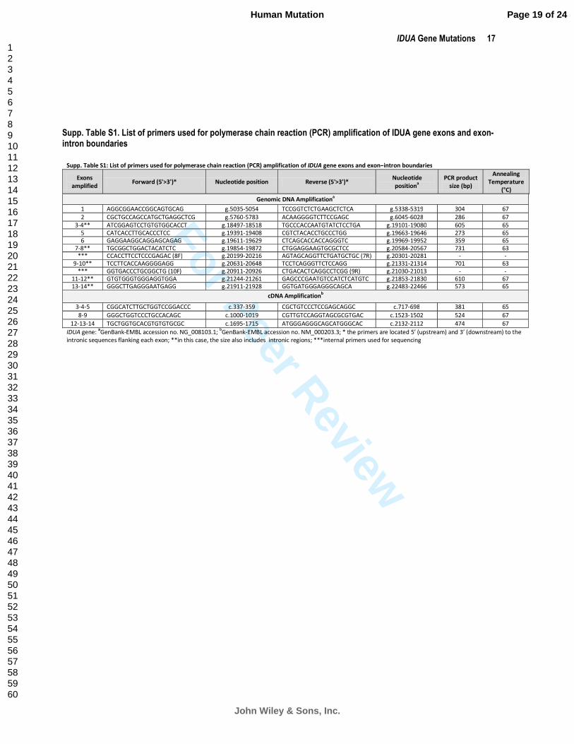
Supp. Table S1. List of primers used for polymerase chain reaction (PCR) amplification of IDUA gene exons and exon-intron boundaries
Supp. Table S1: List of primers used for polymerase chain reaction (PCR) amplification of IDUA gene exons and exon–intron boundaries
Exons
amplified Forward (5'>3')* Nucleotide position Reverse (5'>3')*
Nucleotide
positiona
PCR product
size (bp)
Annealing
Temperature
(°C)
Genomic DNA Amplificationa
1 AGGCGGAACCGGCAGTGCAG g.5035-5054 TCCGGTCTCTGAAGCTCTCA g.5338-5319 304 67
2 CGCTGCCAGCCATGCTGAGGCTCG g.5760-5783 ACAAGGGGTCTTCCGAGC g.6045-6028 286 67
3-4** ATCGGAGTCCTGTGTGGCACCT g.18497-18518 TGCCCACCAATGTATCTCCTGA g.19101-19080 605 65
5 CATCACCTTGCACCCTCC g.19391-19408 CGTCTACACCTGCCCTGG g.19663-19646 273 65
6 GAGGAAGGCAGGAGCAGAG g.19611-19629 CTCAGCACCACCAGGGTC g.19969-19952 359 65
7-8** TGCGGCTGGACTACATCTC g.19854-19872 CTGGAGGAAGTGCGCTCC g.20584-20567 731 63
*** CCACCTTCCTCCCGAGAC (8F) g.20199-20216 AGTAGCAGGTTCTGATGCTGC (7R) g.20301-20281 - -
9-10** TCCTTCACCAAGGGGAGG g.20631-20648 TCCTCAGGGTTCTCCAGG g.21331-21314 701 63
*** GGTGACCCTGCGGCTG (10F) g.20911-20926 CTGACACTCAGGCCTCGG (9R) g.21030-21013 - -
11-12** GTGTGGGTGGGAGGTGGA g.21244-21261 GAGCCCGAATGTCCATCTCATGTC g.21853-21830 610 67
13-14** GGGCTTGAGGGAATGAGG g.21911-21928 GGTGATGGGAGGGCAGCA g.22483-22466 573 65
cDNA Amplificationb
3-4-5 CGGCATCTTGCTGGTCCGGACCC c.337-359 CGCTGTCCCTCCGAGCAGGC c.717-698 381 65
8-9 GGGCTGGTCCCTGCCACAGC c.1000-1019 CGTTGTCCAGGTAGCGCGTGAC c.1523-1502 524 67
12-13-14 TGCTGGTGCACGTGTGTGCGC c.1695-1715 ATGGGAGGGCAGCATGGGCAC c.2132-2112 474 67
IDUA gene: aGenBank-EMBL accession no. NG_008103.1;
bGenBank-EMBL accession no. NM_000203.3; * the primers are located 5’ (upstream) and 3’ (downstream) to the
intronic sequences flanking each exon; **in this case, the size also includes intronic regions; ***internal primers used for sequencing
Page 19 of 24
John Wiley & Sons, Inc.
Human Mutation
123456789101112131415161718192021222324252627282930313233343536373839404142434445464748495051525354555657585960
For Peer Review
18 Bertola et al.
Supp. Table S2: Charac teristics of putative ly neutral SNPs identified in the IDUA gene
Location dbSNP
no.
Site of nucleotide
substitution*
(amino acid change)**
Minor allele
frequency§
( )&
MutPred analysis ���� Polymorphic background in the
Patients#
or control alleles
Probability of
deleterious
mutation
Confident
in-silico hypotheses
Ex.1 rs11248061 c.24C>A (A8A) NA (0.42) .
15,24,26,27,32,35,37,44,60,62,64,
68,69,70,72,73,75,76,79,80,81,82,
85,86,87,89,90, 92 ,93,98,99,102
Ex.1 rs10902762 c.60G>A (A20A) NA (0.44) .
15,24,26,27,32,35,37,44,60,62,64,
68,69,70,72,73,75,76,80,82,85,86,87,89,90,92,93,98,99,102
Ex.1 rs10794537 c.99T>G (Q33H) NA (0.19) 0.21 None 17,20,21,23,25,30,33,39,40,41,43,44,45,46,52,53,54,55,57,60,61,63,
64,65,76,79,91,96,100,101
Intr. 2 c.299+6C>T� (0.04) . control alleles
Intr. 2 c.299+7G>A (0.01) . control alleles
Intr. 2 c.299+9C>T�� (0.00) . 20
Intr. 2 c.300-44C>T� (0.07) . 37,83,72
Ex.3 rs3755955 c.314G>A (R105Q) 0.15 (0 .18) 0.12 None 24,28,29,35,45,51,53,59,60,67,71,75
Ex.3 rs3755954 c.352C>T (L118L) 0.16 (0 .14) .
17,20,21,23,25,27,30,33,34,35,39,40,41,43,44,46,49,52,54,55,57,59,60,61,62,63,64,65,70,71,76,79,83,84,91,96,100,101
Ex.4 c.408C>T (A136A) (0.01) . control alleles
Ex.5 rs6815946 c.543T>C (N181N) 0.14 (0 .18) . 24,28,29,49,51, 59 ,60,67,71
Intr. 5 c.589+20dupG (0.00) . 22
Intr. 5 rs6829789 c.590-45G>C NA (0.18) . 24,28,29,51,53,59,60,67,71
Intr. 5 rs6848974 c.590-8C>T NA (0.18) . 19,24,28,29,45,49,51,53,59,60,67,71
Intr. 6 c.792+12C>T (0.01) . control alleles
Intr. 6 c.792+14C>T (0.00) . 17
Ex. 7 rs11480689 c.891C>T (N297N) NA (0.04) . 21,42,43,52
Ex. 7 rs6830825 c.942G>C (A314A) NA (0.16) . 19,24,28,49,51,59,60,67,71
Intr. 7 rs6811373 c.972+48A>G 0.14 (0 .07) . 28,29, 49,51,53,67,71
Intr. 7 rs6831021 c.973-45G>C NA . 28,29,49,51,53,67
Ex. 8 rs6831280 c.1081G>A (A361T) 0.21 (0 .17) 0.13 None 28,49,53,59,60,67
Ex. 8 rs6836258 c.1164G>C (T388T) NA (0.17) . 28,49,53,59,60,67
Ex. 8 c.1174C>T (L392L)� (0.01) . control alleles
Intr. 8 c.1190-10insC� (0.15) . 19,24,28,29,45,49,51,53,67,71
Ex. 9 rs115790973 c.1230C>G (T410T) NA (0.16) . 19,24,28,29,45,49,51,53, 59,60,67,71
Ex. 9 c.1345C>A (H449N) (0.00) 0.40 None 46
Ex. 9 rs73066479 c.1360G>A (V454I) NA (0.17) 0.10 None 19,24,28,29,45,49,51,53,59,60,67,71
Intr. 9 c.1403+36T>C� (0.17) . 19,24,45,51,59,60,67,71
Ex. 10 rs115929690 c.1467C>T (R489R) NA (0.14) . 19,24,45,49,51,53, 59,60,67,71
Ex. 10 c.1473C>A (G491G) (0.01) . control alleles
Ex. 10 c.1515C>G (R505R) (0.00) . 19
Intr. 10 c.1524+41G>T� (0.11) . 45,49,53,59,60,67,71
Intr. 10 rs113289555 c.1524+53G>T NA (0.29) .
18,19,22,24,26,29,31,32,36,37,39,47,48,50,51,55,56,58,65,66,68,69,70,74,75,76,77,81,82,85,86,88,90,92,95,97,98,100,102
Intr. 10 c.1525-38T>C� (0.09) . 28
Intr. 12 rs2305488 c.1727+72T>G 0.11 . 19,24,29,49,53,67
Intr. 12 rs2305489 c.1727+75G>T 0.04 . 21,42,43,52
Ex. 13 c.1771G>A (A591T)� �� (0.00) 0.15 None 17,19,22,23,30,33
Legend: dbSNP, the Single Nucl eotide Polymorphism database, is ava ilable at http://www.ncbi.nl m.nih.gov/projects/SNP/; *GenBank-EMBL
accession no. NM_000203.3; **GenBank-EMBL accessi on no. NP_000194; the minor allele frequencies were
§ obtained from dbSNP or ( )
&
calculated from 100 Caucasian control alleles (novel polymorphisms, in bold) ; NA: not available; #patient number according to Table 1. �Scott et
al., (1995); �� Venturi et al., (2002); ���neutral change shown in vitro by Beesley et al.(2001); ���� Li et al. , (2009) a nd Mort et al., (2010).
Page 20 of 24
John Wiley & Sons, Inc.
Human Mutation
123456789101112131415161718192021222324252627282930313233343536373839404142434445464748495051525354555657585960

For Peer Review
IDUA Gene Mutations 19
S upp. Table S4. B io info rmatic an alysis of novel missense variants o f uncertain patho genic ity
Nucleot id e s ubs titution
Am in o acid change
Co ns ervation b ase level (phy loP)
MutPred probability of deleteriou s mutat ion
SIFT Prediction
(sco re)
PolyPhen2 (score)
Splicing disrupt ion
c.307A>C p.T103P Conserved
(0.4) 0.36
Tolerated (0.5)
B enig n (0.0)
FALSE
c .1306G >C p.A436P Conserved
(2.8) 0.45
Tolerated (0.2)
Possibly damaging (0.26)
FALSE
Supp. Table S3. MutPred analysis for IDUA missense mutations
Mutation Probability of
deleterious mutation
Molecular mechanism altered
Confident Hypotheses Very Confident Hypotheses
p.M1? 0.99
p.G51D 0.97 Gain of relative solvent accessibility (P = 0.0215)
Gain of solvent accessibility (P=0.0306)
p.Y76C 0.73
p.G84R 0.93 Gain of MoRF binding (P=0.0303)
Gain of solvent accessibility (P=0.0456)
p.R89W 0.96
p.T103P 0.36 See details in Supp. Table S4
p.E178K 0.97 Gain of MoRF binding (P=0.0143)
Gain of ubiquitination at E178 (P = 0.0258) Loss of catalytic residue at W180 (P = 0.0315)
Gain of methylation at E178 (P=0.0036)
p.F188L* 0.95
p.S423R* 0.92 Gain of methylation at S423 (P=0.0279)
p.G219E 0.89 Gain of solvent accessibility (P=0.0145)
Gain of relative solvent accessibility (P=0.0215)
p.G265R 0.95
Gain of methylation at G265 (P=0.0219) Loss of helix (P=0.028) Gain of loop (P=0.0312)
Gain of MoRF binding (P=0.033)
p.E276K 0.76
p.W306L 0.82 Loss of catalytic residue at P309 (P=0.0327)
p.A327P 0.84 Gain of relative solvent accessibility (P=0.0215)
Gain of methylation at K324 (P=0.0487)
p.N348K 0.85
Loss of sheet (P=0.0181) Gain of ubiquitination at N348 (P=0.0192) Gain of methylation at N348 (P=0.0295)
Loss of stability (P=0.0381)
p.P385R 0.82
Gain of relative solvent accessibility (P=0.0215) Loss of sheet (P=0.0315) Loss of loop (P=0.0374) Gain of helix (P=0.0425)
Gain of solvent accessibility (P=0.0055) Gain of MoRF binding (P=0.0081)
p.L396P 0.83 Loss of helix (P=0.0167) Gain of loop (P=0.0195)
Loss of stability (P=0.0047)
p.A436P 0.45 See details in Supp. Table S4
p.L490P 0.86 Gain of disorder (P=0.0154)
Loss of helix (P=0.028) Loss of stability (P=0.0441)
p.R492P 0.83 Loss of MoRF binding (P = 0.0011)
p.P496R 0.95 Gain of MoRF binding (P = 0.0078)
p.P533R 0.94 Gain of MoRF binding (P = 0.0068)
p.L535F 0.59 *mutations in cis on the sam e allele; MoRFs=Molecular Recognition Features
Page 21 of 24
John Wiley & Sons, Inc.
Human Mutation
123456789101112131415161718192021222324252627282930313233343536373839404142434445464748495051525354555657585960
For Peer Review
20 Bertola et al.
Sup p. Table S5. Predictio n of consequences of n ovel missense m utations us in g the 3D h omology model o f the IDUA p rotein constructed by Rempel et al. [2005]
MUTATION PROTEIN POSIT ION MODE LLING ANALYSIS PREDICTION
p .Y76C Solvent accessible Large aromatic and polar side c hain subst ituted by a smaller, hig hly reactive
one.
p.G84R Close to active site Loss of m ain chain flexibility and of solvent accessible surface . G ain of charge.
p.T103P Solvent accessible Introduc tion of a new proline within a long loop (G 101-N110) protruding into
the solvent. Acquisition of loop rigidity. Decrease in residue pola rity.
p .F188L* Active site
F188 be longs to an am ino acid cluster c om prising aroma tic residues (F143, W180, W 306, Y343) and residues bearing indole ring s (H56, H91, H185). The newly acquired 188L residue would maintain the hydrophobic ity but would give r ise to a loss of a rom atic ity. F188 is considered to be an active site residue
p .S423R* Core Increase in size, altera tion in charge. D isruption of local prote in fold due t o
steric hindrance
p.G 219E Core Increase in size, altera tion in c harg e. Desta bilisation of the local prote in fold
due t o steric hindrance
p.E276K Solvent accessible Altera tion in charg e (negative to positive). Cha nge of the protein surface
electrostatic potential
p.W306L Core
W306 belongs to a n amino acid cluster c omprising aroma tic residues (F143,
W180, W 306, Y343) and residues bearing indole ring s (H56, H91, H185). The
newly acquired 306L residue would maintain the hydrophobic ity but would
give rise to a loss of aromaticity.
p.N348K Solvent accessible Altera tion in surfa ce local charge.
p.P385R Core Loss of local rigidity. Destabilisa tion of the local protein fold.
p.L396P Solvent accessible Loss of long and branched hydrophobic side chain (leucine). L oss of local
flexibility in the edge of a turn. Loss of local protein fold.
p .A 436P Solvent accessible
Ga in of m ain chain rigidity (436P) which would disrupt the super-seconda ry
structure of the protein owing to its inability to form the main chain H-bonds
that are required for β-sheet forma tion.
*mutations occur in c is on the same a lle le
Page 22 of 24
John Wiley & Sons, Inc.
Human Mutation
123456789101112131415161718192021222324252627282930313233343536373839404142434445464748495051525354555657585960
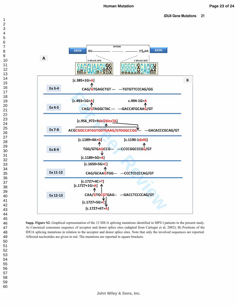
For Peer Review
IDUA Gene Mutations 21
Supp. Figure S2. Graphical representation of the 12 IDUA splicing mutations identified in MPS I patients in the present study.
A) Canonical consensus sequence of acceptor and donor splice sites (adapted from Cartegni et al, 2002); B) Positions of the
IDUA splicing mutations in relation to the acceptor and donor splice sites. Note that only the involved sequences are reported.
Affected nucleotides are given in red. The mutations are reported in square brackets.
INTRON
5’ SPLICE SITE 3’ SPLICE SITE
EXON EXONGU (Y)nAG
A
CAG/GTGAGCTGT --- ---TGTGTTCCCAG/GG
[c.385+1G>A]
Ex 3-4
CAG/GTAGGCTAC --- ---GACCATGCAAG/GT
[c.493+1G>A]
Ex 4-5
c.494-1G>A
TGG/GTGAGCCG--- ---CCCCGGCCCGG/GT
[c.1189+4A>G]
Ex 8-9
[c.1190-1delG]
[c.1189+5G>A]
CAG/GCAAGTGG--- ---CCCTCCCCCAG/GT
[c.1650+5G>C]
Ex 11-12
CAA/GTGCGTGAG-- ---GACCTCCCCAG/GT
[c.1727+1G>A]
Ex 12-13
[c.1727+4C>T]
[c.1727+5G>C]
[c.1727+6T>A]
B
ACGCGGCCATGGTGGTGAAG/GTGGGCCGG --- ---GACACCCGCAG/GT
[c.956_972+9del26insTA]
Ex 7-8
Page 23 of 24
John Wiley & Sons, Inc.
Human Mutation
123456789101112131415161718192021222324252627282930313233343536373839404142434445464748495051525354555657585960

For Peer Review
22 Bertola et al.
Supp Table S6. Genotype-phenotype correlation: comparison with existing data from the literature
NO. OF PATIENTS ANALYZED AND (PHENOTYPE) GENOTYPES*
THIS STUDY
OTHER STUDIES
REFERENCES
[p.W402X]+[p.W402X] 12 (H) 46 (H) Bunge et al. (1994); Gort et al. (1998); Hein et al. (2003); Li et al,
(2002); Matte et al. (2003); Scott et al. (1992); Vazna et al. (2009)
[p.Q70X]+[p.Q70X] 9 (H) 26 (H)
Bunge et al. (1994); Gort et al. (1998); Hein et al. (2003); Scott et
al. (1992); Vazna et al. (2009); Venturi et al. (2002); Voskoboeva et
al. (1998)
[p.W402X]+[p.Q70X] 4 (H) 19 (H) Bunge et al. (1994); Li et al. (2002); Scott et al. (1992)
[p.W402X]+[c.1650+5G>A] 1 (H) 2 (H) Vazna et al. (2009); Venturi et al. (2002);
[p.W402X]+[p.A327P] 1 (H) 2 (H) Bunge et al. (1994); Vazna et al. (2009)
[p.A327P]+[p.A327P] 1 (H) 1 (H-H/S) Gatti et al. (1997)
[p.G51D]+[p.G51D] 2 (H) 1 (H) Gatti et al. (1997)
[p.Q70X]+[c.1333_1335del3] 1 (S) 1 (S) Bunge et al. (1994)
INF
OR
MA
TIV
E
[p.L490P)+[p.L490P] 1 (S) 3 (H/S) Beesley et al. (2001); Tieu et al. (1995)
[p.P533R]+[p.P533R] 2 (H/S) 8 (§) Alif et al. (1999); Gatti et al. (1997); Laradi et al. (2005); Matte et
al. (2003); Scott et al. (1992)
[p.P533R]+[p.P533R] 1 (H) 11 (§) Alif et al. (1999); Hein et al. (2003)
[p.G51D]+[p.P533R] 1 (H/S) 2 (§) Gatti et al. (1997); Venturi et al. (2002)
PA
RT
IALLY
INF
OR
MA
TIV
E
[c.46_57del12]+[c.46_57del12] 2 (H/S) 2 (H) Bunge et al. (1994); Venturi et al. (2002)
Legend: H=Hurler; S=Scheie; *Note that only the comparable genotypes with existing data are reported in this table. For the other
genotypes, refer to Table 1; § various phenotypes
Page 24 of 24
John Wiley & Sons, Inc.
Human Mutation
123456789101112131415161718192021222324252627282930313233343536373839404142434445464748495051525354555657585960