identification of rab7 as a melanosome-associated protein involved in the intracellular transport of...
TRANSCRIPT

Identi®cation of rab7 as a Melanosome-Associated ProteinInvolved in the Intracellular Transport of Tyrosinase-RelatedProtein 1
Paul F. Gomez,* Dong Luo,* Kuninori Hirosaki,² Kyoka Shinoda,² Toshiharu Yamashita,² Jun-ichi Suzuki,³Kaoru Otsu,³ Kiichi Ishikawa,³ and Kowichi Jimbow*²*Division of Dermatology and Cutaneous Sciences, University of Alberta, Edmonton, Alberta, Canada; ³Department of Biochemistry, Yamagata
University, School of Medicine, Yamagata, Japan; ²Department of Dermatology, Sapporo Medical University, School of Medicine, Sapporo, Japan
The melanosome is a unique secretory granule of themelanocyte in which melanin pigments are synthe-sized by tyrosinase gene family glycoproteins.Melanogenesis is a highly regulated process becauseof its inherent toxicity. An understanding of thevarious regulatory mechanisms is important indelineating the pathophysiology involved in pigmen-tary disorders and melanoma. We have puri®ed andanalyzed the total melanosomal proteins from B16mouse melanoma tumors in order to identify newproteins that may be involved in the control of themelanogenesis process. Melanosomal proteins wereresolved by two-dimensional sodium dodecyl sulfatepolyacrylamide gel electrophoresis, a predominantspot (27 kDa with isoelectric point 5.8±6.4) wasexcised and digested with cyanogen bromide, and thefragments were sequenced. Synthetic oligonucleotideprimers were synthesized corresponding to the pep-tide sequences, and reverse transcriptase polymerasechain reaction ampli®cation of total RNA from B16cells was carried out. Sequencing of one of the poly-merase-chain-reaction-mediated clones demonstrated80%±97% sequence homology of 200 bp nucleotidewith GTP-binding proteins at the 3¢-untranslatedregion. GTP-binding assay on two-dimensional gels
of melanosomal proteins showed the presence ofseveral (®ve to six) small GTP-binding proteins,suggesting that small GTP-binding proteins are asso-ciated with the melanosome. Among the knownGTP-binding proteins with similar molecular weightand isoelectric point ranges, rab3, rab7, and rab8were found to be present in the melanosomal fractionby immunoblotting. Confocal immuno¯uorescencemicroscopy showed that rab7 is colocalized with thetyrosinase-related protein 1 around the perinucleararea as well as, in part, in the perikaryon, thereby sug-gesting that rab7 might be involved in the intra-cellular transport of tyrosinase-related protein 1.Tyrosinase-related protein 1 transport was blocked bythe treatment of B16 cells with antisense oligonucleo-tide to rab7. We suggest (i) that rab7 is a melano-some-associated molecule, (ii) that tyrosinase-relatedprotein 1 is present in late-endosome delineatedgranules, and (iii) that rab7 is involved in the trans-port of tyrosinase-related protein 1 from the late-endosome delineated granule to the melanosome. Keywords: intracellular transport/melanogenesis/melanosomes/rab7/small GTP-binding proteins/tyrosinase-related protein1. J Invest Dermatol 117:81±90, 2001
Melanin synthesis (melanogenesis) is a complexprocess that is initiated by the hydroxylation oftyrosine to 3,4-dihydroxyphenylalanine (DOPA)and followed by oxidation to dopaquinone, bothreactions catalyzed by the enzyme tyrosinase.
Dopaquinone undergoes several enzymatic and nonenzymaticreactions after this initial step to form fully polymerized melanin
pigments. Recently a number of genes and their encoded proteinswere identi®ed to be involved in the control of this process (Kwonet al, 1987; Park et al, 1993; del Marmol and Beermann, 1996;Jimbow et al, 1998). Although tyrosinase, the c-locus enzyme, isknown to be of great importance, there appear to be a number ofgenes and encoded proteins that regulate this melanogenesiscascade, e.g., the p-locus and s-locus genes and two tyrosinasegene family proteins, tyrosinase-related proteins 1 and 2 (TRP-1and TRP-2) (Barton et al, 1988; Jackson, 1988; Halaban andMoellmann, 1990; Vijayasaradhi et al, 1990; Kwon et al, 1991;Jackson et al, 1992; Rinchik et al, 1993).
Both TRP-1 and TRP-2 possess about 40% amino acidhomology with tyrosinase, having similar structural features, e.g.,two copper-binding sites, two cysteine-rich regions, a signalpeptide region, and a transmembrane anchor domain (Kwon et al,1987; Muller et al, 1988; del Marmol and Beermann, 1996). TRP-1and TRP-2 have been shown to be involved primarily in the post-
Manuscript received May 30, 2000; revised March 23, 2001; acceptedfor publication March 27, 2001.
Reprint requests to: Dr. Kowichi Jimbow, Department of Dermatology,Sapporo Medical University School of Medicine, S-1, W-16, Chuo-ku,Sapporo 060±8543, Japan. Email: [email protected]
Abbreviations: HMSA-5, human melanosome speci®c antigen-5; LGF,large granule fraction; MPR, mannose-6-phosphate receptor; PI3-kinase,phosphatidylinositol 3-kinase; SDG, sucrose density gradient; SMG-binding protein, small molecular weight GTP-binding protein; TRP,tyrosinase-related protein.
0022-202X/01/$15.00 ´ Copyright # 2001 by The Society for Investigative Dermatology, Inc.
81

tyrosinase steps of the melanogenesis cascade through dihydrox-yindole-2-carboxylic acid (DHICA) oxidase and dopachrometautomerase activities, respectively, and thus may have importantroles in melanin biosynthesis (Tsukamoto et al, 1992; Winder et al,1993, 1994; Kobayashi et al, 1994; Yokoyama et al, 1994; Zhaoet al, 1994b; del Marmol and Bermann, 1996; Jimenez-Cervanteset al, 1997).
Melanogenesis occurs within a subcellular organelle called themelanosome, which is the site of synthesis as well as deposition ofmelanin pigments in the melanocyte. The biosynthesis andmaturation of melanosomes provide more sites for the regulationof melanogenesis. Early electron microscopy studies revealed foursuccessive stages in the maturation of melanosomes, i.e., stage I, aspherical vacuole with ill-de®ned matrix ®laments; stage II, inwhich the melanosome changes to the elliptical shape with a well-de®ned ®lamentous/lamellar matrix; stage III, an ellipsoidal granulewith partial deposition of electron-opaque melanin pigments onthese ®laments; and stage IV, a mature granule with completeopaci®cation of melanosomal contents by melanin pigments. It isthis stage IV melanosome that is ready to be transported to thesurrounding keratinocytes (Jimbow et al, 1998).
The elucidation of the mechanism in the biosynthesis andmaturation of melanosomes is important because this process maybe exploited for the development of new therapeutic strategies inthe cancer of melanocytes, malignant melanoma (Jimbow et al,1993a; Singh et al, 1994). Its alteration may also directly affect thedevelopment of certain hyperpigmentary and hypopigmentarydiseases; hence this characterization may provide the basis for thedevelopment of modalities for diagnosis and treatment of thesepigmentary diseases.
In order to identify new proteins that would be directly orindirectly involved in the biosynthesis, maturation, and eventransfer of melanosomes, this study isolated melanosomal proteinsby two-dimensional sodium dodecyl sulfate (SDS) polyacrylamidegel electrophoresis (2D-PAGE). Reverse transcriptase polymerasechain reaction (RT-PCR) mediated molecular cloning of onegene, corresponding to a spot in 2D-PAGE, demonstrated a strongnucleotide sequence homology with guanosine triphosphate (GTP)binding proteins (Rall and Harris, 1987). Here, we show that atleast ®ve to six small molecular weight GTP-binding proteins(SMG-binding proteins), i.e., rab3, rab7, and rab8, plus two orthree others common to any organelles, are present in themelanosomal fraction, and among them rab7, which has beenassociated with protein transport to the late endosome, was foundto be involved with the transport of TRP-1 to the melanosome. Aswe are also interested in the transport of melanosomal proteins, wepursued our studies toward understanding TRP-1 transport inrelation to rab7.
MATERIALS AND METHODS
Cell culture Mouse B16 melanoma cells and human melanoma celllines G361 and MeWo were obtained from the American Type CultureCollection (ATCC, Rockville, MD). A murine immortal line of melana2 was kindly supplied by Dr. D.C. Bennet (Bennet et al, 1987). Theywere grown in RPMI 1640 medium supplemented with 10% (vol/vol)fetal bovine serum and antibiotics (penicillin, 100 U per ml;streptomycin, 100 mg per ml; Gibco, Grand Island, NY) at 37°C in ahumidi®ed atmosphere with 5% (vol/vol) CO2.
Tumor collection B16 cells (107 per 200 ml phosphate-bufferedsaline, PBS) were inoculated into C57 BL/6 J (6±8-wk-old) micesubcutaneously, and the melanoma tissues were obtained as previouslyreported (M Jimbow et al, 1982). After about 3 wk the tumors werecollected and kept in cold PBS, cut into approximately 3 mm3 pieces,and transplanted to other mice. The tumors were collected after about3 wk (necrotic tissue was discarded) into PBS and melanosomal fractionwas puri®ed from the fresh tumors.
Puri®cation of melanosomal proteins Melanosomes and melano-somal proteins were puri®ed by our previously reported method (KJimbow et al, 1982) with some modi®cations. Brie¯y, 50±60 g of tumortissue was transferred to 150 ml of a solution (puri®cation buffer)
containing 0.5 M sucrose, 4 mM phosphate, pH 6.8, and 2 mMphenylmethylsulfonyl ¯uoride and minced with scissors. The tissue washomogenized with a Te¯on homogenizer. After ®ltering through thegauze, the homogenate was centrifuged through a 0.5 M sucrose solutionat 5000g for 3 min. The supernatant was collected and the centrifugationwas repeated twice more. The ®nal supernatant was centrifuged at25,000g for 10 min and the pelleted large granule fraction (LGF) wascollected. The pellet was rinsed twice with the puri®cation buffer andresuspended in 18 ml of the same buffer. The suspension was slightlyhomogenized to break the aggregated LGF and centrifuged on 1.0±2.0 M discontinuous sucrose density gradient (SDG) at 45,000g for 2 hin a PRS 27-2 rotor (Hitachi Electronic, Japan). The pellet was collectedand subjected to the same puri®cation procedure again. The ®nal pelletwas processed for the dissociation of melanosomal (and membrane-associated) proteins using different solubilizing buffers followed bycentrifugation at 45,000g for 30 min. The supernatant was collected andstored in 90% (vol/vol) acetone in the cold room.
Enzyme assays The activities of tyrosinase, acid phosphatase, andsuccinate dehydrogenase were assayed by the methods of Pomerantz(1963), Trouet (1974), and Ackrell et al (1978), respectively.
Protein assay Protein was measured using Bio-Rad (Richmond, CA)protein assay kit using the manufacturer's recommended procedure.
2D-PAGE A combination of isoelectric focusing and SDS-PAGE wasused to resolve proteins in two dimensions, essentially as described byO'Farrell (1975). For isoelectric focusing, samples (dissolved in 1% wt/vol SDS) were mixed with the same amount of lysis buffer (9.5 M urea2% wt/vol, Nonidet P-40 2% vol/vol) and ampholines comprising 1.6%(vol/vol), pH range 5±7, and 0.4% (vol/vol), pH range 3±10. Tube gelsused for the ®rst dimension were 11 cm long and had an internaldiameter of 4 mm. For the second dimension 10%±12% (vol/vol) SDS-PAGE was used. Gels were visualized by staining with Coomassie-Brilliant Blue (Sigma Chemical, Japan).
Puri®cation and peptide sequencing of melanosomal proteinspots Spots were excised from the 2D-PAGE gels using a disposablescalpel and pooled. The pieces were crushed with a microhomogenizer,rotated overnight at room temperature with 0.5 ml of 0.1% (wt/vol)SDS, and ®ltered through 0.45 mm Millipore ®lter, and the ®ltrates werelyophilized. The pellet residue was suspended in 200 ml of 5% (wt/vol)cyanogen bromide/80% (vol/vol) formic acid and was rotated overnightat room temperature; 800 ml of H2O was added and the solution waslyophilized. The ®nal lyophilized pellet was suspended in 50 ml of 1%(wt/vol) SDS and boiled for 90 s. After centrifugation at 25,000g for2 min, the peptides in the supernatant were separated with an AppliedBiosystems 130 A reverse phase HPLC (Foster City, CA). Individualfractions were collected manually under ultraviolet monitoring.
The amino acid sequence of the resolved peptides was determined byautomated Edman degradation in an Applied Biosystems 470 A gas-phasesequencer equipped with a 120 A phenylthiohydantoin analyzer.
Oligonucleotides were synthesized corresponding to the peptidesequences using an Applied Biosystems 381 A DNA synthesizer accord-ing to the supplier's instruction and puri®ed by Sephadex gel-exclusioncolumn.
Isolation of RNA and RT-PCR Total cellular RNA was extracted,following the method essentially as described by Chomczynski andSacchi (1987). Five micrograms of total RNA were reversed transcribedusing MMLV reverse transcriptase (Stratagene, Ontario, Canada) using50 mM oligo-dT primer in the presence of 20 units of Rnasin (PromegaBiotech, CA). Of the resulting cDNA, 10% served as template for thePCR reaction (ampli®cation of 3¢ cDNA) in 100 ml of 1 3 AmpliTaqpolymerase buffer (Perkin Elmer, Ontario, Canada) in the presence of50 mM dNTP, 100 pmol oligo-dT (21 mer), and internal primers(CA[A/G] TT[T/C] ATI ACI AA[A/G] AA[T/C] GA[T/C] GG) orACI ATI III AA(A/G) ACI GA(T/C) GG and 1 unit of AmpliTaqDNA polymerase (Perkin Elmer). The PCR ampli®cation was carriedout for 40 cycles (Tyler Research Instruments, Edmonton, Canada).Each cycle included denaturation at 94°C for 1 min, annealing ofprimers at 60°C for 1 min, and extension at 72°C for 2 min. After thelast cycle, the samples were incubated at 72°C for 5 min. The PCRfragments were isolated and cloned into pBluescript KS vector(Stratagene). The cloned fragments were sequenced for both strands withan Applied Biosystems 373 A DNA sequencer (University of Alberta,Canada) using universal primers (Stratagene).
Transfer to nitrocellulose paper and (a-32P)-GTP overlay Transferto nitrocellulose and GTP overlay were done according to the method
82 GOMEZ ET AL THE JOURNAL OF INVESTIGATIVE DERMATOLOGY

of Huber et al (1993b). Brie¯y, 70±80 mg of total cellular proteins wereseparated by SDS-PAGE or 2D-PAGE as described above, washed2 3 15 min in 50 mM Tris-HCl, pH 7.5, in 20% (vol/vol) glycerolsolution, and electrophoretically transferred to nitrocellulose paper in10 mM NaHCO3/3 mM Na2CO3 (pH 9.8). The transfer blots wererinsed for 30 min in GTP-binding buffer (50 mM NaH2PO4), pH 7.5,10 mM MgCl2, 2 mM dithiothreitol, 0.2% (wt/vol) Tween-20, and4 mM adenosine-5¢-triphosphate (ATP), and then incubated with(a-32P)-GTP (1 mCi per ml, speci®c activity 2903 Ci per mmol,1 Ci = 37 Gbq) for 2 h. The blots were rinsed for 60 min with severalchanges of binding buffer and air dried. Labeled GTP binding wasvisualized by autoradiography (12±24 h, ±80°C) using Kodak BiomaxMr ®lm with an intensifying screen. Molecular masses were determinedby comparison with prestained SDS-PAGE molecular weight standards(Bio-Rad), electrophoresed in the gels, and transferred to nitrocelluloseblots.
Western blotting For immunoblot analysis, samples were separatedin 10% (vol/vol) SDS-PAGE and transferred onto a nitrocellulosemembrane (Bio-Rad). The membrane was blocked over night with 5%(wt/vol) milk in TBS (150 mM NaCl, 10 mM Tris-HCl, pH 8.0). Theprimary antibodies (rab7, a rabbit polyclonal antibody against a syntheticpeptide, was a gift from Dr. R. Parton, Germany, and rab3 and rab8rabbit polyclonal antibodies were from Santa Cruz Biotechnology, CA)and a secondary antibody, af®nity puri®ed goat antirabbit horseradishperoxidase (HRP) conjugate (Bio-Rad), were added in the blockingbuffer with 0.05% (wt/vol) Tween-20. The bands were visualized withthe enhanced chemiluminescence system (ECL; Amersham, U.K.)according to the manufacturer's instructions.
Immunoprecipitation Immunoprecipitation was done essentially asdescribed by Hara et al (1994).
Immuno¯uorescence microscopy Cells were grown on thecoverslips or on eight-microchamber slides for 24 h prior to treatment.B16 cells were washed once with PBS (pH 7.4) and permeabilized with0.5% (wt/vol) saponin in 80 mM K-PIPES (pH 6.8), 5 mM ethylene-glycol-bis(b-aminoethyl ether)-N,N,N¢,N¢-tetraacetic acid, 1 mM MgCl2for 5 min. The cells were ®xed with 3% (wt/vol) paraformaldehyde inPBS for 15 min and the procedure was continued as described byChavrier et al (1990). The binding with primary antibodies, HMSA-5 (amonoclonal antibody raised in our laboratory, which recognizes TRP-1)and rab7, was visualized with goat antimouse rhodamine and swineantirabbit ¯uorescein isothiocyanate (FITC) or with swine antirabbitFITC and goat antimouse Texas Red, respectively, diluted in 0.5% (wt/vol) saponin±PBS for 1±2 h. The slides were given three washes in PBSand one wash in double distilled water, air dried and mounted inglycerol/paraphenylenediamine.
For double immunostaining, the addition of HMSA-5 antibodyfollowed after rab7 antibody treatment, but the secondary antibodieswere added together.
Electron microscopy Melanosome pellets were processed for theelectron microscopy preparation essentially as described by Jimbow M etal (1982).
Antisense experiment As it has been found that antisenseoligonucleotides are most effective when complementary to intron splicesites or initiation codons (Akhtar and Juliano, 1992), the sequence sitesselected were centered on the initiation codon ATG (ATG-antisense),and deduced from a canine rab7 cDNA (Chavrier et al, 1990). Theoligonucleotides ATG-antisense 5¢ CCTAGAGGTCATCCTTCAAA-CGC3 3¢ and a reversed-ATG-antisense 5¢ GGCGTTTGAAGGATG-ACCTAG 3¢ were synthesized with sulfurization modi®cation (Bio-CanScienti®c, Ontario, Canada). B16 melanoma cells were transfected withthe oligonucleotides, following our previously described method (Luoet al, 1994), with the cationic liposome reagent DOTAP, according tothe supplier's instruction (Boehringer Mannheim, Germany).
RESULTS
Puri®cation of melanosomal fraction Puri®cation ofmelanosomes from melanocytes is an important step in isolatingthe melanosomal proteins to be analyzed. To obtain a suf®cientamount of melanosomal proteins, we raised mouse melanoma byinjecting B16 melanoma cells into syngeneic C57 BL/6 J mice.Melanosomal fractions from freshly prepared mouse melanomawere prepared by SDG centrifugation (K Jimbow et al, 1982).Marker enzyme, tyrosinase, was used to identify melanosomal
fraction. Succinate dehydrogenase and acid phosphatase activitieswere measured to check the contamination, if any, of mitochondriaand lysosomes, respectively. Tyrosinase activity was found mostlyin the pellet of mature melanosomes, whereas succinatedehydrogenase and acid phosphatase activities were found at thetop fractions (Fig 1). Electron microscopy observation of themelanosomal pellet showed highly puri®ed and nativemelanosomes (Fig 2), suggesting that the melanosomal fractionmight be free of likely contaminants such as mitochondria andlysosomes.
Extraction and identi®cation of melanosomal proteins Nextwe labeled the melanosomal proteins of cultured B16 melanomacells with 14C-leucine to analyze the solubility of melanosomalproteins by different detergents to extract the melanosomal
Figure 1. Puri®cation of melanosomes by SDG centrifugation.Puri®cation of melanosomes from B16 melanoma tumor was performedusing 2.0±1.0 M SDG, as explained in Materials and Methods. 0.5 mlfractions were collected from the bottom of the tube and tyrosinase (h),succinate dehydrogenase (e) and acid phosphatase (s) activities weremeasured as marker enzymes for melanosomes, mitochondria, andlysosomes, respectively. Tyrosinase activity for the melanosome washighest in the pellet, whereas succinate dehydrogenase and acidphosphatase activities for mitochondria and lysosomes, respectively, werefound in the top fractions. Enzyme activities are shown as percentactivities to that of the peak values, taken as 100.
Figure 2. Electron micrograph of puri®ed melanosomes.Melanosomal pellets from freshly prepared mouse melanoma wereprepared by discontinuous SDG centrifugation as explained in Materialsand Methods. The highly pigmented melanosomal fraction, devoid oflysosomes and mitochondria, is noted. Bar, 200 nm.
VOL. 117, NO. 1 JULY 2001 rab7 AND TRP-1 TRANSPORT 83

proteins. We found that almost all melanosomal proteins can beextracted from melanosomes with the use of 1% (wt/vol) SDS (datanot shown). Similar to our previous study (K Jimbow et al, 1982),sodium deoxycholate (DOC) and guanidinium hydrochloride werealso very effective in extracting proteins from melanosomes,whereas Brij-35, Nonidet P-40, and Tween-20 were much lesseffective (data not shown). The melanosomal proteins were thenfractionated by 2D-PAGE. On the 2D-PAGE pro®le of B16melanosomal proteins extracted by SDS, more than 60 spots(including the minor spots) were counted (Fig 3). Among them,13 spots were always distinct with Coomassie Blue staining andtheir relative locations were constant. These spots were numberedaccording to their molecular weights, from 76 kDa as #1 to27 kDa as #13 (Table I).
To extract the melanosomal membrane proteins, we treated themelanosomes with 1% (wt/vol) Brij-35 (K Jimbow et al, 1982) andresolved it by 2D-PAGE (Fig 4). We found three very prominentspots (#1, #2, and #3), which were identical in molecular weightand isoelectric point (pI) to the spots #1, #12, and #13 of the 2D-PAGE of the SDS-extracted melanosomal protein.
Cloning of melanosomal proteins by PCR Among the threemajor melanosomal proteins extracted by Brij-35, spot #3 (which isidentical to spot #13 in the 2D-PAGE of the SDS extract) wasexcised from the gels, pooled, puri®ed, and cleaved with cyanogen
bromide. This #3 spot was chosen because it was always seen as oneof the major proteins other than the known spots of the LAMP(lysosome associated membrane protein) and tyrosinase genefamilies. The peptide fragments were separated by reverse phasehigh performance liquid chromatography, and two out of ninepeptide fragments were subjected to the peptide sequencing[fragment #4, T?KTDG (? = could not be determined), andfragment #8, QFITKNDG]. Oligonucleotide primers ACI ATI IIIAA(A/G) ACI GA(T/C) GG and CA(A/G) TT(T/C) ATI ACIAA(T/C) GA(T/C)GG to the corresponding amino acid sequencesTI?KTDG and QFITKNDG, respectively, were designed, basedupon the amino acid sequence data by diversifying the third base ofthe codons. These were then used as PCR primers. For cDNAcloning, the RT-PCR experiments were carried out using totalRNAs from B16 cells. Ampli®cations of 3¢ cDNA ends werecarried out using the oligo-dT and the above internal primers. APCR product about 200 base pairs in size was obtained using theprimer against fragment #8. DNA sequencing using universalprimers (Stratagene) demonstrated sequence homology with thegene for the alpha subunit of Gs protein with more than 90% at the3¢-untranslated region (Rall and Harris, 1987).
Identi®cation of SMG-binding proteins in melano-somes Our PCR product revealed a sequence homology withthe alpha subunit of Gs protein, which is involved in the signaltransduction pathway. On the other hand, we are interested inknowing the biogenesis of the melanosome, i.e., the formation ofthe melanosome and its maturation upon integrating functionalmelanosomal proteins.
As it is already well known that SMG-binding proteins areinvolved in the transport of many proteins in ER±Golgi±lysosome(endosome) systems (Nakano and Muramatsu, 1989; Gravotta et al,1990; Goud and McCaffrey, 1991; Waters et al, 1991; Coleman andSprang, 1996), it is quite likely that SMG-binding proteins are alsopresent in or associated with the melanosomal proteins. To verifythis, a GTP-overlay assay was carried out and bands correspondingto GTP-binding proteins (19±27 kDa range) were obtained.Competition experiments con®rmed that the melanosomal proteinsof 19±27 kDa bind with GTP and GDP speci®cally, and not withGMP, AMP, ADP, or ATP (Fig 5). To resolve the number ofindividual SMG-binding proteins, a GTP-overlay experiment wasperformed on the 2D-PAGE of melanosomal proteins, and at least®ve to six small molecular weight melanosomal proteins werefound to be GTP-binding proteins (Fig 6). These results con-®rmed the presence of SMG-binding proteins in the melanosomalfraction. Huber et al (1994) have reported a similar pattern of SMG-binding proteins of rat brain synaptosomes by GTP-overlay assayon 2D-PAGE, thereby suggesting that some of these proteins, at
Figure 3. 2D-PAGE of melanosomal proteins. Melanosomes werepuri®ed from mouse melanoma by discontinuous SDG centrifugationand the melanosomal proteins were extracted with 1% SDS. The totalproteins (75 mg) were resolved by 2D-PAGE. The major reproducible,distinct proteins that were present both in the melanosomal fractionsfrom mouse B16 melanoma tumor as well as B16 cells were numberedin decreasing order of molecular weight.
Table I. Approximate molecular weight (mol wt) and pIof major distinct melanosomal proteins extracted by 1%
SDS
Spot # pImol wt(3 10±3) Spot # pI
mol wt(3 10±3)
1 7.1 76 8 7.4 562 8.4 74 9 7.1 523 7.9 72 10 7.3 504 8.1 70 11 7.8 405 8.5 68 12 5.8±6.4 276 8.1 60 13 6.5±7.1 277 7.9 60
Figure 4. 2D-PAGE of melanosomal proteins extracted with 1%Brij-35. Melanosomes were puri®ed from mouse melanoma tumor bySDG centrifugation and the melanosomal proteins were extracted with1% Brij-35. The total proteins (100 mg) were resolved by 2D-PAGE.Note that spots #1, #2, and #3 are identical to spots #1, #12, and #13,respectively, of Fig 3.
84 GOMEZ ET AL THE JOURNAL OF INVESTIGATIVE DERMATOLOGY

least two (U-1 and U-2), could be common to different subcellularorganelles and different cell types.
As SMG-binding proteins are known to be involved inintracellular vesicle traf®cking and as melanosome assembly wasan area of our interest, we decided to pursue these melanosomalSMG-binding proteins further. Of the more than 50 SMG-bindingproteins known (Takai et al, 1996), we narrowed down the searchto GTP-binding proteins with similar molecular weight and pI tothose of the SMG-binding proteins in the 2D-PAGE of themelanosomal proteins (rab3, rab7, rab8, and rab11). Rab3 isinvolved in the exocytosis of zymogen granules in the pancreaticacinar cells. Melanosomes are also secreted from melanocytes tosurrounding keratinocytes. We hypothesized that rab3 might beinvolved in melanosome transport and secretion in the samemanner as zymogen granules. The size and pI of rab3 are 25 kDaand 4.8, respectively, being in a similar range to those found in the2D-PAGE of melanosomal proteins (25±27 kDa and 5±8). Rab7may also be recruited not only on late, but also on maturing,epidermal growth factor receptor-containing endosomes. Thisprompted us to speculate that rab7 (MW 24 kDa and pI 6.6) maybe recruited onto maturing melanosomes (Wichmann et al, 1992;Bottger et al, 1996). Finally rab8 and rab11 are involved in vesicular
traf®c between the trans-Golgi network (TGN) and the basolateralplasma membrane. Following maturation, melanosomes are trans-ported to the plasma membrane to be secreted. Rab8 and rab11might be involved in this targeted transport of melanosomes to theplasma membrane. By Western blotting, rab3, rab7, and rab8 werefound to be associated with or present in the melanosome (Fig 7),but not rab11 (data not shown). Based upon the known biologicrole of rab3, rab7, and rab8 (Wichmann et al, 1992; Bottger et al,1996), it was speculated that rab7 would be involved in the proteintransport to the melanosome, whereas rab3 and rab8 might beinvolved in the intracellular transport of the melanosome to theplasma membrane of the melanocyte for melanosome secretion(Huber et al, 1993a; 1993b; Fischer et al, 1994; Valentijn et al, 1996;Mohrmann and der Sluijis, 1999).
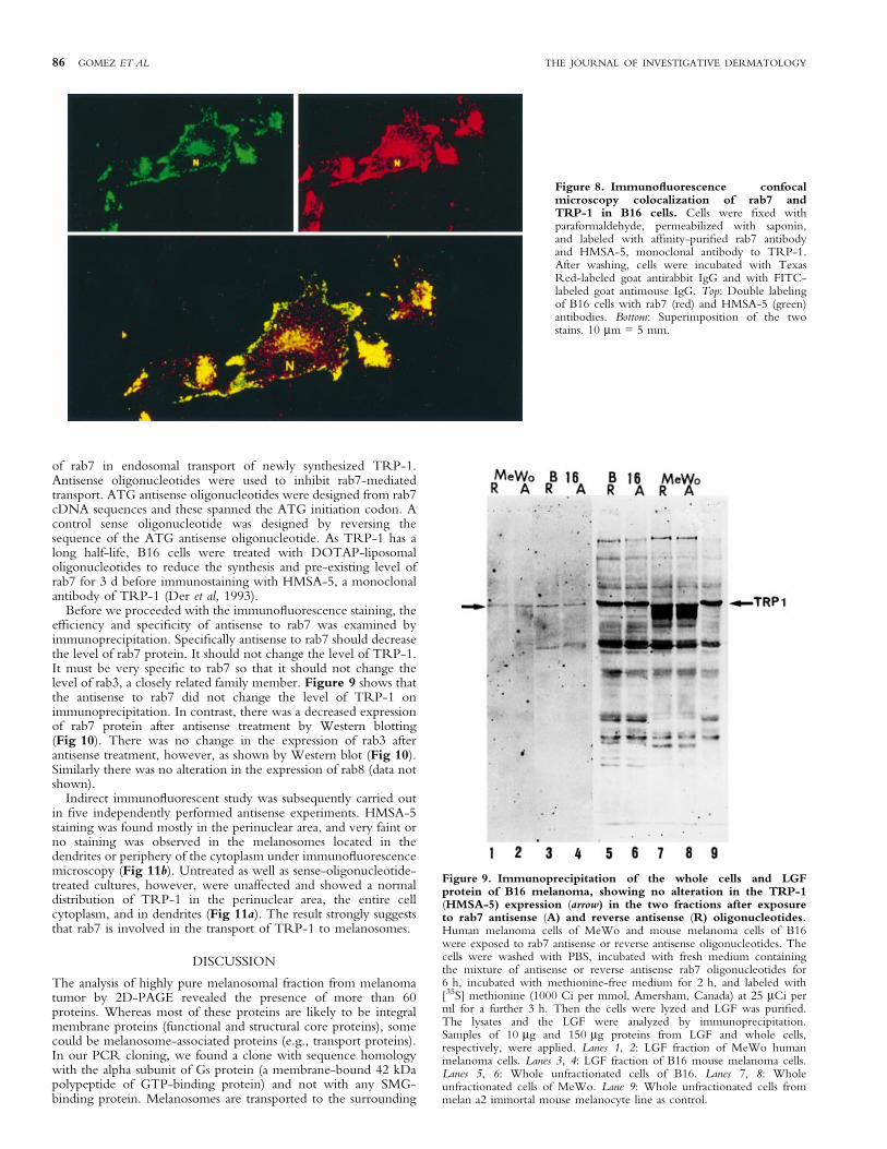
TRP-1 colocalizes with rab7, a late endosomal small GTP-binding protein We next investigated whether SMG-bindingproteins are involved in the intracellular transport of TRP-1, amajor melanosomal protein, to melanosomes. Of the three SMG-binding proteins (rab3, rab7, and rab8) tested by double immuno-¯uorescence, rab7 was found to be strongly immunostained andcolocalized with TRP-1 in the perinuclear area (probably Golgiand endosomal areas as observed by the granular appearance)(Fig 8, top), consistent with the observation by Chavrier et al(1990) that rab7 is localized in Golgi and late endosome. Thecolocalization was further veri®ed by superimposing the twoimages. Optical sectioning of the labeled cells by confocalmicroscopy revealed that the colocalization of rab7 with TRP-1was not uniform and not even. Some peripheral areas did not showcolocalization. Furthermore not all the perinuclear rab7-positivegranules were colocalized with TRP-1 (Fig 8, bottom). Doubleimmunostatining of rab3 or rab8 with TRP-1 was not conclusivein the colocalization. The ®nding suggests that TRP-1 and rab7share a common pathway of movement in a speci®c area oftransport from the TGN to the late endosome, but neither to all thematuration stages of melanosomes nor to all the lysosome±endosome system. This is also consistent with reports that a singleSMG-binding protein regulates the transport of protein only inspeci®c areas, and that the transport of proteins from ER to Golgior to other subcellular organelles required several GTP-bindingproteins (Goud and McCaffrey, 1991).
Antisense nucleotide treatment impairs TRP-1 transport Ascolocalization of TRP-1 and rab7 suggested a possible role of rab7in the intracellular transport of TRP-1, we next analyzed the role
Figure 5. GTP-overlay assay on melanosomal proteins andspeci®city of GTP±GDP binding. GTP-overlay assay on melanosomalproteins of B16 cells was carried out as described in Materials and Methodsin the presence of 10 and 100 molar excess of cold nucleotides ATP,ADP, AMP, GTP, GDP, or GMP. In control, only the labeled GTPwas added whereas in control/block, 4 mM ATP was also added inaddition, to block nonspeci®c binding, if any.
Figure 6. Analysis of SMG-binding proteins by high resolution2D-PAGE and GTP-overlay assay. Melanosomal proteins (75 mg)from B16 cells were resolved on the isoelectric focusing gel at 6±8000 V h,separated by SDS-PAGE, and transferred to nitrocellulose paper, and theGTP-overlay assay was carried out as described in Materials and Methods.Two predominant spots of U-1 and U-2 correspond very probably to twounidenti®ed SMG-binding proteins referred to as A and B by Huber et al(1994).
Figure 7. Immunoblot analysis of rab3, rab7, and rab8 onmelanosomal proteins. Melanosomal proteins (100 mg) from B16 cellswere resolved by SDS-PAGE, electrophoretically transferred tonitrocellulose paper, and incubated with af®nity-puri®ed antirab3,antirab7, and antirab8 peptide antibodies. Bands were visualized usingHRP-conjugated goat antirabbit antibody using the ECL Western blotdetection system.
VOL. 117, NO. 1 JULY 2001 rab7 AND TRP-1 TRANSPORT 85
of rab7 in endosomal transport of newly synthesized TRP-1.Antisense oligonucleotides were used to inhibit rab7-mediatedtransport. ATG antisense oligonucleotides were designed from rab7cDNA sequences and these spanned the ATG initiation codon. Acontrol sense oligonucleotide was designed by reversing thesequence of the ATG antisense oligonucleotide. As TRP-1 has along half-life, B16 cells were treated with DOTAP-liposomaloligonucleotides to reduce the synthesis and pre-existing level ofrab7 for 3 d before immunostaining with HMSA-5, a monoclonalantibody of TRP-1 (Der et al, 1993).
Before we proceeded with the immuno¯uorescence staining, theef®ciency and speci®city of antisense to rab7 was examined byimmunoprecipitation. Speci®cally antisense to rab7 should decreasethe level of rab7 protein. It should not change the level of TRP-1.It must be very speci®c to rab7 so that it should not change thelevel of rab3, a closely related family member. Figure 9 shows thatthe antisense to rab7 did not change the level of TRP-1 onimmunoprecipitation. In contrast, there was a decreased expressionof rab7 protein after antisense treatment by Western blotting(Fig 10). There was no change in the expression of rab3 afterantisense treatment, however, as shown by Western blot (Fig 10).Similarly there was no alteration in the expression of rab8 (data notshown).
Indirect immuno¯uorescent study was subsequently carried outin ®ve independently performed antisense experiments. HMSA-5staining was found mostly in the perinuclear area, and very faint orno staining was observed in the melanosomes located in thedendrites or periphery of the cytoplasm under immuno¯uorescencemicroscopy (Fig 11b). Untreated as well as sense-oligonucleotide-treated cultures, however, were unaffected and showed a normaldistribution of TRP-1 in the perinuclear area, the entire cellcytoplasm, and in dendrites (Fig 11a). The result strongly suggeststhat rab7 is involved in the transport of TRP-1 to melanosomes.
DISCUSSION
The analysis of highly pure melanosomal fraction from melanomatumor by 2D-PAGE revealed the presence of more than 60proteins. Whereas most of these proteins are likely to be integralmembrane proteins (functional and structural core proteins), somecould be melanosome-associated proteins (e.g., transport proteins).In our PCR cloning, we found a clone with sequence homologywith the alpha subunit of Gs protein (a membrane-bound 42 kDapolypeptide of GTP-binding protein) and not with any SMG-binding protein. Melanosomes are transported to the surrounding
Figure 8. Immuno¯uorescence confocalmicroscopy colocalization of rab7 andTRP-1 in B16 cells. Cells were ®xed withparaformaldehyde, permeabilized with saponin,and labeled with af®nity-puri®ed rab7 antibodyand HMSA-5, monoclonal antibody to TRP-1.After washing, cells were incubated with TexasRed-labeled goat antirabbit IgG and with FITC-labeled goat antimouse IgG. Top: Double labelingof B16 cells with rab7 (red) and HMSA-5 (green)antibodies. Bottom: Superimposition of the twostains. 10 mm = 5 mm.
Figure 9. Immunoprecipitation of the whole cells and LGFprotein of B16 melanoma, showing no alteration in the TRP-1(HMSA-5) expression (arrow) in the two fractions after exposureto rab7 antisense (A) and reverse antisense (R) oligonucleotides.Human melanoma cells of MeWo and mouse melanoma cells of B16were exposed to rab7 antisense or reverse antisense oligonucleotides. Thecells were washed with PBS, incubated with fresh medium containingthe mixture of antisense or reverse antisense rab7 oligonucleotides for6 h, incubated with methionine-free medium for 2 h, and labeled with[35S] methionine (1000 Ci per mmol, Amersham, Canada) at 25 mCi perml for a further 3 h. Then the cells were lyzed and LGF was puri®ed.The lysates and the LGF were analyzed by immunoprecipitation.Samples of 10 mg and 150 mg proteins from LGF and whole cells,respectively, were applied. Lanes 1, 2: LGF fraction of MeWo humanmelanoma cells. Lanes 3, 4: LGF fraction of B16 mouse melanoma cells.Lanes 5, 6: Whole unfractionated cells of B16. Lanes 7, 8: Wholeunfractionated cells of MeWo. Lane 9: Whole unfractionated cells frommelan a2 immortal mouse melanocyte line as control.
86 GOMEZ ET AL THE JOURNAL OF INVESTIGATIVE DERMATOLOGY

keratinocytes through the plasma membrane. It is therefore possiblethat we puri®ed both plasma-membrane-bound and unboundmelanosomes and the former could be the case for isolating amembrane-bound 42 kDa polypeptide. There is also a possibility,however, that we might have obtained a nonspeci®c GTP-bindingprotein by PCR cloning, which may be supported by the facts thatwe diversi®ed the third base of the codons to make the internalprimers and that we ampli®ed a product at the 3¢-untranslatedregion of the alpha subunit of G protein (not any translated region).We analyzed the SMG-binding proteins and not the role of Gsprotein because we are interested in knowing the biogenesis of themelanosome, i.e., the formation of the melanosome and itsmaturation upon integrating functional melanosomal proteins.SMG-binding proteins are well known for their role in transportingproteins to subcellular organelles (Deacon and Gelfand, 2001).
Our GTP-overlay assay after denaturing transfer was the mostsuitable assay to identify the SMG-binding proteins and not thealpha subunit of heterotrimeric Gs proteins (Huber et al, 1994).Among the 5±6 SMG-binding proteins in the 2D-GTP-overlayassay, predominantly two spots (U-1 and U-2) correspond veryprobably to two unidenti®ed SMG-binding proteins referred to asA and B by Huber et al (1994). According to Huber et al (1994),these two spots are found in all cells and organelle preparations anddo not appear speci®c for the melanosome. Given their high degreeof conservation, ubiquitous expression, and proven role insubcellular transport, SMG-binding proteins are likely to beimportant players in determining melanosome architecture andtheir maturation. This report demonstrated for the ®rst time thatSMG-binding protein, especially rab7, is very critical for thetransport of melanosomal proteins, e.g., TRP-1, from late-endosome delineated granules to the maturing melanosomes.
Melanosomes are lysosomal organelles specialized for thebiosynthesis and storage of melanin pigments in melanocytes, andcontain many membrane-bound and integral structural proteins(Jimbow, 1995; Orlow, 1995; Jimbow et al, 1998; Raposo et al,2001). Melanosomes may provide a good model for studying theinvolvement of rab protein in the biosynthesis, maturation, andexocytosis of secretory granules. Impairment of this melanosomalcascade is responsible for many pigmentary disorders in humans andanimals (Collier et al, 1979; Zhao et al., 1994a; Valentijn et al,1996). These pigmentary disorders can be due to systemic failure ormalfunction in certain congenital diseases, such as Hermansky±Pudlak syndrome, Chediak±Higashi syndrome, and Griscelli syn-drome. The ®rst two syndromes are both autosomal recessivehuman disorders and are associated with hypopigmentation of theskin and eye (ocuolocutaneous albinism) and systemic defects, suchas bleeding diathesis due to a platelet storage pool defect and anaccumulation of ceroid-like materials in cells in the case ofHermansky±Pudlak syndrome, and recurrent pyrogenic infectionsdue to giant, dysfunctional lysosomes and platelet dense bodyde®ciency in Chediak±Higashi syndrome (Robinson et al, 1975;Frenk and Lattion, 1982; Collier et al, 1985; Novak et al, 1985;McGarry et al, 1986; Summers et al, 1988; Nagle et al, 1996). Chenet al (1997a) recently identi®ed nine novel genes of the rabsubfamily in human melanocytes and melanoma cells, some ofwhich overlapped with the platelets. They speci®cally suggested theinvolvement of rab27a and rab27b in pigment dilution, due to thepresence of large pigment clumps in hair shaft and accumulation of
Figure 10. Western blot analysis of rab7 antisense treatment forB16 cells, showing a signi®cant (at least 30%±50%) decrease ofrab7 expression. B16 cells were transfected with rab7 antisense andreverse antisense oligonucleotides for 72 h using DOTAP liposomalsystem. A hundred micrograms of melanosomal proteins were loaded ineach lane and resolved by SDS-PAGE, transferred onto nitrocellulosemembrane, and incubated with af®nity-puri®ed antirab7 and antirab3antibodies. Lanes 1, 2: Rab7 expression in the cells exposed to antisense(A) and reverse antisense (R) rab7 oligonucleotides. The membrane wasstripped and reprobed with antirab3 antibody showing the speci®city ofantisense action as well as the equal loading of the proteins (lanes 3, 4).Lanes 5, 6: Rab7 expression in an experiment different from that of lanes1 and 2, showing the reproducibility of the result for the decreased rab7expression.
Figure 11. Impairment of TRP-1 transport by rab7 antisenseoligonucleotide. B16 cells were transfected with rab7 antisenseoligonucleotide for 72 h with either reversed ATG-antisense (a) orATG-antisense (b) using the DOTAP liposomal system. Cells were ®xedand HMSA-5 immuno¯uorescence staining was carried out as describedin Materials and Methods. Note that TRP-1 transport is impaired andcon®ned to the perinuclear region of the cell. Scale bar: 10 mm.
VOL. 117, NO. 1 JULY 2001 rab7 AND TRP-1 TRANSPORT 87

melanosomes in the melanocyte as well as a platelet storage pooldefect in Griscelli syndrome (Chen et al, 1997b). In Griscellisyndrome, most patients also develop an uncontrolled T lympho-cyte and macrophage activation syndrome (known as hemopha-gocytic syndrome). Rab27a appears to be involved in the control ofthe immune system, as all patients with rab27a mutations developedGriscelli syndrome (Menasche et al, 2000; Bahadoran et al, 2001).Rab27a appears to be a key effector of cytotoxic granule exocytosis,a pathway essential for immune homeostasis. Rab27a also partici-pates in melanosome transport (Bahadoran et al, 2001), and itsdefect leads to a clustering of melanin pigment in the hair shaft, aswell as defective melanosome transport in the melanocyte (Wu etal, 1998; Menasche et al, 2000; Wilson et al, 2000). Further humanmyosin Va mutations have been identi®ed and shown to beassociated with Griscelli syndrome (Wilson et al, 2000). Rab7appears to be necessary for the recruitment of myosin Va (Hume etal, 2001). Zhao et al suggested abnormal traf®cking of some cellularproteins related to melanosome structure and function in Chediak±Higashi syndrome melanocytes (Zhao et al, 1994a). Davies et alfurther showed that down-regulation of rab7 or rab9 proteinsinduced severe cell vacuolation that resembled the phenotype seenin ®broblasts from patients with Chediak±Higashi syndrome(Davies et al, 1997). It appears that the function of rab proteins(especially rab7) may be critical, not only for normal pigmentation,but also for melanocyte viability.
TRP-1 is one of the major melanogenic glycoproteins, and issynthesized in the rough ER and fully glycosylated in the Golgi andthen transported from the TGN to the early stage melanosomes,e.g., stages I and II (Jimbow, 1995; Jimbow et al, 1998). Little isknown, however, about the biologic process in the transport ofmelanogenic proteins from the TGN to these immature melano-somes. As SMG-binding proteins are involved in vesicular traf®c, itis likely that some SMG-binding proteins may also play animportant role in the transport of melanogenic proteins. ByWestern blotting we detected several known SMG-bindingproteins (rab3, rab7, and rab8) in the melanosomal fraction.Using double immuno¯uorescence and confocal microscopy,however, we found that rab7 colocalizes with TRP-1. Rab7 isbelieved to be present in the late endosome and not in the lysosome(Chavrier et al, 1990). Rab7 has been reported to be present in avesicular compartment connected to the lysosome also (Meresseet al, 1995). Rab7 may be recruited not only on late but also onmaturing epidermal growth factor receptor-containing endosomes(Blagoveshchenskaia and Vinogradova, 1996). This prompts us tospeculate that rab7 may be recruited onto maturing melanosomes.The colocalization observed (Fig 8) was primarily in the peri-nuclear region and occasionally in the periphery of the perikaryon,suggesting the existence of rab7 in the late-endosome granules andimmature melanosomes to which melanosomal glycoproteins,including TRP-1, are transported from the TGN. We haveshown by antisense experiment that TRP-1 transport was blockedby a rab7 antisense oligonucleotide and that TRP-1 is aggregatedperinuclearly (Fig 11), supporting a role for rab7 in the transport ofTRP-1 to the melanosomes.
At present, it is dif®cult to delineate the stages of melanosomalmaturation in which rab7 helps in the transport of TRP-1. Onepossibility is that rab7 is involved in the transport of TRP-1 to thelate-endosome lineaged granules along with some other carrierproteins. Among the four stages of melanosomal maturation, onlystage I melanosomes reveal ®ne structure that is identical or verysimilar to that of late endosomes (Jimbow, 1995; Jimbow et al,1998). It is therefore likely that rab7 is involved in the transport ofTRP-1 from late-endosome delineated granules to melanosomes inthe early stage of maturation. Another study from our group(Jimbow et al, 2000a) showed that TRP-1 also colocalizes withgranules stained with the anti-mannose-6-phosphate receptor (anti-MPR, marker for the late endosome) present only in theperinuclear area after the transfection of TRP-1 cDNA to humanamelanotic melanoma cells, supporting our hypothesis that TRP-1transport from Golgi to melanosomes involves organelles/vesicles
related to late endosomes. Our ®ndings are also consistent with ourprevious observation that the anti-TRP-1 antibody is aggregatedalong with the melanosomal membrane of stages I and II granules(Der et al, 1993; Jimbow et al, 1993b). Phosphatidylinositol 3-kinase (PI3-kinase) may help the transport of lysosomal enzymes byinterfering with MPR-dependent transport in the TGN (Brownet al, 1995), but wortmannin, a PI3-kinase inhibitor, blocked therab7-stimulated transport (Mukhopadhyay et al, 1997). Together,these reports suggest the cooperative functions of rab7, MPR, andPI3-kinase in lysosomal protein transport from the TGN towardsthe endosome±lysosome system, resulting in lysosomal proteins(Jimbow et al, 1997, 2000b).
Our colocalization studies of TRP-1 with rab7 followed byblocking of TRP-1 transport with rab7 antisense oligonucleotides,our colocalization study of TRP-1 with MPR (Jimbow et al, 1997),as well as other colocalization studies of rab7 with MPR (Chavrieret al, 1990) and the functional relationship between rab7 and PI3-kinase (Mukhopadhyaya et al, 1997) favor a transport mechanism ofTRP-1 to the melanosomes similar to that of lysosomal proteins tothe lysosomes through the late endosome-lineage system. PI3-kinase/MPR possibly helps TRP-1 transport from the TGN to thelate endosomes, where rab7 subsequently facilitates the TRP-1sorting to early maturation melanosomes. In this sense, the stage Imelanosome may be identical to the late endosome-lineage granule(Jimbow, 1995). The other less likely possibility is that rab7 inmelanocytes helps the transport of TRP-1 directly from the TGNto the stage I melanosomes without any intermediate lateendosomal compartment, which might be a unique function ofrab7 in melanocytes. The vesicle targeting component, rab7, mustrecycle back. At present, we do not know when and from whichstages of the melanosomal maturation rab7 returns. From thecolocalization studies it is speculated that rab7 returns from the lateendosome-delineated granules and probably also from the earlymaturation stages of melanosomes.
Other groups of macromolecules also appear to be involved inthe transport of melanosomal proteins to early stage melanosomes ascan be seen in the vesicular transport system (Goud and McCaffrey,1991). Arf (another class of SMG-binding proteins) is also believedto be a regulator of vesicular traf®c. Arf is involved in the assemblyof COP-coated vesicles in the Golgi for nonclathrin-coated vesicledependent transport (Orcl et al, 1993). Early electron microscopycytochemistry showed that tyrosinase is transported from the TGNto the melanosomes by coated vesicles (Hatta et al, 1988). WhetherTRP-1 is transported from the TGN with clathrin-coated ornonclathrin-coated vesicles to the melanosomes needs to beclari®ed. A recent study has shown that adapter protein (AP-3) isinvolved in the tyrosinase transport from the TGN to melanosomesthrough nonclathrin-coated vesicles (Odorizzi et al, 1998). Furtherthe altered traf®cking of lysosomal proteins in Hemansky±Pudlaksyndrome was found to be due to mutations in the 3A subunit ofthe AP-3 adapter (Dell'Angelica et al, 1999; Feng et al, 1999).Tyrosinase, TRP-1, and TRP-2 appear to form a functionalcomplex in the melanogenesis cascade after being transported to themelanosomes (Orlow et al, 1994). The presence of several GTP-binding proteins, as revealed by the GTP-overlay experiment,suggests that some other melanogenic proteins are also transportedwith the help of SMG-binding proteins, and these SMG-bindingproteins may also be involved in the melanosomal transport fromthe perinuclear area to the plasma membrane during melanosometransport to the surrounding keratinocytes. Among the otherSMG-binding proteins associated with melanosomes, rab3 may beinvolved in the movement of melanosomes within the melanocytesduring the exocytosis process, inasmuch as rab3A is involved in theexocytosis of zymogen granules in the pancreas (Mohrmann andder Sluijis, 1999). In fact, it was recently found that rab3A isassociated with melanosomes in melanoma cells, possibly beinginvolved in the intracellular transport of melanosomes (Araki et al,2000).
In conclusion, a fairly large number of proteins are probablyinvolved in the transport of melanosomal proteins from the TGN
88 GOMEZ ET AL THE JOURNAL OF INVESTIGATIVE DERMATOLOGY

to melanosomes and in the movement of these granules withinmelanocytes. Speci®cally, recent studies point towards the possi-bility that rab proteins act in concert with molecular motors toregulate directional membrane transport and dynamics of melano-some biogenesis and melanin pigmentation.
The authors would like to thank Dr. R. Parton for providing the rab7 antibody and
Dr. P.D. Thomas and Dr. W.T. Dixon for their critical reading of the manuscript.
This work was supported by grants from the Medical Research Council of Canada
(MT 12866), and the Ministry of Education, Science, Sports, and Culture of Japan
(#08407022 and 12470179). KJ, AHFMR Scientist, is currently an adjunct
professor of the University of Alberta. PFG is now located at the Department of
Pathology, Oregon Health Sciences University, Portland, OR 97201.
REFERENCES
Ackrell BAC, Kerney EB, Singer TP: Mammalian succinate dehydrogenase. MethodsEnzymol 53:466±483, 1978
Akhtar S, Juliano RL: Cellular uptake and intracellular fate of antisenseoligonucleotides. Trends Cell Biol 2:139±144, 1992
Araki K, Horikawa T, Chakraborty AK, et al: Small GTPase Rab3A is associatedwith melanosomes in melanoma cells. Pigment Cell Res 13:33d2±336, 2000
Bahadoran P, Aberdam E, Mantourx F, et al: Rab27a: a key to melanosome transportin human melanocytes. J Cell Biol 152:843±849, 2001
Barton DE, Kwon BS, Francke U: Human tyrosinase gene, mapped to chromosome11 (q14 q21), de®nes second region of homology with mouse chromosome 7.Genomics 3:17±24, 1988
Bennett DC, Cooper PJ, Hart JR: A line of non-tumorgenic mouse melanocytessyngeneic with the B16 melanoma and requiring a tumor promoter for growth.Int J Cancer 39:414±418, 1987
Blagoveshchenskaia AD, Vinogradova NA, Nikol'skii NN: The association of aprotein regulator of intracellular Rab7 membrane transport with endosomescontaining an epidermal growth factor receptor with activated and inactivatedtyrosine kinase. [Russian] Tsitologiia 38:854±862, 1996
Bottger G, Nagelkerken B, Van der Sluijs P: Rab4 and rab7 de®ne distinctnonoverlapping endosomal compartments. J Biol Chem 271:29191±29197,1996
Brown WJ, DeWald DB, Emr SD, Plutner H, Balch WE: Role forphosphatidylinositol 3 kinase in the sorting and transport of newlysynthesized lysosomal enzymes in mammalian cells. J Cell Biol 130:781±796,1995
Chavrier P, Parton RG, Hauri HP, Simons K, Zerial M: Localization of lowmolecular weight GTP-binding proteins to exocytic and endocyticcompartments. Cell 62:317±329, 1990
Chen D, Guo J, Gahl WA: RAB GTPases expressed in human melanoma cells.Biochim Biophys Acta 1355:1±6, 1997a
Chen D, Guo J, Miki T, Tachibana M, Gahl WA: Molecular cloning andcharacterization of rab27a and rab27b, novel human rab proteins shared bymelanocytes and platelets. Biochem Mol Med 60:27±37, 1997b
Chomczynski P, Sacchi N: Single-step method of RNA isolation by acidguanidinium thiocyanate-phenol-chloroform extraction. Anal Biochem162:156±159, 1987
Coleman DE, Sprang SR: How G proteins work: a continuing story. TIBS (TerndsBiochem Sci) 21:41±44, 1996
Collier LL, Bryan GM, Prieur DJ: Ocular manifestations of the Chediak±Higashisyndrome in four species of animals. J Am Vet Med Assoc 175:587±590, 1979
Collier LL, King EJ, Prieur DJ: Aberrant melanosome development in the retinalpigmented epithelium of cats with Chediak±Higashi syndrome. Exp Eye Res41:305±311, 1985
Davies JP, Cotter PD, Ioannou YA: Cloning and mapping of human Rab7 and Rab9cDNA sequences and identi®cation of a Rab9 pseudogene. Genomics 41:131±134, 1997
Deacon S, Gelfand VI: Of yeast, mice, and mean: Rab proteins and organelletransport. J Cell Biol 152:F21±F24, 2001
Dell'Angelica EC, Shotelersuk V, Aguilar RC, Gahl WA, Bonifacino JS: Alteredtraf®cking of lysosomal proteins in Hermansky±Pudlak syndrome due tomutations in the beta 3A subunit of the AP-3 adaptor. Mol Cell 3:11±21, 1999
Der JE, Dixon WT, Jimbow K, Horikoshi T: A murine monoclonal antibody,MoAb HMSA5, against a melanosomal component highly expressed in earlystages, and common to normal and neoplastic melanocytes. Br J Cancer 67:47±57, 1993
Feng L, Seymour AB, Jiang S, et al: The beta3A subunit gene (Ap3b1) of the AP-3adaptor complex is altered in the mouse hypopigmentation mutant pearl, amodel for Hermansky±Pudlak syndrome and night blindness. Human Mol Genet8:323±330, 1999
Fischer VMG, Stahl B, Li C, Sudhop TC, Jahn R: Rab proteins in regulatedexocytosis. TIBS (Trends Biochem Sci) 19:164±168, 1994
Frenk E, Lattion F: The melanin pigmentary disorder in a family with Hermansky±Pudlak syndrome. J Invest Dermatol 78:141±143, 1982
Goud B, McCaffrey M: Small GTP-binding proteins and their role in transport. CurrOpin Cell Biol 3:626±633, 1991
Gravotta D, Adesnik M, Sabatini DD: Transport of in¯uenza HA from the trans-Golgi network to the apical surface of MDCK cells permeabilized in theirbasolateral plasma membranes: energy dependence and involvement of GTP-binding proteins. J Cell Biol 111:2893±2908, 1990
Halaban R, Moellmann G: Murine and human b locus pigmentation genes encode aglycoprotein (gp75) with catalase activity. Proc Natl Acad Sci USA 87:4809±4813, 1990
Hara H, Lee MH, Chen H, Luo D, Jimbow K: Role of gene expression and proteinsynthesis of tyrosinase, TRP-1, lamp-1, and CD63 in UVB-inducedmelanogenesis in human melanomas. J Invest Dermatol 102:495±500, 1994
Hatta S, Mishima Y, Ichihashi M, Ito S: Melanin monomers within coated vesiclesand premelanosomes in melanin synthesizing cells. J Invest Dermatol 91:181±184, 1988
Huber LA, de Hoop MJ, Dupree P, Zerial M, Simons K, Dotti C: Protein transportto the dendritic plasma membrane of cultures neurons is regulated by rab8p. JCell Biol 123:47±55, 1993a
Huber LA, Pimplikar S, Parton RG, Virta H, Zerial M, Simons K: Rab8, a smallGTPase involved in vesicular traf®c between the TGN and the basolateralplasma membrane. J Cell Biol 123:35±45, 1993b
Huber LA, Ullrich O, Takai Y: Mapping of Ras-related GTP-binding proteins byGTP overlay following two-dimensional gel electrophoresis. Proc Natl Acad SciUSA 91:7874±7878, 1994
Hume AN, Collison LM, Rapak A, Gomes AQ, Hopkins CR, Seabra MC: Rab27aregulates the peripheral distribution of melanosomes in melanocytes. J Cell Biol152:795±808, 2001
Jackson IJA: cDNA encoding tyrosinase-related protein maps to the brown locus inmouse. Proc Natl Acad Sci USA 85:4392±4396, 1988
Jackson IJ, Chambers DM, Tsukamoto K, Copeland NG, Gilbert DJ, Jenkins NA,Hearing V: A second tyrosinase-related protein, TRP2, maps to and is mutatedat the mouse slaty locus. EMBO (Eur Mol Biol Organ) J 11:527±535, 1992
Jimbow K: Current update and trends in melanin pigmentation and melanin biology.Keio J Med 44:9±18, 1995
Jimbow K, Jimbow M, Chiba M: Characterization of structural properties formorphological differentiation of melanosomes: II. Electron microscopic andSDS-PAGE comparison of melanosomal matrix proteins in B16 and HardingPassey melanomas. J Invest Dermatol 78:76±81, 1982
Jimbow K, Iwashina T, Alena F, Yamada K, Pankovich J, Umemura T: Exploitationof pigment biosynthesis pathway as a selective chemotherapeutic approach formalignant melanoma. J Invest Dermatol 100:231S±238S, 1993a
Jimbow K, Lee SK, King MG, Hara H, Chen H, Dakour J, Marusyk H: Melaninpigments and melanosomal proteins as differentiation markers unique to normaland neoplastic melanocytes. J Invest Dermatol 100:259S±268S, 1993b
Jimbow K, Gomez PF, Toyofuku K, Chang D, Miura S, Tsujiya H, Park JS:Biological role of tyrosinase related protein and its biosynthesis and transportfrom TGN to stage I melanosome, late endosome, through gene transfectionstudy. Pigment Cell Res 10:206±213, 1997
Jimbow K, Prota G, Quevedo WC: Biology of melanocytes. In: Freedberg IM, EisenAZ, Wolff E, Austen KF, Goldsmith LA, Katz SI, Fitzpatrick TB, eds.Fitzpatrick's Dermatology in General Medicine. 5th edn. McGraw-Hill, 1998:pp192±220
Jimbow K, Park JS, Kato F, Hirosaki K, Toyofuku K, Hua C, Yamashita T:Assembly, target signal and intracellular of tyrosinase gene family protein in theinitial stage of melanosome biogenesis. Pigment Cell Res 13:222±229, 2000a
Jimbow K, Hua C, Gomez PF et al: Intracellular vesicular traf®cking of tyrosinasegene family protein in eu± and pheomelanosome biogenesis. Pigment Cell Res13(Suppl. 8):110±117, 2000b
Jimbow M, Kanoh H, Jimbow K: Characterization of biochemical properties ofmelanosomes for structural and functional differentiation: analysis of thecompositions of lipids and proteins in melanosomes and their subfractions. JInvest Dermatol 79:97±102, 1982
Jimenez-Cervantes C, Solano F, Kobayashi T, Urabe K, Hearing VJ, Lozano JA,GarciaBorron JC: A new enzymatic function in the melanogenic pathway. JBiol Chem 269:17993±18001, 1997
Kobayashi T, Urabe K, Winder A, et al: DHICA oxidase activity of TRP1 andinteractions with other melanogenic enzymes. Pigment Cell Res 7:227±234,1994
Kwon BS, Haq AK, Pomerantz SH, Halaban R: Isolation and sequence of a cDNAclone for human tyrosinase that maps at the mouse c-albino locus. Proc NatlAcad Sci 84:7473±7477, 1987
Kwon BS, Chintamaneni C, Kozak CA, et al: A melanocyte-speci®c gene, Pmel 17,maps near the silver coat color locus on mouse chromosome 10 and is in asyntenic region on human chromosome 12. Proc Natl Acad Sci 88:9228±9232,1991
Luo D, Chen H, Jimbow K: Co-transfection of genes encoding human tyrosinaseand tyrosinase-related protein-1 prevents melanocyte death and enhancesmelanin pigmentation and gene expression of Lamp-1. Exp Cell Res 213:231±241, 1994
del Marmol V, Beermann F: Tyrosinase and related proteins in mammalianpigmentation. FEBS Lett 381:165±168, 1996
McGarry MP, Novak EK, Swank RT: Progenitor cell defect correctable by bonemarrow transplatation in ®ve independent mouse models of platelet storagepool de®ciency. Exp Hematol 14:261±265, 1986
Menasche G, Pastural E, Feldmann J, et al: Mutations in RAB27A cause Griscellisyndrome associated with haemophagocytic syndrome. Nature Genet 25:173±176, 2000
Meresse S, Gorvel JP, Chavrier P: The rab7 GTPase resides on a vesicularcompartment connected to lysosomes. J Cell Sci 108:3349±3358, 1995
VOL. 117, NO. 1 JULY 2001 rab7 AND TRP-1 TRANSPORT 89

Mohrmann K, van der Sluijis P: Regulation of membrane transport through theendocytic pathway by rabGTPases. Mole Membrane Biol 16:81±87, 1999
Mukhopadhyay A, Funato K, Stahl PD: Rab7 regulates transport from early to lateendocytic compartments in xenopus oocytes. J Biol Chem 272:13055±13059,1997
Muller G, Ruppert S, Schmid E, Schutz G: Functional analysis of alternatively splicedtyrosinase gene transcripts. EMBO (Eur Mol Biol Organ) J 7:2723±2730, 1988
Nagle DL, Karim MA, Woolf EA, et al: Identi®cation and mutation analysis of thecomplete gene for Chediak±Higashi syndrome. Nat Genet 14:307±311, 1996
Nakano A, Muramatsu M: A novel GTP-binding protein, Sar1p, is involved intransport from the endoplasmic reticulum to the Golgi apparatus. J Cell Biol109:2677±2691, 1989
Novak EK, McGarry MP, Swank RT: Correction of symptoms of platelet storagepool de®ciency in animal models for Chediak±Higashi syndrome andHermansky±Pudlak syndrome. Blood 66:1196±1201, 1985
Odorizzi G, Cowles CR, Emr SD: The AP-3 complex: a coat of many colours.Trends Cell Biol 8:282±288, 1998
O'Farrell PH: High resolution two-dimensional electrophoresis of proteins. J BiolChem 250:4007±4021, 1975
Orcl L, Palmer DJ, Amherdt M, Rothman JE: Coated vesicle assembly in the Golgirequires only coatomer and ARF proteins from the cytosol. Nature (Lond)364:732±734, 1993
Orlow SJ: Melanosomes are specialized members of the lysosomal lineage of arganells.J Invest Dermatol 105:3±7, 1995
Orlow SJ, Zhou B, Chakraborty A, Drucker M, PifkoHirst S, Pawelek JM: High-molecular-weight forms of tyrosinase and the tyrosinase-related proteins:evidence for a melanogenic complex. J Invest Dermatol 103:196±201, 1994
Park HY, Russakovsky V, Ohno S, Gilchrest BA: The beta isoform of protein kinaseC stimulates human melanogenesis by activating tyrosinase in pigment cells.J Bio Chem 268:11742±11749, 1993
Pomerantz SH: Separation puri®cation, and properties of two tyrosinases fromhamster melanoma. J Biol Chem 238:2351±2357, 1963
Rall T, Harris BA: Identi®cation of the lesion in the stimulatory GTP-bindingprotein of the uncoupled S49 lymphoma. FEBS Lett 224:365±371, 1987
Raposo G, Tenza D, Murphy DM, Berson JE, Marks MS: Distinct protein sortingand localization to premelanosomes, melanosomes, and lysosomes inpigmented melanocytic cells. J Cell Biol 152:809±823, 2001
Rinchik EM, Bultman SJ, Horsthemke B, et al: A gene for the mouse pink-eyeddilution locus and for human type II oculocutaneous albinism. Nature (Lond)361:72±76, 1993
Robinson WGJ, Kuwabara T, Cogan DG: Lysosomes and melanin granules of theretinal pigment epithelium in a mouse model of the Chediak±Higashisyndrome. Invest Ophthalmol 14:312±317, 1975
Singh MV, Price KJ, Bhatnagar R, Malhotra SK: Novel rod-shaped structuresidenti®ed in glioma cell nuclei by immunolabelling and confocal laser¯uorescence microscopy. Biomed Lett 50:163±172, 1994
Summers CG, Knobloch WH, Witkop CJ, King RA: Hermansky±Pudlak syndrome.Ophthalmic ®ndings. Ophthalmology 95:545±554, 1988
Takai Y, Sasaki T, Shirataki H, Nakanishi H: Rab3A small GTP-binding protein inCa(2+)-dependent exocytosis. Genes Cells 1:615±632, 1996
Trouet A: Isolation of modi®ed liver lysosomes. Meth Enzymol 31:323±329, 1974Tsukamoto K, Jackson IJ, Urabe K, Montague PM, Hearing VJ: A second tyrosinase-
related protein, TRP2, is a melanogenic enzyme termed DOPAchrometautomerase. EMBO (Eur Mol Biol Organ) J 11:519±526, 1992
Valentijn JA, Sengupta D, Gumkowski D, Tang LH, Konieczko EM, Jamieson JD:Rab3D localizes to secretory granules in rat pancreatic acinar cells. EJCB (Eur JCell Biol) 70:33±41, 1996
Vijayasaradhi S, Bouchard B, Houghton AN: The melanoma antigen gp75 is thehuman homologue of the mouse b (brown) locus gene product. J Exp Med171:1375±1380, 1990
Waters MG, Griff IC, Rothman JE: Proteins involved in vesicular transport andmembrane fusion. Curr Opin Cell Biol 3:615±620, 1991
Wichmann H, Hengst L, Gallwitz D: Endocytosis in yeast: evidence for theinvolvement of a small GTP-binding protein (Ypt7p). Cell 71:1131±1141,1992
Wilson SM, Yip R, Swing DA, et al: A mutation in Rab27a causes the vesicletransport defects observed in ashen mice. Proc Natl Acad Sci 97:7933±7938,2000
Winder AJ, Wittbjer A, Rosengren E, Rorsman H: The mouse brown (b) locusprotein has dopachrome tautomerase activity and is located in lysosomes intransfected ®broblasts. J Cell Sci 106:153±166, 1993
Winder AJ, Wittbjer A, Odh G, Rosengren E, Rorsman H: The mouse brown (b)locus protein functions as a dopachrome tautomerase. Pigment Cell Res 7:305±310, 1994
Wu X, Bowers B, Rao K, Wei Q: Hammer JA3rd: visualization of melanosomedynamics within wild-type and dilute melanocytes suggests a paradigm formyosin V function in vivo. J Cell Biol 143:1899±1918, 1998
Yokoyama K, Suzuki H, Yasumoto KI, Tomita Y, Shibahara S: Molecular cloningand functional analysis of a cDNA coding for human DOPAchrometautomerase/tyrosinase-related protein-2. Biochim Biophys Acta 1217:317±321,1994
Zhao H, Boissy YL, Abdel-Malek Z, King RA, Nordlund JJ, Boissy RE: On theanalysis of the pathophysiology of Chediak±Higashi syndrome. Lab Invest71:25±34, 1994
Zhao H, Zhou Y, Nordlund JJ, Boissy RE: Human TRP1 has tyrosine hydroxylasebut no DOPA oxidase activity. Pigment Cell Res 7:131±140, 1994
90 GOMEZ ET AL THE JOURNAL OF INVESTIGATIVE DERMATOLOGY