identification of potent drugs and antiviral agents for
TRANSCRIPT
1
Identification of Potent Drugs and Antiviral Agents for the Treatment of the SARS-CoV-2 Infection
N.R. Jena*Discipline of Natural Sciences, Indian Institute of Information Technology, Design, and
ManufacturingDumna Airport Road, Jabalpur-482005, India
*Corresponding author’s email address: [email protected]
AbstractThe recent outbreak of the SARS-CoV-2 infection has affected the lives and economy of
more than 200 countries. The unavailability of vaccines and the virus-specific drugs has
created an opportunity to repurpose existing drugs to examine their efficacy in controlling the
virus activities. Here, the inhibition of the RdRp viral protein responsible for the replication
of the virus in host cells is examined by evaluating the binding patterns of various approved
and investigational antiviral, antibacterial, and anti-cancer drugs by using the molecular
docking approach. Some of these drugs have been proposed to be effective against the SARS-
Cov-2 virus infection. In an attempt to discover new antiviral agents, artificially expanded
genetic alphabets (AEGIS) such as dP, dZ, dJ, dV, dX, dK, dB, dS, dP4, and dZ5 and
different sequences of these nucleotides were also docked into the active site of the RdRp
protein. It is found that among the various approved and investigational drugs, the Clevudine,
N4-hydroxycytidine (EIDD-1931), 2'-C-methylcytidine, EIDD-2801, Uprifosbuvir,
Balapiravir, Acalabrutinib, BMS-986094, Remdesivir (full drug), GS-6620, and Ceforanide
would act as potent inhibitors of the RdRp protein. Similarly, among the AEGIS nucleotides,
dB, dJ, dP, dK, dS, dV, and dZ are found to inhibit the RdRp protein efficiently. It is further
found that sequences containing up to three artificial nucleotides can also inhibit the RdRp
viral protein. As these unnatural nucleotides are foreign molecules for the cells, their
replication in host cells may not occur naturally, and hence these may act as potent drugs for
the treatment of the SARS-CoV-2 infection. However, in vivo evaluation of these drugs and
2
artificial nucleotides are required against SARS-CoV-2 before prescribing them to the
infected patients.
1. Introduction
The recent outbreak of the Coronavirus infection originated from the Wuhan City of China in
December 2019 (Wuhan virus-2019 or COVID-19) [1,2] has led to severe damage to human
lives and the economy of more than 200 countries worldwide. The unavailability of vaccines
and potent drugs specific to COVID-19 has created a challenge and opportunity to identify or
discover potent antiviral agents for the treatment of this pandemic. Although continuous
attempts are going on to design vaccines [3,4] and antiviral agents [5-12], not a single
vaccine has cleared human trials successfully to date. Hence, it is imperative to repurpose
available drugs for the treatment of COVID-19.
It is found that COVID-19 is a positive-strand RNA virus, and the virus protein is structurally
similar to the severe acute respiratory syndrome coronavirus (SARS-CoV-1) [13], which
originated from China in 2002 and several other beta coronaviruses, such as the bat and
pangolin coronaviruses. Due to its structural similarity with the SARS-CoV-1, it is also
popularly known as SARS-CoV-2. The SARS-CoV-2 protein consists of a multi-protein
cascade comprising of several structural and non-structural proteins [14]. Among these
proteins, the RNA-dependent RNA polymerase (RdRp) non-structural protein (NS) is mainly
responsible for the replication of the virus inside host cells [15]. It is recently proposed that
the RdRp requires other non-structural proteins such as NS7 and NS8 to process replication
of the incoming RNA strand [16]. Hence, to block the replication of the SARS-CoV-2, it is
desirable to block the active site of the RdRp NS protein by potent nucleotide inhibitors.

3
Recent cryo-EM studies have resolved the structure of the RdRp NS protein in complex with
NS7 and NS8 [16,17]. These studies have revealed that it contains several vital domains, such
as an N-terminal beta-hairpin (residues 31-50), nidovirus RdRp-associated
nucleotidyltransferase domain (NiRAN, residues 117-250), interface domain (residues 251-
365), and RdRp domain (residues 367-920) [16,17]. The active site is located in the RdRp
domain formed by the conserved motifs ranging from A to G (residues 397-815). Among
these motifs, motif A contains residues 611-626, and motif C includes residues 753-767,
including the catalytically important residues Ser759, Asp760, and Asp 762, which play
essential roles in the synthesis of RNA. The metal ions found in most of the viral RdRps are
coordinated by Asp760 and Asp762. Motif F (residues 397-581) and G (residues 621-679)
interact with the template strand RNA and direct it into the active site [16,17]. The positively
charged hydrophilic residues, such as Lys545, Arg553, and Arg555 of the motif F, helps to
stabilize the +1 base of the incoming strand, thereby putting the RNA strand in the correct
position for catalysis [16]. The residues involved in the RNA binding and catalysis are highly
conserved ad hence crucial for RdRp activities. Therefore, to inhibit replication of the vial
RdRp NS protein, antiviral agents should disrupt the binding of the RNA template with the
above residues.
Recently, the structure of the RdRp, complexed with NS7, NS8, a 50 base template-primer
RNA, and Remdesivir monophosphate (RMP), was resolved by the cryo-EM study [16]. The
RMP is an antiviral prodrug, which is believed to inhibit the RdRp activity by non-obligate
RNA chain termination. It was found that the RMP interacts with the RdRp protein by
interacting with the side chains of Lys545 and Arg555 [16]. The phosphate group of the RMP
was found to interact with the Mg+2. Other than RMP, Favipiravir, Ribavirin, Galidesivir,
EIDD-2801, EIDD-1931 (N4-hydroxycytidine), etc have also been proposed to inhibit RdRp

4
activity by cell-based assays [18,19]. As these are nucleoside- or nucleotide-based drugs, the
mechanism of inhibition of RdRp activity is presumed to be the same as that of RMP.
However, the binding modes of these antiviral drugs are not yet known. Further, the binding
mode of the full drug of Remdesivir with the modified backbone as deposited in the
DRUGBANK database [20] is not known.
To understand the detailed binding modes of various nucleoside and nucleotide-based drugs
and to identify the more potent drugs based on the strong interactions with the active site of
the RdRp, binding of 28 approved and ten investigational drugs were studied herein by using
molecular docking approach. The binding modes of several artificially expanded genetic
information systems (AEGIS) [21-29] with RdRp were also examined to unravel their
possible roles in inhibiting RdRp activity. These unnatural nucleotides were proposed to
make the artificial gene to create artificial life.
2. Computational Methodology
2.1 System Preparation
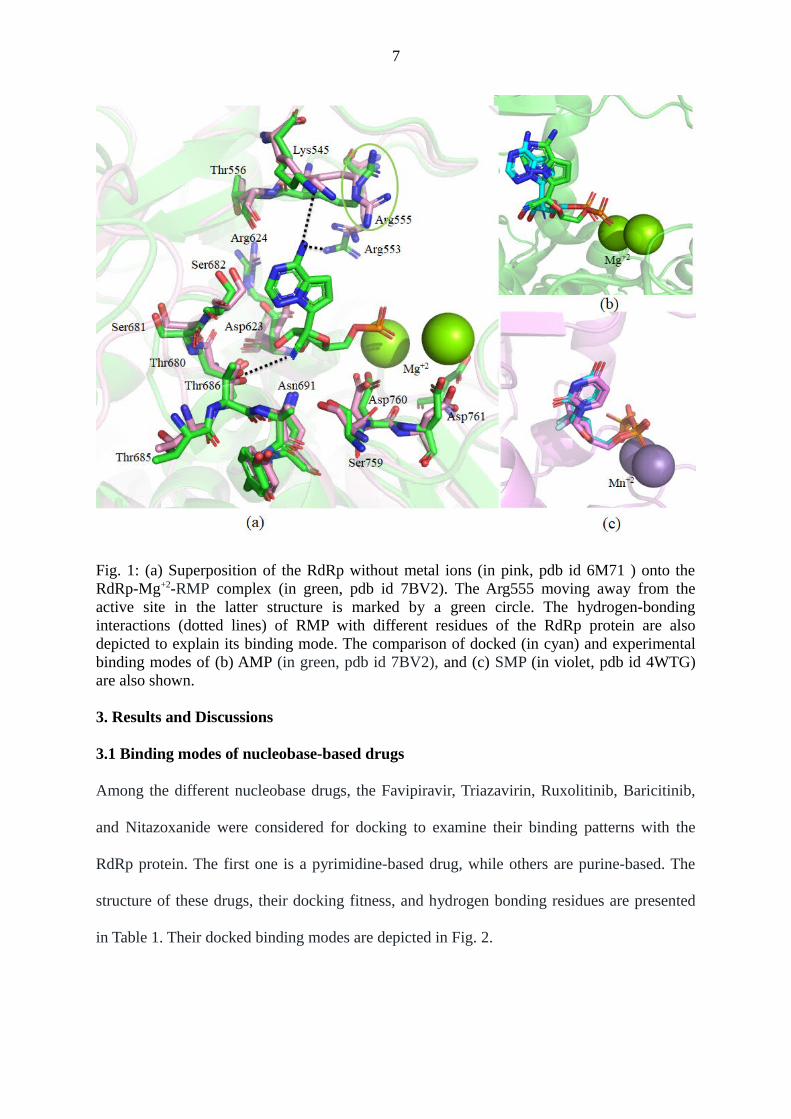
Two Cryo-EM structures of the RdRp protein complexed with NS7 and NS8 are available in
the protein data bank [15,16]. In one of the structures, the RdRp-NS7-NS8 complex was not
bound to metal ions (pdb ID 6M71) [17]. In contrast, in the other one, the RdRp-NS7-NS8
complex was bound to three Mg+2 ions (coordinated by Asp760, Asp761, Asp618, and
pyrophosphate), a double-stranded RNA (14-base template strand and 11-base primer strand),
and Remdesivir in its monophosphate form (RMP) (pdb id 7BV2) [16]. However, in the
latter structure, Arg555 was protruding away from the active site, unlike in the former
structure (Fig. 1a). The superposition of RdRp without metal ions onto the RdRp with metal
ions by considering the Cα atoms produced an RMSD of 0.583 Å (Fig. 1a). As the latter

5
structure contains metal ions, which are conserved in almost all of the RdRp viral proteins,
this structure was considered for docking after removing coordinates of NS7, NS8,
pyrophosphate, and RMP. Before docking, hydrogen atoms were added to the RdRp protein
of SARS-CoV-2 by UCSF Chimera to maintain the neutral pH.
The three-dimensional structures of various nucleobase, nucleoside, and nucleotide drugs
were extracted from the DRUGBANK database (by downloading the SDF files) [20].
Wherever three-dimensional structures were unavailable, two-dimensional structures were
obtained from the DRUGBANK database. Subsequently, three-dimensional structures of
these drugs were generated by using the GaussView 5.0 program [30] as per the standard
geometrical parameters. Subsequently, these molecules were minimized by using the UCSF
Chimera. The coordinates of artificial nucleotides, such as P and Z, were extracted from the
protein databank (pdb id 4XNO) [31]. Subsequently, hydrogen atoms were added to these
nucleotides by using the GaussView program. These nucleotides were modified to create
other nucleotides such as P4, Z5, J, V, B, S, X, and K. Subsequently, these nucleotides were
energy-minimized by using the Gaussian 09 program [32]. Consequently, the minimized
structures were utilized for molecule docking.
2.2 Molecular Docking
Docking of various nucleobase, nucleoside, and nucleotide drugs into the active site of the
RdRp-Mg+2 complex was carried out by using the GOLD 5.0 program [32-34]. The genetic
algorithm [32] was used for the docking purpose to create ten different conformations of the
drug molecule by keeping the protein rigid. The binding site was considered to be situated
within a radius of 10Å from the conserved Thr680. The Chemscore was used for docking,
and the PLP score was used to rescore the binding of different drugs with the RdRp-Mg+2

6
complex. Out of the ten different poses, the one whose purine or pyrimidine interacts with the
residues of motif F (Lys545, Arg553, Arg553, etc) and the sugar-phosphate backbone
interacts with the Mg+2 and residues of motif C (Asp760, Asp761, etc) like the RdRp-RMP
(pdb ID 7BV2) [16] and Hepatitis C virus protein (HCV)-Sofosbuvir monophosphate (SMP)
(pdb id 4WTG) [35] complexes were considered for further analysis.
To examine if the above docking protocol can reproduce experimentally observed binding
modes, the RMP was docked into the binding site of the RdRp-Mg+2 protein of the SARS-
CoV-2 (pdb id 7BV2) [16] after deleting all cofactors. As illustrated in Fig.1b, the above
docking protocol generated the experimental binding mode accurately. It was found that the
purine ring of docked RMP can make two hydrogen bonds with the Arg553 (3.6 Å) and
Lys545 (3.8 Å), while the sugar group can make a hydrogen bond with Thr680 (3.8 Å) as was
observed in the cryo-EM structure (pdb ID 7BV2) (Fig. 1a,b). To re-verify the docking
protocol, the SMP was docked into the active site of the HCV protein (pdb id 4WTG) [35]
after removing all cofactors. Interestingly, the exact experimental binding mode was
reproduced (Fig 1c). These results motivated us to use the same protocol for docking of
various drugs into the active site of the RdRp-Mg+2 complex of the SARS-CoV-2.
7
Fig. 1: (a) Superposition of the RdRp without metal ions (in pink, pdb id 6M71 ) onto theRdRp-Mg+2-RMP complex (in green, pdb id 7BV2). The Arg555 moving away from theactive site in the latter structure is marked by a green circle. The hydrogen-bondinginteractions (dotted lines) of RMP with different residues of the RdRp protein are alsodepicted to explain its binding mode. The comparison of docked (in cyan) and experimentalbinding modes of (b) AMP (in green, pdb id 7BV2), and (c) SMP (in violet, pdb id 4WTG)are also shown.
3. Results and Discussions
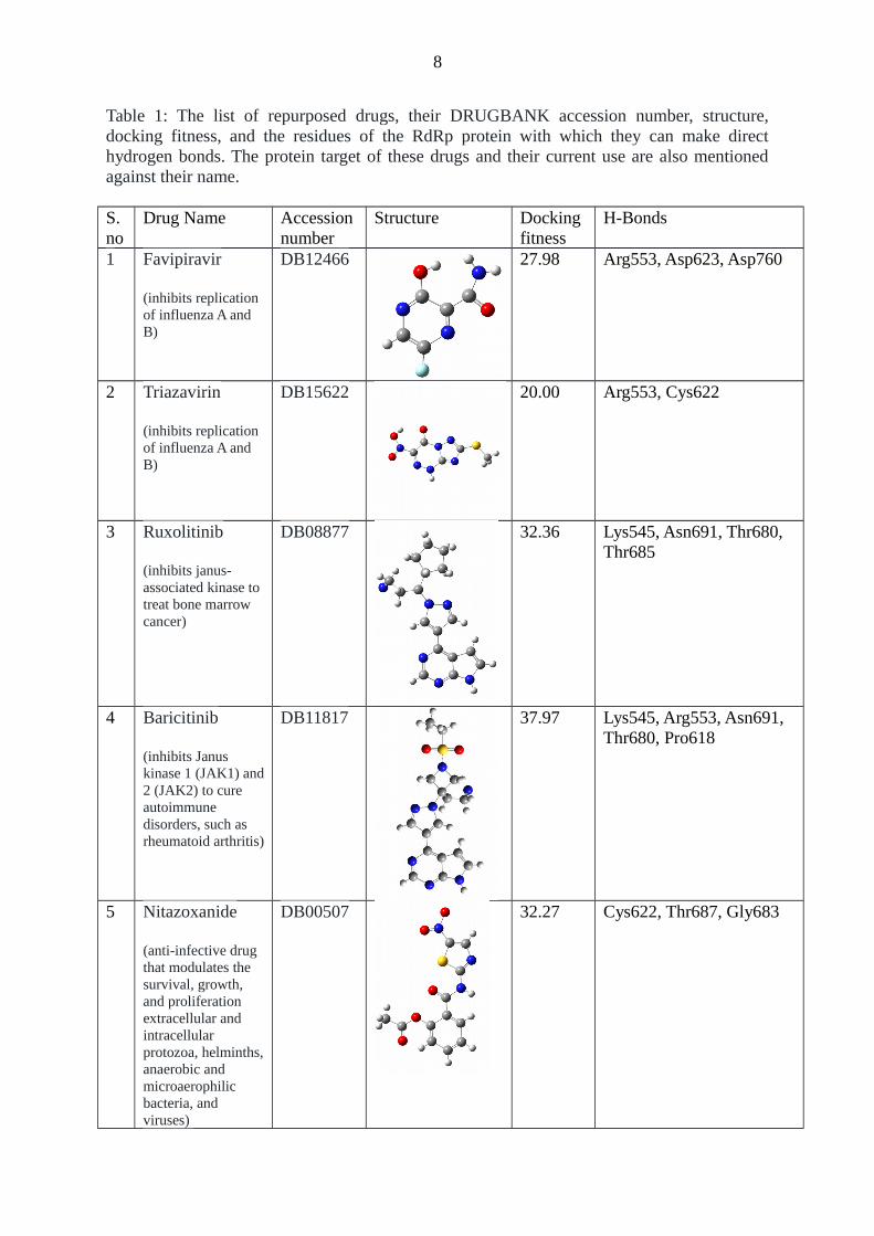
3.1 Binding modes of nucleobase-based drugs
Among the different nucleobase drugs, the Favipiravir, Triazavirin, Ruxolitinib, Baricitinib,
and Nitazoxanide were considered for docking to examine their binding patterns with the
RdRp protein. The first one is a pyrimidine-based drug, while others are purine-based. The
structure of these drugs, their docking fitness, and hydrogen bonding residues are presented
in Table 1. Their docked binding modes are depicted in Fig. 2.
8
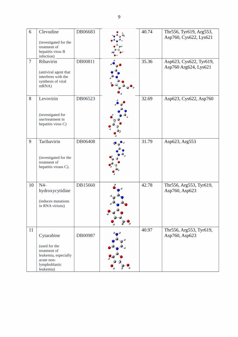
Table 1: The list of repurposed drugs, their DRUGBANK accession number, structure,docking fitness, and the residues of the RdRp protein with which they can make directhydrogen bonds. The protein target of these drugs and their current use are also mentionedagainst their name.
S. no
Drug Name Accession number
Structure Docking fitness
H-Bonds
1 Favipiravir
(inhibits replication of influenza A and B)
DB12466 27.98 Arg553, Asp623, Asp760
2 Triazavirin
(inhibits replication of influenza A and B)
DB15622 20.00 Arg553, Cys622
3 Ruxolitinib
(inhibits janus-associated kinase to treat bone marrow cancer)
DB08877 32.36 Lys545, Asn691, Thr680, Thr685
4 Baricitinib
(inhibits Janus kinase 1 (JAK1) and2 (JAK2) to cure autoimmune disorders, such as rheumatoid arthritis)
DB11817 37.97 Lys545, Arg553, Asn691, Thr680, Pro618
5 Nitazoxanide
(anti-infective drug that modulates the survival, growth, and proliferation extracellular and intracellular protozoa, helminths,anaerobic and microaerophilic bacteria, and viruses)
DB00507 32.27 Cys622, Thr687, Gly683
9
6 Clevudine
(investigated for thetreatment of hepatitis virus B infection)
DB06683 40.74 Thr556, Tyr619, Arg553, Asp760, Cys622, Lys621
7 Ribavirin
(antiviral agent that interferes with the synthesis of viral mRNA)
DB00811 35.36 Asp623, Cys622, Tyr619, Asp760 Arg624, Lys621
8 Levovirin
(investigated for use/treatment in hepatitis virus C)
DB06523 32.69 Asp623, Cys622, Asp760
9 Taribavirin
(investigated for thetreatment of hepatitis viraus C).
DB06408 31.79 Asp623, Arg553
10 N4-hydroxycytidine
(induces mutations in RNA virions)
DB15660 42.78 Thr556, Arg553, Tyr619, Asp760, Asp623
11Cytarabine
(used for the treatment of leukemia, especiallyacute non-lymphoblastic leukemia)
DB00987 40.97 Thr556, Arg553, Tyr619,
Asp760, Asp623
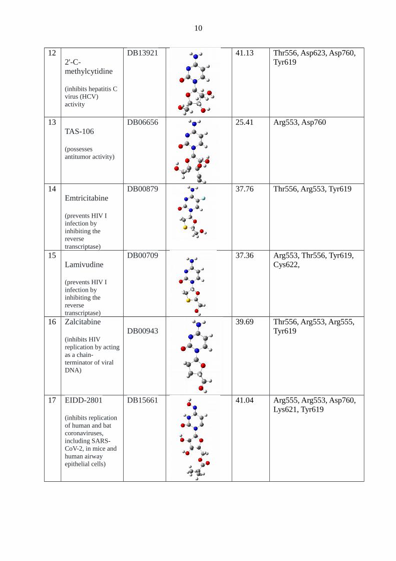
10
122'-C-methylcytidine
(inhibits hepatitis C virus (HCV) activity
DB13921 41.13 Thr556, Asp623, Asp760, Tyr619
13TAS-106
(possesses antitumor activity)
DB06656 25.41 Arg553, Asp760
14Emtricitabine
(prevents HIV I infection by inhibiting the reverse transcriptase)
DB00879 37.76 Thr556, Arg553, Tyr619
15Lamivudine
(prevents HIV I infection by inhibiting the reverse transcriptase)
DB00709 37.36 Arg553, Thr556, Tyr619, Cys622,
16 Zalcitabine
(inhibits HIV replication by actingas a chain-terminator of viral DNA)
DB0094339.69 Thr556, Arg553, Arg555,
Tyr619
17 EIDD-2801
(inhibits replication of human and bat coronaviruses, including SARS-CoV-2, in mice and human airway epithelial cells)
DB15661 41.04 Arg555, Arg553, Asp760, Lys621, Tyr619

11
18Valopicitabine
(inhibits viral RNA chain elongation and viral RNA-dependent RNA polymerase activity)
DB13920 38.77 Thr556, Arg553, Asp623, Tyr619
19Mericitabine
(inhibits HCV RNA polymerase, an enzyme that is necessary for hepatitis C viral replication)
DB1204525.50 Thr556, Arg553, Ser759
20 Galidesivir
(possesses antiviral activity against various negative- and positive-sense RNA viruses including coronaviruses, filoviruses, and arenaviruses)
DB11676 31.45 Thr556, Arg553, Asp623, Asp760
21 Immucillin-G
(investigational nucleoside)
DB02230 36.63 Arg553, Asp623, Tyr619, Asp760
22 Nelarabine
(inhibits DNA synthesis and cytotoxicity and is used to treat acute T-cell lymphoblasticleukemia)
DB01280 38.02 Thr556, Arg553, Asp623, Asp760, Tyr619
23 Vidarabine
(possess antiviral activity against infections caused bya variety of viruses such as the herpes viruses, the vaccinia
DB00194 35.93 Arg553, Asp760, Lys545, Tyr619

12
virus and varicella zoster virus)
24 Inosine
(investigational molecule and may possess neurorestorative, anti-inflammatory, immunomodulatory and cardioprotectiveeffects)
DB04335 39.61 Lys545, Arg553, Asp760, Tyr619
25 Penciclovir
(possesses antiviral activity against herpes simplex virus (HSV) infections)
DB00299 34.08 Thr556, Asp760, Arg624, Arg553, Tyr619
26 Entecavir
(antiviral drug used in the treatment of hepatitis B infection)
DB0044236.92 Lys545, Arg553, Lys624,
Asp623
27 Sofosbuvir
(antiviral drug used as part of combination therapyto treat chronic Hepatitis C, an infectious liver disease caused by infection with Hepatitis C Virus (HCV))
DB08934 39.60 Lys621, Asp760, Cys622, Lys551
28 Uprifosbuvir
(under investigationfor the treatment of Chronic Hepatitis C Genotype (GT)1 and GT2 Infection)
DB15206 47.55 Thr556, Arg553, Lys621, Asp760
29 Adafosbuvir
(under investigationto evaluate the effect of renal impairment on the pharmacokinetics ofAL-335)
DB14906 39.90 Lys545, Arg553, Asp760, Lys551

13
30 Balapiravir
(under in trials for the treatment of Dengue and Hepatitis C viral infections)
DB05060 41.30 Thr556, Arg553, Arg555, Lys621, Asp760
31 Umifenovir
(antiviral agent usedfor the treatment and prophylaxis of influenza and other respiratory infections)investigation into itsuse for a variety of enveloped and non-enveloped RNA andDNA viruses, including Flavivirus,Zika virus, foot-and-mouth disease, Lassa virus,Ebola virus, herpes simplex, hepatitis B and C viruses, chikungunya virus, reovirus, Hantaan virus, and coxsackievirus B5)
DB13609 35.03 Arg555, Arg553, Ser682, Thr687, Asn691
32 Acalabrutinib
(inhibits the Bruton Tyrosine Kinase (BTK) for the treatment of chroniclymphocytic leukemia, small lymphocytic lymphoma)
DB11703 46.49 Thr556, Thr687, Asp760, Lys551, Asn691
33 BMS-986094
(in trials studying the treatment of Hepatitis C, HCV (Genotype 1), and Hepatitis C Virus)
DB11966 41.76 Lys545, Lys621, Arg553, Asp760, Ser759
34 Remdesivir
(used for the treatment of Ebola and coronavirus family of viruses including the SARS-Cov-2)
DB14761 46.73 Ser682, Arg553, Asp760

14
35 GS-6620
(under trial for antiviral activity for the treatment of chronic Hepatitis C virus infection).
DB15222 42.90 Ser622, Arg553, Arg555
36 Sulfaphenazole
(an antibacterial agent)
DB06729 34.39 Asp760
37Cefmetazole
(Possesses antibacterial activityagainst both gram-positive and gram-negative microorganisms. It has a high rate of efficacy in many types of infection and to date no severe side effects have been noted).
DB00274 31.61 Lys545, Arg553, Ser682
38Ceforanide
(anti-bacterial agent)
DB00923 45.33 Ser682, Arg553, Lys551, Thr687
As evident from Fig. 2, Favipiravir, an inhibitor of influenza A and B viruses [36] binds with
the RdRp protein by making strong hydrogen-bonding interactions with Arg553, Asp623, and
Asp760. Its binding position in the RdRp active site is the same as the sugar moiety of the
RMP (Fig. 2c) [16]. This may be the reason for which it was observed to inhibit the RdRp
activity in the cell-based assays. Interestingly, all of the purine-based drugs are found to bind
with the RdRp protein like the RMP. In these drugs, the purine ring makes hydrogen bonding
interactions mainly with the Lys545, while the extended five-membered rings make hydrogen
bonding interactions with Thr680, Thr685, Asn691, Cys622, and Pro618 (Table 1). It is
further found that the CO, S-CH3, SO2, and NO2 groups in Favipiravir, Triazavirin,

15
Baricitinib, and Nitazoxanide, respectively, will make interactions with one of the Mg+2 ions.
As Baricitinib makes more number of hydrogen bonding interactions and its docking score is
more than that of other drugs (Table 1), it may be proposed that the Baricitinib, which is an
inhibitor of the JANUS kinase and is used for the treatment of rheumatoid arthritis [37]
would serve as a better inhibitor of the RdRp protein than that of Fivapiravir and other
nucleobase drugs studied here.
Fig. 2: The binding modes of different nucleobase drugs (represented by sticks) with theRdRp protein (expressed by surface). These drugs include (a) Favipiravir and (b) Triazavirin,Ruxolitinib, Baricitinib, and Nitazoxanide. The first four letters of each drug are mentionedagainst the color code used to represent them. The binding of (c) Favipiravir and (d)Baricitinib with different amino acids of the RdRp protein are also depicted. The dotted lines

16
indicate hydrogen-bonding interactions. For comparison, the crystal structure of the RMP (inlight blue) is also shown by the line representation.
3.2 Binding modes of pyrimidine-based nucleoside drugs
Nucleoside-based drugs can be divided into pyrimidine-based ad purine-based. It is found
that most of the pyrimidine based drugs, such as Clevudine, Taribavirin, N4-hydroxycytidine
(EIDD-1931), Cytarabine, 2'-C-methylcytidine, Emtricitabine, Lamivudine, Zalcitabine,
EIDD-2801, and Valopicitabine (Table 1) bind to the RdRp protein in a similar manner (Fig.
3a). Among these drugs, the clevudine, N4-hydroxycytidine (Fig. 3b), EIDD-2801 (Fig. 3c)
and 2'-C-methylcytidine are found to bind tightly with the RdRp protein by making strong
hydrogen bonds with mainly Thr556, Arg553, Asp623, Asp760 and Tyr619 and possess the
highest docking scores (Table 1). They also interact strongly with the Mg+2 ions. Hence, these
drugs will inhibit RdRp activity very strongly. This is in agreement with the cell-based
assays, where EIDD-2180 was found to inhibit RdRp about 3-6 times more than that of the
Remdesivir [19]. Similarly, the N4-hydroxycytidine has been found to inhibit the Venezuelan
equine encephalitis virus, human coronavirus HCoV-NL63 [38], bat coronaviruses and the
SARS-CoV-2 in mice [39] and human airway epithelial cells [39]. Other pyrimidine-based
drugs, such as Ribavirin, Levovirin, and TAS-106, are found to bind RdRp slightly differently
than the former pyrimidine-based drugs (Fig. 3d). Among these three drugs, Ribavirin is
found to be more promiscuous (Fig. 3e). However, its pyrimidine ring is unable to make any
hydrogen bonding interactions with the F-block residues (Fig. 3e). Interestingly, the five-
membered ring of the Mericitabine binds like the Cytarabine, while the extended carbon rings
connected to the sugar moiety helps it to point toward Lys551 and Ser814 (Fig. 3f). Due to
this reason, their interaction with the Mg+2 ions is weaker compared to other pyrimidine-
based drugs. If we compare the binding of all of these drugs with RdRp, it is clear that the
clevudine, N4-hydroxycytidine, EIDD-2801, and 2'-C-methylcytidine would be more

17
effective than that of other drugs including Favipiravir and Ribavirin that are proposed to be
useful for the treatment of the SARS-CoV-2 infection.
Fig. 3: The binding modes of different pyrimidine-based nucleoside drugs (represented bysticks) with the RdRp protein (represented by surface). These drugs include (a) Clevudine,Taribavirin, N4-hydroxycytidine, Cytarabine, 2'-C-methylcytidine, Emtricitabine,Lamivudine, Zalcitabine, EIDD-2801, and Valopicitabine. The interactions of (b) N4-hydroxycytidine and (c) EIDD-2801 with different amino acids are illustrated. (d) Thebinding modes of Ribavirin, Levovirin, and TAS-106 (in the sticks) with RdRp (in thesurface) are also shown. The interactions of (e) Ribavirin and (f) Mericitabine with differentimportant amino acids are depicted. The dotted lines show important hydrogen bonds. Colorcodes used for different drugs are mentioned in (a) and (d). The line representation shows thecrystal structure of the RMP (in light blue) for the comparison of different binding modes. 3.3 Binding modes of purine-based nucleoside drugs
Binding modes of Galidesivir, Immucillin-G, Nelarabine, Vidarabine, Penciclovir, Entecavir,
and Inosine are shown in Fig. 4. Except Immucillin-G, Entecavir, and Inosine all of these
drugs bind to the RdRp protein almost similarly. The vital amino acids making hydrogen
bonds with these drugs are summarized in Table 1. Among these drugs, Nelabarine and
Inosine have the highest docking fitness and can form strong hydrogen bonds with the

18
protein. Hence, these purine-based nucleosides will act as better inhibitors. However, if we
compare these purine-based nucleoside drugs with those of pyrimidine-based nucleoside
drugs, it is found that the latter drugs, in particular, N4-hydroxycytidine and EIDD-2801
would act as better antiviral drugs.
Fig. 4: The binding modes of (a) Galidesivir, Nelarabine, Vidarabine, and Penciclovir,(represented by sticks) and (b) Immucillin-G, Entecavir, and Inosine (represented by sticks)with the RdRp protein (represented by surface). (c) The dotted lines show importanthydrogen bonds made by Inosine with different amino acids of the RdRp protein. Color codesused for different drugs are mentioned in (a) and (b). The crystal structure of the RMP (inlight blue, pdb id 7BV2)) is shown by the line representation for comparison of differentbinding modes.
3.4: Binding modes of Purine- and Pyrimidine-based drugs with modified backbones
There are several purine- and pyrimidine-based antiviral drugs available, whose sugar-
phosphate backbones are replaced with extended synthetic rings to prohibit their replication
in cells, such as Sofosbuvir, Uprifosbuvir, Remdesivir, etc (Table 1). It should be mentioned
that the recent cryo-EM study of the RdRp-Remdesivir monophosphate complex [16] does
not account for the full structure of the drug mentioned in the DRUGBANK database.
Similarly, the structure of the HCV-Sofosbuvir monophosphate (pdb id 4WTG) [35] does not
include the full structure of the Sofosbuvir stored in the DRUGBANK database. Hence, this
study is expected to unravel the detailed binding modes of these full drugs with the RdRp
protein. It is found that among these drugs, the pyrimidine-based drugs, such as Sofosbuvir,
Uprifosbuvir, Adafosbuvir, and Balapiravir follow a similar binding pattern with the RdRp
19
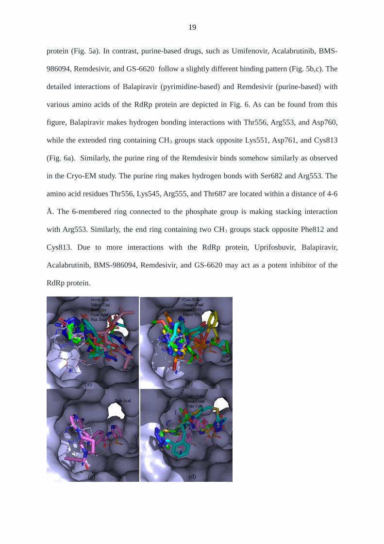
protein (Fig. 5a). In contrast, purine-based drugs, such as Umifenovir, Acalabrutinib, BMS-
986094, Remdesivir, and GS-6620 follow a slightly different binding pattern (Fig. 5b,c). The
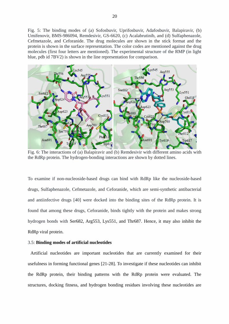
detailed interactions of Balapiravir (pyrimidine-based) and Remdesivir (purine-based) with
various amino acids of the RdRp protein are depicted in Fig. 6. As can be found from this
figure, Balapiravir makes hydrogen bonding interactions with Thr556, Arg553, and Asp760,
while the extended ring containing CH3 groups stack opposite Lys551, Asp761, and Cys813
(Fig. 6a). Similarly, the purine ring of the Remdesivir binds somehow similarly as observed
in the Cryo-EM study. The purine ring makes hydrogen bonds with Ser682 and Arg553. The
amino acid residues Thr556, Lys545, Arg555, and Thr687 are located within a distance of 4-6
Å. The 6-membered ring connected to the phosphate group is making stacking interaction
with Arg553. Similarly, the end ring containing two CH3 groups stack opposite Phe812 and
Cys813. Due to more interactions with the RdRp protein, Uprifosbuvir, Balapiravir,
Acalabrutinib, BMS-986094, Remdesivir, and GS-6620 may act as a potent inhibitor of the
RdRp protein.
20
Fig. 5: The binding modes of (a) Sofosbuvir, Uprifosbuvir, Adafosbuvir, Balapiravir, (b)Umifenovir, BMS-986094, Remdesivir, GS-6620, (c) Acalabrutinib, and (d) Sulfaphenazole,Cefmetazole, and Ceforanide. The drug molecules are shown in the stick format and theprotein is shown in the surface representation. The color codes are mentioned against the drugmolecules (first four letters are mentioned). The experimental structure of the RMP (in lightblue, pdb id 7BV2) is shown in the line representation for comparison.
Fig. 6: The interactions of (a) Balapiravir and (b) Remdesivir with different amino acids withthe RdRp protein. The hydrogen-bonding interactions are shown by dotted lines.
To examine if non-nucleoside-based drugs can bind with RdRp like the nucleoside-based
drugs, Sulfaphenazole, Cefmetazole, and Ceforanide, which are semi-synthetic antibacterial
and antiinfective drugs [40] were docked into the binding sites of the RdRp protein. It is
found that among these drugs, Ceforanide, binds tightly with the protein and makes strong
hydrogen bonds with Ser682, Arg553, Lys551, and Thr687. Hence, it may also inhibit the
RdRp viral protein.
3.5: Binding modes of artificial nucleotides
Artificial nucleotides are important nucleotides that are currently examined for their
usefulness in forming functional genes [21-28]. To investigate if these nucleotides can inhibit
the RdRp protein, their binding patterns with the RdRp protein were evaluated. The
structures, docking fitness, and hydrogen bonding residues involving these nucleotides are

21
presented in Table 2. The binding modes of these artificial nucleotides are illustrated in Fig.
7. As can be seen from this figure, the monophosphate forms of different pyrimidines, such as
dZ, dZ5, dV, dS, and dK possess a similar binding mode. Similarly, the monophosphate forms
of purines, such as dP, dP4, dJ, dB, and dX follow a similar binding mode. As depicted in Fig.
7b,c, dZ and dP, the most studied AEGIS nucleotides can bind with the F-block residues
almost identically. The same trend is also found in the case of other artificial nucleotides. The
residues that make hydrogen bonds with these unnatural nucleotides are Lys545, Arg553,
Arg555, Lys621, Cys622, Asp623, Thr680, Ser682, Thr687, Asn691, Ser759, and Asp760
(Fig. 7c,d, Table 2). The phosphate groups are also found to make tight ionic interactions with
the Mg+2 ions. If we compare binding modes, number of hydrogen bonds, salt-bridge, and
other interactions and docking fitness of these nucleotides with those of the antiviral drugs
studied here, it is evident that these artificial nucleotides would act as better inhibitors of the
RdRp viral protein.
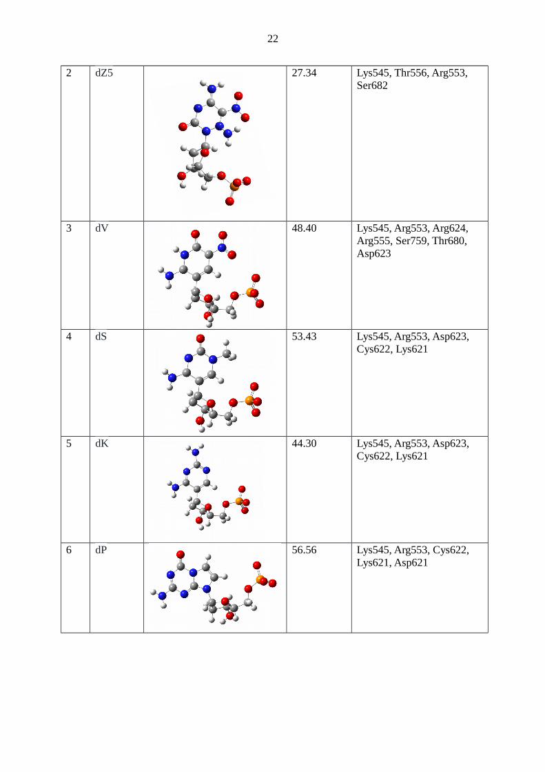
Table 2: Table 1: The list of various AEGIS nucleotides, their structure, docking fitness, andthe residues of the RdRp protein with which they can make direct hydrogen bonds.
S. No.
Sequence Structure Docking Fitness
H-bonds
1 dZ 46.54 Lys545, Thr556, Arg553, Lys612 Cys222
22
2 dZ5 27.34 Lys545, Thr556, Arg553, Ser682
3 dV 48.40 Lys545, Arg553, Arg624, Arg555, Ser759, Thr680, Asp623
4 dS 53.43 Lys545, Arg553, Asp623, Cys622, Lys621
5 dK 44.30 Lys545, Arg553, Asp623, Cys622, Lys621
6 dP 56.56 Lys545, Arg553, Cys622, Lys621, Asp621

23
7 dP4 43.90 Arg553, Cys622, Cys622, Lys621
8 dJ 54.43 Lys545, Thr556, Arg553, Cys622, Lys621
9 dB 52.06 Lys545, Arg553, Cys622Asp623, Ser682, Lys621, Cys622
10 dX 35.13 Lys545 Thr680, Asn691, Ser759
11 5’-dPdZ-3’
59.50 Arg553, Thr556, Thr680, Thr687, Ser682, Ser759,
12 5’-dPdZdZ-3’
53.71 Lys545, Arg553, Arg555, Ser682, Cys622, Asp760, Ser759

24
13 5’-dPdPdZ-3’
55.40 Lys545, Ser682, Arg553, Ser759, Lys621
14 5’-dBdBdZ-3’
35.12 Lys545, Thr680, Arg553, Thr687, Asn691, Ser759
15 5’-dPdPdZdZ-3’
21.14 Lys545, Arg553, Arg555, Asp760, Lys551, Asp760, Tyr619,
Fig. 7: The binding modes of different (a) pyrimidine-based (in the stick) and (b) purine-based (in the stick) artificial nucleotides with the RdRp protein (in surface representation).

25
The detailed interactions of (c) dP and (d) dZ with different amino acid residues of the RdRpprotein are also shown. Dotted lines show the hydrogen bonding interactions.
To further evaluate the binding of more than one artificial nucleotide with the RdRp protein
(Table 2), different sequences of these nucleotides were docked into the binding site of the
RdRp protein. The docking fitness and hydrogen bonding residues are presented in Table 2.
The binding modes of these nucleotides are shown in Fig. 8. As can be found from this figure,
the 5’-dPdZ-3’ can make tight interactions with the F-block residues and the phosphate group
can make stable salt-bridge interactions with the Mg+2 ion (Table 2, Fig. 8a). The dP also
makes stacking interactions with dZ. However, the interbase stacking interaction gets lost
with the increasing size of the sequence (Fig. 8 b-e). Although nucleobases can make more
number of contacts with the protein in large sequences, the phosphate-Mg+2 interaction
becomes weak. This is evident from the docking fitness (Table 2). These results suggest that
more than three nucleotide sequences may reduce the inhibitory activity of the artificial
nucleotides.

26
Fig. 8: The binding modes of (a) 5’-dPdZ-3’, (b) 5’-dPdZdZ-3’, (c) 5’-dPdPdZ-3’, (d) 5’-dBdBdZ-3’, and (e) 5’-dPdPdZdZ-3’ with the RdRp protein (in surface representation).
4. Conclusions
It is revealed that all the approved and investigational drugs studied here can bind to the
RdRp viral protein of the SARS-CoV-2. However, due to the higher binding affinity, some of
the drugs would have more inhibitory activity and can be immediately used for the treatment
of the Wuhan virus infections after a careful clinical trial. These drugs include clevudine, N4-
hydroxycytidine, 2'-C-methylcytidine, EIDD-2801, Uprifosbuvir, Balapiravir, Acalabrutinib,
BMS-986094, Remdesivir, GS-6620, and Ceforanide. It is also found that the artificial
nucleotides that were invented to make functional genes may have better inhibitory activity
than that of the above drugs. Among these nucleotides, dB, dJ, dP, dK, dS, dV, dZ are found
to make tight interactions with the RdRp protein. It is further found that sequences containing
27
up to three artificial nucleotides can also inhibit the RdRp viral protein. Among different
residues, Lys545, Arg553, Arg555, Thr556, Tyr619, Lys621, Cys622, Asp623, Arg624,
Thr680, Ser682, Thr687, Asn691, and Asp760 are found to play a vital role in stabilizing
different inhibitors. Hence, the design of new inhibitors should be done in such a way that
they can make interactions with these residues.
Acknowledgment
NRJ is thankful to the Science and Engineering Research Board (SERB) of the Department of
Science and Technology (DST, NewDelhi) for financial support.
References
1. D. Wang, B. Hu, C. Hu, F. Zhu, X. Liu, J. Zhang, B. Wang, H. Xiang, Z. Cheng, Y.Xiong, Y. Zhao, Y. Li, X. Wang, Z. Peng Clinical characteristics of 138 hospitalizedpatients with 2019 coronavirus-infected pneumonia in Wuhan, China, JAMA, 2020,323, 1061-1069.
2. A.R. Sahin, A. Erdogan, P.M. Agaolglu, Y. Dineri, A. Y, Cakirci, M. E. Senel; R. A.Okyay, A. M. Tasdogan, 2019 Novel coronavirus (COVID-19) outbreak: a review ofthe current literature. Eur. J. Med. Oncol. 2020, 4, 1-7.
3. C. Liu, Q. Zhou, Y. Li, L. V. Garner, S.P. Watkins, L. J. Carter, J. Smoot, A. C. Gregg,A. D. Daniels, S. Jervey, D. Albaiu, Research and development on therapeutic agentsand vaccines for COVID-19 and related human coronavirus diseases. ACS Cent. Sci.2020, 6, 315-331.
4. U.J. Buchholz, A. Bukreyev, L. Yang, E.W. Lamirande, B.R. Murphey, K. Subbarao,P.L. Collins, Contributions of the structural proteins of severe acute respiratorysyndrome coronavirus to protective immunity. Proc. Natl. Acad. Sci. U.S.A. 2004,101, 9804-9809.
5. T. P. Sheahan, A.C. Sims, S.R. Leist, A. Schafer, J. Won, A.J. Brown, S.A.Montgomery, A. Hogg, D. Babusis, M. O. Clarke, J.E. Spahn, L. Bauer, S. Sellers, D.Porter, J.Y. Feng, T. Cihlar, R. Jordan, M.R. Denison, R.S. Baric, Comparativetherapeutic efficacy of remdesivir and combination lopinavir, ritonavir, and interferonbeta against MERS-CoV. Nat. Commun. 2020, DOI: 10.1038/s41467-019-13940-6.
6. R. U. Kadam, I.A. Wilson, Structural basis of influenza virus fusion inhibition by theantiviral drug Arbidol. Proc. Natl. Acad. Sci. U.S. A. 2017, 114, 206-214.
7. Therapeutic options for the 2019 novel coronavirus (2019-nCoV).https://www.nature.com/articles/d41573-020-00016-0.

28
8. The efficacy of Lopinavir Plus Ritonavir and Arbidol against novel coronavirusInfection (ELACOI). https://clinicaltrials.gov/ct2/show/NCT04252885.
9. S. Pant, M. Singh, V. Ravichandran, U.S.N. Murty, H. Srivastava, Peptide-like andsmall molecule inhibitors against COVID-19. Biomol. Struct. Dyn. 2020, DOI:10.1080/07391102.2020.1757510.
10. C. Cava, G. Bertoli, I. Castiglioni, In silico discovery of candidate drugs againstCOVID-19, Viruses, 2020, 12, 404.
11. Y. Zhou, Y. Hou, J. Shen, Y. Huang, W. Martin, F. Cheng, Network-based repurposingfor novel coronavirus 2019-nCoV/SARS-CoV-2. Cell Discovery, 2020, 6, 14.
12. R. Wu, L. Wang, H.C.D. Kuo, A. Shannar, R. Peter, P. J. Chou, S. Li, R. Hudlikar, X.Liu, Z. Liu, G. J. Poiani, L. Amorosa, L. Brunetti, A.N. Kong, Curr. Pharmacol.Rep. 2020, DOI:10.1007/s40495-020-00216-7
13. M. Ceccarelli, M. Berretta, E.V. Rullo, G. Nunnari, B. Cacopardo, Differences andsimilarities between severe acute respiratory syndrome (SARS)-coronaVirus (CoV)and SARS-CoV-2. Would a rose by another name smell as sweet? Eur. Rev. Med.Pharmacol. Sci. 2020, 24, 2781-2783.
14. S.J.R. Da Silva, C.T.A. Da Silva, R.P.G. Mendes, L. Pena, Role of non-structuralproteins in the pathogenesis of SARS-CoV-2. J. Med. Virol. 2020, DOI:10.1002/jmv.25858.
15. E.J. Snijder, E. Decroly, J. Ziebuhr, The non-structural proteins directing coronavirusRNA synthesis and processing. Adv. Virus Res. 2016, 96, 59-126.
16. W. Yin, et al. Structural basis for inhibition of the RNA-dependent RNA polymerasefrom SARS-CoV-2 by remdesivir. Science, 2020, DOI: 10.1126/science.abc1560.
17. Y. Gao, et al. Structure of the RNA-dependent RNA polymerase from COVID-19virus. Science, 2020, DOI:10.1126/science.abb7498.
18. C.-C. Lu, M.-Y. Chen, Y.-L. Chang, Potential therapeutic agents against COVID-19:What we know so far. J. Chin. Med. Assoc. 2020,DOI:10.1097/JCMA.0000000000000318.
19. T. P. Sheahan, A. C. Sims, S. Zhou, R. L. Graham, A. J. Pruijssers, M. L. Agostini, S.R. Leist, A. Schäfer, K. H. Dinnon 3rd, L. J. Stevens, J. D. Chappell, X. Lu, T. M.Hughes, A. S. George, C. S. Hill, S. A. Montgomery, A. J. Brown, G. R. Bluemling,M. G. Natchus, M. Saindane, A. A. Kolykhalov, G. Painter, J. Harcourt, A. Tamin, N.J. Thornburg, R. Swanstrom, M. R. Denison, R. S. Baric, An orally bioavailablebroad-spectrum antiviral inhibits SARS-CoV-2 in human airway epithelial cellcultures and multiple coronaviruses in mice. Sci. Transl. Med. 2020, 12, eabb5883.
20. D.S. Wishart C. Knox, A.C. Guo AC, S. Shrivastava, M. Hassanali, P. Stothard, Z.Chang, J. Woolsey, Drugbank: a comprehensive resource for in silico drug discoveryand exploration. Nucleic Acids Res. 2006 34 (Database issue), D668-D672.
21. S.A. Benner, Z. Yang, F. Chen, Synthetic biology, tinkering biology, and artificialbiology. what are we learning? Comptes Rendus Chim. 2011, 14, 372-387.
22. K.K. Merritt, K.M. Bradley, D. Hutter, M.F. Matsuura, D.J. Rowold, S.A. Benner,Autonomous assembly of synthetic oligonucleotides built from an expanded DNAalphabet. total synthesis of a gene encoding kanamycin resistance. Beilstein J. Org.Chem. 2014, 10, 2348-2360.
23. E. Biondi, S. A. Benner, Artificially expanded genetic information systems for newaptamer technologies. Biomedicines 2018, 6, 53.

29
24. L. Zhang, et al. Evolution of functional six-nucleotide DNA. J. Am. Chem. Soc. 2015,137, 6734-6737.
25. J.J. Voegel, S.A. Benner, Nonstandard hydrogen bonding in duplex oligonucleotides.the base pair between an acceptor-donor-donor pyrimidine analog and a donor-acceptor-acceptor purine analog. J. Am. Chem. Soc. 1994, 116, 6929-6930.
26. N.R. Jena, P. Das, B. Behera, P.C. Mishra, Analogues of P and Z as efficientartificially expanded genetic information system. J. Phys. Chem. B 2018, 122, 8134-8146.
27. B. Behera, P. Das, N.R. Jena, Accurate base pair energies of artificially expandedgenetic information systems (AEGIS): clues for their mutagenic characteristics. J.Phys. Chem. B, 2019, 123, 6728-6739.
28. N.R. Jena, Electron and hole interactions with P, Z, and P:Z and the formation ofmutagenic products by proton transfer reactions, Phys. Chem. Chem. Phys. 2020, 22,919-931.
29. M.M. Georgiadis, I. Singh, W.F. Kellett, S. Hoshika, S.A. Benner, N.G. Richards,Structural basis for a six nucleotide genetic alphabet. J. Am. Chem. Soc. 2015, 137,6947-6955.
30. GaussView,Version5, R. Dennington, T. Keith, J. Millam, J. Inc. Semichem, KSShawnee Mission 2009.
31. Gaussian09, Revision A.1, M.J. Frisch, G.W. Trucks, H.B. Schlegel, G.E. Scuseria,M.A. Robb, J.R. Cheeseman, G. Scalmani, V. Barone, B. Mennucci, G.A. Petersson,H. Nakatsuji, M. Caricato, X. Li, H.P. Hratchian, A.F. Izmaylov, J. Bloino, G. Zheng,J.L. Sonnenberg, M. Hada, M. Ehara, K. Toyota, R. Fukuda, J. Hasegawa, M. Ishida,T. Nakajima, Y. Honda, O. Kitao, V. Nakai, T. Vreven, J. Jr. J.A. Montgomery, J.E.Peralta, F. Ogliaro, M. Bearpark, J.J. Heyd, E. Brothers, K.N. Kudin, V.N. Staroverov,R. Kobayashi, J. Normand, K. Raghavachari, A. Rendell, J.C. Burant, S.S. Iyengar, J.Tomasi, M. Cossi, N. Rega, J.M. Millam, M. Klene, J.E. Knox, J.B. Cross, V.Bakken, C. Adamo, J. Jaramillo, R. Gomperts, R.E. Stratmann, O. Yazyev, A.J.Austin, R. Cammi, C. Pomelli, J.W. Ochterski, R.L. Martin, K. Morokuma, V.G.Zakrzewski, G.A. Voth, P. Salvador, J.J. Dannenberg, S. Dapprich, A.D. Daniels, O.,Farkas, J.B. Foresman, J.V. Ortiz, J. Cioslowski, D.J. Fox, Gaussian, Inc.,WallingfordCT, 2009.
32. G. Jones, P. Willett, R. C. Glen, A. R. Leach and R. Taylor, Development andValidation of a Genetic Algorithm for Flexible Docking. J. Mol. Biol., 1997, 267,727-748.
33. J. W. M. Nissink, C. Murray, M. Hartshorn, M. L. Verdonk, J. C. Cole and R. Taylor,A new test set for validating predictions of protein-ligand interaction. Proteins, 2002,49, 457-471.
34. M. J. Hartshorn, M. L. Verdonk, G. Chessari, S. C. Brewerton, W. T. M. Mooij, P. N.Mortenson, C. W. Murray, Diverse, High-Quality Test Set for the Validation ofProtein-Ligand Docking Performance. J. Med. Chem., 2007, 50, 726-741.
35. T.C. Appleby, J.K. Perry, E. Murakami, O. Barauskas, J. Feng, A. Chao, D. Fox, D.R.Wetmore, M.E. McGrath, A.S. Ray, M.J. Sofia, S. Swaminathan, T.E. Edwards,Structural basis for RNA replication by the hepatitis C virus polymerase. Science,2015, 347, 771-775.
36. F.G. Hayden, N. Shindo, Influenza virus polymerase inhibitors in clinicaldevelopment. Curr. Opin. Infect. Dis. 2019, 32, 176-186.

30
37. B. Kurriya, M.D. Cohen, E. Keystone. Baricitinib in rheumatoid arthritis: evidence-to-date and clinical potential. Theor. Adv. Musculoskelet. Dis. 2017, 9, 37-44.
38. K. Pyrc, B.J. Bosh, B. Berkhout, M.F. Jebbink, R. Dijkman, P. Rottier, L. van derHoek. Inhibition of human coronavirus NL63 infection at the early stages of thereplication cycle. Antimicrob. Agents Chemother. 2006, 50, 2000-2008.
39. T.P. Sheahan wt al. An orally bioavailable broad-spectrum antiviral inhibits SARS-CoV-2 in human airway epithelial cell cultures and multiple coronaviruses in mice.Sci. Transl. Med. 2020, 12, eabb5883.
40. S.L. Barriere, J. Mills Ceforanide: antibacterial activity, pharmacology, and clinicalefficacy. Pharmacotherapy, 1982, 2, 322-327.