identification of high-confidence somatic mutations in whole genome
TRANSCRIPT

Identification of high-confidence somatic mutationsin whole genome sequence of formalin-fixed breastcancer specimensShawn E. Yost1,2, Erin N. Smith1,3, Richard B. Schwab1, Lei Bao1, HyunChul Jung1,2,
Xiaoyun Wang1,3, Emile Voest4, John P. Pierce1, Karen Messer1,5, Barbara A. Parker1,
Olivier Harismendy1,3,6,* and Kelly A. Frazer1,3,6,7,*
1Moores UCSD Cancer Center, 2Bioinformatics and Systems Biology Graduate Program, 3Department ofPediatrics and Rady Children’s Hospital, University of California San Diego, 9500 Gilman Drive, La Jolla, CA92093, USA, 4Department of Medical Oncology, University Medical Center Utrecht, Heidelberglaan 100, POBOX 85500, F02.126, Utrecht 3584CX, The Netherlands, 5Division of Biostatistics and Bioinformatics,Department of Family and Preventive Medicine, 6Clinical and Translational Research Institute and 7Institute forGenomic Medicine, University of California San Diego, 9500 Gilman Drive, La Jolla, CA 92093, USA
Received December 15, 2011; Revised and Accepted March 20, 2012
ABSTRACT
The utilization of archived, formalin-fixed paraffin-embedded (FFPE) tumor samples for massiveparallel sequencing has been challenging due toDNA damage and contamination with normalstroma. Here, we perform whole genome sequencingof DNA isolated from two triple-negative breastcancer tumors archived for >11 years as 5mm FFPEsections and matched germline DNA. The tumorsamples show differing amounts of FFPE damagedDNA sequencing reads revealed as relatively highalignment mismatch rates enriched for C·G>T·Asubstitutions compared to germline samples. Thisincrease in mismatch rate is observable with as fewas one million reads, allowing for an upfront evalu-ation of the sample integrity before whole genomesequencing. By applying innovative quality filtersincorporating global nucleotide mismatch rates andlocal mismatch rates, we present a method to identifyhigh-confidence somatic mutations even in thepresence of FFPE induced DNA damage. Thisresults in a breast cancer mutational profile consist-ent with previous studies and revealing potentiallyimportant functional mutations. Our study demon-strates the feasibility of performing genome-widedeep sequencing analysis of FFPE archived tumorsof limited sample size such as residual cancer aftertreatment or metastatic biopsies.
INTRODUCTION
To date, massively parallel sequencing of cancer genomeshas largely been performed using flash frozen tissue orimmortalized cancer cell lines (1–6). These studies haveprovided tremendous insight into the types of mutationsand genomic rearrangements that occur in cancer cells.However, limiting sequencing to flash frozen tissuesrestricts the types of important clinical questions thatcan be addressed (7). Since formalin fixation andparaffin embedding (FFPE) has been the standardsample preparation for pathologists for decades, theability to perform massively parallel sequencing ofFFPE samples would open up large archived tumorspecimen collections. As these large archives frequentlyhave historical records of patient progression andoutcome, this would allow for powerful retrospectivestudies exploring DNA changes that influence diseaseprogression.RNA isolated from FFPE samples is commonly used for
genome-wide expression studies (8–10), howeverperforming whole-genome analyses of DNA isolatedfrom FFPE samples has two major application-specificchallenges. First is the fact that tumors of biological andclinical interest stored in blocks are often contaminatedwith normal stroma, and thus dissection, which is noteasy to perform in blocks, is required to enrich for tumormaterial. A second challenge is the fact that formalin-fixedtissues exhibit a higher frequency of non-reproducibleDNA sequence alterations than frozen tissues. This islikely due to formalin cross-linking of cytosine nucleotides
*To whom correspondence should be addressed. Tel: +1 858 246 0208. Email: [email protected] may also be addressed to Olivier Harismendy. Tel: +1 858 246 0248. Email: [email protected]
Published online 6 April 2012 Nucleic Acids Research, 2012, Vol. 40, No. 14 e107doi:10.1093/nar/gks299
� The Author(s) 2012. Published by Oxford University Press.This is an Open Access article distributed under the terms of the Creative Commons Attribution Non-Commercial License (http://creativecommons.org/licenses/by-nc/3.0), which permits unrestricted non-commercial use, distribution, and reproduction in any medium, provided the original work is properly cited.
Dow
nloaded from https://academ
ic.oup.com/nar/article/40/14/e107/2414579 by guest on 17 D
ecember 2021

on either strand, resulting in Taq polymerase during PCRnot recognizing the cytosine and incorporating an adeninein place of a guanosine causing an artificial C>T or G>Amutation (11,12). Previous studies have successfullyisolated DNA from FFPE tissue samples stored inparaffin blocks and performed targeted sequencing ofsingle genes (13,14) or whole exome sequencing (15). In afew instances, sequencing was extended to the wholegenome but was limited to copy number analysis orhigh-level mutational profile analysis (16,17). In theKerick study, artificial mutations resulting from theformalin fixation process were observed in the sequencedata by comparison to matched frozen tissues butmethods for removing these false positive calls in theanalysis steps of the sequence data were not presented.In thework presented below, we sequencedDNA isolated
from two FFPE triple-negative breast tumors archived as5mmsections aswell as theirmatched germlineDNA.As thetumor was mounted on slides, it was straightforward toidentify and isolate DNA from areas containing >80–85%malignant cells. By characterizing the patterns of DNAmismatches in the FFPE tumor sequencing reads, wedetermined that one of the samples was more heavilydamaged by fixation than the other and propose guidelinesfor a rapid FFPE integrity test. We then call somaticvariants and implement original filters to remove falsepositive calls specifically resulting from the formalinfixation process, thus leading to a set of high-confidencesomatic mutations in each of the tumors. Finally, weidentify a set of mutations of potential functionalimportance in the progression of the disease (or lackthereof) in each of the two cases.
MATERIALS AND METHODS
Patient information
From the Women’s Healthy Eating and Living (WHEL)cohort (18), we identified two female non-Hispanic whitepatients (06408 and 02542) diagnosed with Stage III histo-logic Grade III infiltrating ductal triple negative breastcancer in 1999 and 1995 at the ages of 38 and 30 years,respectively. All patients provided written informedconsent for enrollment in the WHEL Study and forrelated genomic studies. Triple negative breast cancer indi-cates that the estrogen and progresterone receptor stainingon tumor tissue was negative and Her2/neu over-expression was not observed. Both patients receivedadjuvant chemotherapy and local regional radiationtherapy. Patient 02542’s tumor metastasized 18monthsafter initial diagnosis and she died shortly afterwards.Patient 06408 is still alive without recurrence as of 2006.The patients underwent curative intent surgical resectionand breast tumor material not needed for diagnosis wasformalin fixed, embedded in paraffin, sectioned at 5mmthickness and mounted on slides. Germline DNA was ex-tracted from peripheral blood mononuclear cells (PBMC).
DNA isolationAreas of tumor cells on a hematoxylin and eosin (H&E)stained 5 mm FFPE section were identified and marked by
a pathologist allowing the collection of malignant cellswith a >80–85% purity (Supplementary Figure S1).Additional tumor material from an adjacent unstainedsection was isolated by scraping the area correspondingto the marked section with a sterile scalpel. DNA wasisolated from the FFPE specimens using BiOstic FFPETissue DNA Isolation kit (MO BIO, Carlsbad, CA,USA). The samples were heated at 55�C for an hour inan optimized wax melting buffer and Protease K to com-pletely digest the tissue. Samples were heated at 90�C for1 h to remove protein–DNA cross-links, purified on asilica spin filter and eluted with 10mM Tris pH 8.0.
Tumor cell countingThe H&E stained slides were used to estimate the numberof tumor cells from which DNA was isolated(Supplementary Figure S1). DNA was isolated fromunstained 5 mm thick sections �1.0 and 2.0 cm2 areas forsamples 06408 and 02542, respectively. We used a NikonEclipse E600 microscope to take images of the cells andprocessed the images with MetaMorph 7.7 (MolecularDevices, Sunnyvale, CA, USA). Six random fields withinthe marked areas were taken. We calculated the number ofnuclei in each random field to get an approximate numberof cells per slide. To count the cells, we first separated theconstituent blue, red and green channels from each of the24-bit RGB images. Only the blue channel was used tocount the number of nuclei in the image. Nuclei wereselected by setting the appropriate intensity threshold.The resulting nuclei were filtered by area to removenoise and counted using the morphometry tool inMetamorph. The number of nuclei was used to calculatethe average cell density per image, which was used toextrapolate the number of cells used for sequencing. Thearea of each image was 1360 pixels by 1024 pixels, with1 pixel=0.334355 mm.
SequencingPurified tumor and germline DNA were directly used asstarting material for SOLiD fragment library preparation(Life Technologies, Carlsbad, CA, USA) following manu-facturer’s recommendation. DNA was sheared to �150 bpusing the Covaris S2 system standard fragmentation con-ditions recommended in the SOLiD4 Library Prep UserGuide. After DNA end-repair, P1 and P2 adaptors wereligated, the adaptor-ligated DNA underwent nick transla-tion and then amplification with six and eight PCR cyclesfor germline and tumor DNA, respectively, using LibraryPCR primer 1 and 2, and Platinum PCR amplificationmix. Purified library was quantified by TaqMan assayand used for preparing SOLiD templated beads. Eachsequencing run resulted in �500 million raw 50-bpcolor-space reads per slide. The samples were sequencedover several runs each using both SOLiD3+and SOLiD4platforms generating between 1.3 and 3.1 billion total rawreads per sample (Supplementary Table S1).
Genotyping array data generation and analysisGermline DNA was genotyped on the Illumina Omni2.5M array and processed using GenomeStudio (version2010.3) using standard methods. Genotypes were exported
e107 Nucleic Acids Research, 2012, Vol. 40, No. 14 PAGE 2 OF 12
Dow
nloaded from https://academ
ic.oup.com/nar/article/40/14/e107/2414579 by guest on 17 D
ecember 2021

into reference genome PLUS orientation (build hg19)based on HumanOmni2.5-4v1_D.bpm. As the contenton this array contains new SNPs that are not present indbSNP 132 and were not always named according todbSNP identifiers, we verified that all positions werepresent and consistent with dbSNP 132. We converted1000 Genomes Project (19) SNPs (kgp identifiers) torsIDs by matching chromosome, position and alleles indbSNP132. We excluded 17 959 1000 Genome ProjectSNPs that were duplicates of SNPs with rsID identifiers,11 536 SNPs that had more than two alleles in dbSNP, and405 516 SNPs that were not present in dbSNP 132. Thisresulted in a total of 2 016 729 SNPs. Since the sequencinganalysis was performed in the hg18 reference, weconverted the positions and orientation of the genotypedSNPs from hg19 to hg18 using the LiftOverVCF.pl scriptwithin GATK (20). The 2 015 517 SNPs with successfulcoordinate conversion were used in subsequent analysis.
Calculating concordance between genotyping arrayand sequencing dataTo determine concordance, we used the genotypes of the2 015 517 SNPs described above and the genotypes calledin the sequencing data passing Filter 1.1 (see below) thathad at least the indicated coverage (SupplementaryTable S4). We calculated the total number of the geno-types (homozygous reference, heterozygous and homozy-gous alternate) called in the sequencing data that agreedwith the genotypes called by the array and divided by thetotal number of genotypes called in both data sets.
Initial sequence data analysis
Alignment. All raw color-space reads were aligned to thehuman genome reference sequence (hg18), limited to
chromosomes 1–22, X and Y, as well as mitochondrialgenome. The alignment was carried out using BFASTv0.6.1c with default masks and parameters, except for�M=384 and 10 in the match and local alignmentsteps, respectively, and �K=100 in the match step (21).We identified reads originating from potential PCR dupli-cate fragments (referred to as duplicate reads) as mappingto the same location and showing an identical strandorientation and sequence in the first 40 nt. For all dupli-cate reads, we kept the read with the highest quality score.The reads were then subjected to local realignment usingGATK IndelRealigner (20), to improve the detection ofinsertion–deletions (indel) and remove false positive singlenucleotide variants (SNVs) within 200 bp of indels.
Merging of replicates. Two independent libraries weregenerated and sequenced for both tumor samples 06408and 02542. The sequences generated from these technicalreplicates had similar alignment efficiencies and overallquality metrics (Supplementary Table S1) without anyobvious bias, thus we merged the BAM files resultingfrom the alignments and used the consolidated data inthe rest of our analysis.
Coverage. The coverage was calculated by usingSAMtools v0.1.8-13 (22) ‘pileup’ command and customperl scripts. The normalized coverage was calculated bydividing the coverage at each base by the averagecoverage across the genome for each sample (Supple-mentary Figure S2).
Mismatches. To look for potential DNA damage causedby the formalin fixation process, we analyzed the numberof mismatches in the mapped reads (Figure 1A andSupplementary Table S2a). A mismatch is defined as any
Figure 1. (A) Frequency of mismatches within sequencing reads for germline and FFPE tumor samples. The distribution of reads with 0, 1, 2 or �3mismatches to the reference genome is shown for all sequencing data (All) and a random subset of 50M, 5M and 1M sequencing reads. (B) Readbased global nucleotide mismatch rate for all base substitutions. (C) Read based global nucleotide mismatch rate for each substitution type.
PAGE 3 OF 12 Nucleic Acids Research, 2012, Vol. 40, No. 14 e107
Dow
nloaded from https://academ
ic.oup.com/nar/article/40/14/e107/2414579 by guest on 17 D
ecember 2021

base substitution within an aligned read. The number ofmismatches within realigned reads was calculated by usingthe MD field in the SAM file format (23) and customprograms. The MD field characterizes the location,number and type of mismatches, a read has with the ref-erence sequence.
Calculation of global nucleotide mismatch rates. Wedetermined the global nucleotide mismatch rate profilefor sequencing reads in each tumor sample across all6 nt substitution types; A·T>C·G, A·T>G·C,A·T>T·A, C·G>A·T, C·G>G·C and C·G>T·A(Figure 2 and Supplementary Table S3). To do this, weinvestigated a set of high confidence homozygous refer-ence sites, for each patient, derived from a random setof reference loci across the genome. These homozygousreference sites were chosen by first removing all variantpositions passing Filter 1.2 in both matched germline andFFPE tumor samples (see below). We then removed allsites that are variant in dbSNP132 and/or the 1000genomes project. From the remaining homozygous refer-ence loci, we randomly selected four sets of 100 000A, T,C and G sites that had at least 3� coverage in the sample,making a total of 400 000 random loci selected per sample.In each sample, the expected global nucleotide mismatchrate for each substitution type i! j, pði,jÞ, was thencalculated by summing the number of mismatches for agiven substitution type and dividing it by the totalcoverage at the reference site. For example, for the substi-tution type A·T>C·G, we summed up the number oftimes we saw an A>C or T>G substitution, and thendivided by the total coverage obtained by summing overall 200 000 reference A and T sites.
Somatic variant detection procedure
Step1: variant callingIn each sample, we called the variants from the consensusmodel generated by SAMtools v0.1.8-13 (22) with the fol-lowing two modifications: (i) to correct for the undercalling of homozygous alternate alleles, we set �r to7.0� 10�7 (1); and (ii) to scale the mapping quality tothe BFAST standard, we set �M option to 255.
Filter 1.1: SAMtools varFilters. Our first filter removeslow confidence variants. Variants were filtered usingsamtools.pl varFilter command with the following param-eters: (i) Minimum Root Mean Square of base quality(RMS) set to 43; (ii) Minimum consensus quality set to20 and (iii) the SNP quality set to 50.
Filter 1.2: Coverage thresholds. We next filtered to removefalse positives caused by too low or too high sequencecoverage. To obtain the optimal minimum and maximumcoverage thresholds for calling variants, we used the set of2 015 517 loci assayed by the genotyping array to maximizethe concordance between the array-based genotype callsand the sequence-based genotype calls, for each patient(both germline and tumor). Due to limited amount ofFFPE DNA to carry out genotyping, we compared thetumor FFPE sequencing variant calls to the matchedgermline array genotypes. The results are presented inSupplementary Table S4a and S4b. We determined thatremoving positions with <5� and 10� for germline andFFPE tumor samples, respectively, and >100� depth ofcoverage optimized the concordance while still being ableto call somatic variants in �80% of the FFPE tumorgenomes (Supplementary Table S4a and S4b).
Figure 2. Distribution of substitution types for variants passing Filter 2.1 in germline (G) and FFPE tumor (T) samples and called homozygousalternate (Alt) or heterozygous (Het). Variants identified in public SNP repository (Known) or novel for both patients in this study (Novel) orpassing in both germline and FFPE tumor samples (Paired) or only in one sample (Unique) are distinguished. The fraction of novel heterozygousvariants (C·G>T·A) called between the tumor and germline samples of patient 02542 is substantially different.
e107 Nucleic Acids Research, 2012, Vol. 40, No. 14 PAGE 4 OF 12
Dow
nloaded from https://academ
ic.oup.com/nar/article/40/14/e107/2414579 by guest on 17 D
ecember 2021

Step 2: identification of somatic variantsWe used custom programs to compare the variants calledin Step 1 from the germline and FFPE tumor samples forboth patients. A variant was called somatic if it passed thefollowing successive filters:
Filter 2.1: High quality in matched germline and tumorsamples. This filter removes genomic positions of lowquality in either germline or tumor samples. For eachsubject, we removed the genomic positions that did notpass Filters 1.1 and 1.2 in both germline and tumorsamples. This step removes variants that cannot be confi-dently called somatic due to poor quality or coverage ineither sample.
Filters 2.2 and 2.3 below remove potential germlinevariants.
Filter 2.2: Novel variants. This filter removes previouslyidentified variants present in public databases. Wefiltered somatic variants in the tumor samples that corres-pond to known variants present in either dbSNP132(updated on 18 March 2011) (24) or the 1000 genomesproject (updated on July 2010) (19).
Filter 2.3: Somatic variants. This filter removes variantsthat either are in the germline sample or have supportingreads in the germline sample: (i) all loci called variant inboth the FFPE tumor DNA and the matched germlineDNA and (ii) all tumor variants for which 2 or moresequence reads carrying the alternate allele are present inthe germline data.
Filter 2.4: High supporting read diversity. This filterremoves variants with biased read diversity: Duplicate seq-uencing reads carrying an error can result in false positivecalls. Although duplicate reads were initially removed afteralignment, here we increase the stringency for reads support-ing alternate alleles in candidate somatic variant positions.Filter 2.4 removes candidate somatic variants supported byreads with less than three different start positions.
Filter 2.5: Normal local mismatch rate. This filter removesvariants in regions with significantly elevated localmismatch rate (LMR): The accuracy of Next GenerationSequencing data is very sensitive to sequence context(low-complexity, repeats, di/tri-nucleotide composition)as well as composition (percent GC). We empiricallyestimated the LMR at each somatic variant position (see‘Testing for elevated LMR method’ section) and testedwhether the alternate allele frequency (AAF) supportingthe candidate somatic variant was significantly above theexpected LMR (Q score). We removed any variant wherea Q score was within the 90th percentile of the Q scoredistribution of a gold-standard set of heterozygousvariants.
Filter 2.6: Unbiased global nucleotide mismatchprofile. This filter uses the global nucleotide mismatchrates to remove variants supported by significantlybiased calls. The formalin fixation introduces a bias inthe type of nucleotide substitutions observed (11)(Figure 1C). We used the global nucleotide mismatch
rate profiles to distinguish candidate somatic variantsfrom random substitutions that result from the fixationprocedure. For each genomic position passing Filter 2.5,we calculated a post hoc P-value of a i! j substitutionusing the binomial distribution Bin(x, n, pði,jÞ), where nis the total number of reads covering the position, x is thenumber of reads with the alternate allele j, and pði,jÞ is theglobal nucleotide mismatch rate (see ‘Calculation ofGlobal Nucleotide Mismatch Rates’ section above) forthe given base substitution i! j. We removed all positionswhere the AAF is not significantly different from theexpected global nucleotide mismatch rate using rankedP-values corrected for a false discovery rate (FDR) of0.05 according to Benjamini and Hochberg (25).
Testing for elevated LMRFor the set of candidate somatic variants passing filter 2.4,we calculated the AAF which is the ratio of alternate allelesupporting reads to the total number of reads at thatposition. We then calculated the LMR from positions10 bp upstream and 10-bp downstream of the candidatevariant position LMR=m/(n+m), where (m) is thenumber of positions matching the reference and (n) thenumber of mismatched (excluding the candidate variantposition itself). Notably, mismatches include nucleotidesubstitutions, insertions and deletions. For example, adeletion of 3 bp would result in three mismatch counts.Finally, we inferred a Q score= (AAF�LMR) at eachposition. We generated a gold standard set of heterozy-gous variant positions by selecting the 1 229 492 and986 314 heterozygous SNPs from patient 06408 and02542, respectively, that were called in the sequencingdata and are present in dbSNP132 and/or the 1000genomes project. We calculated the Q scores of thesegold-standard variants in the tumor FFPE DNA andcompared their distribution to the candidate somaticvariants Q score (Filter 2.5, Supplementary Figure S3).
Estimation of alternate allele under-calling
To estimate the false negative rate in the sequencing datafor each sample, we determined the fraction of genotypingarray alternate allele sites not called in the sequencing datathat passed Filter 2.1. The numerator (alternate allele sitesnot called) was calculated by summing the number of sitescalled as AB by the genotyping array and as AA inthe sequencing data; plus the sites called as BB by thegenotyping and AA or AB in the sequencing data. Thedenominator (number of possible sites with an alternateallele) was calculated by summing all AB and BB sites inthe genotyping array excluding sites that were calledmissed variant (MV) or missed called (MC) in thesequencing data (Supplementary Table S5).
Annotation of somatic variantsWe used the SeattleSeq Annotation server (http://gvs.gs.washington.edu/SeattleSeqAnnotation/) for functionalannotation of somatic variants called in FFPE tumorsamples 06408 and 02542. To identify genes carryingsomatic mutations of potential importance for breasttumor initiation and progression, we downloaded thecancer gene census list, updated 22 March 2011, (26)
PAGE 5 OF 12 Nucleic Acids Research, 2012, Vol. 40, No. 14 e107
Dow
nloaded from https://academ
ic.oup.com/nar/article/40/14/e107/2414579 by guest on 17 D
ecember 2021

consisting of 457 genes (27) and created a list of DNAdamage repair genes from the Gene Ontology database(28). Briefly, by searching for ‘DNA damage repair’ inthe GO terms of ‘Biological process’ we identified 5 GOterms and 1049 genes.
Analysis of Illumina sequencing reads for FFPEDNA damageWe downloaded publicly available Illumina sequence dataof 89 FFPE non-small cell lung tumors (29). Thesequencing reads were aligned to the human referencegenome (hg19) using BWA v5.9 (30) with default param-eters, except for a seed length of 25. BWA is morestringent than BFAST in aligning reads that containmismatches; therefore samples with high FFPE damageare expected to have fewer Illumina reads aligning to thegenome. For this reason, to estimate the extent of DNAdamage caused by FFPE we calculated the alignment rateand percent of aligned reads with greater than or equal totwo mismatches. We used a k-means clustering algorithmon the alignment and mismatch rates to separate the 89tumor samples into two groups; one group contained 11samples and the other contained 78 samples.
RESULTS
We sequenced two triple-negative breast cancer tumors(WHEL Study samples 06408 and 02542) and matchedpatient germline DNA. The tumor samples had beenformalin fixed and paraffin wax embedded (FFPE) andstored as 5 mm section for 11 and 16 years, respectively,before DNA was isolated for our study. DNA wasisolated from approximately a 1-cm2 area of 85% tumorcellularity containing �5.4� 105 cells from sample 06408and from approximately a 2-cm2 area of 80% tumor cellu-larity containing about of 1.3� 106 cells from sample 02542(Supplementary Figure S1). We performed technicalreplicates (DNA isolation, library construction andsequencing) for both tumor samples 06408 and 02542.After read alignment, duplicate reads removal and localrealignment, the data resulting from the technical replicateswere checked for consistency before being merged into asingle data set for further analysis (SupplementaryTable S1). This resulted in a coverage depth, respectively,of 13� and 23� for patient 06408 germline and FFPEtumor DNA and 12� and 22� for patient 02542 germline
and FFPE tumor DNA (Table 1). The coverage depth dis-tribution across the genome was similar between FFPEtumor and germline samples (Supplementary Figure S2),indicating that the FFPE process did not create anylarge-scale bias affecting the ability to examine specificintervals of the genome for somatic variants.
Characterizing formalin fixation induced DNA damage
The DNA damage caused by the FFPE process is expectedto lead to a high number of mismatches in the alignedsequencing reads (11,12) confounding the identification ofDNA variants. However, the FFPE damage occurs at dif-ferent nucleotide positions in different cells of the sampleand thus has a random distribution across all DNAsequencing reads. By analyzing the combined signal ofmismatches in sequence reads of the FFPE tumor sample,it is possible for the pattern of random FFPE-induceddamage to be recognized, and then corrected for in thedata analysis. Therefore, in order to comprehensively char-acterize FFPE induced errors, we analyzed mismatches ineach read prior to consensus variant calling. The FFPEtumor DNA showed reduced alignment rates (54–61%)as compared to the germline (66–67%) (SupplementaryTable S1). Moreover, the proportion of reads with �1mismatch was greater in both of the FFPE tumor samples(�32% in 06408 and �51% in 02542) when compared totheir corresponding germline samples (�21%) (Figure 1Aand Supplementary Table S2). These data are all consistentwith formalin fixation induced DNA damage resulting inthe FFPE tumor aligned sequence reads having a highernumber of mismatches.
Interestingly, FFPE tumor sample 02542 had 1.5 timesmore reads with�1 mismatches than FFPE tumor sample06408. This greater number of mismatches was consistentacross technical replicates (Supplementary Table S2),suggesting that the observation was not an artifact ofthe DNA isolation or library preparation process butthat the extent of DNA damage due to formalin fixationis greater in the FFPE tumor sample 02542. Mismatchdistribution differences between the two FFPE tumorsamples were apparent by examining a random set of 50million, 5 million and 1 million non-filtered sequence readsfrom the germline and FFPE tumor samples of bothpatients (Figure 1A and Supplementary Table S2). Thisimplies that by sequencing as few as 1 million reads persample, one can estimate the extent of DNA damage in
Table 1. Sequencing statistics
Patient Sample Sample 06408 Sample 02542
Germline FFPE tumor Germline FFPE tumor
Raw color-space reads 1 352 676 084 2 823 592 370 1 251 754 629 3 174 447 825Fraction of reads aligned to hg18 (%) 67.2 59.3 65.8 54.5Fraction of uniquelya aligned readsb (%) 70 63 70 60Average haploid coverage (�) 12.6� 23.4� 11.7� 22.2�Fraction of genome covered (%) 88 89 87 89Fraction of genome with �3� coverage (%) 85 86 81 87
aReads with only one possible mapping location.bReads after mapping, duplicate removal, local-realignment and merging technical replicates; excluding chrY.
e107 Nucleic Acids Research, 2012, Vol. 40, No. 14 PAGE 6 OF 12
Dow
nloaded from https://academ
ic.oup.com/nar/article/40/14/e107/2414579 by guest on 17 D
ecember 2021

a FFPE tumor from the mismatch distribution. To furtherinvestigate the ability to assess the extent of DNA damagecaused by FFPE in low coverage data we downloadedpublicly available Illumina sequence reads from 89FFPE tumors (29); each sample has about 1 millionreads. We aligned the sequence reads to the human refer-ence genome and then calculated the fraction of reads thataligned and the mismatch rate of the aligned reads. Of the89 samples, 11 had poor mismatch and alignment ratessuggesting that they have a significant amount of DNAdamage from FFPE processing (Supplementary FigureS4). The other 78 samples had moderate to goodmismatch and alignment rates suggesting that the FFPEDNA damage was minimal. Overall these results suggestthat low-coverage data sets can be used to assess theintegrity of the FFPE tumor DNA and thus can serve asan important quality control step before performing costlywhole genome sequencing.
We next determined the global nucleotide mismatch ratein the DNA sequencing reads (Figure 1B), as well as theprofile of each of the six different types of substitutions(Figure 1C). To estimate the global nucleotide mismatchrate profiles, we focused on four sets of 100 000 sites eachcalled as homozygous reference A, T, C and G in eachpatient’s germline genome (based on random high confi-dence reference sites across the genome) and had at least3� coverage in the matched FFPE tumor. While the globalnucleotide mismatch rates were similar in the germlineDNA of the two patients (�11� 10�3), the global nucleo-tide mismatch rates in the FFPE samples were substantiallyhigher (1.6 - and 2.9-fold higher than in the germline, forpatients 06408 and 02542, respectively). The higher relativeglobal nucleotide mismatch rate in the 02542 FFPE tumorsample compared to the 06408 FFPE tumor sample is con-sistent with a greater amount of DNA damage. Across thesix substitution types, the FFPE tumor samples have agreater global nucleotide mismatch rate than the germlinesamples (Figure 1C). The increase in the global nucleotidemismatch rate was particularly prominent for C·G>T·Asubstitutions, which was 1.5 - and 1.8-fold higher than theother substitution types in tumor samples 06408 and 02542,respectively. This is consistent with the types of DNAsequence read mismatches expected to result fromformalin induced cross-linking of cytosine nucleotides.The atypical global nucleotide mismatch rate profiles ofthe FFPE tumor sample suggests that the majority of theDNA sequence read mismatches are due to the formalinfixation process rather than the oncogenic process.Consequently, we used the atypical global nucleotidemismatch rate profiles in the FFPE tumor samples tobetter distinguish high-confidence somatic variants fromformalin fixation induced mismatches (see Filter 2.6 in‘Materials and Methods’ section and ‘Somatic variantcalling and filtering’ section below).
Variant calling and initial quality assessment
As described in ‘Materials and Methods’ section we calledvariants using SAMtools v0.1.8-13 (22) and then appliedtwo filters to remove low confidence variants (Filter 1.1)and to remove false positive variants caused by genomic
regions with too low or too high sequence coverage(Filter 1.2). We used the genotype information obtainedfrom the Illumina Omni 2.5 array analysis of eachpatient’s germline DNA to assess variant calling perform-ance and optimize additional standard and novel filters.After applying Filters 1.1 and 1.2, we called 84–95% of thearray’s SNP positions in all four samples using thesequencing data (Supplementary Table S5). Of note, thisestimation of variant detection sensitivity is likely an over-estimate as variants analyzed on genotyping arrays areeasier to detect using next generation sequencing thanvariants not amendable to array analysis (31). Thegenotype concordance between the array and germlinevariants was 96.9% and 96.8%, respectively, in patients06408 and 02542. For patient 06408, the correspondingFFPE tumor DNA sample had similar concordance withthe genotyping array (96.6%); however for patient 02542the FFPE tumor DNA sample had lower concordance(92.7%). This higher discordance is primarily the resultof under-called alternate alleles, which is more prominentin the 02542 FFPE tumor sample (�21%) than in thematching germline sample (�8%) (SupplementaryTable S5). For patient 06408, the rate of under-callingalternate alleles was similar between the FFPE tumor(�9%) and the germline sample (�8%). A variety ofreasons likely underlie this increased under-calling of thealternate allele in the 02542 FFPE tumor sample includingbiological reasons, such as deletions resulting in loss ofheterozygosity.Because the amount of DNA isolated from the FFPE
tumor samples was low, we examined whether or notcontaminating DNA was introduced during the librarypreparation. For both patients, the FFPE tumorvariants were more concordant with the genotypingarray results of the matched germline sample than withthe other patient’s germline sample (93–97% versus69%, Supplementary Table S6). These data suggest thata contaminating DNA source was not introduced duringlibrary preparation as the cross-sample concordancebetween the germline array genotypes and the FFPEtumor sequence genotypes would have been lower thanwhat we observed and likely have had an expected infla-tion of heterozygous calls (Supplementary Table S5).Thus, we are confident that we sequenced DNA isolatedfrom the FFPE tumor 5 mm sections.To characterize the bias in variant calling introduced by
the formalin fixation process, we compared variants calledin the germline and matched FFPE tumors. In each of thefour samples, we identified �1.8–2.1� 106 variants withhigh sequence quality (Figure 3, passing Filter 2.1).Consistent with the expected findings from the sequencingof a Caucasian individual (20), �95% of the germlinevariants have been previously observed and are in publicdatabases (Figure 3, passing Filter 2.2). The 02542 FFPEtumor sample had a higher number of novel variants(3.8�) than the 06408 FFPE tumor sample or thematched germline samples. These variant data are in align-ment with the observed higher global nucleotide mismatchrate suggesting that the 02542 FFPE tumor sample hasextended damage from formalin fixation. Additionally, itis important to consider the fact that these higher number
PAGE 7 OF 12 Nucleic Acids Research, 2012, Vol. 40, No. 14 e107
Dow
nloaded from https://academ
ic.oup.com/nar/article/40/14/e107/2414579 by guest on 17 D
ecember 2021
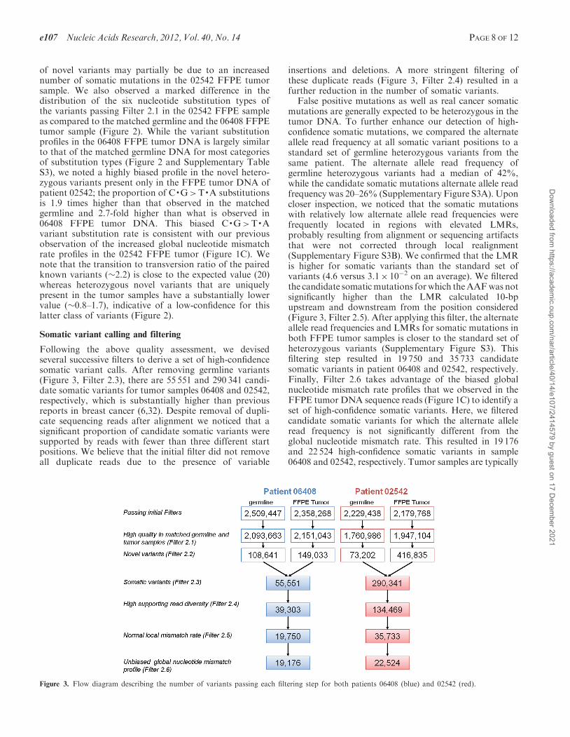
of novel variants may partially be due to an increasednumber of somatic mutations in the 02542 FFPE tumorsample. We also observed a marked difference in thedistribution of the six nucleotide substitution types ofthe variants passing Filter 2.1 in the 02542 FFPE sampleas compared to the matched germline and the 06408 FFPEtumor sample (Figure 2). While the variant substitutionprofiles in the 06408 FFPE tumor DNA is largely similarto that of the matched germline DNA for most categoriesof substitution types (Figure 2 and Supplementary TableS3), we noted a highly biased profile in the novel hetero-zygous variants present only in the FFPE tumor DNA ofpatient 02542; the proportion of C·G>T·A substitutionsis 1.9 times higher than that observed in the matchedgermline and 2.7-fold higher than what is observed in06408 FFPE tumor DNA. This biased C·G>T·Avariant substitution rate is consistent with our previousobservation of the increased global nucleotide mismatchrate profiles in the 02542 FFPE tumor (Figure 1C). Wenote that the transition to transversion ratio of the pairedknown variants (�2.2) is close to the expected value (20)whereas heterozygous novel variants that are uniquelypresent in the tumor samples have a substantially lowervalue (�0.8–1.7), indicative of a low-confidence for thislatter class of variants (Figure 2).
Somatic variant calling and filtering
Following the above quality assessment, we devisedseveral successive filters to derive a set of high-confidencesomatic variant calls. After removing germline variants(Figure 3, Filter 2.3), there are 55 551 and 290 341 candi-date somatic variants for tumor samples 06408 and 02542,respectively, which is substantially higher than previousreports in breast cancer (6,32). Despite removal of dupli-cate sequencing reads after alignment we noticed that asignificant proportion of candidate somatic variants weresupported by reads with fewer than three different startpositions. We believe that the initial filter did not removeall duplicate reads due to the presence of variable
insertions and deletions. A more stringent filtering ofthese duplicate reads (Figure 3, Filter 2.4) resulted in afurther reduction in the number of somatic variants.
False positive mutations as well as real cancer somaticmutations are generally expected to be heterozygous in thetumor DNA. To further enhance our detection of high-confidence somatic mutations, we compared the alternateallele read frequency at all somatic variant positions to astandard set of germline heterozygous variants from thesame patient. The alternate allele read frequency ofgermline heterozygous variants had a median of 42%,while the candidate somatic mutations alternate allele readfrequency was 20–26% (Supplementary Figure S3A). Uponcloser inspection, we noticed that the somatic mutationswith relatively low alternate allele read frequencies werefrequently located in regions with elevated LMRs,probably resulting from alignment or sequencing artifactsthat were not corrected through local realignment(Supplementary Figure S3B). We confirmed that the LMRis higher for somatic variants than the standard set ofvariants (4.6 versus 3.1� 10�2 on an average). We filteredthe candidate somaticmutations forwhich theAAFwas notsignificantly higher than the LMR calculated 10-bpupstream and downstream from the position considered(Figure 3, Filter 2.5). After applying this filter, the alternateallele read frequencies and LMRs for somatic mutations inboth FFPE tumor samples is closer to the standard set ofheterozygous variants (Supplementary Figure S3). Thisfiltering step resulted in 19 750 and 35 733 candidatesomatic variants in patient 06408 and 02542, respectively.Finally, Filter 2.6 takes advantage of the biased globalnucleotide mismatch rate profiles that we observed in theFFPE tumor DNA sequence reads (Figure 1C) to identify aset of high-confidence somatic variants. Here, we filteredcandidate somatic variants for which the alternate alleleread frequency is not significantly different from theglobal nucleotide mismatch rate. This resulted in 19 176and 22 524 high-confidence somatic variants in sample06408 and 02542, respectively. Tumor samples are typically
Figure 3. Flow diagram describing the number of variants passing each filtering step for both patients 06408 (blue) and 02542 (red).
e107 Nucleic Acids Research, 2012, Vol. 40, No. 14 PAGE 8 OF 12
Dow
nloaded from https://academ
ic.oup.com/nar/article/40/14/e107/2414579 by guest on 17 D
ecember 2021

heterogeneous composed of a mixed population of differentclones. Given that the minimum AAF needed to call ahigh-confidence somatic variant after applying Filter 2.6 is�18–21% with the mean around 32–34%, thus in the bestcase scenario we would be able to call a heterozygousmutation found in �50% of tumor cells in a sample withintra-tumor heterogeneity.
Examining the six substitution types (Figure 4), revealsthat this specific filter diminished the C·G>T·A substi-tution bias characteristic of formalin fixation inducedDNA damage and resulted in a distribution of substitu-tion types similar and more balanced for the 02542 FFPEtumor sample. While sample 06408 only had 3% of itscandidate somatic variants filtered by Filter 2.6, 37% ofcandidate somatic variants in sample 02542 were removed(Figure 3). This supports our previous statement thatsample 02542 had greater FFPE induced DNA damagecausing an increase in the number of false positivesomatic variants. On the other hand, both FFPE tumorsamples had >50% of their candidate somatic variantsfiltered by Filter 2.5 which removes false positivevariants caused by sequencing and alignment errors.
To further examine the effects of Filters 2.4, 2.5 and 2.6on the total number of candidate somatic mutations andthe distribution of substitution types, we applied thesefilters in different combinations and determined that allthree filters are necessary (Supplementary Figure S6).These results show the importance of Filters 2.4, 2.5 and2.6 as FFPE tumor samples have increased alignmenterrors compared to matched germline samples mostlikely due to both somatic mutations and formalinfixation induced DNA damage. The succession of filters(2.4–2.6) removed �65% and 92% of the candidatesomatic variants in 06408 and 02542, respectively
(Figure 3). In a recently published framework forsomatic variant calling proposed by the Broad Institute,62% of novel variants were filtered (20). The higherfraction of candidate somatic variants filtered in ourstudy is expected, as our goal is to filter out falsepositive calls due to the formalin fixation induced DNAdamage in both FFPE tumors samples.
Somatic coding variation
The final set of high-confidence somatic mutations con-tained 19 176 and 22 524 variants in tumor samples 06408and 02542, respectively. Of those, 268 and 423 variantswere coding or affect splice sites (Supplementary FigureS5; Supplementary Tables S7 and S8). These numbers arein agreement with previously sequenced whole genomes ofbreast cancer (6,32), which suggests our filtering processhas adequate stringency.We examined457 genes from theCancerGeneCensus (27)
and 1049 genes involved in DNA damage repair for somaticcoding variants. Sample 06408 had 8 high-confidencesomatic mutations in 8 genes (1 nonsense and 7 missense)whereas sample 02542 had 16 high-confidence somaticmutations in 16 genes (1 nonsense, 12 missense and 3coding-synonymous) (Table 2). A number of these changesare of potential biological interest. Both patients carryvariation in TP53: sample 06408 carries a heterozygousnonsense mutation in TP53, suggesting the inactivation ofone copy of this tumor suppressor gene and sample 02542carries a somatic missense mutation. Sample 06408 alsocarries a heterozygous missense mutation in NOTCH1which has been shown to be a recurring mutation inchronic lymphocyte leukaemia, lung squamous cell carcin-oma and breast cancer (5,33,34). The nonsense mutation inTP53 together with the missense mutation in NOTCH1
Figure 4. Filters 2.5 and 2.6 remove false positive somatic variants due to formalin fixation and other systematic and random errors in the process.Shown is the fraction of substitution types for somatic variants after Filter 2.4, after Filter 2.5 and after Filter 2.6 for 06408 and 02542 FFPE tumors.After Filter 2.6 the novel somatic variants of substitution type C·G>T·A called in 02542 tumor have a similar profile to that observed for novelgermline variants in the matched sample (Figure 2).
PAGE 9 OF 12 Nucleic Acids Research, 2012, Vol. 40, No. 14 e107
Dow
nloaded from https://academ
ic.oup.com/nar/article/40/14/e107/2414579 by guest on 17 D
ecember 2021

could be driver mutations for sample 06408’s tumorigenesis.Sample 02542 carries missense mutations in both MLL2and MLL3 which together were recently found as signifi-cantly mutated in 16% of childhood medulloblastomacases (35).
DISCUSSION
Genomic translational research faces a scarcity ofproperly stored and annotated clinical samples. Archivedformalin-fixed tissues in paraffin blocks offer a uniqueopportunity to study thousands of samples with extensiveclinical records and follow-up information. In our study,we show that it is possible to obtain enough DNA from asingle 5 mm FFPE slide (�1–2 cm2) to perform wholegenome sequencing of sufficient coverage depth toidentify potentially important mutations. The FFPEprocess combined with long storage times is known toresult in DNA fragmentation. We show that for the twobreast tumor samples analyzed DNA fragmentation didnot produce large biases in coverage depth distribution(Supplementary Figure S2). However, we observed ahigher global nucleotide mismatch rate within alignedreads from FFPE tumor samples when compared tomatched germline (Figure 1A) and a higher base substitu-tion rate across all 6 different substitution types(Figure 1C). Consistent with damage due to formalinfixation, we observed this increase was biased towardsC·G>T·A mismatches. Interestingly the two samplesstudied were differentially affected by the formalinfixation, tumor 02542 showing a 1.8-fold increase in theglobal nucleotide mismatch rate and greater C·G>T·Abias compared to tumor 06408. This discrepancy can beexplained by the absence of strict standards in theformalin fixation step, where tissue samples are routinely
fixed between 24 and 48 h (11) but sometimes can be fixedfor considerably longer times. The time of the formalinfixation step is not known for the studied samples andnot generally included in pathology reports. Anotherpossible explanation could be the size of the tumortissue, or its density, which also affects the fixation pro-cedure. As formalin fixation-induced DNA damage couldpotentially be so great as to inhibit the ability to analyzean FFPE sample by next generation sequencing we haveestablished a relatively simple test to assess the integrity ofFFPE samples. By simply sequencing from 500 000 to 1million raw reads from a single FFPE tumor, one candetermine the extent of DNA damage and identify thebest preserved samples to conduct larger, more expensivewhole genome sequencing (Figure 1A and SupplementaryFigure S4).
Using a set of innovative filters (Filter 2.4–2.6), weestablish a successful method for filtering false positivesomatic variants caused by the FFPE damage to thetumor DNA, thus increasing our confidence in the finalset of called somatic mutations. It is important to compareour novel filters to existing post-alignment filteringmethods such as GATK (20). Existing methods filter forpoor base quality with a stringent threshold; this is due tothe fact that incorrectly called variants are typically causedby low quality sequence data. The fact that FFPE causesrandom damage, the ‘errors’ do not have poor basequality. Our method filters on the AAF without using athreshold for all substitution types; but rather it uses amismatch error rate across the genome of the givensample. This is important as the amount of FFPE DNAdamage varies from sample to sample. To achieve thesame goal as our novel post-alignment filters, one couldpropose applying more stringent criteria to align the reads.Aligners that trim the reads when their mismatch rate
Table 2. High-confidence FFPE tumor coding somatic variants within cancer associated genes and/or DNA damage repair genes
Patient Gene NCBI ID Chr Position (hg18) Germline Tumor Mutation type Amino acid change
06408 ATRX NM_000489 chrX 76735852 A/A A/C Missense L2027RELN NM_000501 chr7 73109920 G/G G/C Missense A458PKIAA1549 NM_020910 chr7 138253476 T/T T/C Missense Q429RMYH9 NM_002473 chr22 35040266 T/T T/A Missense K475MNOTCH1 NM_017617 chr9 138520141 G/G G/A Missense A1343VNUMA1 NM_006185 chr11 71417948 C/C C/G Missense V27LNUP214 NM_005085 chr9 132998395 A/A A/G Missense D270GTP53 NM_000546 chr17 7517747 G/G G/A Nonsense R306STOP
02542 AKT1 NM_001014431 chr14 104312544 A/A A/G Missense F161LBLM NM_000057 chr15 89105082 T/T T/A Missense F492YCREBBP NM_001079846 chr16 3772787 G/G G/A Missense P453LEXT1 NM_000127 chr8 118886256 G/G G/T Missense D647EGNA11 NM_002067 chr19 3070205 A/A A/G Missense N246SJARID1A NM_001042603 chr12 297581 G/G G/C Missense T950RLPP NM_005578 chr3 190066803 G/G G/T Missense G511VMLL2 NM_003482 chr12 47722022 T/T T/C Missense K2043RMLL3 NM_170606 chr7 151504320 G/G G/C Missense Q3051EPDGFRA NM_006206 chr4 54824777 G/G G/A Missense G185ERET NM_020630 chr10 42921884 G/G G/T Missense G308WRPN1 NM_002950 chr3 129823703 G/G G/T Nonsense C545STOPRUNX1 NM_001001890 chr21 35181094 C/C C/T Coding-synonymous NASTK11 NM_000455 chr19 1171708 G/G G/A Coding-synonymous NATP53 NM_000546 chr17 7519259 C/C C/A Missense K132NZNF521 NM_015461 chr18 21060818 C/C C/G Coding-synonymous NA
e107 Nucleic Acids Research, 2012, Vol. 40, No. 14 PAGE 10 OF 12
Dow
nloaded from https://academ
ic.oup.com/nar/article/40/14/e107/2414579 by guest on 17 D
ecember 2021

becomes too high have been implemented (36,37). As aresult, the global nucleotide mismatch rate wouldimprove, but at the cost of a reduced effective sequencingcoverage depth. Such strategies could also remove bonafide somatic mutations surrounded by extensive DNAdamage therefore limiting the sensitivity to call variants.A second potential alternate approach for achieving a setof high-confidence somatic mutations in FFPE sampleswould be to sequence to greater coverage depth. Sinceformalin fixation is performed on the resected tumorsample and will generally randomly affect differentDNA locations in different cells, elevated global nucleo-tide mismatch rates in DNA sequencing reads should stilllead to accurate variant calls at sufficiently highsequencing coverage depth. In our study, the globalnucleotide mismatch rate was indeed higher than thevariant calling rate, especially in FFPE tumors(18–32� 10�3 versus 10–11� 10�4). In a recent study ofwhole-exome sequencing of FFPE tumors, 40-foldcoverage was insufficient to filter false positives due toformalin fixation DNA damage identified by the substitu-tion profile and discordance with matched frozen tissue(15). Indeed, the authors estimate that 80� coverage isrequired to obtain accurate variant calling in thepresence formalin fixation DNA damage. However, forsamples such as 02542 in our study with substantialamounts of formalin fixation induced DNA damage, thecoverage depths required to overcome the global nucleo-tide mismatch rates in the sequencing reads to achieveaccurate variant calls could be even greater. Thus,applying our series of standard and novel filters willlikely have utility for identifying high-confidence somaticmutations in FFPE tumor samples even when there isrelatively low sequence coverage depth.
In our study, we have not analyzed the tumors forsomatic events such as chromosomal translocations orlarge copy number alterations (CNA). Methods developedfor this purpose (38–40), rely more on the correct mappingof read pairs than accurate sequence. We have onlysequenced single reads, and were thus not able toperform this analysis. We believe that the vast majorityof the reads mapped in our FFPE tumor samples aremapped at the correct location. However, it is possiblethat the sensitivity of translocation or CNA detectionwould be affected as a greater number of reads mighthave ambiguous mappings due to the mismatchesintroduced by the FFPE damage. Various distributionsof insert size in read pairs, especially large ones(1–10 kb) obtained through mate-pair libraries, can alsoimprove the sensitivity of the detection of large deletions.However, the FFPE process fragments the DNA andtherefore would not be adequate for such studies.
Overall, our study demonstrates that a methodical char-acterization and analysis of the sequencing data canreduce the noise resulting from formalin fixation inducedDNA damage and lead to calling a high-confidence set ofsomatic mutations. This opens up the possibility ofsequencing huge archives of stored clinical FFPEsamples of a variety of cancers. Furthermore, we demon-strate that a limited amount of DNA can be used for agenome-wide deep sequencing analysis, which enables
studies on small clusters of tumor cells such residualcancer after treatment or dormant metastases.
SUPPLEMENTARY DATA
Supplementary Data are available at NAR Online:Supplementary Tables 1–8, Supplementary Figures 1–6and Supplementary Programs.
ACKNOWLEDGEMENTS
We would like to thank Kersi Pestonjamasp, Ph.D.,Manager of the UCSD Cancer Center Microscopy Core,who helped isolate cell nuclei from stained FFPE slides forcell counting. We would like to thank Brian Coullahanfrom Life Technologies for support and assistance withlibrary preparation and SOLiD sequencing.
FUNDING
National Center for Research Resources, Center forTranslational Science Award [1UL1RR031980-01];National Cancer Institute [grants 1R21CA152613-01,1R21CA155615-01A1, CA69375]; Safeway Foundation,Breast Cancer Research Foundation and University ofCalifornia Office of the President [grant 6762 Subaward6067sc to Athena Breast Health Network]. Funding foropen access charge: National Institute of Health.
Conflict of interest statement. None declared.
REFERENCES
1. Clark,M.J., Homer,N., O’Connor,B.D., Chen,Z., Eskin,A.,Lee,H., Merriman,B. and Nelson,S.F. (2010) U87MG decoded:the genomic sequence of a cytogenetically aberrant human cancercell line. PLoS Genet, 6, e1000832.
2. Lee,W., Jiang,Z., Liu,J., Haverty,P.M., Guan,Y., Stinson,J.,Yue,P., Zhang,Y., Pant,K.P., Bhatt,D. et al. (2010) The mutationspectrum revealed by paired genome sequences from a lungcancer patient. Nature, 465, 473–477.
3. Ley,T.J., Mardis,E.R., Ding,L., Fulton,B., McLellan,M.D.,Chen,K., Dooling,D., Dunford-Shore,B.H., McGrath,S.,Hickenbotham,M. et al. (2008) DNA sequencing of acytogenetically normal acute myeloid leukaemia genome. Nature,456, 66–72.
4. Pleasance,E.D., Stephens,P.J., O’Meara,S., McBride,D.J.,Meynert,A., Jones,D., Lin,M.L., Beare,D., Lau,K.W.,Greenman,C. et al. (2010) A small-cell lung cancer genome withcomplex signatures of tobacco exposure. Nature, 463, 184–190.
5. Puente,X.S., Pinyol,M., Quesada,V., Conde,L., Ordonez,G.R.,Villamor,N., Escaramis,G., Jares,P., Bea,S., Gonzalez-Dıaz,M.et al. (2011) Whole-genome sequencing identifies recurrentmutations in chronic lymphocytic leukaemia. Nature, 475,101–105.
6. Shah,S.P., Morin,R.D., Khattra,J., Prentice,L., Pugh,T.,Burleigh,A., Delaney,A., Gelmon,K., Guliany,R., Senz,J. et al.(2009) Mutational evolution in a lobular breast tumour profiledat single nucleotide resolution. Nature, 461, 809–813.
7. Pikor,L.A., Enfield,K.S., Cameron,H. and Lam,W.L. (2011) DNAextraction from paraffin embedded material for genetic andepigenetic analyses. J. Vis. Exp., 4, e2763.
8. April,C., Klotzle,B., Royce,T., Wickham-Garcia,E.,Boyaniwsky,T., Izzo,J., Cox,D., Jones,W., Rubio,R., Holton,K.et al. (2009) Whole-genome gene expression profiling of formalin-fixed, paraffin-embedded tissue samples. PLoS One, 4, e8162.
PAGE 11 OF 12 Nucleic Acids Research, 2012, Vol. 40, No. 14 e107
Dow
nloaded from https://academ
ic.oup.com/nar/article/40/14/e107/2414579 by guest on 17 D
ecember 2021

9. Farragher,S.M., Tanney,A., Kennedy,R.D. and Paul Harkin,D.(2008) RNA expression analysis from formalin fixed paraffinembedded tissues. Histochem. Cell Biol., 130, 435–445.
10. Kibriya,M.G., Jasmine,F., Roy,S., Paul-Brutus,R.M., Argos,M.and Ahsan,H. (2010) Analyses and interpretation ofwhole-genome gene expression from formalin-fixedparaffin-embedded tissue: an illustration with breast cancertissues. BMC Genomics, 11, 622.
11. Srinivasan,M., Sedmak,D. and Jewell,S. (2002) Effect of fixativesand tissue processing on the content and integrity of nucleicacids. Am. J. Pathol., 161, 1961–1971.
12. Williams,C., Ponten,F., Moberg,C., Soderkvist,P., Uhlen,M.,Ponten,J., Sitbon,G. and Lundeberg,J. (1999) A high frequency ofsequence alterations is due to formalin fixation of archivalspecimens. Am. J. Pathol., 155, 1467–1471.
13. Ausch,C., Buxhofer-Ausch,V., Oberkanins,C., Holzer,B., Minai-Pour,M., Jahn,S., Dandachi,N., Zeillinger,R. and Kriegshauser,G.(2009) Sensitive detection of KRAS mutations in archivedformalin-fixed paraffin-embedded tissue using mutant-enrichedPCR and reverse-hybridization. J. Mol. Diagn., 11, 508–513.
14. Solassol,J., Ramos,J., Crapez,E., Saifi,M., Mange,A., Vianes,E.,Lamy,P.J., Costes,V. and Maudelonde,T. (2011) KRAS mutationdetection in paired frozen and formalin-fixed paraffin-embedded(FFPE) colorectal cancer tissues. Int. J. Mol. Sci., 12, 3191–3204.
15. Kerick,M., Isau,M., Timmermann,B., Sultmann,H., Herwig,R.,Krobitsch,S., Schaefer,G., Verdorfer,I., Bartsch,G., Klocker,H.et al. (2011) Targeted High Throughput Sequencing in ClinicalCancer Settings: Formaldehyde fixed-paraffin embedded (FFPE)tumor tissues, input amount and tumor heterogeneity. BMC Med.Genomics, 4, 68.
16. Schweiger,M.R., Kerick,M., Timmermann,B., Albrecht,M.W.,Borodina,T., Parkhomchuk,D., Zatloukal,K. and Lehrach,H.(2009) Genome-wide massively parallel sequencing offormaldehyde fixed-paraffin embedded (FFPE) tumor tissues forcopy-number- and mutation-analysis. PLoS One, 4, e5548.
17. Wood,H.M., Belvedere,O., Conway,C., Daly,C., Chalkley,R.,Bickerdike,M., McKinley,C., Egan,P., Ross,L., Hayward,B. et al.(2010) Using next-generation sequencing for high resolutionmultiplex analysis of copy number variation from nanogramquantities of DNA from formalin-fixed paraffin-embeddedspecimens. Nucleic Acids Res., 38, e151.
18. Pierce,J.P., Faerber,S., Wright,F.A., Rock,C.L., Newman,V.,Flatt,S.W., Kealey,S., Jones,V.E., Caan,B.J., Gold,E.B. et al.(2002) A randomized trial of the effect of a plant-based dietarypattern on additional breast cancer events and survival: theWomen’s Healthy Eating and Living (WHEL) Study. ControlClin. Trials, 23, 728–756.
19. Durbin,R.M., Abecasis,G.R., Altshuler,D.L., Auton,A.,Brooks,L.D., Gibbs,R.A., Hurles,M.E. and McVean,G.A. (2010)A map of human genome variation from population-scalesequencing. Nature, 467, 1061–1073.
20. DePristo,M.A., Banks,E., Poplin,R., Garimella,K.V.,Maguire,J.R., Hartl,C., Philippakis,A.A., del Angel,G.,Rivas,M.A., Hanna,M. et al. (2011) A framework for variationdiscovery and genotyping using next-generation DNA sequencingdata. Nat. Genet., 43, 491–498.
21. Homer,N., Merriman,B. and Nelson,S.F. (2009) BFAST: analignment tool for large scale genome resequencing. PLoS One, 4,e7767.
22. Li,R., Li,Y., Fang,X., Yang,H., Wang,J. and Kristiansen,K.(2009) SNP detection for massively parallel whole-genomeresequencing. Genome Res., 19, 1124–1132.
23. Li,H., Handsaker,B., Wysoker,A., Fennell,T., Ruan,J., Homer,N.,Marth,G., Abecasis,G. and Durbin,R. (2009) The SequenceAlignment/Map format and SAMtools. Bioinformatics, 25,2078–2079.
24. Sherry,S.T., Ward,M.H., Kholodov,M., Baker,J., Phan,L.,Smigielski,E.M. and Sirotkin,K. (2001) dbSNP: the NCBIdatabase of genetic variation. Nucleic Acids Res., 29, 308–311.
25. Benjamini,Y. and Hochberg,Y. (1995) Controlling the falsediscovery rate - a practical and powerful approach to multipletesting. J R. Stat. Soc. B. Met., 57, 289–300.
26. Pleasance,E.D., Cheetham,R.K., Stephens,P.J., McBride,D.J.,Humphray,S.J., Greenman,C.D., Varela,I., Lin,M.L.,Ordonez,G.R., Bignell,G.R. et al. (2010) A comprehensivecatalogue of somatic mutations from a human cancer genome.Nature, 463, 191–196.
27. Futreal,P.A., Coin,L., Marshall,M., Down,T., Hubbard,T.,Wooster,R., Rahman,N. and Stratton,M.R. (2004) A census ofhuman cancer genes. Nat. Rev. Cancer, 4, 177–183.
28. Ashburner,M., Ball,C.A., Blake,J.A., Botstein,D., Butler,H.,Cherry,J.M., Davis,A.P., Dolinski,K., Dwight,S.S., Eppig,J.T.et al. (2000) Gene ontology: tool for the unification of biology.The Gene Ontology Consortium. Nat. Genet., 25, 25–29.
29. Belvedere,O., Berri,S., Chalkley,R., Conway,C., Barbone,F.,Pisa,F., MacLennan,K., Daly,C., Alsop,M., Morgan,J. et al.(2012) A computational index derived from whole-genome copynumber analysis is a novel tool for prognosis in early stage lungsquamous cell carcinoma. Genomics, 99, 18–24.
30. Li,H. and Durbin,R. (2009) Fast and accurate short readalignment with Burrows-Wheeler transform. Bioinformatics, 25,1754–1760.
31. Harismendy,O., Ng,P.C., Strausberg,R.L., Wang,X.,Stockwell,T.B., Beeson,K.Y., Schork,N.J., Murray,S.S.,Topol,E.J., Levy,S. et al. (2009) Evaluation of next generationsequencing platforms for population targeted sequencing studies.Genome Biol., 10, R32.
32. Ding,L., Ellis,M.J., Li,S., Larson,D.E., Chen,K., Wallis,J.W.,Harris,C.C., McLellan,M.D., Fulton,R.S., Fulton,L.L. et al.(2010) Genome remodelling in a basal-like breast cancermetastasis and xenograft. Nature, 464, 999–1005.
33. Jiao,X., Wood,L.D., Lindman,M., Jones,S., Buckhaults,P.,Polyak,K., Sukumar,S., Carter,H., Kim,D., Karchin,R. et al.(2012) Somatic mutations in the notch, NF-KB, PIK3CA, andhedgehog pathways in human breast cancers. Genes ChromosomesCancer, 51, 480–489.
34. Wang,N.J., Sanborn,Z., Arnett,K.L., Bayston,L.J., Liao,W.,Proby,C.M., Leigh,I.M., Collisson,E.A., Gordon,P.B., Jakkula,L.et al. (2011) Loss-of-function mutations in Notch receptors incutaneous and lung squamous cell carcinoma. Proc. Natl Acad.Sci. USA, 108, 17761–17766.
35. Parsons,D.W., Li,M., Zhang,X., Jones,S., Leary,R.J., Lin,J.C.,Boca,S.M., Carter,H., Samayoa,J., Bettegowda,C. et al. (2011)The genetic landscape of the childhood cancer medulloblastoma.Science, 331, 435–439.
36. David,M., Dzamba,M., Lister,D., Ilie,L. and Brudno,M. (2011)SHRiMP2: sensitive yet practical SHort Read Mapping.Bioinformatics, 27, 1011–1012.
37. Langmead,B., Trapnell,C., Pop,M. and Salzberg,S.L. (2009)Ultrafast and memory-efficient alignment of short DNAsequences to the human genome. Genome Biol., 10, R25.
38. Medvedev,P., Fiume,M., Dzamba,M., Smith,T. and Brudno,M.(2010) Detecting copy number variation with mated short reads.Genome Res., 20, 1613–1622.
39. Koehler,R., Issac,H., Cloonan,N. and Grimmond,S.M. (2011) Theuniqueome: a mappability resource for short-tag sequencing.Bioinformatics, 27, 272–274.
40. Chen,K., Wallis,J.W., McLellan,M.D., Larson,D.E., Kalicki,J.M.,Pohl,C.S., McGrath,S.D., Wendl,M.C., Zhang,Q., Locke,D.P.et al. (2009) BreakDancer: an algorithm for high-resolutionmapping of genomic structural variation. Nat. Methods, 6,677–681.
e107 Nucleic Acids Research, 2012, Vol. 40, No. 14 PAGE 12 OF 12
Dow
nloaded from https://academ
ic.oup.com/nar/article/40/14/e107/2414579 by guest on 17 D
ecember 2021