identification of cor triatriatum dexter by two-dimensional echocardiography
TRANSCRIPT

In contrast to the 2 patients described above, wbo responded, we have seen 2 other patients who were treated with prednisone after the Fontan procedure and neither responded: one with chylous pleural effu- sions 6 weeks after operation and the other with non- chylous pericardial effusion 12 weeks after surgery.
In summary, our z successfully treated patients show that prednisone can be useful in the manage- ment of chronic pleural effusions in selected cases after modified Fontan procedures.
I. Serratto M. Miiler SA, Tatooies C, Ardekani R. iIemodynamic evuluutiu~i of Fontan operation in tricuspid atresio. Circulation ?976;54:suppl III:III-99- 111-101. 2. Laks H, Milliken JC, Perloff JK, Hellenbrand WE, George BL. Chin A, DiSessa TG, Williams RG. Experience with the Fcmtan procedure. 1 Thoruc Cordivasc Surg 1984;88:939-951. 3. Kreutzer GO, Vargas FJ. Schlichter AJ, Laura JP, Suarez JC, Rodriguez A, Kreutzer EA. Atriopufmonary onastomosis. J Thoroc Cordiovasc Surg 1982; 83:427-436. 4. Lowell JR. Pleural effusions: (I comprehensive review. Baltimore: Universi- ty Park Press, 1977:1x 5. Engle MA, Zabriskie JB, Senterfit LB, Tay DJ, Ebert PA. Jmmunologic ond virologic studies in the postpericardiotomy syndrome [ Pediotr 1975:87:1103- 1108.
Identification of Cor Triatriatum Dexter by Two-Dimensional
Echocardiography
DAVID A. BURTON, MD ALVIN CHIN, MD
PAUL M. WEINBERG, MD JOHN D. PIGOTT, MD
C or triatriatum dexter is an uncommon congenital heart defect that may cause cyanosis in infancy and mimic more complicated forms of congenital heart dis- ease. This lesion occurs when the eustachian valve, or right venous valve of the inferior vena cava, is unusu- ally large and causes obstruction to right ventricular filling. The eustachian valve is frequently seen by 2- dimensional (Z-D] echocardiography and is a normal finding if the valve is smal1.l This report describes the
From the Children’s Hospital of Philadelphia, 34th Street and Civic Center Boulevard, Philadelphia, Pennsylvania 19104. Manuscript reccivcd January 23, 1987; revised manuscript March 9,1987, accepted March 11,1987.
echocardiographic identification of an obstructive eu- stachian valve causing cyanosis.
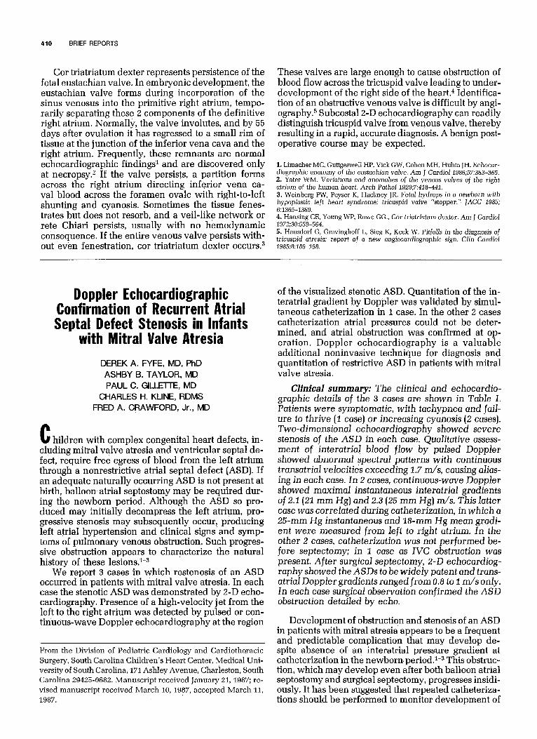
A 1%month-old boy was evaluated for cyanosis. Cardiac catheterization at age I month at another in- stitution was interpreted as showing tricuspid atresia, atrial septal defect and ventricular septal defect. The infant failed to thrive, was cyanotic and was referred to our institution. He weighed 5.7 kg and was mildly cyanotic. The right ventricular impulse was increased. There were no precordial murmurs. An electrocar- diogram revealed slightly diminished right ventricu- lar forces. Chest x-ray showed mild cardiomegaly and normal vascular markings. A 2-D echocardiogram showed a large, discrete venous valve that appeared to separate the right atrium into 2 distinct chambers, es- tablishing the diagnosis of car triatriatum dexter [Fig. 1). A secundum atrial septal defect also was present. The tricuspid valve was normal. Cardiac catheteriza- tion showed a linear density running vertically from the lateral margin of the inferior vena cava identical to the echocardiographic appearance. At surgery, the prominent venous valve was resected and the atrial septal defect was closed. The infant had an uncompli- cated postoperative course.
FIGURE 1. Two-dimensional echocardiogram recorded from the subcostal sagittal view. Left, eustachian valve (arrows), which appears t0 partition the right atrium into 2 separate chambers: the right atrial appendage (RAA) portion, which contains the tricupsid valve (TV) and B venous portion, which receives superior vena cava (SVC), inferlor vena cava (not seen) and coronary sinus (not seen). R@if, tran~~u~~~ tilted slightly to the left separates the eustachian valve from the tricuspid valve. Note eustachian valve obstructing inflow to right v~ntr~ol~ (RV) while permitting caval flow across the atrial septal defect. a = anterior; i = inferior; LA = left atrium; p = ~~stor~~r; nary artery; s = superior.
410 BRIEF REPORTS
Cor triatriatum dexter represents persistence of the fetal eustachian valve. In embryonic development, the eustachian valve forms during incorporation of the sinus venosus into the primitive right atrium, tempo- rarily separating these 2 components of the definitive right atrium. Normally, the valve involutes, and by 55 days after ovulation it has regressed to a small rim of tissue at the junction of the inferior vena cava and the right atrium. Frequently, these remnants are normal echocardiographic finding& and are discovered only at necropsy.z If the valve persists, a partition forms across the right atrium directing inferior vena ca- val blood across the foramen ovale with right-to-left shunting and cyanosis. Sometimes the tissue fenes- trates but does not resorb, and a veil-like network or rete Chiari persists, usually with no hemodynamic consequence. If the entire venous valve persists with- out even fenestration, car triatriatum dexter occurs.3
These valves are large enough to cause obstruction of blood flow across the tricuspid valve leading to under- development of the right side of the hearta Identifica- tion of an obstructive venous valve is difficult by angi- ography.5 Subcostal2-D echocardiography can readily distinguish tricuspid valve from venous valve, thereby resulting in a rapid, accurate diagnosis. A benign post- operative course may be expected.
1. Limacher MC, Guttgeswell HP, Vick GW, Cohen MH, Huhta JH. Echocar- diographic anatomy of the eustachian valve. Am J Cardiol 1986;57:363-365, 2. Yater WM. Variations and anomalies of the venous valves of the right atrium of the human heart. Arch PathoJ 1929:7:418-441. 3. Weinberg PW, Peyser K, Hackney JR. Fetal hydrops in a newborn with hypoplastic left heart syndrome: tricuspid valve “stopper.” JACC 1985; 6236%1369. 4. Hansing CE, Young WP, Rowe GG., Cor triatriatum dexter. Am J Cardiol 1972;30:559-564, 5. Hausdorf G, Gravinghoff L, Sieg K, Keck W. Pitfalls in the diagnosis of tricuspid atresia: report of a new angiocardiographic sign. Clin Cardiol 1985$:189-198.
Doppler Echocardiographic Confirmation of Recurrent Atrial Septal Defect Stenosis in Infants
with Mitral Valve Atrbsia
DEREK A. FYFE, MD, PhD ASHBY B. TAYLOR, MD PAUL C. GILLETTE, MD
CHARLES H. KLINE, RDMS FRED A. CRAWFORD, Jr., MD
C hildren with complex congenital heart defects, in- cluding mitral valve atresia and ventricular septal de- fect, require free egress of blood from the left atrium through a nonrestrictive atria1 septal defect (ASD). If an adequate naturally occurring ASD is not present at birth, balloon atria1 septostomy may be required dur- ing the newborn period. Although the ASD so pro- duced may initially decompress the left atrium, pro- gressive stenosis may subsequently occur, producing left atria1 hypertension and clinical signs and symp- toms of pulmonary venous obstruction. Such progres- sive obstruction appears to characterize the natural history of these lesions.1-3
We report 3 cases in which restenosis of an ASD occurred in patients with mitral valve atresia. In each case the stenotic ASD was demonstrated by 2-D echo- cardiography. Presence of a high-velocity jet from the left to the right atrium was detected by pulsed or con- tinuous-wave Doppler echocardiography at the region
From the Division of Pediatric Cardiology and Cardiothoracic
Surgery, South Carolina Children’s Heart Center, Medical Uni- versity of South Carolina, 171 Ashley Avenue, Charleston, South Carolina 29425-0682. Manuscript received January 21, 1987; re-
vised manuscript received March 10, 1987, accepted March 11,
of the visualized stenotic ASD. Quantitation of the in- teratrial gradient by Doppler was validated by simul- taneous catheterization in 1 case. In the other 2 cases catheterization atria1 pressures could not be deter- mined, and atria1 obstruction was confirmed at op- eration. Doppler echocardiography is a valuable additional noninvasive technique for diagnosis and quantitation of restrictive ASD in patients with mitral valve atresia.
Clinical summary: The clinical and echocardio- graphic details of the 3 cases are shown in Table I. Patients were symptomatic, with tachypnea and fail- ure to thrive (1 case) or increasing cyanosis (2 cases). Two-dimensional echocardiography showed severe stenosis of the ASD in each case. Qualitative assess- ment of interatrial blood flow by pulsed Doppler showed abnormal spectral patterns with continuous transatrial velocities exceeding 1.7 m/s, causing alias- ing in each case. In 2 cases, continuous-wave Doppler showed maximal instantaneous interatrial gradients of 2.2 (21 mm Hg) and 2.3 (25 mm Hg) m/s. This latter case was correlated during catheterization, in which a 25-mm Hg instantaneous and 28-mm Hg mean gradi- ent were measured from left to right atrium. In the other 2 cases, catheterization was not performed be- fore septectomy; in 1 case as IVC obstruction was present. After surgical septectomy, 2-D echocardiog- raphy showed the ASDs to be widely patent and trans- atria1 Doppler gradients ranged from 0.8 to 1 m/s only. In each case surgical observation confirmed the ASD obstruction detailed by echo.
Development of obstruction and stenosis of an ASD in patients with mitral atresia appears to be a frequent and predictable complication that may develop de- spite absence of an interatrial pressure gradient at catheterization in the newborn,period.1-3 This obstruc- tion, which may develop even after both balloon atria1 septostomy and surgical septectomy, progresses insidi- ously. It has been suggested that repeated catheteriza-
1987. tions should be performed to monitor development of