identification of a cis-acting dendritic targeting element ... · protein (egfp) fusion protein...
TRANSCRIPT
Identification of a cis-Acting Dendritic Targeting Element inMAP2 mRNAs
Arne Blichenberg,1 Birgit Schwanke,1 Monika Rehbein,1 Craig C. Garner,2 Dietmar Richter,1 andStefan Kindler1
1Institute for Cell Biochemistry and Clinical Neurobiology, University of Hamburg, D-20246 Hamburg, Germany, and2Department of Neurobiology, University of Alabama at Birmingham, South Birmingham, AL 35213-0021
In neurons, a limited number of mRNAs have been identified indendritic processes, whereas other transcripts are restricted tothe cell soma. Here we have investigated the molecular mech-anisms underlying extrasomatic localization of mRNAs encod-ing microtubule-associated protein 2 (MAP2) in primary neuro-nal cultures. Vectors expressing recombinant mRNAs wereintroduced into hippocampal and sympathetic neurons usingDNA transfection and microinjection protocols, respectively.Chimeric mRNAs containing the entire 39 untranslated region ofMAP2 transcripts fused to a nondendritic reporter mRNA aredetected in dendrites. In contrast, RNAs containing MAP2 cod-ing and 59 untranslated regions or tubulin sequences are re-stricted to the cell soma. Moreover, 640 nucleotides from the
MAP2 39 untranslated region (UTR) are both sufficient andessential for extrasomatic localization of chimeric mRNAs inhippocampal and sympathetic neurons. Thus, a cis-acting den-dritic targeting element that is effective in two distinct neuronalcell types is contained in the 39 UTR of MAP2 transcripts. Theobservation of RNA granules in dendrites implies that extraso-matic transcripts seem to assemble into multimolecular com-plexes that may function as transport units.
Key words: dendritic targeting element; RNA localization intodendrites; subcellular transport; extrasomatic protein synthesis;neuronal cytoskeleton/microtubule-associated protein; primarycultures of neurons; microinjection; transfection
Neurons possess distinct cellular compartments that are highlydiverse with respect to their protein repertoire. Most likely adifferential molecular composition of dendritic microdomains andpostsynaptic structures is not only attributable to selective proteinsorting but also reflects local synthesis of specific proteins (Stew-ard, 1994, 1997; Kindler et al., 1997; Kuhl and Skehel, 1998;Tiedge et al., 1999). This hypothesis first arose with the ultra-structural detection of polyribosomes in dendritic shafts at thebase of dendritic spines (Steward and Levy, 1982; Steward andReeves, 1988) and was subsequently supported by the identifica-tion of distinct mRNAs in dendrites. Dendritic mRNAs encode,among other proteins, the microtubule-associated protein 2(MAP2) (Garner et al., 1988; Bruckenstein et al., 1990; Kleimanet al., 1990), the a subunit of the Ca21/calmodulin-dependentprotein kinase II (a-CaMKII) (Burgin et al., 1990), the productof an activity-regulated gene (arg3.1) (Link et al., 1995), alsodescribed as activity-regulated cytoskeleton-associated protein,arc (Lyford et al., 1995), the neuropeptides vasopressin andoxytocin (Mohr et al., 1995), the inositol 1,4,5-triphosphate re-ceptor type 1 (Furuichi et al., 1993), neurogranin (Landry et al.,1994), amino acid receptors (Miyashiro et al., 1994), the cAMPresponse element binding protein (CREB) (Crino et al., 1998),and dendrin (Herb et al., 1997). The noncoding RNA BC1(Tiedge et al., 1991), ribosomal RNAs (Kleiman et al., 1993), and
tRNAs (Tiedge and Brosius, 1996) are also present in dendrites.Dendritic tran-scripts were detected in synaptosome preparations(Chicurel et al., 1993; Rao and Steward, 1993), and isolateddendrites are capable of protein synthesis (Torre and Steward,1992; Crino and Eberwine, 1996). Moreover, specific forms ofsynaptic plasticity in the rat hippocampus seem to depend ontranslation in dendrites (Kang and Schuman, 1996). These find-ings imply that extrasomatic protein synthesis influences theprotein composition in dendritic compartments and contributesto modulations of synaptic function (for review, see Steward,1994, 1997; Kindler et al., 1997; Kuhl and Skehel, 1998; Tiedge etal., 1999).
Modulation of extrasomatic protein composition and synaptictransmission seems to partially rely on dendritic targeting ofselected transcripts and their regulated decentralized translation.This idea is supported by the observation that arc/arg3.1 mRNAand protein specifically accumulate in dendritic layers in whichsynapses have previously been stimulated (Steward et al., 1998).However, the molecular mechanisms directing selective mRNAtargeting to dendrites and local protein synthesis are poorlyunderstood. In other cell systems, such as Drosophila embryos andXenopus oocytes, regulated interactions between mRNA signalsequences and trans-acting factors govern cytoplasmic transportand site-specific translation of various transcripts (St. Johnston,1995). Our aim was to functionally characterize cis-acting den-dritic targeting sequences in MAP2 mRNAs. This was accom-plished by the expression of chimeric transcripts containing var-ious MAP2 mRNA fragments in two primary neuronal cellsystems and an analysis of their subcellular distribution. A 640nucleotide element contained in the 39 untranslated region (39UTR) of MAP2 transcripts was found to be both sufficient and
Received May 19, 1999; revised July 20, 1999; accepted Aug. 5, 1999.This research was supported by the Deutsche Forschungsgemeinschaft (Ri191-
19-1, Ri192-21-5). This work forms part of a thesis (A.B.).Correspondence should be addressed to Stefan Kindler, Institute for Cell Bio-
chemistry and Clinical Neurobiology, University of Hamburg, D-20246 Hamburg,Germany.Copyright © 1999 Society for Neuroscience 0270-6474/99/198818-12$05.00/0
The Journal of Neuroscience, October 15, 1999, 19(20):8818–8829
essential to mediate efficient dendritic mRNA localization in twodifferent neuronal cell types.
MATERIALS AND METHODSConstruction of eukaryotic expression vectors. The basic vector pNEexpresses a nuclear location signal (NLS)/enhanced green fluorescentprotein (EGFP) fusion protein driven by the b-actin promoter fromchicken. It was derived from the plasmid bact-16 (Fregien and Davidson,1986) provided with additional cloning sites and the GFP cDNA se-quence (pbact-hCGFP; courtesy of A. Matus, Friedrich Miescher Insti-tute, Basel, Switzerland). The NcoI–NotI fragment was replaced by thecorresponding EGFP cDNA fragment from pEGFP-N1 (Clontech Lab-oratories, Palo Alto, CA). A sequence encoding an NLS (MGP-KKKRKVGS) was introduced at the NcoI site upstream of the EGFPcoding region using two oligonucleotides (59-CATGGGGCCCAAG-AAGAAACGCAAAGTGGGAAG-39 and 59-CATGCTTCCCACTT-TGCGTTTCTTCTTGGGCCC-39).
Vectors that express EGFP-encoding mRNAs fused to parts of MAP2mRNAs contain the following cDNA sequences (GenBank/EMBL DataBank accession numbers U30937, X51842, and U30938): pNEc (nucleo-tides 60–5549, X51842); pNEcu (198–367, U30937; 60–5552, X51842;4 –176, U30938); pN Eu (5383–5552, X51842; 4 –3720, U30938);pNEuD2436–3071 (5383–5552, X51842; 4–2435 and 3072–3720, U30938); thenumbers in pNEu181–1963, pNEu1586–3274, pNEu2804–3728, pNEu1586–2435,pNEu2432–3274, pNEu2432–2807, pNEu2804–3274, pNEu2432–3071, pNEu2632–3274, and pNEu2632–3071 refer to the 59 and 39 ends of the inserted MAP2cDNA fragments, respectively, with nucleotide numbering according to the39 UTR sequence (U30938); a-tubulin vector pNEtub (GenBank/EMBLData Bank accession number V01227): nucleotides 1–1617.
Preparation and transfection of primary hippocampal neurons. Culturesof hippocampal neurons were prepared from embryonic day 21 (E21) ratembryos as described (Goslin and Banker, 1991). Briefly, cells weregrown at a density of 400 cells/mm 2 on 18 mm poly-L-lysine (Sigma-Aldrich Chemie, Deisenhofen, Germany)-coated glass coverslips facedown above a confluent layer of glial cells in serum-free medium with N2supplements (Goslin and Banker, 1991) at 37°C and 5% CO2. One dayafter plating, the cells were transfected. Thirty minutes before transfec-tion, the coverslips were transferred into fresh N2 medium containing 1%(v/v) fetal calf serum (FCS) with cells facing up. Fifteen micrograms ofpUC19 carrier DNA prepared over two CsCl gradients (Sambrook et al.,1989) and 5 mg of vector DNA (purified over affinity columns; Qiagen,Hilden, Germany) were mixed in a final volume of 100 ml in 0.25 M
CaCl2. One hundred microliters of 2 3 BBS (50 mM BES, 280 mM NaCl,1.5 mM Na2HPO4, pH 6.96) were slowly added while air was continuouslyblown into the solution with a Pasteur pipette. Twenty minutes later, themixture was distributed dropwise onto the cells (100 ml per coverslip)while the dish was gently swirled. After 6 hr at 37°C, 5% CO2, cells werewashed twice with HBSS (Gibco BRL/Life Technologies, Eggenstein,Germany) buffered with 10 mM HEPES, pH 7.4, and placed back ontothe glial cells. After 14–18 d, neurons were analyzed by in situ hybrid-ization or immunocytochemistry. Transfection rates, as judged by theamount of cells exhibiting a positive in situ hybridization signal, weretypically between 0.1 and 0.5%.
Preparation and microinjection of primary sympathetic neurons. Primarycultures of superior cervical ganglia (SCG) neurons were generated fromE21 rat embryos as described by Higgins et al. (1991). Cells were seededon poly-D-lysine (Sigma-Aldrich Chemie) and laminin (Gibco BRL/LifeTechnologies)-coated coverslips at a density of 5–10 cells/mm 2 in serum-free medium. Three days after plating, dendritic growth was induced bysupplementing the medium with matrigel (75 mg/ml; Roche Diagnostics,Heidelberg, Germany). Non-neuronal cell proliferation was discouragedby adding cytosine b-D-arabinofuranoside (2 mM; Sigma-Aldrich Che-mie). After 14–21 d in culture, differentiated neurons were injected withvector DNA at a concentration of 200 ng/ml using the Eppendorf micro-injector 5242 (Eppendorf, Hamburg, Germany). Cells were placed intoLeibovitz L-15 medium (Gibco BRL/Life Technologies) and viewedwith an Axiovert 135 microscope (Zeiss, Oberkochen, Germany) at 4003magnification. During the injection of nuclei with Femto-tips (Eppen-dorf), the pressure was constantly kept at about 100 hPa. The injectiontime was restricted to 1 hr per coverslip. Afterward the cells wereincubated for 4 hr at 37°C, 5% CO2 in normal SCG cell culture medium,fixed, and analyzed by in situ hybridization or immunocytochemistry.
In situ hybridization analysis. The KpnI–XbaI EGFP cDNA fragmentfrom pEGFP-N1 (Clontech Laboratories) was subcloned intopBluescript-SKII(2) (Stratagene, Heidelberg, Germany). The resultingplasmid pBS-EGFP was linearized with SmaI to transcribe a digoxigenin-labeled antisense EGFP RNA probe with T3 RNA polymerase accordingto the manufacturer’s description (Roche Diagnostics, Mannheim, Ger-many). For in situ hybridization, cells were briefly washed with PBS (5mM NaH2PO4, 5 mM Na2HPO4, 2.7 mM KCl, 137 mM NaCl, pH 7.4)containing 4% (w/v) sucrose and fixed for 15 min in 4% (w/v) parafor-maldehyde, 2 mM MgCl2, 5 mM EGTA, and 4% (w/v) sucrose in PBS atroom temperature, followed by washing in PBS containing 4% (w/v)sucrose (three times, 5 min each). Subsequently, coverslips were irradi-ated in a UV cross-linker (Stratagene) set to 120 mJ. Cells were perme-abilized for 3 min in PBS containing 0.1% (v/v) Triton X-100 and washedthree times for five min each in PBS containing 2 mM MgCl2. Air-driedneurons were prehybridized for 2 hr at 50°C in 50% (v/v) deionizedformamide, 5 3 SSC, 5 3 Denhardt’s solution, 0.2% (w/v) SDS, 50 mg/mlheparin, 100 mg/ml poly(A) homopolymer, 250 mg/ml denatured herringsperm DNA, and 250 mg/ml yeast tRNA. Hybridization was performedovernight at 50°C in the same solution but included ;500 ng/ml of the invitro synthesized digoxigenin-labeled riboprobe. Coverslips were washedtwice in 1 3 SSC and 0.1% (w/v) SDS at room temperature for 5 mineach, and then in 0.2 3 SSC, 0.1% (w/v) SDS at 65°C (twice, 10 mineach). The hybridized probe was detected immunocytochemically usinga sheep anti-digoxigenin antibody coupled to alkaline phosphate (RocheDiagnostics) and nitro blue tetrazolium and 5-bromo-4-chloro-3-indolylphosphate as a substrate according to the protocol provided by themanufacturer. Cells were photographed on a Leitz Aristoplan micro-scope (Ernst Leitz Wetzlar, Wetzlar, Germany). To evaluate the maxi-mal dendritic transport length of mRNA granules, pictures taken with avideo camera were analyzed using the NIH image software (developedat National Institutes of Health, Bethesda, MD, and available on theInternet at http://rsb.info.nih.gov/nih-image/). For each transfected neu-ron the distance between the cell body and the distal-most dendriticsignal was measured using the program’s segmented line tool.
Immunofluorescence microscopy of primary neuronal cultures. Hip-pocampal and sympathetic neurons were prepared and grown on cover-slips as described above, washed in a physiological salt solution [0.9%(w/v) NaCl, 100 mM sodium phosphate buffer, pH 7.4], fixed with 4%paraformaldehyde in high-salt PBS (450 mM NaCl, 20 mM sodium phos-phate buffer, pH 7.4) at room temperature for 15 min, and blocked with5% (v/v) FCS in high-salt PBS for 30 min. For immunofluorescencemicroscopy, coverslips were incubated overnight with monoclonal anti-bodies against MAP2 (1:200 dilution; Chemicon International, Te-mecula, CA), tau (1:200 dilution; Roche Diagnostics) or EGFP (1:500dilution; Clontech Laboratories) at 4°C in 2% (v/v) FCS in high-saltPBS. After three washes in high-salt PBS, coverslips were incubatedovernight at 4°C with goat anti-mouse IgG antibodies conjugated witheither fluorescein, Cy3, or Cy2 (Jackson ImmunoResearch Laboratories,West Grove, PA). Secondary antibodies were diluted 1:100 in 2% (v/v)FCS in high-salt PBS. After three washes in high-salt PBS, cells weremounted and photographed as described above, or a Zeiss laser-scanningmicroscope (Zeiss) was used.
RESULTSPolar differentiation of cultured sympathetic andhippocampal neuronsFor the characterization of molecular determinants involved indendritic mRNA targeting, primary neurons derived from em-bryonic rat hippocampi and superior cervical ganglia were grownin culture. Phase-contrast micrographs presented in Figure 1A,Bshow that the given cell culture conditions promote a polardifferentiation of both sympathetic and hippocampal neurons aswell as the formation of an extended network of processes.Visualization of a recombinant EGFP in individual neuronsindicates that hippocampal cells form a slightly more complexneurite network than sympathetic neurons (Fig. 1C,D). The na-ture of these cell processes was further determined by immuno-cytochemical staining of cultures with antibodies against thedendritic and axonal marker proteins MAP2 and tau, respec-tively. MAP2 antibodies lead to a weak staining of neuronal cell
Blichenberg et al. • Dendritic Targeting Element in MAP2 mRNAs J. Neurosci., October 15, 1999, 19(20):8818–8829 8819
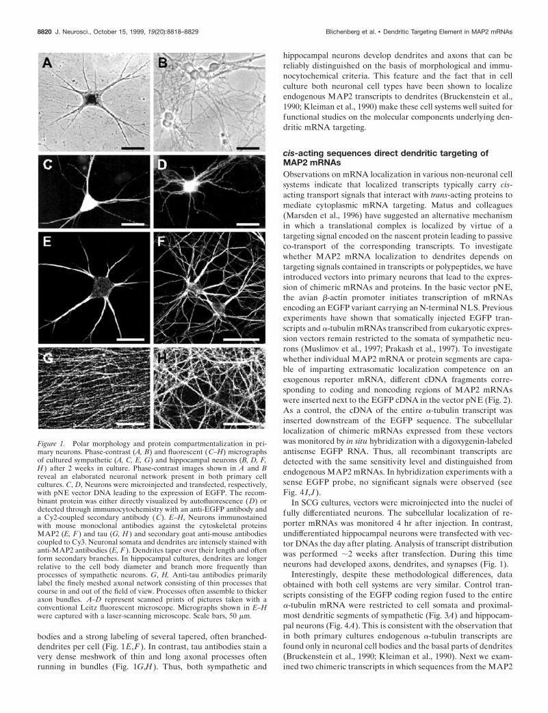
bodies and a strong labeling of several tapered, often branched-dendrites per cell (Fig. 1E,F). In contrast, tau antibodies stain avery dense meshwork of thin and long axonal processes oftenrunning in bundles (Fig. 1G,H). Thus, both sympathetic and
hippocampal neurons develop dendrites and axons that can bereliably distinguished on the basis of morphological and immu-nocytochemical criteria. This feature and the fact that in cellculture both neuronal cell types have been shown to localizeendogenous MAP2 transcripts to dendrites (Bruckenstein et al.,1990; Kleiman et al., 1990) make these cell systems well suited forfunctional studies on the molecular components underlying den-dritic mRNA targeting.
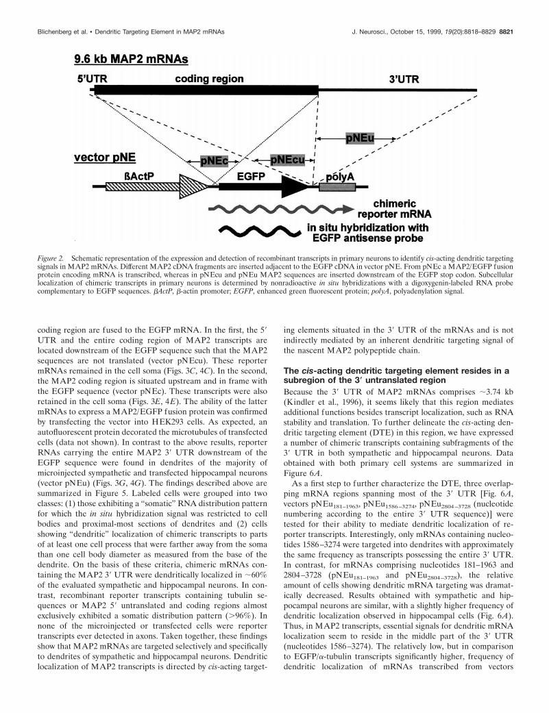
cis-acting sequences direct dendritic targeting ofMAP2 mRNAsObservations on mRNA localization in various non-neuronal cellsystems indicate that localized transcripts typically carry cis-acting transport signals that interact with trans-acting proteins tomediate cytoplasmic mRNA targeting. Matus and colleagues(Marsden et al., 1996) have suggested an alternative mechanismin which a translational complex is localized by virtue of atargeting signal encoded on the nascent protein leading to passiveco-transport of the corresponding transcripts. To investigatewhether MAP2 mRNA localization to dendrites depends ontargeting signals contained in transcripts or polypeptides, we haveintroduced vectors into primary neurons that lead to the expres-sion of chimeric mRNAs and proteins. In the basic vector pNE,the avian b-actin promoter initiates transcription of mRNAsencoding an EGFP variant carrying an N-terminal NLS. Previousexperiments have shown that somatically injected EGFP tran-scripts and a-tubulin mRNAs transcribed from eukaryotic expres-sion vectors remain restricted to the somata of sympathetic neu-rons (Muslimov et al., 1997; Prakash et al., 1997). To investigatewhether individual MAP2 mRNA or protein segments are capa-ble of imparting extrasomatic localization competence on anexogenous reporter mRNA, different cDNA fragments corre-sponding to coding and noncoding regions of MAP2 mRNAswere inserted next to the EGFP cDNA in the vector pNE (Fig. 2).As a control, the cDNA of the entire a-tubulin transcript wasinserted downstream of the EGFP sequence. The subcellularlocalization of chimeric mRNAs expressed from these vectorswas monitored by in situ hybridization with a digoxygenin-labeledantisense EGFP RNA. Thus, all recombinant transcripts aredetected with the same sensitivity level and distinguished fromendogenous MAP2 mRNAs. In hybridization experiments with asense EGFP probe, no significant signals were observed (seeFig. 4 I, J ).
In SCG cultures, vectors were microinjected into the nuclei offully differentiated neurons. The subcellular localization of re-porter mRNAs was monitored 4 hr after injection. In contrast,undifferentiated hippocampal neurons were transfected with vec-tor DNAs the day after plating. Analysis of transcript distributionwas performed ;2 weeks after transfection. During this timeneurons had developed axons, dendrites, and synapses (Fig. 1).
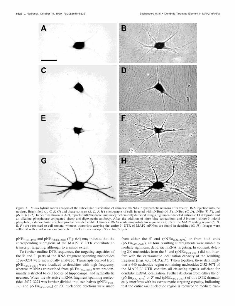
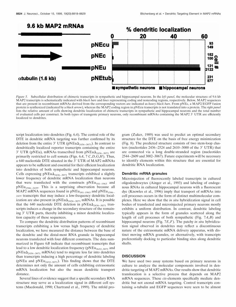
Interestingly, despite these methodological differences, dataobtained with both cell systems are very similar. Control tran-scripts consisting of the EGFP coding region fused to the entirea-tubulin mRNA were restricted to cell somata and proximal-most dendritic segments of sympathetic (Fig. 3A) and hippocam-pal neurons (Fig. 4A). This is consistent with the observation thatin both primary cultures endogenous a-tubulin transcripts arefound only in neuronal cell bodies and the basal parts of dendrites(Bruckenstein et al., 1990; Kleiman et al., 1990). Next we exam-ined two chimeric transcripts in which sequences from the MAP2
Figure 1. Polar morphology and protein compartmentalization in pri-mary neurons. Phase-contrast (A, B) and fluorescent ( C–H) micrographsof cultured sympathetic (A, C, E, G) and hippocampal neurons (B, D, F,H ) after 2 weeks in culture. Phase-contrast images shown in A and Breveal an elaborated neuronal network present in both primary cellcultures. C, D, Neurons were microinjected and transfected, respectively,with pNE vector DNA leading to the expression of EGFP. The recom-binant protein was either directly visualized by autofluorescence (D) ordetected through immunocytochemistry with an anti-EGFP antibody anda Cy2-coupled secondary antibody ( C). E–H, Neurons immunostainedwith mouse monoclonal antibodies against the cytoskeletal proteinsMAP2 (E, F ) and tau (G, H ) and secondary goat anti-mouse antibodiescoupled to Cy3. Neuronal somata and dendrites are intensely stained withanti-MAP2 antibodies (E, F ). Dendrites taper over their length and oftenform secondary branches. In hippocampal cultures, dendrites are longerrelative to the cell body diameter and branch more frequently thanprocesses of sympathetic neurons. G, H, Anti-tau antibodies primarilylabel the finely meshed axonal network consisting of thin processes thatcourse in and out of the field of view. Processes often assemble to thickeraxon bundles. A–D represent scanned prints of pictures taken with aconventional Leitz fluorescent microscope. Micrographs shown in E–Hwere captured with a laser-scanning microscope. Scale bars, 50 mm.
8820 J. Neurosci., October 15, 1999, 19(20):8818–8829 Blichenberg et al. • Dendritic Targeting Element in MAP2 mRNAs
coding region are fused to the EGFP mRNA. In the first, the 59UTR and the entire coding region of MAP2 transcripts arelocated downstream of the EGFP sequence such that the MAP2sequences are not translated (vector pNEcu). These reportermRNAs remained in the cell soma (Figs. 3C, 4C). In the second,the MAP2 coding region is situated upstream and in frame withthe EGFP sequence (vector pNEc). These transcripts were alsoretained in the cell soma (Figs. 3E, 4E). The ability of the lattermRNAs to express a MAP2/EGFP fusion protein was confirmedby transfecting the vector into HEK293 cells. As expected, anautofluorescent protein decorated the microtubules of transfectedcells (data not shown). In contrast to the above results, reporterRNAs carrying the entire MAP2 39 UTR downstream of theEGFP sequence were found in dendrites of the majority ofmicroinjected sympathetic and transfected hippocampal neurons(vector pNEu) (Figs. 3G, 4G). The findings described above aresummarized in Figure 5. Labeled cells were grouped into twoclasses: (1) those exhibiting a “somatic” RNA distribution patternfor which the in situ hybridization signal was restricted to cellbodies and proximal-most sections of dendrites and (2) cellsshowing “dendritic” localization of chimeric transcripts to partsof at least one cell process that were farther away from the somathan one cell body diameter as measured from the base of thedendrite. On the basis of these criteria, chimeric mRNAs con-taining the MAP2 39 UTR were dendritically localized in ;60%of the evaluated sympathetic and hippocampal neurons. In con-trast, recombinant reporter transcripts containing tubulin se-quences or MAP2 59 untranslated and coding regions almostexclusively exhibited a somatic distribution pattern (.96%). Innone of the microinjected or transfected cells were reportertranscripts ever detected in axons. Taken together, these findingsshow that MAP2 mRNAs are targeted selectively and specificallyto dendrites of sympathetic and hippocampal neurons. Dendriticlocalization of MAP2 transcripts is directed by cis-acting target-
ing elements situated in the 39 UTR of the mRNAs and is notindirectly mediated by an inherent dendritic targeting signal ofthe nascent MAP2 polypeptide chain.
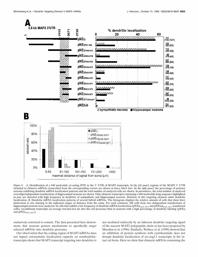
The cis-acting dendritic targeting element resides in asubregion of the 3* untranslated regionBecause the 39 UTR of MAP2 mRNAs comprises ;3.74 kb(Kindler et al., 1996), it seems likely that this region mediatesadditional functions besides transcript localization, such as RNAstability and translation. To further delineate the cis-acting den-dritic targeting element (DTE) in this region, we have expresseda number of chimeric transcripts containing subfragments of the39 UTR in both sympathetic and hippocampal neurons. Dataobtained with both primary cell systems are summarized inFigure 6A.
As a first step to further characterize the DTE, three overlap-ping mRNA regions spanning most of the 39 UTR [Fig. 6A,vectors pNEu181–1963, pNEu1586–3274, pNEu2804–3728 (nucleotidenumbering according to the entire 39 UTR sequence)] weretested for their ability to mediate dendritic localization of re-porter transcripts. Interestingly, only mRNAs containing nucleo-tides 1586–3274 were targeted into dendrites with approximatelythe same frequency as transcripts possessing the entire 39 UTR.In contrast, for mRNAs comprising nucleotides 181–1963 and2804–3728 (pNEu181–1963 and pNEu2804–3728), the relativeamount of cells showing dendritic mRNA targeting was dramat-ically decreased. Results obtained with sympathetic and hip-pocampal neurons are similar, with a slightly higher frequency ofdendritic localization observed in hippocampal cells (Fig. 6A).Thus, in MAP2 transcripts, essential signals for dendritic mRNAlocalization seem to reside in the middle part of the 39 UTR(nucleotides 1586–3274). The relatively low, but in comparisonto EGFP/a-tubulin transcripts significantly higher, frequency ofdendritic localization of mRNAs transcribed from vectors
Figure 2. Schematic representation of the expression and detection of recombinant transcripts in primary neurons to identify cis-acting dendritic targetingsignals in MAP2 mRNAs. Different MAP2 cDNA fragments are inserted adjacent to the EGFP cDNA in vector pNE. From pNEc a MAP2/EGFP fusionprotein encoding mRNA is transcribed, whereas in pNEcu and pNEu MAP2 sequences are inserted downstream of the EGFP stop codon. Subcellularlocalization of chimeric transcripts in primary neurons is determined by nonradioactive in situ hybridizations with a digoxygenin-labeled RNA probecomplementary to EGFP sequences. bActP, b-actin promoter; EGFP, enhanced green fluorescent protein; polyA, polyadenylation signal.
Blichenberg et al. • Dendritic Targeting Element in MAP2 mRNAs J. Neurosci., October 15, 1999, 19(20):8818–8829 8821
pNEu181–1963 and pNEu2804–3728 (Fig. 6A) may indicate that thecorresponding subregions of the MAP2 39 UTR contribute totranscript targeting, although to a minor extent.
To further outline DTE sequences, the targeting capacities ofthe 59 and 39 parts of the RNA fragment spanning nucleotides1586–3274 were individually analyzed. Transcripts derived frompNEu2432–3274 were localized to dendrites with high frequency,whereas mRNAs transcribed from pNEu1586–2435 were predom-inantly restricted to cell bodies of hippocampal and sympatheticneurons. When the cis-active mRNA fragment spanning nucleo-tides 2432–3274 was further divided into two halves (pNEu2432–
2807 and pNEu2804–3274) or 200 nucleotide deletions were made
from either the 59 end (pNEu2632–3274) or from both ends(pNEu2632–3071), all four resulting subfragments were unable tomediate significant dendritic mRNA targeting. In contrast, delet-ing 200 nucleotides from the 39 end (pNEu2432–3071) did not inter-fere with the extrasomatic localization capacity of the resultingfragment (Figs. 6A, 7A,B,E,F). Taken together, these data implythat a 640 nucleotide region containing nucleotides 2432–3071 ofthe MAP2 39 UTR contains all cis-acting signals sufficient fordendritic mRNA localization. Further deletions from either the 59(pNEu2632–3071) or 39 end (pNEu2432–2807) of this DTE dramati-cally interferes with its extrasomatic targeting capacity, indicatingthat the entire 640 nucleotide region is required to mediate tran-
Figure 3. In situ hybridization analysis of the subcellular distribution of chimeric mRNAs in sympathetic neurons after vector DNA injection into thenucleus. Bright-field (A, C, E, G) and phase-contrast (B, D, F, H ) micrographs of cells injected with pNEtub (A, B), pNEcu (C, D), pNEc (E, F ), andpNEu (G, H ). In neurons shown in A–H, reporter mRNAs were immunocytochemically detected using a digoxigenin-labeled antisense EGFP probe andan alkaline phosphatase-conjugated sheep anti-digoxigenin antibody. After the addition of nitro blue tetrazolium and 5-bromo-4-chloro-3-indolylphosphate, a dark-colored reaction product was detectable. Chimeric RNAs containing a-tubulin sequences (A, B) or the MAP2 coding region (C, D,E, F ) are restricted to cell somata, whereas transcripts carrying the entire 39 UTR of MAP2 mRNAs are found in dendrites (G, H ). Images werecollected with a video camera connected to a Leitz microscope. Scale bar, 50 mm.
8822 J. Neurosci., October 15, 1999, 19(20):8818–8829 Blichenberg et al. • Dendritic Targeting Element in MAP2 mRNAs
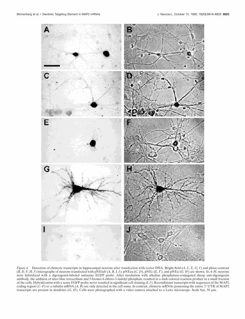
Figure 4. Detection of chimeric transcripts in hippocampal neurons after transfection with vector DNA. Bright-field (A, C, E, G, I ) and phase-contrast(B, D, F, H, J ) micrographs of neurons transfected with pNEtub (A, B, I, J ), pNEcu (C, D), pNEc (E, F ), and pNEu (G, H ) are shown. In A–H, neuronswere hybridized with a digoxigenin-labeled antisense EGFP probe. After incubation with alkaline phosphatase-conjugated sheep anti-digoxigeninantibody, the addition of nitro blue tetrazolium and 5-bromo-4-chloro-3-indolyl phosphate resulted in a dark-colored reaction product in a small fractionof the cells. Hybridization with a sense EGFP probe never resulted in significant cell staining (I, J ). Recombinant transcripts with sequences of the MAP2coding region (C–F) or a-tubulin mRNA (A, B) are only detected in the cell soma. In contrast, chimeric mRNAs possessing the entire 39 UTR of MAP2transcripts are present in dendrites (G, H ). Cells were photographed with a video camera attached to a Leitz microscope. Scale bar, 50 mm.
Blichenberg et al. • Dendritic Targeting Element in MAP2 mRNAs J. Neurosci., October 15, 1999, 19(20):8818–8829 8823
script localization into dendrites (Fig. 6A). The central role of theDTE in dendritic mRNA targeting was further confirmed by itsdeletion from the entire 39 UTR (pNEuD2436–3071). In contrast todendritically localized reporter transcripts containing the entire39 UTR (pNEu), mRNAs transcribed from pNEuD2436–3071 areprimarily restricted to cell somata (Figs. 6A, 7 C,D,G,H). Thus,a 640 nucleotide DTE situated in the 39 UTR of MAP2 mRNAsappears to be sufficient and essential for their efficient localizationinto dendrites of both sympathetic and hippocampal neurons.Cells expressing pNEuD2436–3071 transcripts exhibited a slightlylower frequency of dendritic mRNA localization than neuronsthat were transfected with the constructs pNEu181–1963 andpNEu1586–2435. This is a surprising observation because allMAP2 mRNA sequences found in pNEu181–1963 and pNEu1586–
2435 transcripts that may induce a low-frequency dendritic local-ization are also present in pNEuD2436–3071 mRNAs. It is possiblethat the 640 nucleotide DTE deletion in pNEuD2436–3071 tran-scripts induces a change in the secondary structure of the remain-ing 39 UTR parts, thereby inhibiting a minor dendritic localiza-tion capacity of these sequences.
To compare the dendritic distribution patterns of recombinanttranscripts exhibiting a low versus high frequency of dendriticlocalization, we have measured the distance between the base ofthe dendrite and the distal-most RNA granule in hippocampalneurons transfected with four different constructs. The data sum-marized in Figure 6B indicate that recombinant transcripts thatlead to a low dendritic localization frequency (pNEu2632–3071 andpNEuD2436–3071 mRNAs) tend to migrate less far into dendritesthan transcripts inducing a high percentage of dendritic labeling(pNEu and pNEu2432–3071). This finding shows that the DTEdetermines not only the amount of cells exhibiting extrasomaticmRNA localization but also the mean dendritic transportdistance.
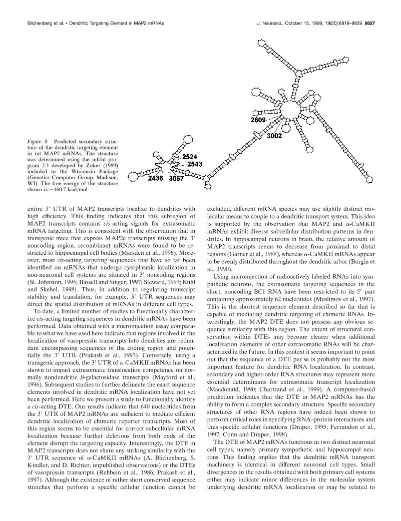
Several lines of evidence suggest that a specific secondary RNAstructure may serve as a localization signal in different cell sys-tems (Macdonald, 1990; Chartrand et al., 1999). The mfold pro-
gram (Zuker, 1989) was used to predict an optimal secondarystructure for the DTE on the basis of free energy minimization(Fig. 8). The predicted structure consists of two stem-loop clus-ters (nucleotides 2436–2524 and 2610–3000 of the 39 UTR) thatare connected via a long double-stranded region (nucleotides2544–2609 and 3002–3067). Future experiments will be necessaryto identify elements within this structure that are essential fordendritic RNA localization.
Dendritic mRNA granulesMicroinjection of fluorescently labeled transcripts in culturedoligodendrocytes (Ainger et al., 1993) and labeling of endoge-nous RNAs in cultured hippocampal neurons with a fluorescentdye (Knowles et al., 1996) imply that transport of mRNAs intocell processes occurs in the form of distinct multimolecular com-plexes. Here we show that the in situ hybridization signal in cellbodies of transfected and microinjected primary neurons mostlyexhibits a uniform distribution. In contrast, dendritic labelingtypically appears in the form of granules scattered along thelength of cell processes of both sympathetic (Fig. 7A,B) andhippocampal neurons (Fig. 7E,F). The patchy in situ hybridiza-tion signal observed in dendrites may reflect a discontinuousnature of the extrasomatic mRNA delivery apparatus, with dis-tinct moving mRNA granules, or alternatively, with transcriptspreferentially docking to particular binding sites along dendriticshafts.
DISCUSSIONWe have used two assay systems based on primary neurons inculture to analyze the molecular components involved in den-dritic targeting of MAP2 mRNAs. Our results show that dendritictranslocation is a selective process that depends on MAP2mRNA sequences. These cis-elements specifically mediate den-dritic but not axonal mRNA targeting. Control transcripts con-taining a-tubulin and EGFP sequences were seen to be almost
Figure 5. Subcellular distribution of chimeric transcripts in sympathetic and hippocampal neurons. In the lef t panel, the molecular structure of 9.6 kbMAP2 transcripts is schematically indicated with black bars and lines representing coding and noncoding regions, respectively. Below, MAP2 sequencesthat are present in recombinant mRNAs derived from the corresponding vectors are indicated as heavy black bars. From pNEc, a MAP2/EGFP fusionprotein is synthesized (indicated by a black arrow), whereas the MAP2 coding region in pNEcu transcripts is not translated into a protein. The right panellists the relative amount of cells showing dendritic localization of chimeric transcripts in sympathetic and hippocampal neurons and the total numberof evaluated cells per construct. In both types of transgenic primary neurons, only recombinant mRNAs containing the MAP2 39 UTR are efficientlylocalized to dendrites.
8824 J. Neurosci., October 15, 1999, 19(20):8818–8829 Blichenberg et al. • Dendritic Targeting Element in MAP2 mRNAs
exclusively restricted to somata. The data presented here demon-strate that neurons possess mechanisms to specifically targetselected mRNAs into dendritic processes.
Our observation that the coding region of MAP2 mRNAs doesnot impart extrasomatic localization capacity on nondendritic-transcripts shows that MAP2 transcript targeting into dendrites is
not mediated indirectly by an inherent dendritic targeting signalof the nascent MAP2 polypeptide chain as has been proposed byMarsden et al. (1996). Similarly, Wallace et al. (1998) showed thatan inhibition of protein synthesis with cycloheximide does notdisrupt dendritic localization of arc/arg3.1 transcripts in the in-tact rat brain. Here we show that chimeric mRNAs containing the
Figure 6. A, Identification of a 640 nucleotide cis-acting DTE in the 39 UTR of MAP2 transcripts. In the lef t panel, regions of the MAP2 39 UTRincluded in chimeric mRNAs transcribed from the corresponding vectors are shown as heavy black bars. In the right panel, the percentage of primaryneurons exhibiting dendritic mRNA localization patterns and the total number of analyzed cells are shown. In parentheses, the total number of analyzedcoverslips/independent transfections of hippocampal neurons are shown. Only chimeric transcripts containing a 640-nucleotide-long sequence highlightedin gray are detected with high frequency in dendrites of sympathetic and hippocampal neurons. Deletion of this targeting element omits dendriticlocalization. B, Dendritic mRNA localization patterns of several hybrid mRNAs. The histogram displays the relative amount of cells that show theirdistal-most in situ staining in the indicated ranges of distance from the soma. For each construct, 200 cells from two independent transfections ofhippocampal neurons were analyzed. In cells that exhibit a low frequency of dendritic mRNA localization (pNEu2632–3071 and pNEuD2436–3071 transfectedcells), recombinant transcripts on average traveled less far into the cell processes than in neurons with a high percentage of dendritic labeling (pNEuand pNEu2432–3071).
Blichenberg et al. • Dendritic Targeting Element in MAP2 mRNAs J. Neurosci., October 15, 1999, 19(20):8818–8829 8825
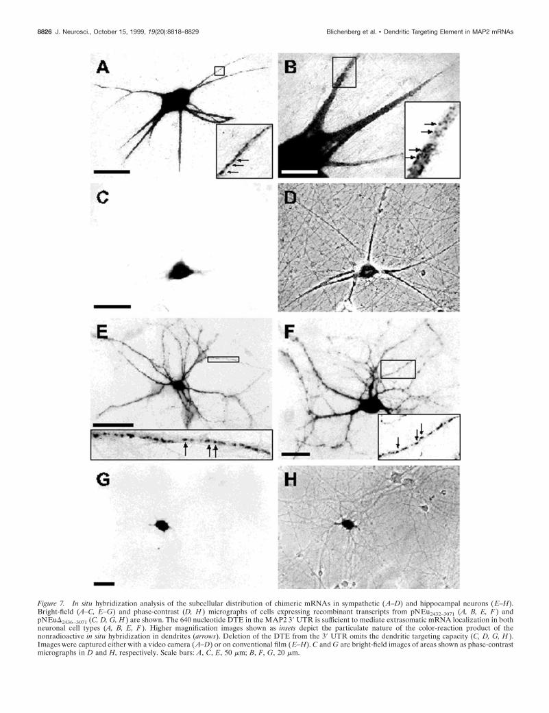
Figure 7. In situ hybridization analysis of the subcellular distribution of chimeric mRNAs in sympathetic ( A–D) and hippocampal neurons ( E–H).Bright-field (A–C, E–G) and phase-contrast (D, H ) micrographs of cells expressing recombinant transcripts from pNEu2432–3071 (A, B, E, F ) andpNEuD2436–3071 (C, D, G, H ) are shown. The 640 nucleotide DTE in the MAP2 39 UTR is sufficient to mediate extrasomatic mRNA localization in bothneuronal cell types (A, B, E, F ). Higher magnification images shown as insets depict the particulate nature of the color-reaction product of thenonradioactive in situ hybridization in dendrites (arrows). Deletion of the DTE from the 39 UTR omits the dendritic targeting capacity (C, D, G, H ).Images were captured either with a video camera ( A–D) or on conventional film ( E–H). C and G are bright-field images of areas shown as phase-contrastmicrographs in D and H, respectively. Scale bars: A, C, E, 50 mm; B, F, G, 20 mm.
8826 J. Neurosci., October 15, 1999, 19(20):8818–8829 Blichenberg et al. • Dendritic Targeting Element in MAP2 mRNAs
entire 39 UTR of MAP2 transcripts localize to dendrites withhigh efficiency. This finding indicates that this subregion ofMAP2 transcripts contains cis-acting signals for extrasomaticmRNA targeting. This is consistent with the observation that intransgenic mice that express MAP2c transcripts missing the 39noncoding region, recombinant mRNAs were found to be re-stricted to hippocampal cell bodies (Marsden et al., 1996). More-over, most cis-acting targeting sequences that have so far beenidentified on mRNAs that undergo cytoplasmic localization innon-neuronal cell systems are situated in 39 noncoding regions(St. Johnston, 1995; Bassell and Singer, 1997; Steward, 1997; Kuhland Skehel, 1998). Thus, in addition to regulating transcriptstability and translation, for example, 39 UTR sequences maydirect the spatial distribution of mRNAs in different cell types.
To date, a limited number of studies to functionally character-ize cis-acting targeting sequences in dendritic mRNAs have beenperformed. Data obtained with a microinjection assay compara-ble to what we have used here indicate that regions involved in thelocalization of vasopressin transcripts into dendrites are redun-dant encompassing sequences of the coding region and poten-tially the 39 UTR (Prakash et al., 1997). Conversely, using atransgenic approach, the 39 UTR of a-CaMKII mRNAs has beenshown to impart extrasomatic translocation competence on nor-mally nondendritic b-galactosidase transcripts (Mayford et al.,1996). Subsequent studies to further delineate the exact sequenceelements involved in dendritic mRNA localization have not yetbeen performed. Here we present a study to functionally identifya cis-acting DTE. Our results indicate that 640 nucleotides fromthe 39 UTR of MAP2 mRNAs are sufficient to mediate efficientdendritic localization of chimeric reporter transcripts. Most ofthis region seems to be essential for correct subcellular mRNAlocalization because further deletions from both ends of theelement disrupt the targeting capacity. Interestingly, the DTE inMAP2 transcripts does not share any striking similarity with the39 UTR sequence of a-CaMKII mRNAs (A. Blichenberg, S.Kindler, and D. Richter, unpublished observations) or the DTEsof vasopressin transcripts (Rehbein et al., 1986; Prakash et al.,1997). Although the existence of rather short conserved sequencestretches that perform a specific cellular function cannot be
excluded, different mRNA species may use slightly distinct mo-lecular means to couple to a dendritic transport system. This ideais supported by the observation that MAP2 and a-CaMKIImRNAs exhibit diverse subcellular distribution patterns in den-drites. In hippocampal neurons in brain, the relative amount ofMAP2 transcripts seems to decrease from proximal to distalregions (Garner et al., 1988), whereas a-CaMKII mRNAs appearto be evenly distributed throughout the dendritic arbor (Burgin etal., 1990).
Using microinjection of radioactively labeled RNAs into sym-pathetic neurons, the extrasomatic targeting sequences in theshort, noncoding BC1 RNA have been restricted to its 59 partcontaining approximately 62 nucleotides (Muslimov et al., 1997).This is the shortest sequence element described so far that iscapable of mediating dendritic targeting of chimeric RNAs. In-terestingly, the MAP2 DTE does not possess any obvious se-quence similarity with this region. The extent of structural con-servation within DTEs may become clearer when additionallocalization elements of other extrasomatic RNAs will be char-acterized in the future. In this context it seems important to pointout that the sequence of a DTE per se is probably not the mostimportant feature for dendritic RNA localization. In contrast,secondary and higher-order RNA structures may represent moreessential determinants for extrasomatic transcript localization(Macdonald, 1990; Chartrand et al., 1999). A computer-basedprediction indicates that the DTE in MAP2 mRNAs has theability to form a complex secondary structure. Specific secondarystructures of other RNA regions have indeed been shown toperform critical roles in specifying RNA–protein interactions andthus specific cellular functions (Draper, 1995; Ferrandon et al.,1997; Conn and Draper, 1998).
The DTE of MAP2 mRNAs functions in two distinct neuronalcell types, namely primary sympathetic and hippocampal neu-rons. This finding implies that the dendritic mRNA transportmachinery is identical in different neuronal cell types. Smalldivergences in the results obtained with both primary cell systemseither may indicate minor differences in the molecular systemunderlying dendritic mRNA localization or may be related to
Figure 8. Predicted secondary struc-ture of the dendritic targeting elementin rat MAP2 mRNAs. The structurewas determined using the mfold pro-gram 2.3 developed by Zuker (1989)included in the Wisconsin Package(Genetics Computer Group, Madison,WI). The free energy of the structureshown is 2160.7 kcal /mol.
Blichenberg et al. • Dendritic Targeting Element in MAP2 mRNAs J. Neurosci., October 15, 1999, 19(20):8818–8829 8827

differences in methodology, such as the developmental time pointof vector DNA introduction into neurons.
In both cultured sympathetic and hippocampal neurons, chi-meric mRNAs exhibit a granular pattern in dendrites. This is inaccordance with several other studies using primary neuronal cellculture systems. In cortical neurons, Knowles et al. (1996) visu-alized individual moving granules in neurites after fluorescentlabeling of the entire RNA population. In sympathetic neurons,chimeric transcripts containing sequences of dendritically local-ized BC1 or vasopressin RNAs were observed to assemble intoclusters along dendritic shafts (Muslimov et al., 1997; Prakash etal., 1997). The endogenous b-actin mRNA was found to assembleinto complexes in growth cones of early differentiating hippocam-pal neurons (Bassell et al., 1998). Moreover, after microinjectioninto cultured oligodendrocytes, fluorescently labeled myelin basicprotein mRNA forms granules that undergo anterograde move-ment into cell processes (Ainger et al., 1993). Taken togetherthese observations imply that in distinct neuronal and non-neuronal cells, mRNA molecules are delivered to cytoplasmicsubregions in the form of multimolecular transport complexes.
REFERENCESAinger K, Avossa D, Morgan F, Hill SJ, Barry C, Barbarese E, Carson JH
(1993) Transport and localization of exogenous myelin basic proteinmRNA microinjected into oligodendrocytes. J Cell Biol 123:431–441.
Bassell G, Singer RH (1997) mRNA and cytoskeletal filaments. CurrOpin Cell Biol 9:109–115.
Bassell GJ, Zhang H, Byrd AL, Femino AM, Singer RH, Taneja KL,Lifshitz LM, Herman IM, Kosik KS (1998) Sorting of b-actin mRNAand protein to neurites and growth cones in culture. J Neurosci18:251–265.
Bruckenstein DA, Lein PJ, Higgins D, Fremeau Jr RT (1990) Distinctspatial localization of specific mRNAs in cultured sympathetic neurons.Neuron 5:809–819.
Burgin KE, Waxham MN, Rickling S, Westgate SA, Mobley WC, KellyPT (1990) In situ hybridization histochemistry of Ca 21/calmodulin-dependent protein kinase in developing rat brain. J Neurosci10:1788–1798.
Chartrand P, Meng XH, Singer RH, Long RM (1999) Structural ele-ments required for the localization of ASH1 mRNA and of a greenfluorescent protein reporter particle in vivo. Curr Biol 9:333–336.
Chicurel ME, Terrian DM, Potter H (1993) mRNA at the synapse:analysis of a synaptosomal preparation enriched in hippocampal den-dritic spines. J Neurosci 13:4054–4063.
Conn GL, Draper DE (1998) RNA structure. Curr Opin Struct Biol8:278–285.
Crino PB, Eberwine J (1996) Molecular characterization of the den-dritic growth cone: regulated mRNA transport and local protein syn-thesis. Neuron 17:1173–1187.
Crino PB, Khodakhah K, Becker K, Ginsberg S, Hemby S, Eberwine J(1998) Presence and phosphorylation of transcription factors in devel-oping dendrites. Proc Natl Acad Sci USA 95:2313–2318.
Draper DE (1995) Protein-RNA recognition. Annu Rev Biochem64:593–620.
Ferrandon D, Koch I, Westhof E, Nusslein-Volhard C (1997) RNA-RNAinteraction is required for the formation of specific bicoid mRNA39UTR-STAUFEN ribonucleoprotein particles. EMBO J 16:1751–1758.
Fregien N, Davidson N (1986) Activating elements in the promoterregion of the chicken b-actin gene. Gene 48:1–11.
Furuichi T, Simon-Chazottes D, Fujino I, Yamada N, Hasegawa M,Miyawaki A, Yoshikawa S, Guenet JL, Mikoshiba K (1993) Wide-spread expression of inositol 1,4,5-trisphosphate receptor type 1 gene(Insp3r1) in the mouse central nervous system. Receptors Channels1:11–24.
Garner CC, Tucker RB, Matus A (1988) Selective localization of mes-senger RNA for cytoskeletal protein MAP2 in dendrites. Nature336:674–677.
Goslin K, Banker G (1991) Rat hippocampal neurons in low densityculture. In: Culturing nerve cells (Banker G, Goslin K, eds), pp 251–281. Cambridge, MA: MIT.
Herb A, Wisden W, Catania M, Marechal D, Dresse A, Seeburg P (1997)Prominent dendritic localization in forebrain neurons of a novelmRNA and its product, dendrin. Mol Cell Neurosci 8:367–374.
Higgins D, Lein PJ, Osterhout DJ, Johnson MI (1991) Tissue culture ofmammalian autonomic neurons. In: Culturing nerve cells (Banker G,Goslin K, eds), pp 177–205. Cambridge, MA: MIT.
Kang H, Schuman E (1996) A requirement for local protein synthesis inneurotrophin-induced hippocampal synaptic plasticity. Science273:1402–1406.
Kindler S, Muller R, Chung WJ, Garner CC (1996) Molecular charac-terization of dendritically localized transcripts encoding MAP2. MolBrain Res 36:63–69.
Kindler S, Mohr E, Richter D (1997) Quo vadis: extrasomatic targetingof neuronal mRNAs. Mol Cell Endocrinol 128:7–10.
Kleiman R, Banker G, Steward O (1990) Differential subcellular local-ization of particular mRNAs in hippocampal neurons in culture. Neu-ron 5:821–830.
Kleiman R, Banker G, Steward O (1993) Subcellular distribution ofrRNA and poly(A) RNA in hippocampal neurons in culture. Brain ResMol Brain Res 20:305–312.
Knowles RB, Sabry JH, Martone ME, Deerinck TJ, Ellisman MH,Bassell GJ, Kosik KS (1996) Translocation of RNA granules in livingneurons. J Neurosci 16:7812–7820.
Kuhl D, Skehel P (1998) Dendritic localization of mRNAs. Curr OpinNeurobiol 8:600–606.
Landry CF, Watson JB, Kashima T, Campagnoni AT (1994) Cellularinfluences on RNA sorting in neurons and glia: an in situ hybridizationhistochemical study. Brain Res Mol Brain Res 27:1–11.
Link W, Konietzko U, Kauselmann G, Krug M, Schwanke B, Frey U,Kuhl D (1995) Somatodendritic expression of an immediate earlygene is regulated by synaptic activity. Proc Natl Acad Sci USA92:5734–5738.
Lyford GL, Yamagata K, Kaufmann WE, Barnes CA, Sanders LK,Copeland NG, Gilbert DJ, Jenkins NA, Lanahan AA, Worley PF(1995) Arc, a growth factor and activity-regulated gene, encodes anovel cytoskeleton-associated protein that is enriched in neuronal den-drites. Neuron 14:433–445.
Macdonald PM (1990) Bicoid mRNA localization signal: phylogeneticconservation of function and RNA secondary structure. Development110:161–171.
Marsden KM, Doll T, Ferralli J, Botteri F, Matus A (1996) Transgenicexpression of embryonic MAP2 in adult mouse brain: implications forneuronal polarization. J Neurosci 16:3265–3273.
Mayford M, Baranes D, Podsypanina K, Kandel ER (1996) The 39untranslated region of CaMKII alpha is a cis-acting signal for thelocalization and translation of mRNA in dendrites. Proc Natl Acad SciUSA 93:13250–13255.
Miyashiro K, Dichter M, Eberwine J (1994) On the nature and differ-ential distribution of mRNAs in hippocampal neurites: implications forneuronal function. Proc Natl Acad Sci USA 91:10800–10804.
Mohr E, Morris JF, Richter D (1995) Differential subcellular mRNAtargeting: deletion of a single nucleotide prevents the transport to axonsbut not to dendrites of rat hypothalamic magnocellular neurons. ProcNatl Acad Sci USA 92:4377–4381.
Muslimov IA, Santi E, Homel P, Perini S, Higgins D, Tiedge H (1997)RNA transport in dendrites: a cis-acting targeting element is containedwithin neuronal BC1 RNA. J Neurosci 17:4722–4733.
Prakash N, Fehr S, Mohr E, Richter D (1997) Dendritic localization ofrat vasopressin mRNA: ultrastructural analysis and mapping of target-ing elements. Eur J Neurosci 9:523–532.
Rao A, Steward O (1993) Evaluation of RNAs present in synaptoden-drosomes: dendritic, glial, and neuronal cell body contribution. J Neu-rochem 61:835–844.
Rehbein M, Hillers M, Mohr E, Ivell R, Morley S, Schmale H, Richter D(1986) The neurohypophyseal hormones vasopressin and oxytocin.Precursor structure, synthesis and regulation. Biol Chem 367:695–704.
Sambrook J, Fritsch EF, Maniatis T (1989) Molecular cloning: a labora-tory manual. Cold Spring Harbor, NY: Cold Spring HarborLaboratory.
St. Johnston D (1995) The intracellular localization of messengerRNAs. Cell 81:161–170.
Steward O (1994) Dendrites as compartments for macromolecular syn-thesis. Proc Natl Acad Sci USA 91:10766–10768.
Steward O (1997) mRNA localization in neurons: a multipurpose mech-anism? Neuron 18:9–12.
8828 J. Neurosci., October 15, 1999, 19(20):8818–8829 Blichenberg et al. • Dendritic Targeting Element in MAP2 mRNAs

Steward O, Levy WB (1982) Preferential localization of polyribosomesunder the base of dendritic spines in granule cells of the dentate gyrus.J Neurosci 2:284–291.
Steward O, Reeves TM (1988) Protein-synthetic machinery beneathpostsynaptic sites on CNS neurons: association between polyribosomesand other organelles at the synaptic site. J Neurosci 8:176–184.
Steward O, Wallace CS, Lyford GL, Worley PF (1998) Synaptic activa-tion causes the mRNA for the IEG Arc to localize selectively nearactivated postsynaptic sites on dendrites. Neuron 21:741–751.
Tiedge H, Brosius J (1996) Translational machinery in dendrites of hip-pocampal neurons in culture. J Neurosci 16:7171–7181.
Tiedge H, Fremeau Jr RT, Weinstock PH, Aranchio O, Brosius J (1991)
Dendritic localization of neural BC1 RNA. Proc Natl Acad Sci USA88:2093–2097.
Tiedge H, Bloom FE, Richter D (1999) RNA, whither goest thou?Science 283:186–187.
Torre ER, Steward O (1992) Demonstration of local protein synthesiswithin dendrites using a new cell culture system that permits theisolation of living axons and dendrites from their cell bodies. J Neurosci12:762–772.
Wallace CS, Lyford GL, Worley PF, Steward O (1998) Differential in-tracellular sorting of immediate early gene mRNAs depends on signalsin the mRNA sequence. J Neurosci 18:26–35.
Zuker M (1989) On finding all suboptimal foldings of an RNA molecule.Science 244:48–52.
Blichenberg et al. • Dendritic Targeting Element in MAP2 mRNAs J. Neurosci., October 15, 1999, 19(20):8818–8829 8829