identification guide to larval stages of ticks of medical
TRANSCRIPT

Georgia Southern UniversityDigital Commons@Georgia Southern
University Honors Program Theses
2015
Identification Guide to Larval Stages of Ticks ofMedical Importance in the USAKymbreana ColeyGeorgia Southern University
Follow this and additional works at: https://digitalcommons.georgiasouthern.edu/honors-theses
Part of the Parasitic Diseases Commons
This thesis (open access) is brought to you for free and open access by Digital Commons@Georgia Southern. It has been accepted for inclusion inUniversity Honors Program Theses by an authorized administrator of Digital Commons@Georgia Southern. For more information, please [email protected].
Recommended CitationColey, Kymbreana, "Identification Guide to Larval Stages of Ticks of Medical Importance in the USA" (2015). University HonorsProgram Theses. 110.https://digitalcommons.georgiasouthern.edu/honors-theses/110

Identification guide to larval stages of ticks of medical importance in the USA
An Honors Thesis submitted in partial fulfillment of the requirements for Honors in Biology
By Kymbreana Coley
Under the mentorship of Lance A. Durden
ABSTRACT Scanning Electron Micrographs were prepared of four morphologically diagnostic regions (dorsal capitulum, ventral capitulum, scutum, leg coxae) for the larval stage of the 16 species of ixodid (hard) ticks known to parasitize humans in the USA. These species are: Amblyomma americanum, A. maculatum, A. mixtum, A. tuberculatum, Dermacentor albipictus, D. andersoni, D. occidentalis, D. variabilis, Haemaphysalis leporispalustris, Ixodes angustus, I. cookei, I. pacificus, I. scapularis, I. spinipalpis, I. texanus, and Rhipicephalus sanguineus. Based on the morphological characters observed, a dichotomous identification key to ixodid larvae that parasitize humans in the USA was prepared. Common names, hosts and geographical distributions are included for each tick species.
Thesis Mentor:___________________________
Dr. Lance A. Durden
Honors Director:___________________________
Dr. Steven Engel
April 2015 Biology Department
University Honors Program Georgia Southern University

2
Introduction
Wherever present, ticks pose a threat to humans and animals. They are vectors of
numerous pathogenic agents that are responsible for disease and fatalities worldwide and
are second only to mosquitoes as arthropods of medical-veterinary importance (Oliver
1989). During blood-feeding, ticks can transmit of the causative agents of Rocky
Mountain Spotted Fever, Human Monocytic Ehrlichiosis, Human Granulocytic
Anaplasmodsis, Lyme disease, Q-fever, Tularemia, Colorado tick fever, and several
other vector-borne diseases, in North America alone (Nicholson et al. 2009). Some
feeding ticks can also cause paralysis or various forms of toxicosis in their hosts (Durden
and Mans, 2015). In addition to affecting human health, ticks and tick-borne diseases
can decimate livestock and have a severe monetary impact (Nicholson et al. 2009).
Many tick larvae cannot currently be identified to species so their medical-
veterinary importance including their role in pathogen transmission is insufficiently
known. However, ticks of all stages (larvae, nymphs, adult males and females) have
distinct morphological attributes that can allow them to be accurately identified. A
combination of morphological characters of the capitulum (including the hypostome), leg
coxae, and scutum (Figures 1, 2) can be used to identify ticks of all stages to species.
However, to date, very few identification guides to tick larvae have been available and
none have used Scanning Electron Micrographs SEMs), which accurately illustrate key
morphological characters, to differentiate species. For the eastern United States, Clifford
et al. (1961) produced a key to ixodid (hard tick) larvae and included some line drawings
of various species but not all known eastern U.S. species were included in that guide.
Similarly, Robbins and Keirans (1992) provided SEMs for the larvae of all 5 U.S. species

3
in the subgenus Ixodiopsis (genus Ixodes) and Webb et al. (1990) and Kleinjan and Lane
(2008) provided diagnostic illustrations and keys to tick larvae belonging to the genus
Ixodes in California. However, there is no available guide to the larval stages of hard
ticks that parasitize humans across the USA and, except for ticks belonging to the
subgenus Ixodiopsis, none of these larvae have been illustrated using SEMs. Being able
to identify tick larvae to species will allow future researchers to determine the medical-
veterinary importance of tick larvae of various species including their role in the
transmission of pathogens. Knowing exactly which species of ticks can transmit which
pathogens in their larval stage would also be highly beneficial for implementing control
and treatment programs. Because some tick-borne diseases are only transmitted by
certain species, the identification of ticks of all stages is essential to linking tick-borne
illnesses to their vectors. This information can also then be used to estimate the
geographical range for those particular tick-borne diseases. Being able to match human-
biting tick larvae with particular tick-borne diseases will also be important for elucidating
the transmission dynamics of recently discovered tick-borne pathogens in the USA
including Heartland virus and Bourbon virus (Swei et al. 2013, Kosoy et al. 2015).
As an example, the lone star tick, Amblyomma americanum, is known to transmit
the causative agents of Human Monocytic Ehrlichiosis, Southern Tick Associated Rash
Illness (STARI), and a recently discovered bunyavirus belonging to the genus
Phlebovirus named Heartland virus (HRTV). As of April 2013, there were two cases of
HRTV reported in the United States, both in Missouri (Swei, 2013). Because HRTV is
fairly new to the United States, there is little information on how to treat, diagnose, and
prevent the disease. If lone star tick larvae can be distinguished from larvae of other
4
species and their populations can be reduced or removed from the area, the transmission
of HRTV can be intercepted and prevented from spreading to other parts of the United
States.
While some tick species are relatively indiscriminate feeders and will feed on a
wide variety of different host species, others are more selective and some are host-
specific to a single host species, especially as adult ticks. A North American example of
tick-host specificity involves Amblyomma tuberculatum, the adults of which exclusively
parasitize the gopher tortoise, Gopherus polyphemus (Ennen and Qualls, 2011).
However, the nymphs and larvae of many tick species are progressively less discriminate
in their feeding habits (Durden, 2006). For example, the larvae of A. tuberculatum are
known to feed on a variety of reptiles, birds and mammals, including humans (Clifford et
al., 1961).
Vectors can be difficult to control and the case fatality rates associated with the
disease agents they transmit can be remarkably high (Schmidt, 2013). Wherever present,
ticks pose a threat to humans and animals. This makes the understanding of tick species
identification, hosts and geographical distributions especially important.
The main aim of this project was to prepare an identification guide to the larval
stages of medically important ticks of the United States. The guide will be useful to the
medical and veterinary community including researchers across North America. This
aim was met by:
1. Matching larval ticks with conspecific identified adults.
2. Using a scanning electron microscope to prepare SEM images of morphological
characters of larvae.
5
3. Preparing a dichotomous identification key to larvae for the species included in
this project.
4. Recording information about the geographical range, hosts, and medical/veterinary
importance for each tick species included in the guide.
Tick species included in this study are those hard tick (family Ixodidae) species
recorded to attach to humans in the United States by Merten and Durden (2000) in
their survey of human-infesting tick species. The following U.S. tick species of
major medical importance and a few additional species of lesser importance are
therefore included in this identification guide:
- Amblyomma americanum (lone star tick)
-Amblyomma mixtum (cayenne tick)
- Amblyomma maculatum (Gulf Coast tick)
-Amblyomma tuberculatum (gopher tortoise tick)
-Dermacentor albipictus (winter tick)
- Dermacentor andersoni (Rocky Mountain Wood Tick)
- Dermacentor occidentalis (West Coast Tick)
- Dermacentor variabilis (American Dog Tick)
- Haemaphysalis leporispalustris (Rabbit tick)
-Ixodes angustus (no common name)
- Ixodes cookei (no common name)
- Ixodes pacificus (Western Blacklegged Tick)
- Ixodes scapularis (Blacklegged Tick)
- Ixodes spinipalpis (no common name)

6
-Ixodes texanus (no common name)
- Rhipicephalus sanguineus (Brown Dog Tick)
Materials and Methods
Tick specimens were selected from collections in the United States National Tick
Collection (Georgia Southern University) or the United States Department of Agriculture
(Agricultural Research Service – Veterinary Services) Tick Collection (USDA) (Ames,
Iowa).
Two principal techniques for identifying larval ticks were employed.
Firstly, the identity of some larvae was known because they represented the progeny
(from eggs) of identified adult female ticks. Secondly, the identity of some tick larvae
used in this project was confirmed by previous molecular matching with DNA from
identified adult ticks (Beati & Keirans, 2001). This involved using the Polymerase Chain
Reaction (PCR) to amplify and match DNA sequences from selected genes of identified
adult ticks and larvae. Some larvae of host-specific ticks were tentatively identified
because they were co-collected (on the same host individual) with conspecific nymphs or
adults. This latter technique further involved confirmation of larval identities via
molecular matching.
Selected, identified tick larvae were prepared for Scanning Electron Microscopy
(SEM) firstly by removing debris from individual specimens with fine probes while they
were observed under a low-power binocular microscope (MEIJI Model. EMZ-TR).
Individual larvae were then positioned on SEM specimen stubs using SEM double-stick
tape (Ted Pella Company, Redding, CA) and sputter-coated with a fine coat of gold using

7
a DENTON DESK II sputter coater. Coated specimens were then examined using a
JOEL JSM-6610 LV scanning electron microscope (SEM) in the GSU Biological
Sciences Building. SEM image files were saved on CDs and then organized into plates
and labeled using Adobe IllustratorTM and Microsoft Word. Four images for the larva of
each species were prepared and organized into plates. These images show the Dorsal
Capitulum, Ventral Capitulum, Scutum, and Leg Coxae, respectively, for each species.
Based on morphological characters shown in the SEMs, a dichotomous identification key
was prepared to the larvae included in this study. Information on hosts, range, and
medical importance was recorded from literature searches and from specimens
accessioned into the U.S. National Tick Collection and USDA Tick Collection databases.
Results
SEM plates were prepared to show key morphological characters for the larval
stage for all 16 human-biting ticks included in this study (Figs. 5-20). Based on these
characters, the following identification key to human-biting hard tick larvae in the USA is
presented.

8
Fig 1. SEM of dorsal view of larval ixodid tick with key morphological characters identified
Fig 2. SEM of ventral view of larval ixodid tick SEM with key morphological characters identified
coxa I
Basis capituli
scutum
coxa II coxa III
anus
hypostome
basis capituli
cervical groove
palp

9
Fig 3. non Ixodes tick Fig 4. Ixodes tick

10
DICHOTOMOUS IDENTIFICATION KEY TO IXODID (HARD) TICK LARVAE
PARASITIZING HUMANS IN THE USA
1A. Anal groove not extending anteriorly around anus (Figure 3)………………….....…7
1B. Anal groove extending anteriorly around anus (Figure 4) (genus Ixodes)…...………2
2A. Palpal segment 2 extending anteriorly and posteriorly (Fig. 14B) ….. Ixodes angustus
2B. Palpal segment 2 not extending anteriorly and posteriorly ……………….…….……3
3A. Palps broad and relatively short (Figs. 15B, 19B) ……………………………….…..4
3B. Palps narrow and long (Figs. 16B, 17B, 18B) ……………………………………….5
4A. Dorsal basis capituli almost triangular (Fig. 15A); small internal spur on coxa I (Fig.
15D) ………………………………………………………………..………. Ixodes cookei
4B. Dorsal basis capituli squarish (Fig. 19A); no coxal spurs (Fig. 19D)………………….
……………………………………………………………………..…..……Ixodes texanus
5A. Tip of hypostome pointed (Fig. 17B); tiny extensions (auriculae) present on ventral
basis capituli (Fig. 17B) ………………………………………………....Ixodes scapularis
5B. Tip of hypostome rounded (Figs. 16B, 18B); large shelf-like extensions (auriculae)
present on ventral basis capituli (Figs. 16B, 18B)…………………………...……………6
6A. Palps longer (Fig. 18A) …………………………………...………..Ixodes spinipalpis
6B. Palps shorter (Fig. 16A)……………………………………….…….Ixodes pacificus
7A. Basis capituli almost hexagonal (Fig. 20A) ……………….Rhipicephalus sanguineus
7B. Basis capituli almost triangular or squarish (e.g., Figs. 8A, 12A, 13A)………...……8
8A. Palpal segment II greatly expanded laterally so palps appear triangular (Fig.
13AB)……………………………………….…….………Haemaphysalis leporispalustris
8B. Palpal segment II not greatly expanded laterally; palps not triangular ………..……..9

11
9A. Palpal segment II much longer than broad (Figs. 5AB, 6AB, 7AB, 8AB) (genus
Amblyomma) …………………………………………………………………………….10
9B. Palpal segment II about as long as broad (Figs.9AB, 10AB, 11AB, 12AB) (genus
Dermacentor)…………………………………………………………………………….13
10A. Palps and hypostome short (Fig. 7AB); postero-lateral angles of dorsal basis capituli
acute (Fig. 7A)……………………………………………..……Amblyomma maculatum
10B. Palps and hypostome long (Figs. 5AB, 6AB, 8AB); postero-lateral angles of dorsal
basis capituli rounded (Figs. 5A, 6A, 8A)……………………………………………….11
11A. External spur on coxa I pointed and distinctly larger than internal spur (Fig. 5D)
…………………………………………………..……………… Amblyomma americanum
11B. External spur on coxa I rounded and not distinctly larger than internal spur (Figs.
6D, 8D)………………………………………………………………....………………..12
12A. Posterior margin of scutum rounded (Fig. 8C)………..….Amblyomma tuberculatum
12B. Posterior margin of scutum slightly concave on either side (Fig. 6C)………………..
………………………………………………………………...……... Amblyomma mixtum
13A. Palps very broad (Fig. 9AB); postero-lateral angles of dorsal basis capituli not
extended laterally (Fig. 9A) …………………………...………….Dermacentor albipictus
13B. Palps less broad (Figs. 10AB, 11AB, 12AB); postero-lateral angles of dorsal basis
capituli extended laterally (Figs. 10A, 11A, 12A)……………………………...………..14
14A. Posterior margin of scutum slightly concave on either side (Fig. 11C)………………
……………………………………………………….………… Dermacentor occidentalis
14B. Posterior margin of scutum rounded (Figs. 10C, 12C)…………….………………15

12
15A. Postero-lateral angles of dorsal basis capituli acute (Fig. 10A) ……………………...
……………………………………………………………...……..Dermacentor andersoni
15B. Postero-lateral angles of dorsal basis capituli not acute (Fig. 12A) ………………...
……………………………………………………………………. Dermacentor variabilis

13
B A
D C
Fig. 5 Amblyomma americanum. A, Capitulum, dorsal view. B, Capitulum, ventral view. C, scutum. D, Coxae I-III.

14
Fig. 6 Amblyomma mixtum. A, Capitulum, dorsal view. B, Capitulum, ventral view. C, scutum. D, Coxae I-III.
C
A
D
B

15
Fig. 7 Amblyomma maculatum. A, Capitulum, dorsal view. B, Capitulum, ventral view. C, scutum. D, Coxae I-III.
A B
C D

16
A B
C D
Fig. 8 Amblyomma tuberculatum. A, Capitulum, dorsal view. B, Capitulum, ventral view. C, scutum. D, Coxae I-III.

17
C
A
D
B
Fig. 9 Dermacentor albibictus. A, Capitulum, dorsal view. B, Capitulum, ventral view. C, scutum. D, Coxae I-III.

18
Fig. 10 Dermacentor andersoni. A, Capitulum, dorsal view. B, Capitulum, ventral view. C, scutum. D, Coxae I-III.
B A
C D

19
D C
A B
Fig. 11 Dermacentor occidentalis. A, Capitulum, dorsal view. B, Capitulum, ventral view. C, scutum. D, Coxae I-III.

20
Fig. 12 Dermacentor variabilis. A, Capitulum, dorsal view. B, Capitulum, ventral view. C, scutum. D, Coxae I-III.
B
D
A
C

21
Fig. 13 Haemaphysalis leporispalustris. A, Capitulum, dorsal view. B, Capitulum, ventral view. C, scutum. D, Coxae I-III.
B
D
A
C

22
Fig. 14 Ixodes angustus. A, Capitulum, dorsal view. B, Capitulum, ventral view. C, scutum. D, Coxae I-III.
A B
C D

23
Fig 15 Ixodes cookei. A, Capitulum, dorsal view. B, Capitulum, ventral view. C, scutum. D, Coxae I-III.
A B
C D
24
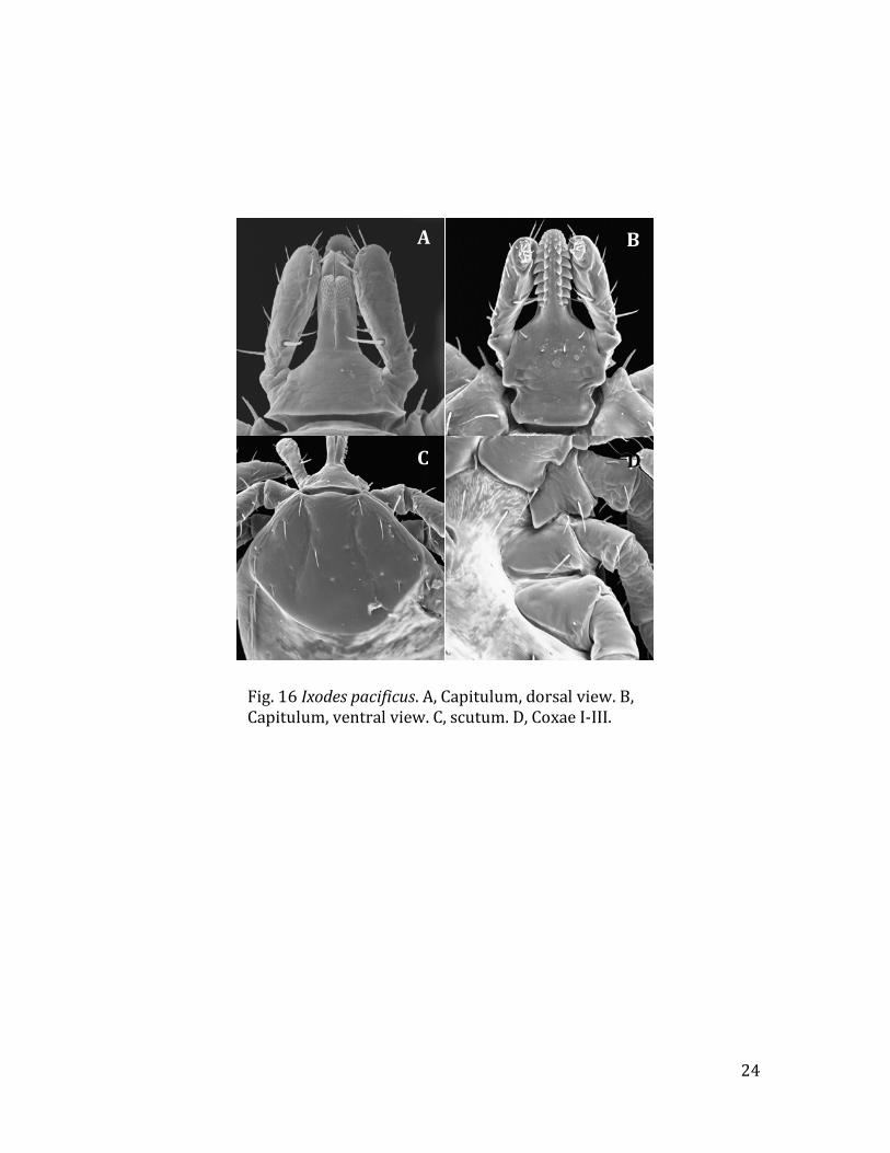
Fig. 16 Ixodes pacificus. A, Capitulum, dorsal view. B, Capitulum, ventral view. C, scutum. D, Coxae I-III.
C
A
D
B

25
Fig. 17 Ixodes scapularis. A, Capitulum, dorsal view. B, Capitulum, ventral view. C, scutum. D, Coxae I-III.
C
A
D
B

26
Fig. 18 Ixodes spinipalpis. A, Capitulum, dorsal view. B, Capitulum, ventral view. C, scutum. D, Coxae I-III.
C
A
D
B

27
Fig. 19 Ixodes texanus. A, Capitulum, dorsal view. B, Capitulum, ventral view. C, scutum. D, Coxae I-III.
C
A
D
B

28
A B
C D
Fig. 20 Rhipicephalus sanguineus. A, Capitulum, dorsal view. B, Capitulum, ventral view. C, scutum. D, Coxae I-III.

29
Discussion
An illustrated identification key to the larval stages of the human-biting hard ticks
of the USA is presented for the first time. Although SEMs have been used to illustrate
important morphological characters for each tick species, a scanning electron microscope
is not needed in order to identify these ticks. The SEMs merely provide high quality
images of features that can easily be observed using a low-power binocular microscope.
The benefits of being able to identify the larval stage of these ticks has already been
emphasizes in the Introduction. The following notes provide information on the
medical/veterinary importance, hosts and geographical distribution of the 16 ticks
included in this identification guide.
Tick Species Main Host(s) for larvae Distribution Disease(s)
Transmitted Amblyomma americanum (Lone Star Tick) (Fig. 5)
White-tailed deer, humans, raccoons, opossums, birds
Southeastern and Eastern United States
Heartland virus, Human Monocytic Ehrlichiosis, Tularemia, Southern Ttick Associated Rash Illness
Amblyomma mixtum (Cayenne Tick) (Fig. 6)
Birds, small mammals
Texas Spotted fever
Amblyomma maculatum (Gulf Coast Tick) (Fig. 7)
Birds, rodents Coastal areas of the United States along the Atlantic coast and the Gulf of Mexico
Spotted fever
Amblyomma tuberculatum (Gopher Tortoise Tick)
Reptiles, birds, mammals
Southeastern United States
None to humans

30
(Fig. 8) Dermacentor albipictus (Winter Tick) (Fig. 9)
Moose, deer, elk, cattle
Throughout the U.S.
None to humans
Dermacentor andersoni (Rocky Mountain Wood Tick) (Fig. 10)
Rodents Rocky Mountain states
Rocky Mountain spotted fever, tick paralysis, Colorado tick fever
Dermacentor occidentalis (West Coast Tick) (Fig. 11)
Rodents Western U.S. Rocky Mountain spotted fever, tick paralysis
Dermacentor variabilis (American Dog Tick) (Fig. 12)
Rodents East of Rocky Mountains; limited areas of Pacific Coast
Rocky Mountain spotted fever, tularemia, tick paralysis
Haemaphysalis leporispalustris (Rabbit Tick) (Fig. 13)
Rabbits, birds
Eastern United States; also some western states
Rocky Mountain spotted fever, Tularemia
Ixodes angustus (no common name) (Fig. 14)
Rodents, insectivores, rabbits
Higher elevations and latitudes in Western and Eastern U.S.
Lyme disease
Ixodes cookei (no common name) (Fig. 15)
Rodents, raccoons Eastern United States
Lyme disease, Powassan virus
Ixodes pacificus (Western Blacklegged Tick (Fig. 16)
Lizards,birds, rodents
Pacific coast of the United States
Human Granulocytic Anaplasmosis, Lyme disease, tick paralysis
Ixodes scapularis (Blacklegged Tick) (Fig. 17)
Lizards, birds, mammals
Eastern U.S. States Human Granulocytic Anaplasmosis, babesiosis, Lyme disease
31
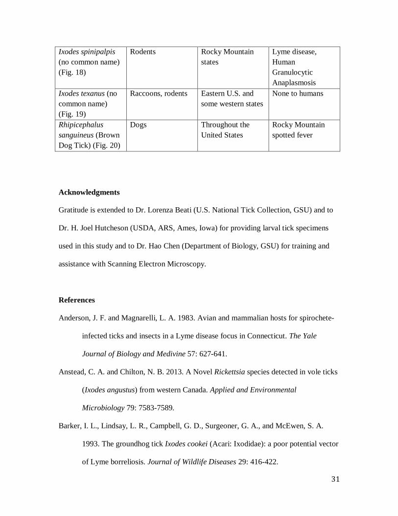
Ixodes spinipalpis (no common name) (Fig. 18)
Rodents Rocky Mountain states
Lyme disease, Human Granulocytic Anaplasmosis
Ixodes texanus (no common name) (Fig. 19)
Raccoons, rodents Eastern U.S. and some western states
None to humans
Rhipicephalus sanguineus (Brown Dog Tick) (Fig. 20)
Dogs Throughout the United States
Rocky Mountain spotted fever
Acknowledgments
Gratitude is extended to Dr. Lorenza Beati (U.S. National Tick Collection, GSU) and to
Dr. H. Joel Hutcheson (USDA, ARS, Ames, Iowa) for providing larval tick specimens
used in this study and to Dr. Hao Chen (Department of Biology, GSU) for training and
assistance with Scanning Electron Microscopy.
References
Anderson, J. F. and Magnarelli, L. A. 1983. Avian and mammalian hosts for spirochete-
infected ticks and insects in a Lyme disease focus in Connecticut. The Yale
Journal of Biology and Medivine 57: 627-641.
Anstead, C. A. and Chilton, N. B. 2013. A Novel Rickettsia species detected in vole ticks
(Ixodes angustus) from western Canada. Applied and Environmental
Microbiology 79: 7583-7589.
Barker, I. L., Lindsay, L. R., Campbell, G. D., Surgeoner, G. A., and McEwen, S. A.
1993. The groundhog tick Ixodes cookei (Acari: Ixodidae): a poor potential vector
of Lyme borreliosis. Journal of Wildlife Diseases 29: 416-422.

32
Beati, L., and Keirans, J. E. 2001. Analysis of the systematic relationships among ticks of
the genera Rhipicephalus and Boophilus (Acari: Ixodidae) based on 12S
ribosomal DNA gene sequences and morphological characters. Journal of
Parasitology 87: 32-48.
Beati, L., Nava, S., Burkman, E. J., Barros-Battesti, D. M., Labruna, M. B., Guglielmone,
A. A., Cáceres, A. G., Guzmán-Cornejo, C. M., León, R., Durden, L. A., and
Faccini, J. L. H. 2013. Amblyomma cajennense (Fabricius, 1787) (Acari:
Ixodidae), the Cayenne tick: phylogeography and evidence for allopatric
speciation. BMC Evolutionary Biology 13: 267.
Burkot, T. R., Maupin, G. O., Schneider, B. S., Denatale, C., Happ, C. M., Rutherford, J.
S., and Zeidner, N. S. 2001. Use of a sentinel host system to study the questing
behavior of Ixodes spinipalpis and its role in the transmission of Borrelia bissettii,
Human Granulocytic Ehrlichiosis, and Babesia microti. American Journal of
Tropical Medicine and Hygiene 65: 293-299.
Clifford, C. M., Anastos, G., and Elbl, A. 1961. The larval ixodid ticks of the eastern
United States. Miscellaneous Publications of the Entomological Society of
America 2: 213-237.
Damrow, T., Freedman, H., Lane, R. S., and Preston, K. L. 1989. IsIxodes (Ixosiopsis)
angustus a vector of Lyme disease in Washington State? Western Journal of
Medicine 150: 580-582.

33
Durden, L. A. 2006. Taxonomy, host associations, life cycles and vectorial importance
of ticks parasitizing small mammals, pp. 91-102, In: S. Morand, B. R. Krasnov &
R. Poulin (editors), Micromammals and macroparasites: from evolutionary
ecology to management. Springer, Tokyo.
Durden, L. A., and Mans, B. J. 2015. Tick paralysis: some tick and host perspectives, In:
A Century of Parasitology, J. Janovy & G. W. Esch (editors). Cambridge
University Press, Cambridge.
Ennen, J. R. and Qualls, C. P. 2011. Distribution and habitat utilization of the gopher
tortoise tick (Amblyomma tuberculatum) in southern Mississippi. Journal of
Parasitology 97: 202-206.
Kleinjan & Lane. 2008. Larval keys to the genera of Ixodidae (Acari) and species of
Ixodes (Latreille) ticks established in California. Pan-Pacific Entomologist 84:
121-142.
Kosoy, O. L., Lambert, A. J., Hawkinson, D. J., Pastula, D. M., Goldsmith, C. S, Hunt,
D. C., and Staples, J. E. 2015. Novel thogotovirus associated with febrile illness
and death, United States, 2014. Emerging Infectious Diseases 21.
Ktawczak, F. S., Nieri-Bastos, F. A., Nunes, F. P., Soares, J. F., Moraes-Filho, J.,and
Labruna, M. B. 2014. Rickettsial infection in Amblyomma cajennense ticks and
capybaras (Hydrochoerus hydrochaeris) in a Brazilian spotted fever-endemic
area. Parasites & Vectors 7: 7.
Lopes, C. M. L., Leite, R. C., Labruna, M. B., de Oliveira, P. R., Borges, L. M. F.,
Rodrigues, Z. B., Carvalho, F. A., de Freitas, C. M. V. & Júnior, C. R. V. 1998.
Host specificity of Amblyomma cajennense (Fabricius, 1787) (Acari: Ixodidae)

34
with comments on the drop-off rhythm. Memorias do Instituto Oswaldo Cruz 93:
347-351.
Magnarelli, L. A. and Swihart, R. K. 1991. Spotted fever group rickettsiae or Borrelia
burgdorferi in Ixodes cookei (Ixodidae) in Connecticut. Journal of Clinical
Microbiology 29: 1520-1522.
Merten, H. A., and Durden, L. A. 2000. A state-by-state survey of records of ticks
parasitizing humans in the United States. Journal of Vector Ecology 25: 102-
113.
Nicolson, W. L., Sonenshine, D. E., Lane, R. S., and Uilenberg, G. 2009. Ticks
(Ixodoidea), pp. 493-542, In: Medical and Veterinary Entomology (2nd edition),
G. R. Mullen & L. A. Durden (editors). Elsevier, Amsterdam.
Oliver, J. H., Jr. 1989. Biology and systematics of ticks (Acari: Ixodida). Annual Review
of Ecology and Systematics 20: 397-420.
Schmidt, K., Dressel, K.M., Niedrig, M., Mertens, M., Schuele, S.A. & Groschup, M.H.
2013, "Public Health and Vector-Borne Diseases - A New Concept for Risk
Governance", Zoonoses and Public Health 60: 528-538.
Swei, A., Russell, B.J., Naccache, S.N., Kabre, B., Veeraraghavan, N., Pilgard, M.A.,
Johnson, B.J.B. & Chiu, C.Y. 2013. The genome sequence of Lone Star Virus, a
highly divergent Bunyavirus found in the Amblyomma americanum tick. Plos One
8: e62083.
Webb, Bennett & Challet. 1990. The larval ticks of the genus Ixodes Latreille (Acari:
Ixodidae) of California. Bulletin of the Society for Vector Ecology 15: 73-124.