identification of a human scarb2 region that is important
TRANSCRIPT

JOURNAL OF VIROLOGY, May 2011, p. 4937–4946 Vol. 85, No. 100022-538X/11/$12.00 doi:10.1128/JVI.02358-10Copyright © 2011, American Society for Microbiology. All Rights Reserved.
Identification of a Human SCARB2 Region That Is Important forEnterovirus 71 Binding and Infection�
Seiya Yamayoshi and Satoshi Koike*Neurovirology Project, Tokyo Metropolitan Institute of Medical Science, Tokyo Metropolitan Organization for Medical Research,
2-1-6, Kamikitazawa, Setagaya-ku, Tokyo 156-8506, Japan
Received 10 November 2010/Accepted 28 February 2011
We previously identified human scavenger receptor class B, member 2 (SCARB2), as a cellular receptor forenterovirus 71 (EV71). Expression of human SCARB2 (hSCARB2) permitted mouse L929 cells to efficientlybind to virions and to produce both viral proteins and progeny viruses upon EV71 infection. Mouse Scarb2(mScarb2) exhibited 85.8% amino acid identity and 99.9% similarity to hSCARB2. The expression of mScarb2in L929 cells conferred partial susceptibility. Very few virions bound to mScarb2-expressing cells. The viraltiter in L929 cells expressing mScarb2 was approximately 40- to 100-fold lower than that in L929 cellsexpressing hSCARB2. Using hSCARB2–mScarb2 chimeric mutants, we attempted to map the region that wasimportant for efficient EV71 infection. L929 cells expressing chimeras that carried amino acids 142 to 204 fromthe human sequence were susceptible to EV71, while chimeras that carried the mouse sequence in this regionwere not. Moreover, this region was also critical for binding to virions. The determination of this region inhSCARB2 that is important for EV71 binding and infection greatly contributes to the understanding ofvirus-receptor interactions. Further studies will clarify the early steps of EV71 infection.
Enterovirus 71 (EV71), together with coxsackievirus A16(CVA16), belongs to human enterovirus species A of the genusEnterovirus within the family Picornaviridae (28). The viruscontains a single-stranded, positive-sense RNA surrounded byan icosahedral capsid assembled from 60 copies of each of thefour structural proteins: VP1, VP2, VP3, and VP4 (32). EV71was first isolated from patients with neurological diseases, in-cluding fatal encephalitis and aseptic meningitis, in Californiafrom 1969 to 1972 (33). Later studies revealed that EV71 isassociated with hand-foot-and-mouth disease (HFMD) inyoung children and infants (6, 13). Although HFMD is gener-ally considered a mildly infectious disease, HFMD caused byEV71, but not by other enteroviruses, is sometimes involvedwith severe neurological diseases, including brain stem enceph-alitis and acute flaccid paralysis (23). In recent years, epi-demic or sporadic outbreaks of neurovirulent EV71 infec-tions have been reported mainly in Southeast or East Asia,including Taiwan, Malaysia, Singapore, Japan, and China (1,8, 11, 17, 27, 38). In particular, the epidemic outbreaks thatoccurred in 2008 and 2009 in China resulted in a total of 488,955and 1,155,525 HFMD cases, including 126 and 353 fatal casesper year, respectively (http://www.moh.gov.cn/publicfiles/business/htmlfiles/mohbgt/s3582/201002/46043.htm) (40). Moreover, theEV71 epidemic has since continued in China, with 987,779HFMD cases, including 537 fatal cases reported as of 22June 2010 (http://www.moh.gov.cn/publicfiles/business/htmlfiles/mohbgt/s3582/201006/47871.htm).
Human RD cells are highly susceptible to EV71 infection,whereas mouse L929 cells exhibit very low susceptibility. We
previously reported the identification of EV71 receptors bygenetic complementation of L929 cells with genes transferredfrom human RD cells (37). Briefly, we established two EV71-susceptible cell lines that carried a portion of human genomicDNA. By identifying the integrated human DNA in one of thetransformants, Ltr051 cells, we have shown that scavenger re-ceptor class B, member 2 (SCARB2), is a cellular receptor forEV71. SCARB2 serves as a receptor for all EV71 isolatestested. EV71 infection in the SCARB2-expressing L929 cellline is as efficient as that in the RD cell line (37). We have alsofound that virus replication efficiency in another transformantcell line, Ltr246, is slightly lower than that in Ltr051 cells (37)and that Ltr246 cells are susceptible to only a subset of EV71strains (S. Yamayoshi et al., unpublished data). AlthoughLtr246 cells appear to express another, unknown receptor (re-ceptor X), we have not yet identified the human DNA se-quence that confers susceptibility on Ltr246 cells. In addition,Nishimura et al. also have identified the selectin P ligand(SELPLG, also known as P-selectin glycoprotein ligand-1[PSGL-1]) as an EV71 receptor from human T cell leukemiaJurkat cells by using the panning assay, which enriched formolecules with strong binding affinity to EV71 particles (26).SELPLG also confers susceptibility only to some EV71 strainson L929 cells. In Jurkat cells and L929 cells expressingSELPLG, however, the appearance of cytopathic effect (CPE)and the expression of viral proteins after EV71 infection oc-curred more slowly than in RD cells and L929 cells expressingSCARB2 (26). We have confirmed that Ltr246 cells do notcarry the human SCARB2 and SELPLG genes. Another studyhas shown that the depletion of O-linked glycans or pretreat-ment with sialidase reduced EV71 infection in DLD-1 humancolon cancer cells (39). These results suggest that EV71 canenter the cell via multiple pathways. Because all EV71 strainstested used SCARB2 as the receptor (37), whereas only asubset of EV71 strains used SELPLG (26) or receptor X,
* Corresponding author. Mailing address: Neurovirology Project,Tokyo Metropolitan Institute of Medical Science, Tokyo MetropolitanOrganization for Medical Research, 2-1-6, Kamikitazawa, Setagaya-ku, Tokyo 156-8506, Japan. Phone: 81-3-5316-3312. Fax: 81-3-5316-3224. E-mail: [email protected].
� Published ahead of print on 9 March 2011.
4937
Dow
nloa
ded
from
http
s://j
ourn
als.
asm
.org
/jour
nal/j
vi o
n 19
Feb
ruar
y 20
22 b
y 1.
36.1
3.18
2.

SCARB2 may play a central role in the early steps of EV71infection. Therefore, characterizations of the role of SCARB2during EV71 infection will contribute to the understanding ofEV71 entry.
SCARB2 (also known as lysosomal integral membrane pro-tein II, or CD36b like-2) belongs to the CD36 family and hastwo transmembrane domains, with the N and C termini locatedin the cytosol (10). SCARB2 is one of the most abundantproteins in the lysosomal membrane and participates in mem-brane transport and the reorganization of the endosomal/lyso-somal compartment (21). SCARB2 also works as the receptorfor the mannose-6-phosphate-independent transport of �-glu-cocerebrosidase (�-GC) to the lysosome (5, 30). The bindingof �-GC to SCARB2 occurs within the luminal domain ofSCARB2, particularly in the coiled-coil motif at amino acids152 to 167 (30). SCARB2 deficiency in mice causes uretero-pelvic junction obstruction, deafness, and peripheral neuropa-thy (12). Although the motifs in some cellular receptors forpicornaviruses that are important for binding and/or infectionhave been elucidated, SCARB2 has no motifs common toother picornavirus receptors, such as an immunoglobulin (Ig)-like motif. Thus, SCARB2 is a new class of picornavirus re-ceptor. The identification of important regions in SCARB2would contribute to the elucidation of the virus-receptor inter-action.
In this report, we compared the susceptibilities of cells ex-pressing human SCARB2 (hSCARB2) or mouse Scarb2(mScarb2) to EV71. Additionally, we mapped the region inhSCARB2 that is important for EV71 infection by usinghSCARB2–mScarb2 chimeras.
MATERIALS AND METHODS
Cells. Human RD cells, mouse L929 cells, and African green monkey Verocells were cultured in Dulbecco’s modified Eagle medium (DMEM; Sigma)supplemented with 5% fetal bovine serum (FBS) and a penicillin-streptomycinsolution (Invitrogen) (5% FBS-DMEM).
Viruses. EV71 strain SK-EV006/Malaysia/97 was propagated in Vero cells foruse in this study (25). EV71-GFP, which expresses green fluorescent protein(GFP) upon viral replication, was recovered from an infectious cDNA clone,pSVA-EV71-GFP, which has been described previously (37).
Plasmids. The cDNA fragment of mScarb2 was amplified by reverse transcrip-tion-PCR (RT-PCR) from L929 cells with primers mSCARB2-Eco(�) (CAGAATTCACCATGGGCAGATGCTGCTTCTACA) and mSCARB2-Xba(�) (CACATCTAGATTAGGTTCGTATGAGGGGTGCT), and the PCR product wasinserted into pCAGGS-PUR (14). The resulting construct was designated pCA-mScarb2. A cDNA fragment encoding mScarb2 or hSCARB2 (37) was subclonedinto pCAGGS.MCS (19, 36) to create a FLAG tag at the C terminus, and theconstruct was designated pCA-mScarb2-F or pCA-hSCARB2-F, respectively.
Chimeric hSCARB2–mScarb2 mutants (see Fig. 6A and 7A) were constructedusing standard PCR-based methods and were cloned into pCAGGS.MCS with aFLAG tag at the C terminus. The mutants did not have unexpected mutations ordeletions.
Viral spread in cell culture. RD cells were infected with either EV71-GFP orEV71. At 24 h postinfection, cells infected with EV71-GFP were imaged withIX70 and DP70 cameras (Olympus) and were analyzed using DP Controllersoftware (Olympus). The cells infected with EV71 or EV71-GFP were subse-quently fixed with 4% paraformaldehyde and were probed with a mouse anti-EV71 antibody (clone 422-8D-4C-4D; Millipore), followed by incubation with anAlexa Fluor 488 donkey anti-mouse IgG (Invitrogen). Images were acquiredusing the IX70 camera.
PNGase F digestion. L929 cells were transfected with the plasmid encodinghSCARB2, mScarb2, hSCARB2-F, mScarb2-F, H(M4)-F, M(H2)-F, or M(H3)-Fusing the FuGENE 6 transfection reagent. After 48 h, the transfected cells werelysed with glycoprotein denaturing buffer (New England Biolabs [NEB]) andwere then incubated for 10 min at 99°C. Denatured samples were mixed with G7
reaction buffer (NEB) supplemented with 1% NP-40 and 500 U peptide N-gly-cosidase F (PNGase F) (NEB) and were then incubated for 2 h at 37°C fordigestion. These samples were mixed with a sodium dodecyl sulfate (SDS) sam-ple buffer and were then incubated for 5 min at 95°C before being resolved on a12% Tris-glycine gel. Resolved proteins were probed with a goat anti-hSCARB2antibody (R&D Systems), a goat anti-mScarb2 antibody (R&D Systems), or arabbit anti-FLAG antibody (Sigma), followed by incubation with a horseradishperoxidase (HRP)-conjugated anti-goat or anti-rabbit antibody (Jackson Immuno-Research).
Single-round infection assay. L929 cells were transfected with the indicatedplasmid in triplicate to analyze (i) transfection efficiency, (ii) protein expression,and (iii) EV71-GFP infection. Transfected cells in the first well were fixed at theplate bottom with 4% paraformaldehyde at 48 h posttransfection and wereprobed with the mouse anti-FLAG antibody (Sigma), followed by incubationwith Alexa Fluor 488 donkey anti-mouse IgG. Images were acquired using theIX70 and DP70 cameras. Transfected cells in the second well were mixed withSDS sample buffer (Invitrogen) at 48 h posttransfection, and these samples wereincubated for 5 min at 95°C before being resolved on a 12% Tris-glycine gel(Invitrogen). Resolved proteins were probed with the anti-hSCARB2 antibody,the rabbit anti-FLAG antibody (Sigma), or a mouse antibody against �-actin(ACTB) (clone AC-74; Sigma), followed by incubation with the appropriateHRP-conjugated secondary antibody (Jackson ImmunoResearch). Transfectedcells in the third well were infected with EV71-GFP at 24 h after transfection andwere incubated for another 24 h at 37°C. Subsequently, images were acquiredusing the IX70 and DP70 cameras and were analyzed using DP Controllersoftware. Infected cells, including GFP-positive cells, were detached in trypsin-EDTA (0.05% trypsin, 0.53 mM EDTA � 4Na) (GIBCO) and were incubatedwith 4% paraformaldehyde for 30 min at room temperature. After being washedwith phosphate-buffered saline (PBS) containing 2% FBS, the cells were ana-lyzed with a FACSCalibur flow cytometer and CellQuest Pro software (bothfrom Becton Dickinson and Company).
Multistep infection assay. L929 cells were transfected with the indicated plas-mid using the FuGENE 6 transfection reagent. After 24 h, the transfected cellswere infected with EV71 (1 � 103 50% tissue culture infective doses [TCID50])and were incubated for 0, 12, 24, 36, or 48 h at 37°C. At each time point, viraltiters were determined and expressed as the TCID50 according to the Reed-Muench method (31).
Immunoprecipitation assay. Pulldown assays were performed as reportedpreviously (37) with some modifications. EV71 (1.87 � 108 TICD50) was incu-bated with bovine serum albumin (BSA) (10 �g), human IgG Fc (control Fc) (10�g; R&D Systems), hSCARB2-Fc (1, 3, or 10 �g; R&D Systems), or mScarb2-Fc(1, 3, or 10 �g; R&D Systems) and anti-human IgG (Fc specific)-agarose (Sigma)in 1 ml of 5% FBS DMEM for 2 h at 4°C. The beads were then washed twice with5% FBS-DMEM, suspended in SDS sample buffer, and incubated for 10 min at95°C. After the beads were removed, the samples were loaded onto a 12%Tris-glycine gel, followed by Western blotting with a rabbit anti-EV71 antibody(25) or an anti-human IgG Fc� fragment-specific antibody (Jackson Immuno-Research). For deglycosylation, control Fc (3 �g) and hSCARB2-Fc (3 �g) in thenative form were treated with 1,500 U PNGase F in G7 reaction buffer and wereincubated for 24 h at 37°C before being mixed with anti-human IgG (Fc specific)-agarose.
Virus attachment assay. L929 cells transfected with the indicated plasmid weredetached in PBS containing 0.05% EDTA. These cells were mixed with EV71(1.87 � 108 TICD50) for 1 h at 4°C, washed twice with 5% FBS-DMEM,suspended in SDS sample buffer, and then incubated for 10 min at 95°C. Thesamples were resolved by SDS-polyacrylamide gel electrophoresis (PAGE) usingthe 12% Tris-glycine gel, followed by Western blot analysis with an anti-EV71,anti-FLAG, or anti-ACTB antibody.
Biotinylation of cell surface proteins. L929 cells transfected with the indicatedplasmids were washed twice with ice-cold PBS and were biotinylated twice withSulfo-N-hydroxysuccinimide (NHS)-SS-Biotin (Pierce) for 15 min each time at4°C. After two washes with ice-cold PBS, free thiol groups were quenched withquenching solution (Pierce), and the cells were solubilized in lysis buffer (20 mMTris-HCl [pH 7.5], 150 mM NaCl, 1 mM EDTA, 1% Triton X-100, and CompleteMini protease inhibitor cocktail [Roche]) and were precipitated with NeutrAvi-din agarose resin (Pierce). Precipitated proteins and cell lysates were analyzed byWestern blotting with the anti-FLAG antibody.
RESULTS
Comparison of amino acid sequences of hSCARB2 andmScarb2. The cDNA encoding mScarb2 was prepared from
4938 YAMAYOSHI AND KOIKE J. VIROL.
Dow
nloa
ded
from
http
s://j
ourn
als.
asm
.org
/jour
nal/j
vi o
n 19
Feb
ruar
y 20
22 b
y 1.
36.1
3.18
2.

L929 cells, and its amino acid sequence was compared withthat of hSCARB2 (Fig. 1A). Both hSCARB2 and mScarb2 arecomposed of 478 amino acids, which are encoded in 12 exons.The level of amino acid identity between hSCARB2 andmScarb2 was 85.8%, and the level of similarity was 99.9%. Wetransiently expressed hSCARB2 and mScarb2 in L929 cells anddetected the expressed proteins using anti-human and anti-mouse SCARB2 antibodies. We detected a single band that
migrated at approximately 80 kDa in the Western blot of RDcells with anti-hSCARB2 antibody. mSCARB2, which is en-dogenously expressed in L929 cells, was detected in empty-vector-transfected L929 cells with an anti-mScarb2 antibody(Fig. 1B). We detected two major molecular species migratingat approximately 70 kDa and 80 kDa with an anti-hSCARB2antibody in cells transfected with pCA-hSCARB2 (Fig. 1B,left). Similarly, we also detected two major molecular species
FIG. 1. Human SCARB2 and mouse Scarb2. (A) Comparison of the amino acid sequence of hSCARB2 with that of mScarb2. Divergent aminoacids are highlighted. Two transmembrane domains are underlined, and the amino acids encoded in the mRNA from SCARB2 exons 1, 3, 5, 7,9, and 11 are outlined with red dashed lines. The amino acids encoded in the mRNA from exons 2, 4, 6, 8, 10, and 12 are outlined with black dashedlines. (B) Expression of hSCARB2 and mScarb2. L929 cells were transfected with plasmids encoding hSCARB2 or mScarb2. Transfected cells andRD cells were analyzed by Western blotting with an anti-hSCARB2 or anti-mScarb2 antibody. (C) Schematic diagrams of hSCARB2 and mScarb2.Red lines represent the amino acid sequences encoded in exons 1, 3, 5, 7, 9, and 11, and black lines represent those encoded in exons 2, 4, 6, 8,10, and 12 (numbering from the N terminus to the C terminus). Potential N-glycosylation sites are indicated by lollipops. An asterisk marks theN-glycosylation site that is present only in mScarb2. (D) PNGase F treatment of hSCARB2 and mScarb2. L929 cells were transfected with plasmidsencoding hSCARB2 or mScarb2. The cells were either left untreated (Non Treat) or treated with PNGase F, and the samples were analyzed byWestern blotting with an anti-hSCARB2 or an anti-mScarb2 antibody.
VOL. 85, 2011 HUMAN SCARB2 REGION AND ENTEROVIRUS 71 BINDING 4939
Dow
nloa
ded
from
http
s://j
ourn
als.
asm
.org
/jour
nal/j
vi o
n 19
Feb
ruar
y 20
22 b
y 1.
36.1
3.18
2.

migrating at approximately 70 kDa and 80 kDa with an anti-mScarb2 antibody in cells transfected with pCA-mScarb2 (Fig.1B, right). The apparent molecular sizes of these bands werelarger than that deduced from the amino acid sequence (ap-proximately 54 kDa). hSCARB2 and mScarb2 are reported tobe N-glycosylated proteins in human and mouse cells, respec-tively (5, 30). hSCARB2 was found to have 10 potential N-gly-cosylation sites, whereas mScarb2 was found to have 11 (Fig.1C). Potential N-glycosylation sites were found in the aminoacid sequences encoded in exons 2, 3, 5, 6, 7, 10, and 11 of bothhSCARB2 and mScarb2. mScarb2 had one additional potentialN-glycosylation site in exon 3. To confirm whether exogenouslyexpressed SCARB2s were glycosylated in L929 cells, cell ly-sates were treated with PNGase F, which removes all N-linkedcarbohydrate residues. After PNGase F treatment, hSCARB2and mScarb2 were each detected as a single band at the cal-culated molecular mass (approximately 54 kDa) (Fig. 1D).These results indicate that hSCARB2 and mScarb2 are ex-pressed as N-glycosylated proteins in mouse L929 cells andthat the glycosylation pattern differs slightly from that of theendogenous proteins.
Strategy for the comparison of infection efficiencies in L929cells expressing hSCARB2 or mScarb2. To compare the effi-ciency of a single round of EV71 infection via hSCARB2versus mScarb2, we used EV71-GFP as a challenge virus.EV71-GFP, when used to infect cells, expresses sufficient levelsof GFP to monitor the establishment of infection. This virus,however, had a defect in growth kinetics and in spreading. Itwas impossible to determine the viral titer either by plaqueformation or by the TCID50 method. When RD cells wereinfected with EV71-GFP at an appropriate dilution, GFP-pos-itive cells became visible at approximately 16 to 18 h postin-fection, suggesting that the kinetics of viral protein expressionwas unusual compared with that of wild-type virus. Clusters ofGFP-positive or viral antigen-positive cells, which had spreadfrom the initial infectious center, were not observed at 24 hpostinfection (Fig. 2A, left and center). In contrast, when RDcells were infected with wild-type EV71, we observed clustersof viral antigen-positive cells that had formed due to spreadingfrom the infectious centers at 24 h postinfection (Fig. 2A, rightpanel). Therefore, we evaluated the efficiency of EV71 infec-tion at the initial round of infection by using EV71-GFP andcounting the number of GFP-positive cells at 24 h postinfec-tion.
To monitor the expression levels of hSCARB2 and mScarb2by Western blotting, we added a FLAG epitope tag to the Ctermini of the proteins (5). To examine whether the C-terminalFLAG tag affected EV71 infection, we compared the numbersof GFP-positive cells after infection with EV71-GFP in L929cells expressing hSCARB2 with or without the FLAG tag (Fig.2B to E). L929 cells were transfected with pCA-hSCARB2 orpCA-hSCARB2-F, and after 24 h, the transfection efficienciesand expression of hSCARB2 and hSCARB2-F were confirmedby immune staining and Western blot analysis, respectively(Fig. 2B and C). The numbers of SCARB2-positive cellsin L929 cells transfected with pCA-hSCARB2 or pCA-hSCARB2-F were comparable (Fig. 2B). Two major species ofhSCARB2 and hSCARB2-F with similar intensities were de-tected by Western blotting using the anti-hSCARB2 antibody(Fig. 2C). Thus, the transfection efficiency, expression level,
and glycosylation pattern of hSCARB2 were not affected bythe addition of the FLAG tag. Under these conditions, thesetransfected L929 cells were infected with EV71-GFP and wereimaged at 24 h postinfection (Fig. 2D). No GFP-positive cellswere observed in the mock-transfected or empty-plasmid-transfected cells, whereas many GFP-positive cells were de-tected in the hSCARB2- and hSCARB2-F-transfected cells(Fig. 2D). To quantify the microscopic observations, these cellswere analyzed by fluorescence-activated cell sorting (FACS) to
FIG. 2. Strategy for comparing infection efficiencies in L929 cellsexpressing hSCARB2 or mScarb2. (A) EV71-GFP is defective in viralspread. RD cells were infected with EV71 or EV71-GFP. Infected cellswere imaged at 24 h postinfection in order to observe GFP via fluo-rescence microscopy (GFP) and were then stained with an anti-EV71antibody. (B to E) Efficiency of EV71-GFP infection via hSCARB2with or without the FLAG tag. (B) Transfection efficiency was assessedby immunostaining with an anti-hSCARB2 antibody. (C) Expression ofhSCARB2 or hSCARB2-F was confirmed by Western blot analysiswith an anti-hSCARB2 or an anti-FLAG antibody. Expression of �-ac-tin (ACTB) was used as a loading control. (D and E) Cells infectedwith EV71-GFP were imaged via fluorescence microscopy (D) andwere concomitantly analyzed by FACS to quantify the number ofGFP-positive cells (E). The FACS data are shown as mean counts withstandard deviations (n � 3). Statistical significance was determined byStudent’s t test. There was no significant difference between hSCARB2and hSCARB2-F (P � 0.823).
4940 YAMAYOSHI AND KOIKE J. VIROL.
Dow
nloa
ded
from
http
s://j
ourn
als.
asm
.org
/jour
nal/j
vi o
n 19
Feb
ruar
y 20
22 b
y 1.
36.1
3.18
2.

count the number of GFP-positive cells (Fig. 2E). As reportedpreviously, the number of GFP-positive cells was significantlygreater in cells expressing hSCARB2 or hSCARB2-F than inmock- or empty plasmid-transfected cells (P � 0.01) (37).There was no significant difference in the number of GFP-positive cells between hSCARB2- and hSCARB2-F-expressingcells (P � 0.823). Similar results were obtained usingnontagged and FLAG-tagged mScarb2 (data not shown).These results clearly show that EV71 infection via SCARB2was not affected by the C-terminal FLAG tag. From theseresults, we could compare the efficiencies of EV71-GFP infec-tion using this assay.
EV71-GFP infection of L929 cells expressing hSCARB2 andmScarb2. We conducted the single-round infection assay tocompare the efficiencies of EV71 infection in L929 cells ex-pressing hSCARB2-F versus mScarb2-F (Fig. 3). L929 cellswere either mock transfected or transfected with pCA-hSCARB2-F, pCA-mScarb2-F, or an empty plasmid. Afterconfirmation of transfection efficiency and expression ofhSCARB2-F and mScarb2-F by immune staining and Westernblotting (Fig. 3A and B), the transfected cells were infectedwith EV71-GFP and were observed at 24 h postinfection (Fig.3C). No GFP-positive cells were found in mock- or empty-plasmid-transfected cells. A large number of GFP-positive
cells were detected in hSCARB2-F-transfected cells, whereas amodest number were detected in mScarb2-F-transfected cells(Fig. 3C). By FACS analysis, the number of GFP-positive cellswas significantly greater in L929 cells expressing hSCARB2-Fthan in those expressing mScarb2-F (P � 0.0032) (Fig. 3D).These results indicate that EV71-GFP infected efficiently viahSCARB2 and less efficiently via mScarb2 and that the effi-ciencies of infection by use of the two receptors were signifi-cantly different.
Multistep EV71 infection of L929 cells expressing hSCARB2and mScarb2. To validate the results obtained with the single-round infection experiment using EV71-GFP, we performed amultistep infection assay with wild-type EV71 infecting at a lowmultiplicity of infection (MOI) to examine the spread of thevirus. We confirmed by immune staining and Western blottingthat hSCARB2-F and mScarb2-F were transfected and ex-pressed at similar levels (data not shown). L929 cells express-ing hSCARB2-F and mScarb2-F were infected with EV71, andthe viral titers were determined at each time point (Fig. 4).EV71 grew efficiently in L929 cells expressing hSCARB2-F butonly moderately in L929 cells expressing mScarb2-F. At thelast time point, the viral titer in L929 cells expressinghSCARB2-F was approximately 100-fold higher than that inL929 cells expressing mScarb2-F. EV71 propagated minimallyin mock-transfected and empty-plasmid-transfected L929 cells.These results indicate that EV71 infected more efficiently viahSCARB2 than via mScarb2.
Binding of hSCARB2 and mScarb2 to EV71. To elucidatethe functional difference between hSCARB2 and mScarb2 inthe early steps of infection, we conducted pulldown assays tocompare the binding affinities of soluble hSCARB2-Fc andsoluble mScarb2-Fc for EV71 (Fig. 5A). EV71 was incubatedwith BSA, control Fc, hSCARB2-Fc, or mScarb2-Fc and withanti-Fc-agarose beads, and precipitated proteins were analyzedby Western blotting. EV71 VP1 was detected at all concentra-tions of hSCARB2-Fc, and the amount of precipitated VP1increased in a concentration-dependent manner, as reportedpreviously (37). However, while mScarb2-Fc was precipitatedat a level similar to that of hSCARB2-Fc at each concentration
FIG. 3. Efficiencies of EV71-GFP infection via hSCARB2 ormScarb2. Single-round infection assays were performed to comparethe efficiencies of EV71 infection via hSCARB2-F versus mScarb2-F.(A and B) The transfection efficiencies and expression of hSCARB2-Fand mScarb2-F were confirmed by immunostaining (A) and Westernblot analysis with an anti-FLAG antibody (B), respectively. ACTB wasused as a loading control for the Western blot. (C and D) The trans-fected cells infected with EV71-GFP were imaged via fluorescencemicroscopy (C) and were then analyzed by FACS (D). The FACS dataare shown as mean counts with standard deviations (n � 3). Statisticalsignificance was determined by Student’s t test.
FIG. 4. Multistep infections of EV71 in L929 cells expressinghSCARB2 or mScarb2. Mock-transfected L929 cells and cells trans-fected with an empty plasmid or with a plasmid encoding hSCARB2-For mScarb2-F were infected with EV71 at a low MOI. Viral titers inVero cells were determined at 0, 24, and 48 h postinfection. The dataare shown as mean viral titers with standard deviations (n � 3).
VOL. 85, 2011 HUMAN SCARB2 REGION AND ENTEROVIRUS 71 BINDING 4941
Dow
nloa
ded
from
http
s://j
ourn
als.
asm
.org
/jour
nal/j
vi o
n 19
Feb
ruar
y 20
22 b
y 1.
36.1
3.18
2.

(Fig. 5A, lower panel), VP1 was not detected at any concen-tration of mScarb2-Fc (Fig. 5A, upper panel). VP1 did notprecipitate with BSA or control Fc. These data show that thebinding affinity of hSCARB2 for EV71 was stronger than thatof mScarb2.
To determine whether the N-linked carbohydrate chains ofhSCARB2 contribute to the binding to virions, the native formof hSCARB2-Fc was treated with PNGase F. The removal ofthe carbohydrate chains from hSCARB2-Fc was confirmed bythe 25-kDa downward size shift observed by Western blotting
in the protein treated with PNGase F. PNGase F-treatedhSCARB2-Fc or untreated hSCARB2-Fc was mixed with viri-ons. After immunoprecipitation, bound virions and Fc proteinswere analyzed by Western blotting (Fig. 5B). EV71 coprecipi-tated with both PNGase F-treated and nontreated hSCABR2-Fc; however, the intensity of the EV71 VP1 band coprecipi-tating with PNGase F-treated hSCARB2-Fc was onlymoderately lower than that of the band coprecipitating withnontreated hSCARB2-Fc. These results indicate that the car-bohydrate chains of hSCARB2 are not essential for the EV71–hSCARB2 interaction.
To assess the binding of EV71 to SCARB2 at the plasmamembrane, we performed virus attachment assays using a tran-sient expression system (Fig. 5C). L929 cells were either mocktransfected or transfected with an empty plasmid, pCA-hSCARB2-F, or pCA-mScarb2-F. The expression level ofhSCARB2-F was similar to that of mScarb2-F (Fig. 5C, cen-ter). After 48 h posttransfection, these cells were mixed withEV71, washed to remove unbound virus, lysed together withbound viruses, and then analyzed by Western blotting. Theamount of EV71 that was bound to cells transfected withpCA-hSCARB2 was substantially greater than that bound tomock-transfected cells or to cells transfected with an emptyplasmid or pCA-mScarb2 (Fig. 5C, top). These data show thatthe binding affinity of EV71 for hSCARB2 in the membranewas stronger than that for mScarb2 in the membrane.
Determination of the hSCARB2 region that mediates EV71infection. Because EV71 infected L929 cells more efficientlyvia hSCARB2 than via mScarb2 (Fig. 4), we mapped the re-gion(s) that was important for EV71 infection by using chime-ric hSCARB2–mScarb2 mutants. We hypothesized that theefficiency of EV71 infection via hSCARB2 would be loweredby replacement of the important region(s) with the corre-sponding region(s) in mScarb2 and vice versa. For this pur-pose, we prepared 6 chimeric mutants in which some of theexons were replaced (Fig. 6A). In the H(M1-4)-F mutant, forexample, the human amino acid sequence 1 to 204, encoded inhSCARB2 exons 1 to 4, was replaced with the correspondingregion from mScarb2. Other chimeric mutants were con-structed according to the same strategy. We performed thesingle-round infection assay to compare the efficiencies ofEV71-GFP infection via these chimeras. All mutants weretransfected and expressed at similar levels (Fig. 6B). Trans-fected cells were infected with EV71-GFP and were imagedwith a microscope at 24 h postinfection (data not shown).These cells were analyzed by FACS to count the number ofGFP-positive cells (Fig. 6C). Among the hSCARB2 backbonechimeric mutants, the H(M1-4)-F mutant failed to mediateefficient EV71-GFP infection. Among the mScarb2 backbonechimeras, the M(H1-4)-F mutant mediated efficient EV71-GFP infection. These data indicate that the region from aminoacids 1 to 204, encoded in human SCARB2 exons 1 to 4, isimportant for EV71-GFP infection.
To refine the region important for EV71-GFP infection, weconstructed an additional 8 chimeric mutants in each of whichone of exons 1 to 4 was replaced (Fig. 7A). The transfectionefficiencies of all chimeric mutants were comparable (data notshown). Expression of chimeric mutants in L929 cells was con-firmed by Western blotting (Fig. 7B). We found that the levelsof expression of mutants H(M1)-F, H(M2)-F, H(M3)-F,
FIG. 5. Binding of hSCARB2 to EV71. (A) hSCARB2-Fc bound toEV71. A total of 10 �g of BSA, 10 �g of control Fc, or 1, 3, or 10 �gof hSCARB2-Fc or mScarb2-Fc was bound to anti-human Fc-agaroseand was incubated with EV71. The precipitated proteins were analyzedby Western blotting with an anti-EV71 or anti-Fc antibody. The anti-EV71 antibody detected primarily the viral VP1 protein.(B) hSCARB2-Fc with or without N-linked carbohydrate chains boundto EV71. EV71 was incubated with 3 �g of control Fc or 3 �g ofhSCARB2-Fc treated with PNGase F, and the proteins were precipi-tated with an anti-Fc antibody. The precipitated proteins were ana-lyzed by Western blotting with an anti-EV71 or anti-Fc antibody.(C) EV71 attached to hSCARB2-expressing L929 cells. L929 cellswere either mock transfected or transfected with an empty plasmid,pCA-hSCARB2-F, or pCA-mScarb2-F. The transfected cells were in-cubated with EV71 at 4°C. After the wash steps, the cells were lysedand analyzed by Western blotting with an anti-EV71, anti-FLAG, oranti-ACTB antibody. ACTB was used as a loading control.
4942 YAMAYOSHI AND KOIKE J. VIROL.
Dow
nloa
ded
from
http
s://j
ourn
als.
asm
.org
/jour
nal/j
vi o
n 19
Feb
ruar
y 20
22 b
y 1.
36.1
3.18
2.

M(H1)-F, and M(H4)-F were comparable to that ofhSCARB2-F, and the ratios of the two major molecular speciesmigrating at approximately 80 kDa and 70 kDa were similar tothat for hSCARB2-F. Two species of approximately 80 kDaand 70 kDa for H(M4)-F and M(H3)-F were detected; how-ever, the intensity of the 80-kDa species in H(M4)-F andM(H3)-F was lower than that in hSCARB2. In M(H2)-F-trans-fected cells, we detected at least 3 species migrating at approx-imately 55 kDa, 80 kDa, and 90 kDa. Treatment of wild-typeSCARB2-F and the three chimeric mutants with PNGase F
resulted in similar apparent molecular masses (Fig. 7D). Thisresult shows that the differences in the mobility and the ratiosof these species between these three mutants were caused bydiffering N-glycosylation patterns and not by protein degrada-tion. Replacement of a region in hSCARB2 with the corre-sponding region in mScarb2, and vice versa, may affect proteinfolding and it may contribute to the difference in N-glycosyla-tion patterns. We performed the single-round infection assayto determine the infection efficiency. The transfected cellswere infected with EV71-GFP and were analyzed by FACS tocount the number of GFP-positive cells (Fig. 7C). In a series ofexperiments using the hSCARB2 backbone chimeras, thenumber of GFP-positive cells in cells expressing H(M4)-F wassignificantly decreased (P � 0.000039). In experiments usingthe mScarb2 backbone chimeras, the number of GFP-positivecells in cells expressing M(H4)-F was significantly greater (P �0.00071). These data indicate that the region from amino acids142 to 204, encoded in SCARB2 exon 4, is important for EV71-GFP infection.
To further refine the region important for EV71 infection,we constructed 4 chimeric mutants in which the first or secondhalf of exon 4 was replaced with the sequence of the otherspecies of origin, and we analyzed infection efficiency with thesingle-round infection assay. Each mutant showed an infectionefficiency intermediate between those of hSCARB2 andmScarb2 (data not shown). These data indicate that the im-portant amino acids for EV71 infection are localized through-out the region from amino acids 142 to 204.
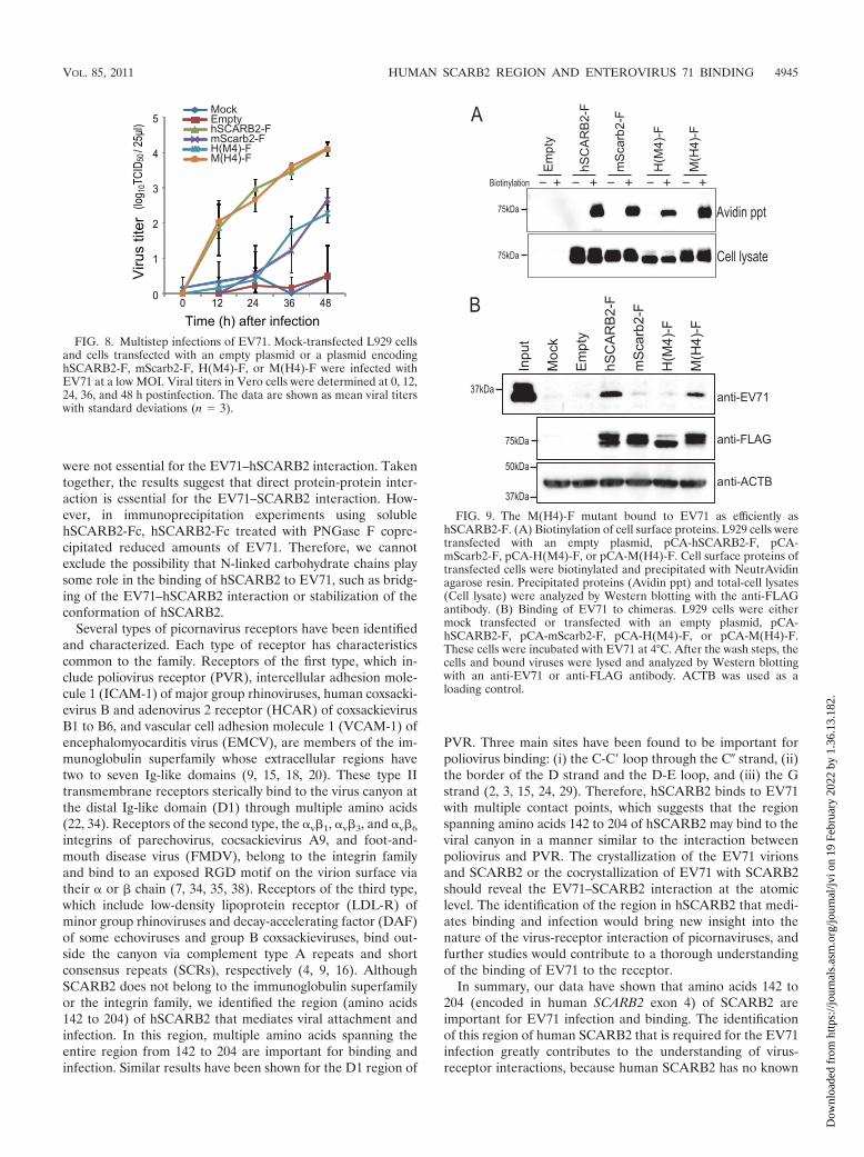
Multistep infection of L929 cells expressing H(M4)-F orM(H4)-F. In order to confirm the important region identifiedby the single-round infection assay, we carried out multistepinfection assays to determine the spread of wild-type EV71 inchimera-expressing L929 cells. L929 cells were transfected withplasmids encoding hSCARB2-F, mScarb2-F, H(M4)-F, orM(H4)-F and were subsequently infected with EV71. Viraltiters were determined at each time point (Fig. 8). EV71 grewas efficiently in L929 cells expressing M(H4)-F as in L929 cellsexpressing hSCARB2-F, whereas EV71 grew at a moderatelevel in L929 cells expressing M(H4)-F or mScarb2-F. The finalviral titer in L929 cells expressing hSCARB2-F or M(H4)-Fwas approximately 40-fold higher than that in L929 cells ex-pressing mScarb2-F or H(M4)-F. These results indicate thatthe M(H4)-F mutant functioned as a receptor for EV71 withan efficiency similar to that of hSCARB2-F.
Binding of H(M4)-F and M(H4)-F to EV71. Because theregion from amino acids 142 to 204 was important for thesingle and multistep EV71 infections, we presumed that thisregion played a critical role in the binding of EV71. Before avirus attachment assay was performed, the expression ofhSCARB2-F, mScarb2-F, H(M4)-F, and M(H4)-F on the cellsurface was confirmed by biotinylation of cell surface proteins(Fig. 9A). Similar amounts of biotinylated hSCARB2-F,mScarb2-F, H(M4)-F, and M(H4)-F were precipitated withNeutrAvidin agarose resin. Without biotinylation, no FLAG-tagged receptors were precipitated. These results indicate thatthese receptors were expressed comparably at the cell surface.Next, L929 cells transfected with a plasmid encodinghSCARB2-F, mScarb2-F, H(M4)-F, or M(H4)-F were mixedwith EV71, and bound viruses were detected by Western blot-ting. The amount of EV71 VP1 that bound to cells expressing
FIG. 6. Amino acids 1 to 204 of hSCARB2 are important for effi-cient EV71 infection. (A) Schematic diagram of chimeric hSCARB2–mScarb2 mutants. A series of mutants was constructed by the sequen-tial substitution of a set of 4 exons. A FLAG tag was added at the Cterminus of the open reading frame. (B and C) Single-round infectionassays were performed to compare the efficiencies of EV71 infection asdescribed in the legend to Fig. 3. FACS data are shown as mean countswith standard deviations (n � 3). Statistical significance was deter-mined by Student’s t test.
VOL. 85, 2011 HUMAN SCARB2 REGION AND ENTEROVIRUS 71 BINDING 4943
Dow
nloa
ded
from
http
s://j
ourn
als.
asm
.org
/jour
nal/j
vi o
n 19
Feb
ruar
y 20
22 b
y 1.
36.1
3.18
2.

hSCARB2-F or M(H4)-F was clearly larger than that for cellsthat were either mock transfected or transfected with an emptyplasmid, mScarb2-F, or H(M4)-F. These results indicate thatM(H4)-F bound to EV71 at a level similar to that ofhSCARB2-F.
DISCUSSION
The viral receptor plays multiple roles, including the keysteps for viral entry, viral attachment, possible internalization,and/or uncoating. We have shown that EV71 efficiently in-fected mouse cells via hSCARB2 but not via mScarb2. Usingsoluble receptors and receptor-expressing cells, we demon-strated that hSCARB2, but not mScarb2, bound efficiently toEV71. The binding of mScarb2 to EV71 was too weak to detectunder our experimental conditions. We conclude that the abil-ity of SCARB2 molecules to bind to EV71 virions is at leastone of the major functional differences between hSCARB2and mScarb2. Other receptor roles, such as internalization anduncoating, in the SCARB2 molecule require elucidation infurther studies.
We have mapped the region of human SCARB2 that isimportant for both efficient virus binding and the establish-ment of infection to amino acids 142 to 204 by using human
SCARB2 and mouse Scarb2 chimeric receptors. The overallamino acid identity between hSCARB2 and mScarb2 was85.8%, while the local amino acid identity of amino acids 142to 204 was 76.2%. EV71 binds a region that is divergent for thehuman and mouse sequences. Amino acids 142 to 204 mayform the core region of the binding site for virions. We pre-pared a fusion protein consisting of amino acids 142 to 204 ofhSCARB2 and the Fc region of human IgG (H4-Fc) in orderto determine whether this region was sufficient for virus bind-ing. However, an attempt to precipitate EV71 with H4-Fc wasnot successful (data not shown). This failure suggested twopossibilities: either that that H4 region, which would have beensufficient for virus binding, was not folded appropriately in thefusion protein or that some amino acids outside this region,which are common to the human and mouse sequences, par-ticipate in the interaction. Recently, it was reported that acoiled-coil domain of SCARB2/LIMPII at positions 152 to 167,which included the region important for EV71 infection andbinding, is necessary for �-GC binding (5). This report indi-cates that the region from amino acids 142 to 204 is potentiallyexposed on the surface of the protein.
There is no potential N-glycosylation site in the region fromamino acids 142 to 204 of hSCARB2 or mScarb2. Moreover,we showed that the N-linked carbohydrate chains of hSCARB2
FIG. 7. The region from amino acids 142 to 204 of hSCARB2 is important for efficient EV71 infection. (A) Schematic diagram of chimerichSCARB2–mScarb2 mutants. A series of mutants was constructed by the substitution of exons 1 to 4 individually. A FLAG tag was added at theC terminus of the open reading frame. (B and C) Single-round infection assays. (B) Expression of hSCARB2-F, mScarb2-F, and chimeric receptorswas confirmed by Western blot analysis with an anti-FLAG antibody. ACTB was used as a loading control for the Western blot. (C) Transfectedcells infected with EV71-GFP were analyzed by FACS. The FACS data are shown as mean counts with standard deviations (n � 3). Statisticalsignificance was determined by Student’s t test. (D) PNGase F treatment of H(M4)-F, M(H2)-F, and M(H3)-F. L929 cells were transfected witha plasmid encoding H(M4)-F, M(H2)-F, or M(H3)-F. The cells were treated with PNGase F, and the samples were analyzed by Western blottingwith an anti-FLAG antibody.
4944 YAMAYOSHI AND KOIKE J. VIROL.
Dow
nloa
ded
from
http
s://j
ourn
als.
asm
.org
/jour
nal/j
vi o
n 19
Feb
ruar
y 20
22 b
y 1.
36.1
3.18
2.
were not essential for the EV71–hSCARB2 interaction. Takentogether, the results suggest that direct protein-protein inter-action is essential for the EV71–SCARB2 interaction. How-ever, in immunoprecipitation experiments using solublehSCARB2-Fc, hSCARB2-Fc treated with PNGase F copre-cipitated reduced amounts of EV71. Therefore, we cannotexclude the possibility that N-linked carbohydrate chains playsome role in the binding of hSCARB2 to EV71, such as bridg-ing of the EV71–hSCARB2 interaction or stabilization of theconformation of hSCARB2.
Several types of picornavirus receptors have been identifiedand characterized. Each type of receptor has characteristicscommon to the family. Receptors of the first type, which in-clude poliovirus receptor (PVR), intercellular adhesion mole-cule 1 (ICAM-1) of major group rhinoviruses, human coxsacki-evirus B and adenovirus 2 receptor (HCAR) of coxsackievirusB1 to B6, and vascular cell adhesion molecule 1 (VCAM-1) ofencephalomyocarditis virus (EMCV), are members of the im-munoglobulin superfamily whose extracellular regions havetwo to seven Ig-like domains (9, 15, 18, 20). These type IItransmembrane receptors sterically bind to the virus canyon atthe distal Ig-like domain (D1) through multiple amino acids(22, 34). Receptors of the second type, the v�1, v�3, and v�6
integrins of parechovirus, cocsackievirus A9, and foot-and-mouth disease virus (FMDV), belong to the integrin familyand bind to an exposed RGD motif on the virion surface viatheir or � chain (7, 34, 35, 38). Receptors of the third type,which include low-density lipoprotein receptor (LDL-R) ofminor group rhinoviruses and decay-accelerating factor (DAF)of some echoviruses and group B coxsackieviruses, bind out-side the canyon via complement type A repeats and shortconsensus repeats (SCRs), respectively (4, 9, 16). AlthoughSCARB2 does not belong to the immunoglobulin superfamilyor the integrin family, we identified the region (amino acids142 to 204) of hSCARB2 that mediates viral attachment andinfection. In this region, multiple amino acids spanning theentire region from 142 to 204 are important for binding andinfection. Similar results have been shown for the D1 region of
PVR. Three main sites have been found to be important forpoliovirus binding: (i) the C-C loop through the C� strand, (ii)the border of the D strand and the D-E loop, and (iii) the Gstrand (2, 3, 15, 24, 29). Therefore, hSCARB2 binds to EV71with multiple contact points, which suggests that the regionspanning amino acids 142 to 204 of hSCARB2 may bind to theviral canyon in a manner similar to the interaction betweenpoliovirus and PVR. The crystallization of the EV71 virionsand SCARB2 or the cocrystallization of EV71 with SCARB2should reveal the EV71–SCARB2 interaction at the atomiclevel. The identification of the region in hSCARB2 that medi-ates binding and infection would bring new insight into thenature of the virus-receptor interaction of picornaviruses, andfurther studies would contribute to a thorough understandingof the binding of EV71 to the receptor.
In summary, our data have shown that amino acids 142 to204 (encoded in human SCARB2 exon 4) of SCARB2 areimportant for EV71 infection and binding. The identificationof this region of human SCARB2 that is required for the EV71infection greatly contributes to the understanding of virus-receptor interactions, because human SCARB2 has no known
FIG. 8. Multistep infections of EV71. Mock-transfected L929 cellsand cells transfected with an empty plasmid or a plasmid encodinghSCARB2-F, mScarb2-F, H(M4)-F, or M(H4)-F were infected withEV71 at a low MOI. Viral titers in Vero cells were determined at 0, 12,24, 36, and 48 h postinfection. The data are shown as mean viral titerswith standard deviations (n � 3).
FIG. 9. The M(H4)-F mutant bound to EV71 as efficiently ashSCARB2-F. (A) Biotinylation of cell surface proteins. L929 cells weretransfected with an empty plasmid, pCA-hSCARB2-F, pCA-mScarb2-F, pCA-H(M4)-F, or pCA-M(H4)-F. Cell surface proteins oftransfected cells were biotinylated and precipitated with NeutrAvidinagarose resin. Precipitated proteins (Avidin ppt) and total-cell lysates(Cell lysate) were analyzed by Western blotting with the anti-FLAGantibody. (B) Binding of EV71 to chimeras. L929 cells were eithermock transfected or transfected with an empty plasmid, pCA-hSCARB2-F, pCA-mScarb2-F, pCA-H(M4)-F, or pCA-M(H4)-F.These cells were incubated with EV71 at 4°C. After the wash steps, thecells and bound viruses were lysed and analyzed by Western blottingwith an anti-EV71 or anti-FLAG antibody. ACTB was used as aloading control.
VOL. 85, 2011 HUMAN SCARB2 REGION AND ENTEROVIRUS 71 BINDING 4945
Dow
nloa
ded
from
http
s://j
ourn
als.
asm
.org
/jour
nal/j
vi o
n 19
Feb
ruar
y 20
22 b
y 1.
36.1
3.18
2.

picornavirus receptor motifs. Further studies would clarify theearly steps of EV71 infection. The replacement of exon 4 ofmouse Scarb2 with the corresponding human SCARB2 se-quence will lead to the development of a new mouse modelsusceptible to EV71. These mice will greatly contribute to theunderstanding of EV71 neuropathogenicity in vivo and possi-bly to the development of a vaccine and/or an antiviral drug.
ACKNOWLEDGMENTS
We thank H. Shimizu and Y. Nishimura (NIDD of Japan) forproviding us with EV71 strain SK-EV006/Malaysia/97 and for helpfuldiscussions, Y. Kawaoka (University of Tokyo) for providing us withpCAGGS.MCS, and K. Fujii for useful discussions and critical readingof the manuscript.
This work was supported in part by a Grant-in-Aid for ScientificResearch (C) (20590243) from the Japan Society for the Promotion ofScience, in part by a Grant-in-Aid for Young Scientists (B) (21790454)from the Ministry of Education, Culture, Sports, Science and Tech-nology (MEXT), and in part by a Grant-in-Aid for Research onEmerging and Re-emerging Infectious Diseases from the Ministry ofHealth, Labor and Welfare of Japan.
REFERENCES
1. Ahmad, K. 2000. Hand, foot, and mouth disease outbreak reported in Sin-gapore. Lancet 356:1338.
2. Aoki, J., S. Koike, I. Ise, Y. Sato-Yoshida, and A. Nomoto. 1994. Amino acidresidues on human poliovirus receptor involved in interaction with poliovi-rus. J. Biol. Chem. 269:8431–8438.
3. Belnap, D. M., et al. 2000. Three-dimensional structure of poliovirus recep-tor bound to poliovirus. Proc. Natl. Acad. Sci. U. S. A. 97:73–78.
4. Bergelson, J. M., et al. 1994. Decay-accelerating factor (CD55), a glycosyl-phosphatidylinositol-anchored complement regulatory protein, is a receptorfor several echoviruses. Proc. Natl. Acad. Sci. U. S. A. 91:6245–6248.
5. Blanz, J., et al. 2010. Disease-causing mutations within the lysosomal integralmembrane protein type 2 (LIMP-2) reveal the nature of binding to its ligandbeta-glucocerebrosidase. Hum. Mol. Genet. 19:563–572.
6. Blomberg, J., et al. 1974. New enterovirus type associated with epidemic ofaseptic meningitis and-or hand, foot, and mouth disease. Lancet ii:112.(Letter.)
7. Burman, A., et al. 2006. Specificity of the VP1 GH loop of foot-and-mouthdisease virus for v integrins. J. Virol. 80:9798–9810.
8. Chan, L. G., et al. 2000. Deaths of children during an outbreak of hand, foot,and mouth disease in Sarawak, Malaysia: clinical and pathological charac-teristics of the disease. For the Outbreak Study Group. Clin. Infect. Dis.31:678–683.
9. Coyne, C. B., and J. M. Bergelson. 2006. Virus-induced Abl and Fyn kinasesignals permit coxsackievirus entry through epithelial tight junctions. Cell124:119–131.
10. Eskelinen, E. L., Y. Tanaka, and P. Saftig. 2003. At the acidic edge: emergingfunctions for lysosomal membrane proteins. Trends Cell Biol. 13:137–145.
11. Fujimoto, T., et al. 2002. Outbreak of central nervous system disease asso-ciated with hand, foot, and mouth disease in Japan during the summer of2000: detection and molecular epidemiology of enterovirus 71. Microbiol.Immunol. 46:621–627.
12. Gamp, A. C., et al. 2003. LIMP-2/LGP85 deficiency causes ureteric pelvicjunction obstruction, deafness and peripheral neuropathy in mice. Hum.Mol. Genet. 12:631–646.
13. Hagiwara, A., I. Tagaya, and T. Yoneyama. 1978. Epidemic of hand, foot andmouth disease associated with enterovirus 71 infection. Intervirology 9:60–63.
14. Hatakeyama, S., et al. 2005. Enhanced expression of an 2,6-linked sialicacid on MDCK cells improves isolation of human influenza viruses and
evaluation of their sensitivity to a neuraminidase inhibitor. J. Clin. Microbiol.43:4139–4146.
15. He, Y., et al. 2000. Interaction of the poliovirus receptor with poliovirus.Proc. Natl. Acad. Sci. U. S. A. 97:79–84.
16. Hewat, E. A., et al. 2000. The cellular receptor to human rhinovirus 2 bindsaround the 5-fold axis and not in the canyon: a structural view. EMBO J.19:6317–6325.
17. Ho, M., et al. 1999. An epidemic of enterovirus 71 infection in Taiwan.Taiwan Enterovirus Epidemic Working Group. N. Engl. J. Med. 341:929–935.
18. Huber, S. A. 1994. VCAM-1 is a receptor for encephalomyocarditis virus onmurine vascular endothelial cells. J. Virol. 68:3453–3458.
19. Kobasa, D., M. E. Rodgers, K. Wells, and Y. Kawaoka. 1997. Neuraminidasehemadsorption activity, conserved in avian influenza A viruses, does notinfluence viral replication in ducks. J. Virol. 71:6706–6713.
20. Kolatkar, P. R., et al. 1999. Structural studies of two rhinovirus serotypescomplexed with fragments of their cellular receptor. EMBO J. 18:6249–6259.
21. Kuronita, T., et al. 2002. A role for the lysosomal membrane protein LGP85in the biogenesis and maintenance of endosomal and lysosomal morphology.J. Cell Sci. 115:4117–4131.
22. Lin, J. Y., et al. 2009. Viral and host proteins involved in picornavirus lifecycle. J. Biomed. Sci. 16:103.
23. McMinn, P. C. 2002. An overview of the evolution of enterovirus 71 and itsclinical and public health significance. FEMS Microbiol. Rev. 26:91–107.
24. Morrison, M. E., Y. J. He, M. W. Wien, J. M. Hogle, and V. R. Racaniello.1994. Homolog-scanning mutagenesis reveals poliovirus receptor residuesimportant for virus binding and replication. J. Virol. 68:2578–2588.
25. Nagata, N., et al. 2002. Pyramidal and extrapyramidal involvement in exper-imental infection of cynomolgus monkeys with enterovirus 71. J. Med. Virol.67:207–216.
26. Nishimura, Y., et al. 2009. Human P-selectin glycoprotein ligand-1 is afunctional receptor for enterovirus 71. Nat. Med. 15:794–797.
27. Qiu, J. 2008. Enterovirus 71 infection: a new threat to global public health?Lancet Neurol. 7:868–869.
28. Racaniello, V. 2007. Picornaviridae: the viruses and their replication, p.795–838. In D. M. Knipe, P. M. Howley, D. E. Griffin, R. A. Lamb, M. A.Martin, B. Roizman, and S. E. Straus (ed.), Fields virology, 5th ed. Lippin-cott Williams & Wilkins, Philadelphia, PA.
29. Racaniello, V. R. 1996. Early events in poliovirus infection: virus-receptorinteractions. Proc. Natl. Acad. Sci. U. S. A. 93:11378–11381.
30. Reczek, D., et al. 2007. LIMP-2 is a receptor for lysosomal mannose-6-phosphate-independent targeting of beta-glucocerebrosidase. Cell 131:770–783.
31. Reed, L. J., and H. Muench. 1938. A simple method of estimating 50 percentendpoints. Am. J. Hyg. (Lond.) 27:493–497.
32. Rossmann, M. G., and J. E. Johnson. 1989. Icosahedral RNA virus structure.Annu. Rev. Biochem. 58:533–573.
33. Schmidt, N. J., E. H. Lennette, and H. H. Ho. 1974. An apparently newenterovirus isolated from patients with disease of the central nervous system.J. Infect. Dis. 129:304–309.
34. Semler, B., and E. Wimmer. 2002. Molecular biology of picornaviruses. ASMPress, Washington, DC.
35. Williams, C. H., et al. 2004. Integrin v�6 is an RGD-dependent receptor forcoxsackievirus A9. J. Virol. 78:6967–6973.
36. Yamayoshi, S., et al. 2008. Ebola virus matrix protein VP40 uses the COPIItransport system for its intracellular transport. Cell Host Microbe 3:168–177.
37. Yamayoshi, S., et al. 2009. Scavenger receptor B2 is a cellular receptor forenterovirus 71. Nat. Med. 15:798–801.
38. Yan, J. J., J. R. Wang, C. C. Liu, H. B. Yang, and I. J. Su. 2000. An outbreakof enterovirus 71 infection in Taiwan 1998: a comprehensive pathological,virological, and molecular study on a case of fulminant encephalitis. J. Clin.Virol. 17:13–22.
39. Yang, B., H. Chuang, and K. D. Yang. 2009. Sialylated glycans as receptorand inhibitor of enterovirus 71 infection to DLD-1 intestinal cells. Virol. J.6:141.
40. Yang, F., et al. 2009. Enterovirus 71 outbreak in the People’s Republic ofChina in 2008. J. Clin. Microbiol. 47:2351–2352.
4946 YAMAYOSHI AND KOIKE J. VIROL.
Dow
nloa
ded
from
http
s://j
ourn
als.
asm
.org
/jour
nal/j
vi o
n 19
Feb
ruar
y 20
22 b
y 1.
36.1
3.18
2.