ida vol 3 - ida nagpur
TRANSCRIPT
Hon. EditorDr. Anand N. WankhedeISSN0976-9277
DENTAL PROBEJOURNAL
DENTAL PROBEJOURNAL
...................... ............................................
Dental Probe Journal Vol 17 (3) 2017
Executive Committee 2017
INDIAN DENTAL ASSOCIATION, NAGPUR BRANCH
PresidentDr. Manoj Chandak
44 Jeevan-Chhaya Building, New Ramdaspeth, Behind Hotel
Centre Pont, Nagpur- 10
Hon. SecretaryDr. Vaibhave Karemore66/11, Vastavya, VIP Road,Dharampeth, Nagpur - 10
Emil: [email protected]
Hon. EditorDr. Anand N. WankhedeOpp. Lok Vidhyalaya School,
Bachlor Road, Wardha - 442001Email : [email protected]
Editorial Committee
Editorial Committee
Editorial
President’s Message
Secretary’s Message
practice “Denture Adhesive” a review
Infection Control in The Dental Clinic And Labratory : A Reveiw
Methods used to assess implant stability : Current Status
Most Neglected yet important in clinical
Dental Prob Journal Vol 17 (3) 2017
DR . MANOJ CHANDAK President
DR . VAIBHAV KAREMOREHon. Secretary
DR . KETAN GARG Treasurer
DR . TUSHAR SHRIRAOPresident Elect.
DR . SANDIP N. FULADI Imm Past President
DR . GIRISH BHUTADA1st Vice President
DR . KRISHNAKUMAR LAHOTI 2nd Vice President
DR. YOGESH S. INGOLEJoint Secretary
DR . SHRADDHA AGRAWALAsst. Secretary
DR. ANSHUL MAHAJANAsst. Treasurer
DR. POONAM HUDIYA Rep. to CDE
DR. VIVEK THOMBRERep to CDH
DR. ANAND WANKHEDE Hon. Editor (Dental Probe)
DR. MANGESH PHADNAIKEditor News Letter
DR. DEEPAK H. KAMDARRep. to IDA MSB
DR. ANIL Y. CHAUDHARI Rep. to IDA MSB
DR. ABHAY KOLTERep. to IDA MSB
DR. ZUBAIR QUAZI Rep. to IDA MSB
DR. JAYSHREE JOSHIRep. to IDA MSB
DR. ANKUR DHOOTRep. to IDA MSB
DR. SHARD KABRA Librarian
DR. MITUL MISHRA
EC Member
DR. GANESH BAJAJEC Member
DR. ANAND RATHI
EC Member
DR. ANURAG SHENDREEC Member
DR. PRAFUL SHUDDHALWAR
EC Member
DR. DEOKI KHATIEC Member
DR. ROHIT MUDEEC Member
Dr. Usha Radake
Dr. Ashok Pakhan
Dr. Manoj Chandak
Dr. Girish Bhutada
Dr. Abhay Kolte
Dr. Mangesh Padanaik
Dr. Sunita Kulkarni
Dr. Rakhi Chandak
Dr. Devendra Palve
Dr. Meenal Choudhary
Dr. Shweta Chandak
Dr. Pushpa Hazarey
Dr. Sindhu Ganvir
Dr. Vandana Gade
Dr. Abhay Datarkar
Dr. Chandrashekhar Bande
This views/ opinions express by the authors are entirely their own. The journal bears no responsibility, whatsoever about them. The readers are welcome to comment on the issues or subjects raised in the journal. No article/ write up in full or in part may be reproduced without the permission of the Hon. Editor. Any Legal issue/ matter subject to Nagpur Jurisdiction.
2
2
2
4
9
14
1
...................... ............................................
Dental Probe Journal Vol 17 (3) 2017
Dental probe journal is committed to continuously reporting
developments in the field of dental sciences that would help dentists to
recognize & address the patients problem in an efficient and comfortable
manner.
Hon. Secretary’s Message
Your’s In IDA
Dr. Vaibhav Karemore
Hon. Secretary, IDA - Nagpur Branch
EDITORIALSharing of information & knowledge, exchange of experience
and expertises are very important for successful dental practice.Dental probe brings a new research work and advances in
dentistry which is mandatory for the growth and success of day to day dental practice. We have been making sincere efforts to bring to you articles with new knowledge & information.
Your’s In IDA
Dr. Anand N. Wankhede
Hon. Editor, IDA - Nagpur Branch
2
President’s Message
I am happy to know that Dental Probe Journal is
committed to continuously reporting new research finding & exploring
new idea, concepts, methods & technology. We are confident that our
journal will devote to bring the new update and advances in dentistry from
clinical aspect and academic point of view. Your’s In IDA
Dr. Manoj Chandak
President, IDA - Nagpur Branch
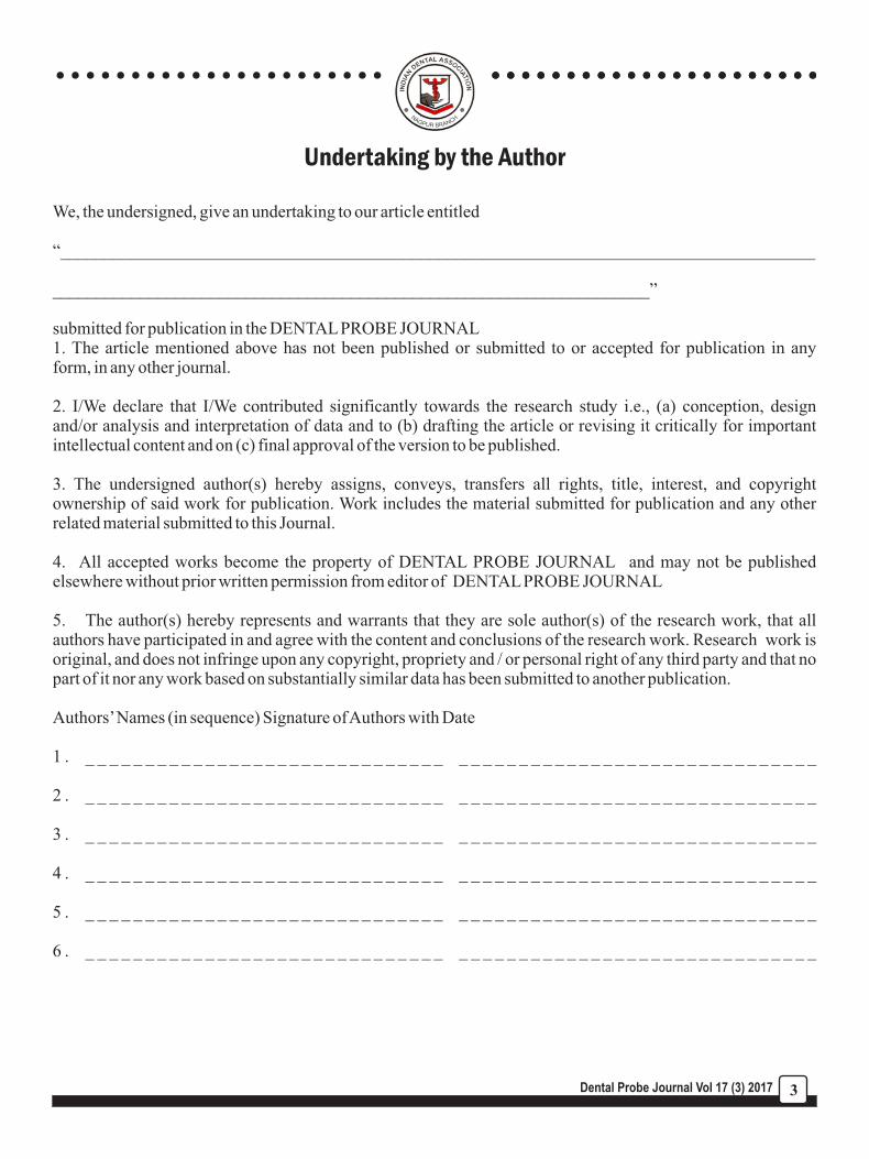
We, the undersigned, give an undertaking to our article entitled
“______________________________________________________________________________________
____________________________________________________________________”
submitted for publication in the DENTAL PROBE JOURNAL 1. The article mentioned above has not been published or submitted to or accepted for publication in anyform, in any other journal. 2. I/We declare that I/We contributed significantly towards the research study i.e., (a) conception, designand/or analysis and interpretation of data and to (b) drafting the article or revising it critically for important intellectual content and on (c) final approval of the version to be published.
3. The undersigned author(s) hereby assigns, conveys, transfers all rights, title, interest, and copyright ownership of said work for publication. Work includes the material submitted for publication and any other related material submitted to this Journal.
4. All accepted works become the property of DENTAL PROBE JOURNAL and may not be published elsewhere without prior written permission from editor of DENTAL PROBE JOURNAL
5. The author(s) hereby represents and warrants that they are sole author(s) of the research work, that all authors have participated in and agree with the content and conclusions of the research work. Research work is original, and does not infringe upon any copyright, propriety and / or personal right of any third party and that no part of it nor any work based on substantially similar data has been submitted to another publication.
Authors’ Names (in sequence) Signature of Authors with Date
1 . _ _ _ _ _ _ _ _ _ _ _ _ _ _ _ _ _ _ _ _ _ _ _ _ _ _ _ _ _ _ _ _ _ _ _ _ _ _ _ _ _ _ _ _ _ _ _ _ _ _ _ _ _ _ _ _ _ _ _ _
2 . _ _ _ _ _ _ _ _ _ _ _ _ _ _ _ _ _ _ _ _ _ _ _ _ _ _ _ _ _ _ _ _ _ _ _ _ _ _ _ _ _ _ _ _ _ _ _ _ _ _ _ _ _ _ _ _ _ _ _ _
3 . _ _ _ _ _ _ _ _ _ _ _ _ _ _ _ _ _ _ _ _ _ _ _ _ _ _ _ _ _ _ _ _ _ _ _ _ _ _ _ _ _ _ _ _ _ _ _ _ _ _ _ _ _ _ _ _ _ _ _ _
4 . _ _ _ _ _ _ _ _ _ _ _ _ _ _ _ _ _ _ _ _ _ _ _ _ _ _ _ _ _ _ _ _ _ _ _ _ _ _ _ _ _ _ _ _ _ _ _ _ _ _ _ _ _ _ _ _ _ _ _ _
5 . _ _ _ _ _ _ _ _ _ _ _ _ _ _ _ _ _ _ _ _ _ _ _ _ _ _ _ _ _ _ _ _ _ _ _ _ _ _ _ _ _ _ _ _ _ _ _ _ _ _ _ _ _ _ _ _ _ _ _ _
6 . _ _ _ _ _ _ _ _ _ _ _ _ _ _ _ _ _ _ _ _ _ _ _ _ _ _ _ _ _ _ _ _ _ _ _ _ _ _ _ _ _ _ _ _ _ _ _ _ _ _ _ _ _ _ _ _ _ _ _ _
Undertaking by the Author
...................... ............................................
Dental Probe Journal Vol 17 (3) 2017 3
Abstract
Since people began using denture adhesives
more than 200 years ago, dentists have been slow to
acknowledge their place in prosthetic dentistry.
Denture Adhesives are material used to adhere a
denture to the oral mucosa. Most dentist advice the
use adhesive in the clinical practice but yet not have
clear idea of denture adhesive literature states it is
used in clinical practice yet a neglected topic of
discussion. In current article it is an attempt to discuss
various adhesive available in market, their
composition, its mechanism of action, method of
application and removal of denture adhesive and the
side effects of it.
Introduction
With increasing life expectancy, complete
denture is one of the major treatment modalities in
Prosthodontics for Indian scenario.One of the major
contributing factors for the success of a complete
denture is perceived retention of the prosthesis by the 1patient. Those who wear complete dentures, are often
confronted with varying degrees of looseness of their
prosthesis and complain of discomfort and/or reduced 2
masticatory function or speech.
Retention is defined in GPT as that quality inherent in
the dental prosthesis acting to resist the force of
dislodgment along the path of placement.The
enhancement of retention and stability, which are
major properties that determine the performance of a
removable prosthesis, has always been a goal of 2prosthetic dentistry. Retention of dentures in the oral
cavity is controlled by a complex interrelationship of
physical, biological, physiological and mechanical
properties.Denture adhesive has become a common
adjuvant in complete denture treatment which not
only improves the retention but also positively
impacts the patient comfort and confidence level.
Denture adhesive refers to a commercially available,
nontoxic, soluble material that is applied to the tissue
surface of the denture to enhance denture retention, 1
stability, and performance.
In the present article denture adhesive has been
reviewed in detail.
History
The use of denture adhesives, started in the late 18th century, before which adhesives were not part of the dentist’s armamentarium. Adhesives or fixatives used in the 19th century,were formulated by mixing vegetable gums toproduce a material that absorbed moisture from the salivaand swelled to a mucilaginous substrate that adhered to the mucosa of the mouth and denture. The earliest patent pertainingto adhesives was issued in 1913 followed by othersin 1920s and 1930s. The first reference by the American Dental association to denture adhesives
Most Neglected yet important in clinical practice
“Denture Adhesive” a review
Address for correspondences :Dr. Pratik Hodar
nd2 year post graduate student Department of Prosthodontics Dr Hedgewar Smruti Rugna Seva Mandal’s Dental College and Hospital, Hingoli.
1) Dr. Pratik Hodar 2) Dr. Dilip Dhamankar 3) Dr. Sashi Purna 4) Dr. Brijesh Dammani
...................... ............................................
Dental Probe Journal Vol 17 (3) 20174

camefrom the Accepted Dental Remedies of 1935 in which theCouncil of Dental Materials, Instruments, and Equipmentadmitted that these products were
3nonmedical.1
Ideal Requirements of Denture Adhesive
1. It should be biocompatible and non-toxic
odour less and tasteless.
2. It should be easy to remove, clean and replace.
3. It should not cause damage to denture base
material and soft liner.
4. It should exhibit antimicrobial properties.
5. It should have a longer shelf life.
6. It should be long lasting (8-12 hours)
7. It should be economic.
Classification
Denture adhesive can be classified in various ways such as on based on its solubility as soluble and insoluble. It can also be classified on bases of its composition as, zinc containing and zinc free, with medicaments and without medicaments some of denture adhesive has antifungal agent which can be used to treat denture stomatitis. It can also be classified on its base composition as oil based and water based adhesive (Table 1)
Composition
Denture adhesive mainly contains adhesive group i.e carboxymethylcellulose it is a short acting polymer, polymethylvinyl ethermaleic anhydride it is a long acting polymer. These adhesives are mainly used in food industry. But due to its short acting action divalent salts like zinc and calcium were added to denture adhesive to prolong its action by Shah et al. there have been several reports that excess of zinc intake from denture adhesive, results in bone marrow suppression.4,9 These haematological abnormalities are due to zinc induced copper deficiency; copper supplementation with removal of external zinc source, which was identified as zinc containing denture adhesive promptly resolved the symptoms.4,9Copper deficiency has the potential to induced neurological disorder in human and animal, but this requires relatively long duration of exposure to zinc.1,4,9 Zinc containing formulations of denture adhesive are being phased out of the market; however, the adverse effect resulting from improper use of denture adhesive emphasise the importance of patient education when using these material.1 Adhesive contain a binder in it bindsall the composition together binder such as Petrolatum, mineral oil, polyethylene. For prevention of clumping it has
Insoluble e.g. secure comfort stripZinc Free
e.g. Super Poligrip Free Super Poligrip Comfort Seal Strips Super Poligrip Powder Protefix Adhesive Cream, Extra-Strong Fittydent Super Adehesive Cream
Without Medicament e.g. Fixodent plus, Fitty dentWater based eg Fixodent plus, Fitty dent
Soluble e.g. Fixodent, fitty dentZinc Containing
e.g. Super Poligrip Original Fixodent Original Fixodent Fresh Fixodent Control Fixodent Complete Fixodent Comfort Fixodent Control Plus Scope FlavorSuper-Haftcreme Extra StarkWith Medicament e.g. GeiserpharmaOil based eg Olivafix
Based on SolubilityBased on Composition
Base on base compostion
Table 1 Classification of Denture Adhesive
...................... ............................................
Dental Probe Journal Vol 17 (3) 2017 5
Mechanism of action
The physical factors effecting denture
retention are based on a principle derived by Stefan
over a century ago, which states that the force
required to pull two disks or plates apart is directly
proportional to the viscosity of the liquid between
them. Denture adhesive increases the viscosity of the
saliva, it swells 50-150% by volume in presence of
water, and fills space between the intaglio surface of
denture and basal seat. Thus enhance the physical
factors adhesion, cohesion, interfacial surface
tension.
The correct application of denture adhesive to a
denture is as follows:
1. Clean and dry the tissue-bearing surface of the
denture.
2. A proper amount of denture adhesive should be
used. For maxillary denture 3-4 pea sized increments
of the adhesive cream can be applied to the anterior
ridge, midline and palate. For mandible, 3 pea sized
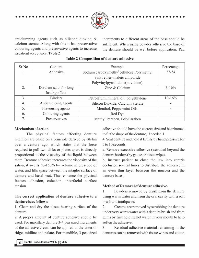
anticlumping agents such as silicone dioxide & calcium sterate. Along with this it has preservative colouring agents and preservative agents to increase itspatient acceptance. Table 2
increments to different areas of the base should be
sufficient. When using powder adhesive the base of
the denture should be wet before application. Pad
adhesive should have the correct size and be trimmed
to fit the shape of the denture, if needed.1
4. Seat denture and hold it firmly by hand pressure for
5 to 10 seconds.
a. Remove excessive adhesive (extruded beyond the
denture borders) by gauze or tissue wipes.
b. Instruct patient to close the jaw into centric
occlusion several times to distribute the adhesive in
an even thin layer between the mucosa and the
denture bases.
Method of Removal of denture adhesive.
1. Powders removed by brush from the denture
using warm water and from the oral cavity with a soft
brush and toothpaste.
2. Creams are removed by scrubbing the denture
under very warm water with a denture brush and from
gums by first holding hot water in your mouth to help
soften the adhesive.
3. Residual adhesive material remaining in the
dentures can be removed with tissue wipes and cotton
Table 2 Composition of denture adhesive
Sr No
1.
2.
3.
4.
5.
6.
7.
Content
Adhesive
Divalent salts for long
lasting effect
Binders
Anticlumping agents
Flavouring agents
Colouring agents
Preservatives
Examplel
Sodium carboxymethy cellulose Polymethyl
vinyl ether–maleic anhydride
Polyvinylpyrrolidone(povidone).
Zinc & Calcium
Petrolatum, mineral oil, polyethylene
Silicon Dioxide, Calcium Sterate
Menthol, Peppermint Oils.
Red Dye
Methyl Paraben, PolyParaben
Percentage
27-54
3-16%
10-16%
-
-
-
...................... ............................................
Dental Probe Journal Vol 17 (3) 20176
applicators soaked with orange solvent.
4. Avoid scratching or mutilating the tissue-
bearing surface of the denture.
Literature on denture adhesive says
There is no longitudinal report of local tissue 1reaction to denture adhesive. However, there have
been several reports that excess of zinc intake from
denture adhesive results in bone marrow 4 , 9
suppression. Various studies based on
biocompatibility of denture adhesive show that,
keratin level of mucosa decrease with prolong use of
adhesive. Studies did not show any inflammatory
response of adhesive to the mucosa except in those 6, 7
patient with poor oral hygiene.
Studies also indicate that there is no statistical
alteration in population of C.albicans, streptococcus
M in saliva, palate denture between adhesive uses & 8, 10, 11non users.
Studies showed that there is an increased denture base 12,13,19,21retention and stability, but substantial loss of
retention in mandibular dentures after mastication 12 and sipping. There was no recognizable effect of
denture adhesives on denture mobility in patients with 14
mild alveolar resorption but have more significant
results in individuals with poor denture bearing 13, 15, 22 16
tissues. Improvement in occlusal forces &
Significant improvement of maximum incisal force
seen for both new and previous complete dentures 17, 20patients with unfavourable bearing tissues.
Indication
1. To improve retention stability and masticatory
efficiency.
2. Provide psychological benefits to the patient.
3. Secure interim immediate or new dentures.
4. Apply medications via the oral mucosa.
5. Simplify placement for specific condition, like
patients with xerostomia, geriatric patients, patient
with poor muscle tone (such as those with Parkinson’s
disease).
6. Stabilize trial bases during jaw relation and trial
denture.57. Retention of maxillofacial prosthesis.
Contraindication
1. Denture adhesive should aid but not substitute
well-fitting denture.
2. Denture adhesive should not be used with ill-fitting
dentures or by patients who tend to overuse denture
adhesives.
3. Denture adhesive should not be used by patients
who have medication induced xerostomia where
adhesives require ample saliva.
4. Not for use with immediate, temporary or
transitional dentures where trauma could result from
inadequate hygiene or adherence to suture.
Conclusion
As dentists, it is our responsibility to be
knowledgeable and caring enough to assist each
patient in adapting to dental prostheses. This may
require recommendation of denture adhesives and
counselling on their use. Also, continued research and
vigilance into the use of denture adhesives is
essential.
Reference
1. Zarb GA, Bolender CL. Boucher`s Prosthodontic
treatment of the edentulous patient, 13th edition, St
Louis; Mosby Year book 1997, pp155.
2. Papadiochou S: Denture adhesives: A systematic
review. J Prosthet Dent 2015; 11; pp1-7.3. American Dental Association. Accepted dental remedies. Chicago: American Dental Association. 1935; 172.
...................... ............................................
Dental Probe Journal Vol 17 (3) 2017 7
4. Tezvergil-Mutluay A, Carvalho RM, Pashley DH: Hyperzincemia from ingestion of denture adhesives. J Prosthet Dent 103:380-383, 20105. Amato L, Asher ES: Use of denture adhesive to retain an extra oral facial prosthetic wax pattern for trial placement. J Prosthet Dent 2002;88:542-36. Abdelmalek RG, Michael CG. The effects of denture adhesives on the palatal mucosa under complete dentures. A clinical and histological investigation. Egypt Dent J 1978; 24:419-30.7. Tarbet WJ, Grossman E. Observations of denture-supporting tissue during six months of denture adhesive wearing. J Am Dent Assoc 1980;101:789-918. Kim E, Driscoll CF, Minah GE. The effect of a denture adhesive on the colonization of Candida species in vivo. J Prosthodont 2003; 12:187-91.9. Hedera P, Peltier A, Fink JK, Wilcock S, London Z, Brewer GJ. Myelopolyneuropathy and pancytopenia due to copper deficiency and high zinc levels of unknown origin. II. The denture cream is a primary source of excessive zinc. Neurotoxicology 2009; 30:996-9.10. Özkan YK, Uçankale M, Ozcan M, Uner N. Effect of denture adhesive on the micro-organisms in vivo. Gerodontology 2012; 29:9-16.11. Leite AR, Mendoza-Marin DO, Paleari AG, Rodriguez LS, Roccia AA, Policastro VB, et al. Crossover clinical trial of the influence of the use of adhesive on biofilm formation. J Prosthet Dent 2014; 112:349-5612. Kapur KK. A clinical evaluation of denture adhesives. J Prosthet Dent 1967; 18:550-8. 13. Chew CL, Boone ME, Swartz ML, Phillips RW. Denture adhesives: their effects on denture retention and stability. J Dent 1985; 13:152-9.14. Karlsson S, Swartz B. Effect of a denture adhesive on mandibular denture dislodgment. Quintessence Int 1990; 21:625-7. 15. Fujimori J, Hirano S, Hayakawa I. Effects of a denture adhesive on masticatory functions for complete denture wearers-consideration for the
condition of denture-bearing tissues. J Med Dent Sci 2002; 49:151-6. 16. Psillakis JJ, Wright RF, Grbic JT, Lamster IB. In practice evaluation of a denture adhesive using a gnathometer. J Prosthodont 2004; 13:244-50. 17. De Baat C, van’t Hof M, Van Zeghbroeck L, Ozcan M, Kalk W. An international multicenter study on the effectiveness of a denture adhesive in maxillary dentures using disposable gnathometers. Clin Oral Investig 2007; 11:237-43. 18. Figueiral MH, Fonseca PA, Pereira-Leite C. The effect of different adhesive materials on retention of maxillary complete dentures. Int J Prosthodont 2011; 24:175-7. 19. Mañes J, Selva E, De-Barutell A, Bouazza K. Comparison of the retention strengths of three complete denture adhesives: an in vivo study. Med Oral Patol Oral Cir Bucal 2011; 16:132-6. 20. Kalra P, Nadiger R, Shah FK. An investigation into the effect of denture adhesives on incisal bite force of complete denture wearers using pressure transducers-a clinical study. J Adv Prosthodont 2012; 4:97-102. 21. Munoz CA, Gendreau L, Shanga G, Magnuszewski T, Fernandez P, Durocher J. A clinical study to evaluate denture adhesive use in well-fitting dentures. J Prosthodont 2012; 21:123-9.22. Gonçalves TM, Viu FC, Gonçalves LM, Garcia RC. Denture adhesives improve mastication in denture wearer. Int J Prosthodont 2014; 27:140-6. 23. Grasso JE. Denture adhesives. Dent Clin North Am 2004; 48:721-3.24. Grasso JE. Denture adhesives: changing attitudes. J Am Dent Assoc 1996; 127: 90-6.
...................... ............................................
Dental Probe Journal Vol 17 (3) 20178
Introduction :
The oral cavity contains a wide variety of
microorganisms which can cause various infectious
diseases. Since dental professionals work in an
environment that is bathed by blood and saliva of the
patients, they are at a higher risk of contacting 1infectious diseases. In 2003, the Centre for Disease
Control and Prevention of the United States of
America (CDC) updated their guidelines for infection 2
control in dental settings.
The use of effective infection control
procedures and universal precautions in the dental
office will prevent cross contamination that could
extend to dental professionals, dental office staff, 3
dental technicians and the patients.
Technicians are particularly vulnerable to microbial
cross-contamination from the impressions they
receive from dental offices. Casts poured from
impressions can also harbour infectious
microorganisms that can be distributed throughout
the laboratory when the casts or dies are trimmed. 4
The aim of this review is to provide a
background about the possible ways of transmission
of infection spreading, and procedures recommended
for preventing their spread in the discipline of
prosthodontics.
Sterilizationis a process by which all forms of
microorganisms are destroyed, including virus, 5bacteria, fungi, and spores. Disinfectionis a less
lethal process than sterilization. It eliminates all
recognized pathogenic microorganisms but not
necessarily all microbial forms i.e. bacterial 5
endospores on inaminate objects.
6Infection control procedures:
R.R. Runnelsin 1988 gave basic infection
control procedures as mandatory for the control of
infectious diseases in dental practice. These are:
* All dental treatment personnel should wear latex
gloves during treatment.
* All dental treatment personnel should wear masks
covering the nose and mouth during treatment.
* All dental treatment personnel should wear
protective eyewear during treatment.
* All items used in the oral cavity should be
sterilized.
* All “touch & splash” surfaces should be
disinfected with an EPA registered disinfectant.
* Contaminated material should be carefully
disposed by placing it in sealed and marked
containers.
Management of Instruments
According to the CDC, dental instruments are
classified into three categories depending on the risk
1) Dr. Ishani P. Bindra
2) Dr. Sham M. Gundawar
3) Dr. Usha M Radke
4) Dr. Sneha Mehta
5) Dr. Trupti Bangare
Address for correspondences :Dr. Ishani P. BindraLecturer, Dept. of Prosthodontic, Swargiya Dadasaheb Kalmegh Smruti Dental College and Hospital, Nagpur.
Infection Control in The Dental Clinic And Labratory : A Reveiw
...................... ............................................
Dental Probe Journal Vol 17 (3) 2017 9
7of transmitting infection:
1) Critical instruments such as forceps, scalpels,
bone chisels, scalers and surgical burs which
penetrate soft tissue or bone, or enter into or contact
the bloodstream should be sterilized after each use.
2) Semi-critical instruments like mirrors,
impression trays and amalgam condensers that do not
penetrate soft tissues or bone but contact mucous
membranes or non- intact skinshould also be
sterilized after each use.
3) Non-critical instruments that come into
contact only with intact skin such as external
components of x-ray heads have a relatively low risk
of transmitting infection; and, therefore should be
disinfected.
8Disinfection of Impressions
Many studies have been performed to
evaluate effects of various disinfectants on different
types ofimpression materials.No single disinfectant is
compatible with all impression materials.
Impression must be rinsed to remove saliva,
blood and debris and disinfected before being sent to
the laboratory. Simple washing removes 90% surface
bacteria. By disinfection 100% removal is achieved.
Disinfection by spraying: The rinsed impression is
sprayed with an acceptable disinfectant and put in
plastic zipped bag and sprayed. The bag is sealed to
create a “charged atmosphere”.After the end of the
exposure time, the impression is rinsed in running
water to remove the excess.
Advantages of this method include use of less
disinfectant as compared to immersion but the
disadvantage is that it is not as effective as immersion
and the disinfectant may be released into air
increasing occupational exposure.
Disinfection by immersion :
Preferred over spraying as it provides a
constant contact of the disinfectant with all surfaces
of the impression. It is done by placing the impression
in a zipped plastic bag with the appropriate
disinfectant.
9,10Disnfection of Impressions
1) Irreversible hydrocolloid: Alginate
Current CDC protocol is to use synthetic
phenolsas disinfectants. Ten minutes spray with
sodium hypochlorite is also effective. Spraying with
disinfectants does not affect the dimensional stability
as compared to immersion.
Recommended: Chlorine compounds or iodophors
2) Reversible hydrocolloid: Agar
Spraying with sodium hypochlorite 1 : 10 for
10 min or Iodophor 1 : 213 is effective. The
impression should not be immersed in alkaline
glutraldehyde.
3) Polysulfide
The impression should be rinsed and
immersed in sodium hypochlorite 1:10 for 10 min.
Disinfectants requiring more than 30 minute
exposure time are not recommended.
Recommended: Glutraldehyde, chlorine compounds,
iodophors , phenols
4) Addition silicone
The material is susceptible to damage by
neutral gluteraldehyde.Immersion longer than 15 min
because longer time may cause the surfactant in the
hydrophilic polysiloxane to leach out and render the
impression less hydrophilic.
Alternative-iodophor
5) Condensation silicone
...................... ............................................
Dental Probe Journal Vol 17 (3) 201710
The impression material is unaffected by
immersion disinfectant provided that the disinfection
time is short.
Recommended: 2% gluteraldehyde, Sodium
hypochlorate- 1 : 10 for 10 min
6) Polyether
The impression is subjected to dimensional
change if immersed for more than 10 minutes because
of the hydrophilic nature of the material. ADA
recommends any of the disinfectant classes, with
short term exposure to avoid distortion2%
Glutraldehyde provides satisfactory disinfection.
Recommended: chlorine compounds or iodophors
Zinc oxide eugenol impression material
Immersion is preferred.Spraying may be used
for bite registrations. The material is not compatible
with chlorine compounds.
Recommended:Glutraldehydes or iodophors
Impression compound
For the disinfection of this material, phenolic
spray can be used.
Recommended: Iodophors or chlorine compounds.
Impression trays
Plastic disposable trays should be discarded
after single use. Sodium hypochlorite can be used as a
disinfectant on aluminium or chrome-plated trays but
these should be monitored for corrosion.Impression
trays can also be heat-sterilized.
Custom acrylic resin impression trays should
be disinfected by spraying with surface disinfectants
or immersing in either 1:213 iodophor or 1:10 sodium
hypochlorite. After disinfection, they should be
rinsed to remove any residual disinfectant.
Disnfection of Other Materials or Instruments 11,12,13,14
Disinfection of gypsum cast
It is preferable to disinfect the impression so
that the cast will not have to be disinfected as these
are difficult to disinfect without causing damage
ADA recommends that stone casts be
disinfected by the spraying until wet or immersing in
a 1:10 dilution of sodium hypochlorite or an
iodophor.. Investigators submerged die stone models
in a variety of disinfectants and found that with 1:10
sodium hypochlorite and 1:213 iodophor, undesirable
physical effects on set die stone ranged from none to
minimal.
Disinfection of prosthesis
The ADA recommends disinfection by
immersion in iodophors or chlorine compounds.
Although both of these disinfectants are corrosive,
studies have shown little effect on chrome cobalt
alloy with short-term exposure (10 minutes) to
iodophors or 1:10 hypochlorite. Damage of heat
cured denture base resin has been shown to occur after
only 10 minutes of immersion in a glutaraldehyde
with phenol buffer, although immersion in 2%
alkaline glutaraldehyde did not damage the acrylic
surfaces. Fixed metal/porcelain prosthesis may be
disinfected by immersion in gluteraldehydes ,diluted
hypochlorite.
Oral Safe is a germicide-deodorant that is
harmless if ingested. It destroys 99 percent of
microbes on removable appliances during 10 minutes
of submersion.A three-minute procedure that
combines the use of a germicide-deodorant with
ultrasonic energy kil ls 10 t imes more
microorganisms than passive submersion. Also,
microwave disinfection for 3 minutes at 650 kilowatts
has shown good result. After disinfection and
...................... ............................................
Dental Probe Journal Vol 17 (3) 2017 11
ZOE).
Non-sterilizable equipments such as some
face bow components must be cleaned with soap and
water and disinfected with a hospital-level
disinfectant if they become contaminated.. The
method of choice is spraying or soaking these items in
the disinfectant in a separate container or bag.
Iodophors, chlorine solutions, glutaraldehydes or
phenols are all acceptable for this step. It is important
to remember that most immersion disinfectants can
only be used once.
Conclusion
The increased awareness of the consequences
of cross- contamination with hepatitis B virus (HBV)
and HIV during dental procedures is having a
growing impact on attitudes towards infection control
in the dental clinic and laboratory.
Dentists must ensure that a basic infection
control procedure is observed when treating patients
and additional control procedures are observed in the
fabrication and handling of the impressions and
dental prosthesis.
Dental offices and dental laboratories should
co-ordinate to control the potential cross-infections
between the two disciplines.
References
1) Naveen B H, et al. Infection control in
prosthodontics. Journal of Dental Sciences and
Research 2011; 2: 93-107.
2) Kohn WG, Collins AS, Cleveland JL, Harte
JA, Eklund KJ, Malvitz DM, et al. Guidelines for
infection control in dental healthcare settings.
MMWR Recomm Rep.2003; 52(RR-17): 1 –61.
3) Kumar R, Maller S. Infection control in
prosthodontics. JIADS 2010; 1(2) : 22-24.
4) Vidya S. Bhat VS, Shetty MS, Shenoy KK.
thorough rinsing, acrylic items can be stored in
diluted mouthwash until inserted.
Polishing lathes (pumice and dry) should be
disinfected by pumice solution which can be made by
suspending the pumice in tincture of green soap or
other surfactant and adding an effective disinfectant
solution to the mix.
Rubber items (spatula & rubber bowls) and
saliva ejectors are sterilised byethylene oxide
sterilization. Dry or moist heat sterilization may cause
damage to these, hence are avoided.
Hand pieces may be sterilised by steam, dry
heat or ethylene oxide sterilization and Airrotor burs
may be sterilised by either moist heat or dry heat
sterilisation.
Shade guides should be cleaned and
disinfected to avoid cross contamination. If iodophors
are used on shadeguides, they should be wiped with
water or alcohol after the exposure time to remove
any residual disinfectant.
Wax bites / rims, bite registrations should be
disinfected by the spray wipe spray method using an
iodophor as recommended by the ADA. Rinse spray
may be more appropriate for wax bites. These
itemsshould be rinsed again after disinfection to
remove any residual disinfectant.
Bite registrations made of various materials
such as ZOE or compound can be handled in the same
manner as impressions of the same materials. These
registrations also can be disinfected, using the rinse
spray rinse technique, with most EPA registered
hospital level tuberculocidal disinfectants used as
sprays (chlorine compounds should not be applied to
...................... ............................................
Dental Probe Journal Vol 17 (3) 201712
Infection control in the prosthodontic laboratory.
Journal of Indian Prosthodontic Society 2007 Vol 7
Issue 2 page 62-5.
5) W. Patrick Naylor. Infection control in fixed
prosthodontics. DCNA July 1992; 36(3):809-31.
6) Runnells. R.R. “An over view of infection
control in dental practice”. J. Prosthet. Dent., 1988;
59: 625.
7) Sterilization & Disinfection In Prosthodontics
Indian Journal of Dental Sciences. October 2014
Supplementary Issue Issue:4, Vol.:6
All rights are reservedSiddharth Phull 2 Arvind Arora
Yashendra
8) Jain S. et al., Int J Dent Health Sci 2014;
1(5):779-787
9) Philips science of dental materials. Eleventh
edition.
10) Science of dental materials – Skinner
11) Wood PR. Cross infection control in dentistry
a practical illustrated guide.
12) Rowell GL, Runnells RD, Saxon BA,
Whisenant BK. The presence and identification of
organisms transmitted to dental laboratories. J
Prosthet Dent 1990;64:235-7.
13) Dental laboratory relationship working Group
OSAP Position paper. Laboratory Asepsis: November
1998.
14) Infection control recommendations for the
dental office and the dental laboratory. ADA Council
on Scientific Affairs and ADA Council on Dental
Practice. J Am Dent Assoc 1996;127:672-80.
...................... ............................................
Dental Probe Journal Vol 17 (3) 2017 13
Abstract:
Successful osseointegration is a prerequisite
for functional dental implants. Continuous
monitoring in an objective and quantitative manner is
important to determine the status of implant stability.
Historically, the gold standard method used to
evaluate degree of osseointegration was microscopic
or histologic analysis. However, due to the
invasiveness of this method and related ethical issues,
various other methods of analysis have been
proposed: radiographs, cutting torque resistance,
reverse torque, modal analysis, and resonance
frequency analysis. This review focuses on the
methods currently available for the evaluation of
implant stability.
Key words: cutting resistance analysis, implant
stability evaluation, radiographic assessment,
resonance frequency analysis, reverse torque test
Introduction:
Fruitful osseointegration is primary for
practical dental implants, and primary implant
Methods used to assess implant stability : Current Status
1) Dr. G. Soni
2) Dr. G. Bhutada
3) Dr. M. K. Mishra
4) Dr. G. Niswade
5) Dr. S. Ansari
stability isprime for effective osseointegration.
Implant stability is the absence of clinical mobility.
Implant instability could bring about fibrous
encapsulation with resultant disappointment. Primary
implant stability at placement is a mechanical wonder
that is identified with the nearby bone quality and
amount, the sort of implant and arrangement
procedure utilized. Secondaryimplant stability is the
expansion in dependability inferable from bone
arrangement and rebuilding at the implant /tissue [1,2]interface and in the encompassing bone.
Under defined conditions, early and
immediate loadingprotocols have now been
perceived to be reasonable other options to the
established 1-or 2-stage delayed loading approaches.
Hence, the clinician needs dependable and strong
target rules to decide on an individual premise the
visualization of a given implant, if immediately
loaded, early loaded within 6-8 weeks or left [3]traditionally to heal for a 3-6 months’ time span.
Generally, the gold standard technique used to
assess the level of osseointegration was microscopic [4]or histologic analysis. However, due to the
invasiveness of this strategy and related moral issues,
different techniques for analysis have been proposed;
clinically checking for mobility with the assistance of
limit finished instruments, radiographs, cutting
torque resistance, reverse torque and resonance
Address for correspondences :Dr. G. Soni
Swargiya Dadasaheb Kalmegh Smruti Dental College and Hospital Nagpur
Post Graduate Student
...................... ............................................
Dental Probe Journal Vol 17 (3) 201714

frequency analysis (RFA).
Measuring insert stability underpins using sound
judgment about when to load, permits profitable
convention decision on a patient-to-patient basis,
demonstrates circumstances in which it is best to
unload, bolsters great correspondence and expanded [5]
trust and gives better case documentation. The
techniques to decide implant stability clinically are
clinical observation, percussion test, switch torque
test, cutting torque protection analysis, Periotest,
RFA.
Clinical Perception:
The clinical view of primaryimplant stability
is much of the time considering the portability
identified by limit finished instruments. It's an
exceptionally inconsistent and irregular strategy. It
can likewise be checked by the cutting protection of
the implant amid its addition. The sentiment "great"
stability might be highlighted if there is the feeling of
an unexpected stop at the seating of the implant. Root
types of tapered implants frequently have a geometry
that will give a firm stop and maybe a bogus [6]
impression of high security.
Percussion Test
The percussion test may include the tapping of
a mirror handle against the implant carrier and is
intended to evoke a ringing sound from the implant as
a sign of good stability or osseointegration.
Percussion tests most likely give more data about the
tapping instrument, and will, best case scenario just [6] yield poor subjective data.
Reverse torque test
Use of a reverse or unscrewing torque has
additionally been proposed for the appraisal of [7]implant stability at the time of abutment connection.
Implants that turn under the connected torque are
considered disappointments and are then removed.
Nevertheless, the implant surface during the time
spent osseointegrating, though gradually, may break
under the connected torque stretch. Besides, as animal
tests have exhibited the re-integration of released and
rotationally mobile implants, the invert/reverse [8]
torque testing has fallen into notoriety
Cutting torque protection analysis
The energy required for a current-fed electric
engine in removing a unit volume of bone amid [9,10,11]
implant surgery is measured. The energy
corresponds to bone thickness, which is one of the
variables deciding implant stability. Nonetheless, as
far as possible value has not been set up, which can
indicate potential disappointment of the implant.
Also, it must be utilized amid the surgery and not as an
analytic guide, and it can't evaluate the secondary
stability by new bone arrangement and remodeling [3]around the implant.
Periotest
It is a gadget which is an electrically
determined and electronically observed tapping head
that percusses the implant for a total of 16 times. The
whole measuring methodology takes around 4
seconds. The instrument incorporates a tapping bar
that effects the projection/implant gathering. The bar
is drawn by an impetus curl toward the affecting
surface and basically moves at a steady speed from
the minute it leaves the hand piece until the point that
it impacts the surface. This implies over a specific
separation (around 4 mm), the tapping pole is moving
at a similar speed and is intended to affect the surface
whenever amid this steady speed travel. The finish of
...................... ............................................
Dental Probe Journal Vol 17 (3) 2017 15

the pole inside the hand piece is inflexibly associated
with an accelerometer, which creates a yield relative
to its increasing speed. The readings are from −8 to
+50 and are deciphered as in [Table 1].
Reading Interpretation
-8 to 0 Good osseointegration, implant can
be loaded
+1 to +9 Clinical examination is required,
in most cases loading is not possible
+10 to +50 Osseointegration is not sufficient,
implant cannot be loaded
The variables that impact the Periotest value are the
nature of the hard tissue in the locale of the implant,
with the goal that no values can be considered as
proper for higher or bring down degrees of [12]coordination. It is an element of the separation from
the implant spine to the time when the bar impacts the
projection. These varieties recommend that for
implants, there is no supreme value that can be viewed
as satisfactory; rather, varieties that happen after
some time might be more significant.
In vitro assessments uncovered that no
measurably significant distinction existed in
measuring Periotestvalues from the administrator to
administrator, and in addition abnormal state of
repeatability between various Periotest units.
Effectively incorporated dental implants have yielded
an extensive variety of security readings with the
Periotest. This range in values is accepted to reflect
bone thickness at the implant interface, which is
identified with implant area.
The estimations are primarily influenced by
excitation conditions, for example, heading and
position. The estimations must be made in the mid
buccal district and be opposite to the implant
tomahawks. Considering the intra oral condition, it is
extensively simple to make estimations on front
implants while it isn't feasible for molars inferable
from the buccal mucosa. The Periotest can't analyze a
"marginal" case or "an implant in the process of [13]osseointegration." It doesn't reflect the level of
peri-implant bone and in this way, can't be substituted
for radiography.
Resonance Frequency Analysis
It is a noninvasive indicative strategy that
measures implant stability and bone thickness at
different time focuses utilizing vibration and
auxiliary guideline analysis. [14] Two industrially
gadgets have been produced to survey implant
soundness. The first (electrical) technique utilizes an
immediate association (wire) between the transducer
and the resonance frequency analyzer. The second
strategy utilizes attractive frequencies amongst
transducer and resonance frequency analyzer. In the
electronic gadget, the transducer is L formed
cantilever shaft which associates with the implant by
means of a screw connection. A piezoelectrical crystal
on the vertical bit of the L shaft is utilized to animate
the implant /transducer complex; second
piezoelectric crystal on the inverse side of the pillar is
utilized as an accepting component to recognize the
reaction of the bar.
The new attractive RFA gadget has a
transducer, a metallic pole with a magnet to finish
everything, which is screwed onto an implant or
abutment. The magnet is energized by an attractive
heartbeat from a remote test. The beat span is around 1
ms. After excitation, the peg vibrates unreservedly,
and the magnet prompts an electric voltage in the test
curl. That voltage is the estimation flag inspected by
the resonance frequency analyzer. The electronic
gadget and the attractive gadget are fit for measuring
comparative changes; however, the attractive gadget
...................... ............................................
Dental Probe Journal Vol 17 (3) 201716

brings about higher implant stability quotient (ISQ)
value when measuring the stability of non-submerged
dental implant.
With this strategy, implant stability is
measured either by deciding the resonance frequency
of the implant bone complex or by perusing an ISQ
value given by the Osstell device (Integration
Diagnostics AB, Gothenburg, Sweden) or Penguin
RFA (Neoss, Gothenburg, Sweden). Traditionally, the
ISQ has been found to fluctuate near 40 and 80, the [3]
higher the ISQ, the higher the implant stability. A
significant increment or reduction in implant stability
could be recognized with this strategy that generally
couldn't be clinically seen. The components
influencing the readings are powerful implant length,
bone quality and amount, implant length, distance
across and shape. Successful implant length is the
length of the exposed threads and abutment height. It [15]is contrarily relative to the resonance frequency.
Implant stability can be resolved for implants
with an ISQ of 47. All implants with an ISQ more than
49 osseointegrated when left to heal for 3 months. All
implants with an ISQ more than 54 osseointegrated
when quickly loaded. For implants with low ISQ
values a reduction in implant stability should caution
the specialist to present these implants to a more
tightly follow-up plan and to take extra prudent
estimations as far as emptying until implant strength
is recovered or if non-loaded to check for mechanical
injury or potentially disease. For implants with high
ISQ values, diminishment of implant strength amid
the first 12 weeks of mending ought to be considered
as a typical occasion that ought not require change of [3]routine development.
The downsides with this innovation are that
the transducer is restricted to an arrangement of 60
estimations, along these lines making the strategy
rather costly. With a specific end goal to play out the
RFA, a transducer is fixed to the implant.This bar
observing all implants that help a solidified
reclamation.
Conclusion:
Even though there are different techniques
which help to decide implant soundness, the number
factors influencing the outcomes makes it hard to go
to a basic value which can decide the achievement,
disappointment or long-haul guess of an implant.
Thus, more research is required to devise a precise
instrument which will help gage the implant stability
References
1. Meredith N. Assessment of implant stability
as a prognostic determinant. Int J Prosthodont 1998;
11:491-501.
2. Brunski JB. Biomechanical factors affecting
the bone-dental implant interface. Clin Mater 1992;
10:153-201.
3. Nedir R, Bischof M, Szmukler-Moncler S,
Bernard JP, Samson J. Predicting osseointegration by
means of implant primary stability. Clin Oral
Implants Res 2004; 15:520-8.
4. Atsumi M, Park SH, Wang HL. Methods used
to assess implant stability: Current status. Int J Oral
Maxillofac Implants 2007; 22:743-54.
5. The Implant Stability Quotient Whitebook:
The Relationship between Reliable Diagnostics and
Safe, Successful Dental Implant Procedures. 1 st ed.
6. Sennerby L, Meredith N. Implant stability
measurements using resonance frequency analysis:
Biological and biomechanical aspects and clinical
implications. Periodontol 2000 2008; 47:51-66.
7. Sullivan DY, Sherwood RL, Collins TA,
Krogh PH. The reverse-torque test: A clinical report.
Int J Oral Maxillofac Implants 1996; 11:179-85.
8. Ivanoff CJ, Sennerby L, Lekholm U.
...................... ............................................
Dental Probe Journal Vol 17 (3) 2017 17
Reintegration of mobilized titanium implants. An
experimental study in rabbit tibia. Int J Oral
Maxillofac Surg 1997; 26:310-5.
9. Friberg B, Sennerby L, Roos J, Johansson P,
Strid CG, Lekholm U. Evaluation of bone density
using cutting resistance measurements and
microradiography: An in vitro study in pig ribs. Clin
Oral Implants Res 1995; 6:164-71.
10. Friberg B, Sennerby L, Meredith N, Lekholm
U. A comparison between cutting torque and
resonance frequency measurements of maxillary
implants. A 20-month clinical study. Int J Oral
Maxillofac Surg 1999; 28:297-303.
11. Friberg B, Sennerby L, Gröndahl K,
Bergström C, Bäck T, Lekholm U. On cutting torque
measurements during implant placement: A 3-year
clinical prospective study. Clin Implant Dent Relat
Res 1999; 1:75-83.
12. Teerlinck J, Quirynen M, Darius P, van
Steenberghe D. Periotest: an objective clinical
diagnosis of bone apposition toward implants. Int J
Oral Maxillofac Implants 1991; 6:55-61.
13. Hürzeler MB, Quiñones CR, Schüpbach P,
Vlassis JM, Strub JR, Caffesse RG. Influence of the
suprastructure on the peri-implant tissues in beagle
dogs. Clin Oral Implants Res 1995; 6:139-48.
14. Valderrama P, Oates TW, Jones AA, Simpson
J, Schoolfield JD, Cochran DL. Evaluation of two
different resonance frequency devices to detect
implant stability: A clinical trial. J Periodontol 2007;
78:262-72.
15. Meredith N, Alleyne D, Cawley P.
Quantitative determination of the stability of the
implant-tissue interface using resonance frequency
analysis. Clin Oral Implants Res 1996; 7:261-7.
...................... ............................................
Dental Probe Journal Vol 17 (3) 201718