ics tracheostomy standards - · pdf filetracheostomy standards and guidelines. tracheostomy...
TRANSCRIPT

Standards for the careof adult patients with atemporarytracheostomy
STANDARDS AND GUIDELINES

TRACHEOSTOMY CARE
INTENSIVE CARE SOCIETY STANDARDS © 2008
Neither the Intensive Care Society nor the authors accept any responsibility for any loss of ordamage arising from actions or decisions based on the information contained within this publication.Ultimate responsibility for the treatment of patients and interpretation of the published material lieswith the medical practitioner. The opinions expressed are those of the authors and the inclusion inthis publication of material relating to a particular product or method does not amount to anendorsement of its value, quality, or the claims made by its manufacturer.
Prepared on behalf of the Council of the Intensive Care Society by:Simon MackenziePaul MurphyAndrew BodenhamDominic BellSteve BonnerFiona BranchDeborah DawsonPaul Morgan
Ratified by ICS Council: July 2008Review date July 2011

TRACHEOSTOMY CARE
INTENSIVE CARE SOCIETY STANDARDS © 2008
Contents
1. Introduction
2. Insertion
3. Tracheostomy tube types and choice
4. Routine care of the established tracheostomy
5. Changing tracheostomy tubes
6. Emergencies
7. Weaning and longer term follow up

TRACHEOSTOMY CARE
INTENSIVE CARE SOCIETY STANDARDS © 2008
1. Introduction
Tracheostomy is a common procedure in intensive care. As with all procedures, the benefits are
associated with risk, both during and after insertion. The most common problems, in both general
wards and critical care, are related to obstruction or displacement. Recent developments have
increased the significance of these issues for the NHS: longer-term respiratory support for a range
of conditions, the increased use of tracheostomy, and the drive to de-escalate intensity of care as
soon as possible.
Historically, the indications for temporary tracheostomy in the intensive care environment have
centred upon treatment for upper airway obstruction, the avoidance of the laryngeal complications of
prolonged tracheal intubation and the continued need to protect and maintain the airway in patients
with severe neurological injury. More recently temporary tracheostomy has become regarded as
beneficial for the general critical care population. This has coincided with the development of
percutaneous techniques that enable a temporary tracheostomy to be inserted by the critical care
physician as a bedside procedure. The result is that temporary tracheostomy has become a more
commonplace, and frequently early, intervention in critical care units.
At the same time, pressure on intensive care beds and a desire to use resources effectively has
encouraged earlier discharge to intermediate and ward care. The very effectiveness of
tracheostomy in accelerating weaning from mechanical ventilation and discharge from level 3 care
often results in patients with temporary tracheostomies being cared for in multiple locations
throughout an organisation. This creates a risk that they are cared for separately from the clinical
services that are best placed to identify and treat the potentially life threatening complications
associated with a temporary tracheostomy. It is therefore very important that there is clear
documentation and communication, together with explicit responsibility and training for the
healthcare staff involved.
The main focus of this document is on the care of adult patients with temporary tracheostomies.
This is the largest group of patients, but the standards should be applicable to patients in other
situations. The authors acknowledge that there is limited evidence for many of the
recommendations. To make the document useful, it has often been simplest to use trade and
manufacturer names. This is for convenience, and the ICS is neither endorsing nor deprecating any
particular product. The document is not intended to be a manual. For the sake of brevity, standard
procedures (for example with regard to infection control) have been assumed. It has been produced
in conjunction with the NPSA and staff may find it useful to also refer to the NPSA website for
sample policies and integrated care pathways. This is particularly the case for specific nursing
procedures.

TRACHEOSTOMY CARE
INTENSIVE CARE SOCIETY STANDARDS © 2008
2. InsertionA tracheostomy may be fashioned surgically or percutaneously (PCT), and as an emergency or
elective procedure, for a range of indications. This document is primarily concerned with planned
temporary tracheostomy in the critically ill, rather than emergency procedures for airway obstruction.
There is no conclusive evidence to justify recommending a surgical or percutaneous technique over
the other [1, 2, 3]. The choice will be affected by available expertise, and individual patient
characteristics. Surgical techniques are in the realm of the specialist surgeon and will not be further
described. At present, intensivists using a percutaneous technique perform the majority of
tracheostomies undertaken in the critically ill in the UK. Precise techniques are beyond the scope of
a standards document and are covered in other publications but there are some important principles
that should be covered, including:
• indications for the procedure, including indications for a surgical rather than percutaneous
approach
• contra-indications and cautions
• issues surrounding consent (particularly since many patients will lack capacity)
• equipment and monitoring
• staffing issues, particularly relating to training and competence
• documentation, follow-up and audit
1. Indications for temporary tracheostomy
The only indication for an emergency tracheostomy is an obvious imperative to bypass an
obstructed airway. The majority of tracheostomies on the ICU will be planned, and result from the
perceived need for a (relatively) long term artificial airway. The timing of a ‘planned’ tracheostomy
versus continued trans-laryngeal intubation is currently a matter for clinical judgement, and although
the ICS is currently sponsoring a randomised trial [4] to assess the benefits of early or delayed
tracheostomy, it is inevitable that individual patient factors will continue to influence timing to some
degree. The recognised indications for tracheostomy in the critically ill are listed in the box below.
Indications for tracheostomy
• to maintain airway; e.g. reduced level of consciousness, upper-airway obstruction, intubation
difficulties
• to protect airway; e.g. bulbar palsy
• for bronchial toilet; e.g. excessive secretions/inadequate cough
• for weaning from IPPV; e.g. patient comfort, reduction of sedation

TRACHEOSTOMY CARE
INTENSIVE CARE SOCIETY STANDARDS © 2008
2. Cautions and contraindicationsIn the absence of airway obstruction, the only absolute contraindication to either an open or
percutaneous tracheostomy is severe local sepsis or an uncontrollable coagulopathy. More
commonly, the clinician will be required to decide between the convenience of employing a
percutaneous technique at the bedside and the potential benefits of conducting the procedure in the
operating theatre (either surgically, or with the option to convert to an open surgical procedure
should difficulties with a percutaneous approach be encountered), recognising the hazards
associated with transfer to the operating theatre, and the declining general surgical experience in
formal tracheostomies.
Cautions and relative contraindications to percutaneous tracheostomy
• difficult anatomy: e.g. morbid obesity, lack of neck mobility, proven or potential cervical spine
injury, known difficult intubation, tracheal pathology, thyroid pathology, aberrant vessels, friable
tissues, COPD with hyper-expansion or bullae
• moderate coagulopathy• proximity to site of recent surgery or trauma: e.g. carotid endarterectomy, anterior cervical
fixation, sternotomy, oesophageal drainage, burn
• potential aggravated morbidity: e.g. patients unable to tolerate cardiovascular or respiratory
changes, such as those with unstable intra-cranial pressure (ICP) after brain injury
• severe gas exchange problems: e.g. FiO2 >0.6 and PEEP >10 cm H2O
• age: children under 12 years of age
3. Provision of Information & Consent / Assent
Very few critically ill patients have the capacity to give informed consent during the acute phase of
their illness, but attempts should be made to seek their understanding and approval where this is
possible. The role of the next of kin in healthcare decision-making is being formalised under the new
Mental Capacity Act (England and Wales) and the Adults with Incapacity Act (Scotland). Current
directives from the GMC and Department of Health specify their involvement using Consent Form 4;
‘Form for Adults who are Unable to Consent to Investigation or Treatment’. This process requires
provision of information on the nature of the procedure, proposed benefits, potential hazards and
alternatives, ideally written and visual in the first instance, and an example of this is provided in
Appendix 1.
4. Equipment and monitoring
a. tracheostomy equipment
There are a number of commercially available tracheostomy kits and tubes, which are constantly
evolving, but those presently available all follow a similar principle. There is no evidence to justify
recommending a particular product but tubes with an inner cannula are preferable. Clinicians who

TRACHEOSTOMY CARE
INTENSIVE CARE SOCIETY STANDARDS © 2008
find that a particular technique or product carries an increased risk of any particular complication,
should notify the relevant authority. This should include the MHRA (Medical and Healthcare
products Regulatory Agency, which incorporates the previous Medical Devices Agency, and
regulates the manufacturers of medical devices), the NPSA. Reports to MRHA can be made on-line
at www.mrha.gov.uk. The choice of tracheostomy tube is discussed in the following section, but an
important consideration in this section is whether the choice of kit inappropriately restricts the choice
of tracheostomy tube to one which is not necessarily fit for longer term purpose. A key feature in
minimising the trauma associated with tube insertion is close and even contact between tube and
obturator, and a smooth tapered end to the tube, such that a wide rim does not create an
impediment to entry within the trachea. There is increasing realisation that in many patients,
particularly those with marked obesity, conventional length tracheostomy tubes are too short. The
need for a longer adjustable flange tube should be assessed on an individual patient basis.
b. airway rescue
Regardless of which percutaneous technique is chosen, clinicians must be prepared for the
possibility of losing the airway during insertion and difficulties in re-securing it. Intensive care units
must have a comprehensive ‘difficult airway’ trolley equivalent to that found in operating theatres,
the completeness and availability of which is confirmed prior to performing a percutaneous
tracheostomy. The contents of the trolley should include a range of tracheal tubes, laryngoscopes,
bougies, airway exchange catheters, laryngeal mask airways and cricothyroid needles for
emergency oxygen insufflation.
All Critical Care Areas should have a Difficult Airway trolley
c. monitoring
Routine intensive care monitoring of ECG and oxygen saturation can be assumed for these patients.
Invasive blood pressure monitoring should be in place, given the potential for abrupt changes in
blood pressure with either administration of anaesthetic agents or the stimulation of the procedure.
Capnography should be considered mandatory given the potential for accidental extubation, and
subsequent need for re-intubation, assessment of the adequacy of ventilation with obstruction of the
airway by a bronchoscope or as a leak develops, as well as for confirmation of correct placement.
The limits of capnography in monitoring arterial PaCO2 should be appreciated (particularly in the
presence of air leaks, tube obstruction and lung disease) and an arterial line is also indicated for
ABG analysis.
d. ultrasound
Ultrasound is of value to identify blood vessels (the first marker of an anterior jugular vein in the line
of the proposed stoma may be uncontrollable bleeding), the size of the thyroid and the tracheal
rings. Unfortunately, ultrasound cannot be used to visualise within the trachea so it is somewhat
limited for the actual cannulation procedure itself, particularly since this requires two hands, and the
probe furthermore tends to obstruct the small operating field.

TRACHEOSTOMY CARE
INTENSIVE CARE SOCIETY STANDARDS © 2008
e. endoscopy
Although there is no evidence that the routine use of endoscopy during the procedure is essential, it
offers a number of potential benefits:
• confirmation of tracheal cannulation in the midline, between the tracheal rings, without cartilage
fracture or posterior wall trauma
• confirmation from above that the tracheostomy tube is correctly sited with the cuff fully inside the
trachea
• confirmation from within the tracheostomy tube that this is sitting within the tracheal lumen,
orientated parallel to the tracheal walls and that there is no active bleeding into the trachea (any
blood should be suctioned out)
• light enhancement to aid the identification of a deep-seated trachea,
The problems associated with endoscopy include:
• impairment of ventilation,
• increased risk of tube displacement during endoscopic manipulation
• expensive damage by the seeker needle
The preferable system for endoscopy is a large screen such that all present can see the procedure.
The choice of flexible or rigid bronchoscope is open to the individual unit, but a rigid scope which
can be fashioned to the curve of the tracheal tube is clearly resistant to puncture, and if designed for
vision rather than suction, will have a significantly smaller cross-sectional area with less impediment
to ventilation.
Bronchoscopy should be available and its routine use may well be beneficial.
5. The EnvironmentThe basic requirements for a procedure with the known potential for complications include:
1. adequate space and lighting
2. sufficiently clean to support an aseptic approach
3. a bed capable of tipping and suitable for resuscitation
4. monitoring facilities
5. support staff, familiar with the requirements of the procedure and resuscitation
6. anaesthetic/emergency drugs and full resuscitation equipment including chest drains
7. access to x-ray equipment
6. AnaesthesiaTracheostomy is a surgical procedure that requires adequate anaesthesia. Although it can be
performed under local anaesthesia comfort for the patient and operating conditions are generally
superior under general anaesthesia with paralysis and assisted ventilation. Levels of sedation and
analgesia utilised to tolerate tracheal intubation and assisted ventilation in the average patient on
ICU do not equate with general anaesthesia. The same care should be taken with prevention of

TRACHEOSTOMY CARE
INTENSIVE CARE SOCIETY STANDARDS © 2008
awareness as occurs in the operating theatre with adequate doses of intravenous or inhalational
agents.
7. StaffingTwo trained medical operators are required, one to administer anaesthesia and related airway care
and a second to perform the procedure itself. They must be supported by a third member of staff,
who will frequently be a nurse, who is familiar with both the procedure and environment and able to
support clinicians in the management of any complications that might develop.
The practitioner responsible for anaesthesia also has to manipulate and control the airway. Direct
laryngoscopy should be performed prior to starting the procedure in order to assess the ease of
translaryngeal re-intubation. If a difficult intubation is anticipated, due to pre-existing abnormalities
or glottic oedema, which is very common in the ventilated patient, then active consideration should
be given to how the patient could be re-intubated should the translaryngeal tube become displaced.
Percutaneous tracheostomy should only be performed by those competent in the procedure, or a
doctor under direct supervision by a competent doctor. Every hospital should have a list of
competent practitioners, and a training programme if appropriate.
Two trained medical practitioners plus a third member of staff to assist is the minimum
staffing. Hospitals should maintain a list of practitioners competent in the procedure.
Expertise in this most interventional of anaesthetic/intensivist skills goes beyond the simple insertion
of the tracheostomy tube and embraces anticipation, avoidance and competent management of
complications. It is inevitable that such expertise can only be acquired with experience, but this
principle should never be considered an excuse for exposing patients to potential harm from the
relative novice. Any practitioner should be aware of all the potential complications and have a
strategy to address these including knowing when to abort the procedure and seek assistance. The
key elements for training for percutaneous tracheostomy are detailed in the box below.
• maximise use of simulation and cadaveric animal models
• demonstrate the procedure for the benefit of trainees
• systematically question the theoretical knowledge of trainees before allowing actual intervention,
• ensure appropriate patient selection and supervise the trainee
• consider beforehand the criteria for taking over the procedure, including the number of
unsuccessful passes with a needle
• formally assess the performance and debrief the trainee
• clarify the point at which the trainee may carry out these procedures without direct supervision.
8. Tracheostomy Technique.

TRACHEOSTOMY CARE
INTENSIVE CARE SOCIETY STANDARDS © 2008
A brief summary of technique for percutaneous tracheostomy is given here. Itis not intended as a comprehensive manual, but defines standards.
1. Tracheostomy ‘Cockpit Check’ including ‘Right patient, right kit’, consent, withholding of
anticoagulation, stopping of NG feed.
2. Anaesthetic sequence;
a. Preoxygenate, induce anaesthesia, and paralyse the patient.
b. Ventilate with adequate PEEP and FiO2 1.0.
c. Optimise the position of the patient for access - extend the head by repositioning
the pillow (or ‘sandbag’) under the shoulders.
d. Check the airway using direct laryngoscopy, aspirate any secretions within the
pharynx and down the tracheal tube, and assess difficulty of re-intubation.
e. Pull back the tracheal tube under direct vision and re-secure so that the cuff lies
within the larynx and the tip is at the level of the cricoid cartilage - or alternatively,
consider the use of an LMA.
f. Assess position of a tracheal tube and anatomy of the trachea using an appropriate
endoscope
3. Operator Sequence;
a. The Operator should wear sterile gown, gloves and surgical mask. Eye protection
from aerosolised blood and respiratory secretions is recommended for both
anaesthetist and operator.
b. Disinfect the skin, using 2% chlorhexidine or iodine preparations, for an area of at
least 10 cm around the proposed incision site and apply surgical drapes. Ensure
however that the anaesthetist can visualise and has access to the upper airway.
c. Ensure all essential equipment is available, functional, and laid out ready for use
d. The incision site should be chosen according to the shape of the patient’s neck,
rather than rigidly adhering to a quoted optimal level of T2-3. Most authorities
recommend avoiding the cricoid and first ring due to risks of stenosis. Low stomas
however increase the risk of erosion of great vessels in the thoracic inlet, are
technically more difficult as the trachea becomes deeper and make tube changes
more problematic. Furthermore, if the tube becomes wedged against the sternal
notch on resumption of a neutral neck position, this causes difficulty and discomfort
for the patient on swallowing. In practice therefore, decisions need to be
individualised with documentation of the reason why a particular approach has been
adopted.
e. Local anaesthetic with adrenaline 1 in 200,000 should be injected into the pre-
tracheal tissues, but not in such a volume that the anatomy is distorted.
TRACHEOSTOMY CARE
INTENSIVE CARE SOCIETY STANDARDS © 2008
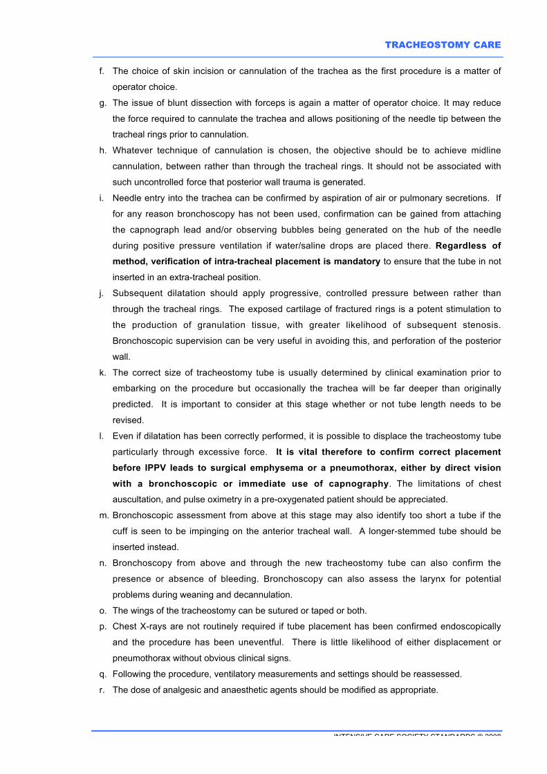
f. The choice of skin incision or cannulation of the trachea as the first procedure is a matter of
operator choice.
g. The issue of blunt dissection with forceps is again a matter of operator choice. It may reduce
the force required to cannulate the trachea and allows positioning of the needle tip between the
tracheal rings prior to cannulation.
h. Whatever technique of cannulation is chosen, the objective should be to achieve midline
cannulation, between rather than through the tracheal rings. It should not be associated with
such uncontrolled force that posterior wall trauma is generated.
i. Needle entry into the trachea can be confirmed by aspiration of air or pulmonary secretions. If
for any reason bronchoscopy has not been used, confirmation can be gained from attaching
the capnograph lead and/or observing bubbles being generated on the hub of the needle
during positive pressure ventilation if water/saline drops are placed there. Regardless ofmethod, verification of intra-tracheal placement is mandatory to ensure that the tube in not
inserted in an extra-tracheal position.
j. Subsequent dilatation should apply progressive, controlled pressure between rather than
through the tracheal rings. The exposed cartilage of fractured rings is a potent stimulation to
the production of granulation tissue, with greater likelihood of subsequent stenosis.
Bronchoscopic supervision can be very useful in avoiding this, and perforation of the posterior
wall.
k. The correct size of tracheostomy tube is usually determined by clinical examination prior to
embarking on the procedure but occasionally the trachea will be far deeper than originally
predicted. It is important to consider at this stage whether or not tube length needs to be
revised.
l. Even if dilatation has been correctly performed, it is possible to displace the tracheostomy tube
particularly through excessive force. It is vital therefore to confirm correct placement
before IPPV leads to surgical emphysema or a pneumothorax, either by direct vision
with a bronchoscopic or immediate use of capnography. The limitations of chest
auscultation, and pulse oximetry in a pre-oxygenated patient should be appreciated.
m. Bronchoscopic assessment from above at this stage may also identify too short a tube if the
cuff is seen to be impinging on the anterior tracheal wall. A longer-stemmed tube should be
inserted instead.
n. Bronchoscopy from above and through the new tracheostomy tube can also confirm the
presence or absence of bleeding. Bronchoscopy can also assess the larynx for potential
problems during weaning and decannulation.
o. The wings of the tracheostomy can be sutured or taped or both.
p. Chest X-rays are not routinely required if tube placement has been confirmed endoscopically
and the procedure has been uneventful. There is little likelihood of either displacement or
pneumothorax without obvious clinical signs.
q. Following the procedure, ventilatory measurements and settings should be reassessed.
r. The dose of analgesic and anaesthetic agents should be modified as appropriate.

TRACHEOSTOMY CARE
INTENSIVE CARE SOCIETY STANDARDS © 2008
s. Documentation of the process should be completed. This is an invasive procedure
with recognised immediate and late complications. As a minimum, documentation
should record the staff involved, the technique employed, the size and type of tube
inserted, and difficulties or immediate complications and post-procedure
instructions.
TRACHEOSTOMY CARE
INTENSIVE CARE SOCIETY STANDARDS © 2008
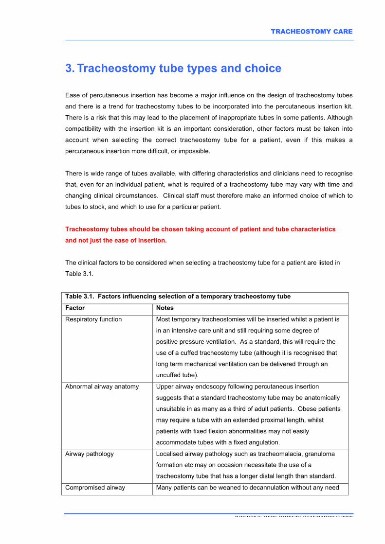
3. Tracheostomy tube types and choice
Ease of percutaneous insertion has become a major influence on the design of tracheostomy tubes
and there is a trend for tracheostomy tubes to be incorporated into the percutaneous insertion kit.
There is a risk that this may lead to the placement of inappropriate tubes in some patients. Although
compatibility with the insertion kit is an important consideration, other factors must be taken into
account when selecting the correct tracheostomy tube for a patient, even if this makes a
percutaneous insertion more difficult, or impossible.
There is wide range of tubes available, with differing characteristics and clinicians need to recognise
that, even for an individual patient, what is required of a tracheostomy tube may vary with time and
changing clinical circumstances. Clinical staff must therefore make an informed choice of which to
tubes to stock, and which to use for a particular patient.
Tracheostomy tubes should be chosen taking account of patient and tube characteristics
and not just the ease of insertion.
The clinical factors to be considered when selecting a tracheostomy tube for a patient are listed in
Table 3.1.
Table 3.1. Factors influencing selection of a temporary tracheostomy tube
Factor Notes
Respiratory function Most temporary tracheostomies will be inserted whilst a patient is
in an intensive care unit and still requiring some degree of
positive pressure ventilation. As a standard, this will require the
use of a cuffed tracheostomy tube (although it is recognised that
long term mechanical ventilation can be delivered through an
uncuffed tube).
Abnormal airway anatomy Upper airway endoscopy following percutaneous insertion
suggests that a standard tracheostomy tube may be anatomically
unsuitable in as many as a third of adult patients. Obese patients
may require a tube with an extended proximal length, whilst
patients with fixed flexion abnormalities may not easily
accommodate tubes with a fixed angulation.
Airway pathology Localised airway pathology such as tracheomalacia, granuloma
formation etc may on occasion necessitate the use of a
tracheostomy tube that has a longer distal length than standard.
Compromised airway
protection and weaning
problems
Many patients can be weaned to decannulation without any need
to change from the cuffed tracheostomy tube that was initially
inserted. In problematic cases however, it may be useful to
consider options such as downsizing, to an uncuffed or
fenestrated tube, or a tube with the option for sub-glottic
aspiration of airway secretions. The introduction of a speaking
valve may also aid swallowing and secretion control.

TRACHEOSTOMY CARE
INTENSIVE CARE SOCIETY STANDARDS © 2008
protection and weaning
problems
to change from the cuffed tracheostomy tube that was initially
inserted. In problematic cases however, it may be useful to
consider options such as downsizing, to an uncuffed or
fenestrated tube, or a tube with the option for sub-glottic
aspiration of airway secretions. The introduction of a speaking
valve may also aid swallowing and secretion control.
Obstructed / absent upper
airway
Patients with an obstructed or absent upper airway are at
particular risk should a tracheostomy become obstructed or
misplaced. This has implications for both the choice of
tracheostomy tube as well as the method by which the stoma is
fashioned.
Clinical environment Obstruction of a cuffed tracheostomy tube is a potentially life-
threatening emergency. Wherever possible a dual cannula tube
(i.e. a tube with an inner cannula) should be used, particularly for
patients in HDU or ward environments who may not have
immediate access to clinicians with emergency airway skills.
Ward staff can change inner tubes easily and quickly to relieve
obstruction with secretions.
The characteristics of the tube to be considered when selecting a tracheostomy tube for temporary
use include:
• construction
• dimensions
o internal and outer diameter (ID and OD respectively)
o proximal and distal length (i.e. the length of the tube proximal and distal to its
angulation)
o shape and angulation
• compatibility with percutaneous insertion kit
• presence and nature of tube cuff
• presence of inner cannula (dual cannula tracheostomy)
• fixed versus adjustable flange
• presence of fenestration
• specialist features, e.g. low contour on deflation tight to shaft cuffs, subglottic secretion
control systems, voice enhancement tubes etc
It is essential that the staff caring for a patient with a tracheostomy know the type of tube inplace at any time, and this should be clearly documented in the patient’s notes.

TRACHEOSTOMY CARE
INTENSIVE CARE SOCIETY STANDARDS © 2008
ConstructionMaterial
Tracheostomy tubes can be constructed of either metal or plastic, and thereby vary considerably in
rigidity, durability and kink resistance. This may be clinically relevant.
Metal tracheostomy tubes are constructed of either silver or stainless steel, but are seldom used in
the critical care environment, and will not be discussed further.
Temporary tracheostomies are constructed of polyurethane, polyvinyl chloride or silicone. Products
made of polyurethane are more rigid than those constructed of silicone, whilst those of polyvinyl
chloride construction are of an intermediate stiffness (although some become softer at body
temperature).
Shape
Many tubes have an inherent curvature or angulation; others are completely flexible and only
assume a correct anatomical alignment through appropriate stabilisation at the stoma and the level
of the cuff. Some flexible tubes are reinforced with a spiral wire in order to avoid kinking and airway
obstruction. Whilst the design of some tracheostomy tubes has been modified to facilitate
percutaneous introduction, others are only appropriate for insertion once a formal track has been
formed (either surgically or through the prior placement of an alternative tube). Various aspects of
the construction of some commonly used tracheostomy tubes are described in appendix 2.
Dimensions
In most circumstances a tracheostomy tube is both described and selected on the basis of its size,
or more specifically its diameter. This is simple in theory but may easily be confusing in practice.
The European Standard for the basic requirements and method of size designation of
tracheostomy tubes are those defined by the International Standards Organisation in EN ISO 5366-
1:2004. This standard requires that tracheostomy tubes are sized according to their functional
internal diameter (ID) at the narrowest point, quoting the ID of the outer cannula for the case of
single cannula tubes and the ID of the inner cannula for double cannula tubes, but only if this is
required for connection to a ventilator or other breathing circuit. This has the potential to cause
confusion: for example, the Portex® Blue Line Ultra® tracheostomy tube is provided with an inner
cannula that sits flush within the outer tube and which is not necessary for connection to a ventilator
circuit, it is primarily described according to the ID of the outer tube, even though the inner tube
reduces the functional internal diameter by 1.5mm for tubes of 7mm ID upwards (Table 3.2). The
standard also requires that machine end of the neck plate of the tube displays the size of the

TRACHEOSTOMY CARE
INTENSIVE CARE SOCIETY STANDARDS © 2008
tracheostomy tube (i.e. the ID), the tubes outside diameter (in mm) and the name or trademark of
the manufacturer.
Most tracheostomy tubes are sized by internal diameter in millimetres, but this may not takeaccount of inner cannula in all cases.
Table 3.2. Dimensions of dual cannula Portex® Blue Line Ultra® tracheostomy tube
Equivalent
Jackson size
(approx.)
ID without inner
cannula (mm)
ID with inner
cannula (mm)
OD (mm) length (mm)
5 6.0 5.0 9.2 64.5
6 7.0 5.5 10.5 70.0
7 7.5 6.0 11.3 73.0
8 8.0 6.5 11.9 75.5
- 8.5 7.0 12.6 78.0
- 9.0 7.5 13.3 81.0
10 10.0 8.5 14.0 87.5
The dual cannula products from Shiley© are still primarily described according to the Chevalier
Jackson sizing system originally developed for metal tubes (Table 3.3), although the appropriate
information is available on the neckplate of the tube. Note also that, for a given ID, there can be
considerable variation between manufacturers in the both the external diameter and length of their
tracheostomy tube, an issue that may be of some clinical significance should (as commonly occurs)
a transition from one tube to another be made.
Tracheostomy tubes with the same internal diameter (‘size’) may have quite different externaldiameters and length.
Table 3.3. Dimensions of dual cannula Shiley® tracheostomy tubes
Jackson size ID without inner
cannula (mm)
ID with inner
cannula (mm)
OD (mm) length (mm)*
4 6.7 5.0 9.4 62 – 65
6 8.1 6.4 10.8 74 – 76
8 9.1 7.6 12.2 79 – 81
10 10.7 8.9 13.8 79 – 81*varying according to the precise model selected

TRACHEOSTOMY CARE
INTENSIVE CARE SOCIETY STANDARDS © 2008

TRACHEOSTOMY CARE
INTENSIVE CARE SOCIETY STANDARDS © 2008
Choosing the correct sizeDiameter
When selecting the size of tube for a patient, there is an unavoidable compromise to be made
between a desire to maximise the functional internal diameter (and thereby reduce airway
resistance and the work of breathing during weaning) and a need to limit the OD to approximately
three-quarters of the internal diameter of the trachea (in order to facilitate airflow through the upper
airway when the cuff is deflated). Furthermore, selection of a tube that is too small may result in the
need to over-inflate the cuff, thereby increasing the risk of mucosal pressure necrosis, which in turn
increases the risk of complications such as tracheal stenosis and tracheo-oesophageal fistula. A
need to exceed the quoted nominal cuff volume is often an early indicator that too small a tube has
been selected. As a general rule, most adult females can accommodate a tube with an OD of
10mm, whilst a tube with an OD of 11mm is suitable for most adult males.
Need for excessive cuff volume or pressure suggests that the tube size may be too small orit may be misplaced.
Length and shape
Although a temporary tracheostomy is most commonly selected on the basis of its diameter, there
may be situations where the length, angulation or curvature of a tube is of relevance. Thus, whilst
many tracheostomy tubes are smoothly curved, others are clearly angulated, thereby allowing a
distinction to be made between the proximal (or horizontal) length of a tube (i.e. the distance
between the neckplate and the mid-point of the angulation) and the distal length (i.e. the distance
from the mid-point of the angulation and the tip). It should be appreciated that these respective
lengths are quite short in standard tubes and may be too short even in the patient with apparently
normal anatomy.
There may be occasions where the proximal length of a standard tube is inadequate (e.g. in the
obese, or when cellulitis around a tracheostomy site increases the depth of the anterior cervical
tissues after insertion), and leads to the tube tip then abutting against the posterior tracheal wall.
This can result in
• obstruction of the tube, and consequent difficulties with ventilation and weaning
• injury to the posterior tracheal wall, thereby increasing the risk of tracheo-oesophageal fistula
formation
• suboptimal positioning of the tube cuff, with the associated risk of aspiration, inadequate
ventilation and high cuff pressures
There may also be occasions where the proximal length is too long, with the result that the knuckle
of the tube abuts against the posterior tracheal wall, whilst the tip is pivoted towards the anterior
tracheal wall (the latter increasing the risk of granuloma formation and the development of a tracheal
– innominate arterial fistula). Less commonly, there may be a need to review the distal length of a
temporary tracheostomy. For instance, there may be occasions where there is a need for additional

TRACHEOSTOMY CARE
INTENSIVE CARE SOCIETY STANDARDS © 2008
distal length in order to bypass fistulae or obstructing lesions such as tracheomalacia, tracheal
stenosis or excessive granuloma formation.
Clinically significant anatomical and pathological variances such as these can be circumvented by
using either extended length tracheostomy tubes with an adjustable flange or pre-formed extended
tubes which are offered with a range of extended proximal or distal lengths. Extended length
adjustable flange tubes, examples of which are given in Appendix 2 are suitable for short term
situations but are often difficult to introduce percutaneously and are not currently supplied with an
inner cannula.
Whilst adjustable flange devices are considered suitable for short terms problems, patients who are
likely to need airway access for a considerable length of time may be better served with pre-formed
non standard products such as the Shiley® Tracheosoft XLT range or the Portex® Blue Line®
Extra Horizontal and Vertical length products (appendix 2). Some manufacturers offer a bespoke
service should none of their stock items be suitable.
Inner cannula (dual cannula tracheostomies)Many tracheostomies are now manufactured with an inner cannula. The design of some makes the
use of this optional (e.g. Portex® Blue Line Ultra®), whilst for others it is mandatory, as it is the inner
cannulae that has a standard 15mm attachment to connect to the breathing circuit of a mechanical
ventilator (e.g. Shiley®, Kapitex Tracoetwist®). Whilst some inner cannulae are disposable and
designed for single use, others can be cleaned and re-used.
The principal (and very major) advantage of an inner cannula is that it allows the immediate relief of
life-threatening airway obstruction in the event of blockage of a tracheostomy tube with blood clot or
encrusted secretions. Whilst traditionally, this has been seen to be particularly advantageous for
patients once they have been discharged to a ward environment, it is now recognised that tube
obstruction can occur even while the patient is in a critical care facility, and that in such
circumstances removal of an obstructed inner cannula may be preferable to removal and repeat
tracheal intubation.
The principal disadvantage of dual cannula tubes is that the inner cannula may significantly reduce
the effective inner diameter of the tracheostomy tube (Table 3.4), and thereby increase the work of
breathing and impair weaning. Failure to properly lock the inner tube in place may also result in
disconnection of the breathing circuit in circumstances where it is connected to this rather than the
outer cannula.
Tracheostomy tubes with an inner cannula are inherently safer and are normally preferred

TRACHEOSTOMY CARE
INTENSIVE CARE SOCIETY STANDARDS © 2008
Table 3.4. Comparison of ID and OD of tracheostomy tubes with and without inner cannulae
Portex® Blue Line® Portex® Blue Line Ultra® Shiley® Dual Cannula
Tube
Kapitex® Tracoetwist®
ID
with
(mm)
ID
without(mm)
OD
(mm)
ID
with
(mm)
ID
without(mm)
OD
(mm)
ID
with
(mm)
ID
without
(mm)
OD
(mm)
ID
with
(mm)
ID
without
(mm)
OD
(mm)
4.0 5.8 7.2
5.0 7.0 8.6
n/a 6.0 8.3 5.0 6.0 9.2 5.0 6.7 9.4 6.0 8.1 9.2
5.0 7.0 9.7 5.5 7.0 10.5 7.0 8.9 10.4
6.0 7.5 11.3
6.0 8.0 11.0 6.5 8.0 11.9 6.4 8.1 10.8 8.0 10.1 11.4
7.0 8.5 12.6
7.0 9.0 12.4 7.5 9.0 13.3 7.6 9.1 12.2 9.0 10.8 12.5
n/a 10.0 13.8 8.5 10.0 14.0 8.9 10.7 13.8 10.0 11.9 13.8
Bold type indicates the dimension used to describe the tube commercially. Data for the Shiley® dual
cannula tube refers to Jackson sizes 4, 6, 8 and 10 respectively.
Fenestrated tracheostomy tubesMany tracheostomy tubes come with the option of a fenestration in the posterior wall of the intra-
tracheal component of shaft above the cuff. Manufacturers do not recommend the use of such
tubes at the time of percutaneous tracheostomy, and generally they should not be used whilst a
patient still requires mechanical ventilation because of significant risk of surgical emphysema (even
when a non-fenestrated inner cannula is in place) [5,6,7}. Although a correctly sited fenestrated
tube may aid both phonation and weaning (by facilitating the flow of air both through as well as
around the tracheostomy tube), in practice the fenestrations are frequently poorly positioned within
the trachea, and when abutting against the posterior tracheal wall may increase airway resistance
and as well as promote the development of granulation tissue in the tracheal mucosa. The design
of the fenestrations varies between manufacturers, with some clinicians favouring tubes with
multiple smaller openings to a single large one, although both patterns are at risk of blockage by
encrusted secretions. If a fenestrated tube is used, it is vital that the position and on-going potency
of the fenestration(s) are checked regularly if the patient is to benefit from this option. In practice
down sizing or switching to an uncuffed tracheostomy tube is enough to improve flow of air through
the upper airway in most patients.
Fenestrated tracheostomy tubes should be used with caution in mechanically ventilated
patients, and only with patients who are weaning from ventilation.

TRACHEOSTOMY CARE
INTENSIVE CARE SOCIETY STANDARDS © 2008
Cuffed tracheostomy tubesIn the ICU setting, most patients will require an air-filled cuffed tracheostomy tube initially, both to
facilitate effective mechanical ventilation and also to protect the lower respiratory tract against
aspiration. The cuff should be of a “high volume / low pressure” design, and should effectively seal
the trachea at a pressure of no more than 20 – 25 cmH2O in order to minimise the risk of tracheal
mucosal ischaemia and subsequent tracheal stenosis. Although many manufacturers offer
tracheostomy tubes with suitable cuffs, there is considerable variation between them in the length of
the cuff and its precise shape when inflated. Furthermore, there is now considerable evidence to
suggest that the current high volume / low pressure design is unable to guarantee isolation of the
lower respiratory tract.
The intra-cuff pressure should be monitored regularly (see section 4).
Causes of excessive cuff pressures include:
the use of a tube that is too small (an indication for which would be a need to inflate with more
than the nominal cuff volume in order to achieve an effective seal)
poor tube positioning in the trachea
tracheal dilatation
over inflation of the cuff
One proposed solution to the problem of cuff-related tracheal mucosal ischaemia is the use of a
foam rather than air-filled cuff (Bivona® Adult Fome-Cuf® tracheostomy tube). The cuff is
constructed of air-filled polyurethane foam within a silicone envelope that is deflated prior to
insertion and then allowed to expand once sited by opening the pilot port to atmosphere. Correct
sizing and placement is crucial to their use, which tends to be limited to patients with existing tube-
related tracheal injury. The prevention of phonation is a further significant drawback to the device.
Most patients can wean to decannulation by simply deflating the existing cuff (even though the
residual profile of the deflated cuff can still significantly restrict airflow around the tube), or
alternatively by changing to a smaller or uncuffed tube. In circumstances where a patient still
requires periods of airway protection, but is unable to satisfactorily breathe past a deflated cuff, it
may be advantageous to switch to a so-called “tight to shaft” tube in which the deflated cuff makes
no distinguishable contribution to the external profile of the tracheostomy tube.
Percutaneous tracheostomy tubes / kitsSeveral manufacturers have modified aspects of the construction of their standard tracheostomy
tubes such as cuff and distal tube profiles in order that they are more easily introduced as part of a
percutaneous dilatational technique (e.g. Portex® Per-Fit and Shiley® PERC). Such changes
frequently go unnoticed when the tube comes as a component of a percutaneous tracheostomy kit

TRACHEOSTOMY CARE
INTENSIVE CARE SOCIETY STANDARDS © 2008
and may have unpredictable functional consequences. Clinicians are advised to carefully evaluate
situations where such product consolidation occurs.
Specialist functionalitySome specialist tracheostomy tubes incorporate a facility for aspiration of sub-glottic secretions (e.g.
Portex® Blue Line Ultra® “Suctionaid” tracheostomy tube, Tracoetwist® 306). Initially advocated in
the prevention of ventilator-associated pneumonia, they may also benefit patients with poor bulbar
function who are unable to effectively clear secretions that accumulate above the tracheostomy
tube, although they carry a high risk of significant laryngeal injury if suction is applied continuously.
When considering changing to one of these devices clinicians are also advised that in order to
accommodate the additional suction channel these tubes may have a wider OD than the standard
device that it is replacing.
Other options include features that enhance phonation such as the Portex® “Vocalaid” cuffed Blue
Line® tracheostomy, in which an external air source is used to deliver gas via a separate pilot
channel to the sub-glottic area, or the introduction one-way speaking valves such as the Passy-
Muir® valve, the latter also improving swallowing and secretion control.
Descriptions of some of the currently available tubes can be found in appendix 2.
Summary recommendations1. Critical care clinicians need to be aware of the wide variability in the construction, design and
functionality of the tracheostomy tubes that are currently available, and recognise that the
anatomical variation of their patients limits the universal applicability of a single tube type.
2. Most adult patients who require a temporary tracheostomy as part of their critical illness will
initially need a non-fenestrated semi-soft tube with an air-filled cuff. As a standard, a dual
cannula tube (i.e. a tube with an inner cannula) should be used from the outset unless there is a
requirement to insert an extended adjustable flange tracheostomy (which currently do not have
inner tubes available).
3. Patients with single cannula adjustable flange tracheostomies should not be discharged from a
level 3 critical care environment without review of their on-going need for a device that puts
them at particular risk of the consequences of tube obstruction. A change to a pre-formed
extended tracheostomy with an inner cannula should be considered as soon as the patient is
actively weaning from mechanical ventilation and certainly before discharge from critical care.
4. When considering changing an existing tracheostomy to that of another type or manufacturer,
clinicians should compare the relative lengths and external diameters of the two tubes,
particularly if the proposed new tracheostomy has a wider OD (because the existing stoma may
not accommodate it), shorter length (in case cuff related granulation tissue obstructs the tube) or
different curvature / angulation.

TRACHEOSTOMY CARE
INTENSIVE CARE SOCIETY STANDARDS © 2008
5. Specialist features such as fenestrations, foam or tight to shaft cuffs or a sub-glottic suction
facility may be useful in specific circumstances, although are not recommended as a routine.

TRACHEOSTOMY CARE
INTENSIVE CARE SOCIETY STANDARDS © 2008
4. Routine care of the established tracheostomy
Patients immediately post tracheostomy placement usually return either to a critical care area or a
specialist ward environment used to dealing with tracheostomies. Routine care of an established
tracheostomy is a basic ward skill, but unfamiliarity with tracheostomies and the simple rules
governing their routine care often leads to anxiety for both patient and carer. Good routine care is
not difficult and will avoid almost all emergencies, but patients with tracheotomies should only be
cared for in areas where staff are competent in such care. Regular review and good communication
are the keys to both avoidance of problems, and effective treatment. Consideration should be given
to a form such as in appendix 3 to ensure that staff are always aware of all relevant information.
4.1 Essential EquipmentThe following equipment should be immediately available at all times for a patient with a
tracheostomy, both by the bedside as well as during transfers:
• Operational suction unit, which should be checked at least daily, with suction tubing attached and
Yankeur sucker
• Appropriately sized suction catheters
• Non-powdered latex free gloves, aprons and eye protection
• Spare tracheostomy tubes of the same type as inserted: one the same size and one a size
smaller
• Tracheal dilators
• Rebreathing bag and tubing
• Catheter mount or connection Tracheostomy disconnection wedge
• Tracheostomy tube holder and dressing
• 10ml syringe (if tube cuffed)
• Artery forceps
• Resuscitation equipment
One approach is to ensure that all these are in a ‘tracheostomy box’ that goes with the patient from
critical care to the ward.
4.2 Cuff managementThe tracheostomy cuff provides a seal to enable positive pressure ventilation and also provides
some protection against aspiration of secretions. Over-inflation of the cuff may cause ischaemia of
the tracheal mucosa and thereby lead to tracheal stenosis, tracheomalacia and arterial erosion. The
pressure within the cuff should be checked regularly with a hand held pressure manometer and
should be maintained ideally below 20 – 25cm H2O. It is good practice to document cuff pressure
and inflating volume on a daily basis and following any tracheostomy-related intervention.
• Cuff pressure should not exceed 25 cm H2O.

TRACHEOSTOMY CARE
INTENSIVE CARE SOCIETY STANDARDS © 2008
• If an air leak occurs with the cuff pressure at the maximum recommended, the
tracheostomy may have become displaced or may require changing: medical or other
professionals who are competent in tracheostomy management should review thepatient.
See section 3 for further information on types and choice of tube. Finger tip pressure on the
external pilot balloon is not an accurate method of measuring cuff pressure.
The cuff should be deflated to remove the tube, to allow the patient to eat or drink and when a
speaking valve or decannulation cap is secured to the tube.
• Failure to deflate the cuff when the speaking valve or decannulation cap is secured to
the tube will result in a total occlusion of the patient’s airway.
4.3 HumidificationInadequate humidification may lead to life-threatening blockage of the tracheostomy with tenacious
sputum, keratinisation and ulceration of the tracheal mucosa, sputum retention, atelectasis and
impaired gas exchange. The provision of adequate humidification of inspired gases is therefore
essential, and can be achieved in patients with minimal or low oxygen requirements using a heat-
moisture exchanger (HME) or cold water venturi humidifier system connected to a T-piece or
tracheostomy mask. HMEs may also suffice in patients who remain mechanically ventilated and
offer the additional advantage of bacterial filtration. Patients with more tenacious sputum, or who
require high flow oxygen therapy will require additional saline nebulisers and may require heated
water humidification.
Humidification is essential for patients with temporary tracheostomies.
4.4 SuctioningTracheal suction is an essential component of secretion control and maintenance of tube patency.
However, it may be both painful and distressing for the patient, and can also be complicated by
hypoxaemia, bradycardia (particularly in patients with autonomic dysfunction such as spinal
injuries), tracheal mucosal damage, bleeding, and introduction of infection. As a result, the suction
requirements of an individual patient should be re-assessed each shift, and where possible patients
should be encouraged to expectorate their own secretions. Basic guidelines for effective, safe
suctioning are shown in the box below.
Guidelines for tracheal suction
• Suctioning should be performed using aseptic techniques, with the patient upright and in a
neutral head alignment
• Always suction with the inner tube in situ, and change to a non-fenestrated inner tube
• The suction catheter should have a diameter no greater than half internal diameter of the
tracheostomy tube

TRACHEOSTOMY CARE
INTENSIVE CARE SOCIETY STANDARDS © 2008
tracheostomy tube
Suction catheter size (Fg) = 2 x (Size of tracheostomy tube – 2)
For example, 8.0mm ID tube: 2 x (8 – 2) = 12 Fg
• The lowest possible vacuum pressure should be used - ≤ 100-120mmHg (13-16kPa) to
minimise atelectasis.
• Patients with high oxygen requirements may require pre-oxygenation
• Insert the suction catheter approximately 10-15cm (or less, depending on the length of the
tracheostomy tube) into the tube before applying suction and slowly withdrawing the
catheter.
• Suction should be applied for a maximum of 10 seconds.
• Installation of saline to ‘aid’ suctioning is not recommended
Any difficulty in passing the suction catheter should lead to consideration that the tube may
be partially blocked, badly orientated or misplaced and requires immediate attention.
4.5 Inner cannulaeWherever possible, tracheostomy tubes with an inner cannula should be used for all patients, even
for those who are still within in a critical care area (see chapter 3). The inner cannula must be
removed, inspected and when necessary cleaned at regular intervals, the frequency of inspection
being determined by the volume and tenacity of a patient’s secretions. Disposable inner cannulae
should be discarded if soiled and replaced with a new one. Basic guidelines for changing the inner
cannula of a tracheostomy tube are listed in the box below:
Guidelines for changing inner cannula• Position patient with neck slightly extended
• Preoxygenate and suction as necessary
• Using a sterile technique, remove or change inner cannula. If non disposable clean cannula
with sterile saline 0.9% or water and dry thoroughly
• Clear persistent secretions on cannula in line with manufacturer’s instructions, rinsing theinner cannula thoroughly with normal saline or water before re-insertion. Do not leave innercannula to soak.
4.6 DressingsSecretions that collect above the cuff ooze out of the stoma site producing a moist environment
leading to excoriation and infection. The site should be assessed and stoma cleaned at least once in
every 24 hours using a clean technique. Routinely a thin pre cut dressing e.g. metaline is ideal, but
for exuding stomas allevyn is more absorbent. Where the skin around the stoma is excoriated, a
thin layer of duoderm may prevent further deterioration. The tracheostomy tube should normally be
secured effectively with a commercial tracheostomy holder rather than thin tape; this protects the

TRACHEOSTOMY CARE
INTENSIVE CARE SOCIETY STANDARDS © 2008
patient from pressure on the back of the neck and is easily adjusted. Red, excoriated or exuding
stomas should have microbiology swabs sent for culture.
4.7 Swallowing
Patients with tracheostomies may experience problems with swallowing. Whilst oral intake may be
permitted with an inflated or partially deflated cuff for psychological well being and to help establish
enteral feeding early, the presence of an inflated cuff compresses the oesophagus, and makes
swallowing difficult for some patients, increasing the risk of aspiration. The risk is greatest in those
patients with associated neurological or mechanical causes of dysphagia, or those with significant
on-going respiratory failure. The decisions to allow feeding with an inflated cuff should be made on
an individual patient basis after a swallowing assessment, and the patient should be regularly
reviewed for evidence of aspiration. Sips of sterile water are initially given and if tolerated without
coughing, desaturation, fatigue or signs of aspiration on tracheal suctioning thickened fluids may be
introduced followed by a soft diet. Guidance for initiating oral intake, along with risk factors for likely
problematic patients, is shown in the boxes below.
Guidelines for the initiation of oral intake in patients with a tracheostomy
• Confirm that patient can tolerate cuff deflation (see above for exceptions)
• Sit patient up with head slightly flexed and deflate cuff
• Start with sips of water, moving on to thickened fluids and then soft diet providing
patient shows no signs of respiratory distress (coughing, desaturation, increased
tracheal secretions, increased respiratory rate etc)
• In problematic cases consider referral to Speech and Language therapy
Risk factors for swallowing problems in patients with a tracheostomy
• Neurological injury e.g. bulbar palsy
• Disuse atrophy
• Head & neck surgery
• Evidence of aspiration of enteral feed or oral secretions on tracheal suctioning
• Increased secretion load, or persistent wet / weak voice, when cuff is deflated
• Coughing and / or desaturation following oral intake
• Patient anxiety or distress during oral intake

TRACHEOSTOMY CARE
INTENSIVE CARE SOCIETY STANDARDS © 2008

TRACHEOSTOMY CARE
INTENSIVE CARE SOCIETY STANDARDS © 2008
5. Changing tracheostomy tubesChanging a tracheostomy tube is potentially hazardous, but so is failure to do so. Unfortunately,
recommendations for the frequency of changing tracheostomy tubes are inconsistent and
unsupported by evidence. Basic principles guiding replacement of a tracheostomy tube are listed in
the box below. A more detailed example is given in appendix 4.
Basic principles for changing a tracheostomy tube
• Tracheostomy tubes without an inner cannula should be changed every 7-14 days, the
frequency then decreasing once the patient is free of pulmonary secretions and has a
well formed clean stoma
• A European Economic Community Directive (1993) states that tracheostomy tubes with
an inner cannula can remain in place for a maximum of thirty days.
• The first tracheostomy tube change:
o Should not be performed within 72 hours following a surgical tracheostomy and
not before 3 – 5 (and ideally 7 – 10) days after a percutaneous tracheostomy to
allow the stoma to become established.
o The decision to change the tube must be made in conjunction with a medical
practitioner competent in the care of tracheostomies.
o Must be carried out by either:
a medical practitioner with appropriate, advanced airway skills, or
Another suitably skilled practitioner.
• Subsequent changes can be made by experienced personnel trained in tracheostomy
tube changes (e.g. specialist tracheostomy nurse)
• In practice, the frequency with which the tube needs to be changed will be affected by
the individual patient’s condition and the type of tube used. Elective changes are
inherently safer than those done in a crisis.
Tracheostomy tubes should only be changed by staff who are competent to do so. Most will
be doctors, but there are specialist non-medical practitioners who have the necessary skills.
Doctors who have not had relevant training will not have competency in this area, and shouldnot undertake the procedure unsupervised.
The first tube changeChanges within the first 72 hours should be avoided unless absolutely essential and should only be
performed in an environment in which trans-laryngeal intubation can immediately be established.
TRACHEOSTOMY CARE
INTENSIVE CARE SOCIETY STANDARDS © 2008
Although the risk of stomal closure lessens somewhat thereafter, the first change remains
hazardous, particularly following a percutaneous procedure, and exchange over a bougie or an
airway exchange catheter that allows the continued insufflation of oxygen such as the Frova
catheter or Cook TM airway exchange catheter should be considered.
Whilst problems can occur in any patient, they are particularly likely in the obese, those with a deep
trachea, other anatomical difficulties.
Second and subsequent changesA standard procedure for subsequent changes of a tracheostomy tube is described in appendix 4. It
is reasonable to expect that two skilled practitioners should be present when changing the tube,
particularly when a previous change has been difficult. Resuscitation equipment must be readily
available, as should a smaller tube.
If a patient has a smooth long term stoma and has regular tracheostomy changes, it is reasonable to
introduce the new tube using the tracheostomy introducer rather than an airway exchange device.
The practitioner must however be aware of the risk of paratracheal tube placement with any blind
technique and be able to recognise the clinical features of failure to re-cannulate the trachea.
It is essential that if the new tube cannot be inserted or is misplaced, there is an agreed
procedure for handling the situation.

TRACHEOSTOMY CARE
INTENSIVE CARE SOCIETY STANDARDS © 2008
6. Emergencies, Common Complications and theirManagement
The main life threatening complications associated with a tracheostomy are blockage, dislodgement
and bleeding.
Blockage and displacementA blocked or displaced tracheostomy tube generally presents with respiratory difficulty. The nature
of the problem will often be obvious, but if not, it is important to adopt a systematic approach and be
aware that acutely ill patients may have other cardio-respiratory reasons for difficulty in breathing.
Every hospital must have a procedure for managing patients whose tracheostomy is blocked
or displaced. Staff must be aware of this and receive appropriate training to manage the
problem.
Tracheostomy tubes may become dislodged or displaced for a number of reasons. This can be a
stressful event for both patients and staff, and prevention is better than cure. It is important to
ensure that the tracheostomy tube holder is adjusted regularly so that the tube is fixed in a secure,
comfortable position all times. Tracheostomy tubes may become dislodged when a ventilated
patient is turned, or moved from their bed to a trolley. Restless or agitated patients may pull at their
tracheostomy tubes, or ventilator tubing attached to the tracheostomy.
A partly dislodged tracheostomy tube is just as dangerous, if not more dangerous, as a completely
removed tracheostomy tube.
Airway: is the airway (at least partially) patent?
• If the tube is displaced, the patient may be breathing through their nose or mouth. The
patient may be safe in the short term, requiring urgent but not emergency action. Only
experienced staff should try to replace the tube under such circumstances. If in doubt it will
usually be safer to remove a partly dislodged tube, although a suction catheter or airway
exchange catheter may be first advanced through it to allow oxygen administration. The
airway should be maintained by other methods until experienced help arrives.
• If tube is partially occluded, the patient may still be able to breathe through it, but with
difficulty. If the tube has an inner cannula, this should be removed and changed. If the tube
does not have an inner cannula, but a suction catheter can be passed down the
tracheostomy tube, then it must be at least partially patent. It may be possible to change the
tube over a catheter or other airway exchange device.

TRACHEOSTOMY CARE
INTENSIVE CARE SOCIETY STANDARDS © 2008
Staff treating patients with a tracheostomy in an emergency situation need to be aware that
‘bag and mask’ ventilation via the mouth is not possible with a cuffed tracheostomy tube in
situ.
Individual hospitals are advised to develop their own specific guidelines with bleep numbers, taking
account of available staff and recognising this may happen day or night.
Chest X rays do not give useful information about the position of the tube in this situation. Fibreoptic
bronchoscopy may be useful if it is immediately available, but this should not delay treatment.
Displaced tracheostomy tube
This guidance is appropriate for patients capable of spontaneous respiration.In a patient who is fully ventilated, critically ventilator dependent or
paralysed, prompt replacement or removal and replacement by a trachealtube will be required. Such a situation is only likely to arise in a level 3 area,but can also occur in a cardio-respiratory arrest elsewhere. Remember tubedisplacement as a possible cause of cardio-respiratory arrest.
1. DON’T PANIC!
2. Call for help – senior medical and nursing staff, other AHPs with tracheostomy care skills
(e.g. physiotherapist). If in doubt, call for anaesthetic assistance
3. Reassure patient.
4. Assess patency of airway (A) and patient’s breathing (B). Is air passing through the
tracheostomy tube or stoma? Is the patient breathing via mouth or nose? Check oxygen
saturation with a pulse oximeter.
5. If airway is not patent, it must be cleared immediately.
• ONLY SENIOR STAFF WITH APPROPRIATE EXPERIENCE OF
TRACHEOSTOMY MANAGEMENT SHOULD ATTEMPT TO RE-INSERT A
DISLODGED OR DISPLACED TRACHEOSTOMY.
• ANY SUCH ATTEMPT SHOULD BE QUICKLY ABANDONED IF
UNSUCCESSFUL.
• MULTIPLE ATTEMPTS SHOULD NOT BE MADE.
• IF IN ANY DOUBT, REMOVE TRACHEOSTOMY TUBE AND ALLOWBREATHING THROUGH THE STOMA OR THE MOUTH/NOSE.
• APPLY OXYGEN MASK OVER ONE OR BOTH SITES OF AIR ENTRY.
Appropriately experienced staff may attempt to re-insert a tracheostomy tube in a patient
with a well-formed track from skin to the tracheal stoma. Typically, this would be more than
72 hours following surgical tracheostomy or more than 7 days after percutaneous

TRACHEOSTOMY CARE
INTENSIVE CARE SOCIETY STANDARDS © 2008
tracheostomy. It is advisable to attempt any such re-insertion using a gum elastic bougie or,
if time permits, a fibre-optic bronchoscope.
6. Re-establish the airway in the usual fashion – tilt the head back to extend the head on the
neck, perform a jaw thrust. If necessary insert an oral airway (Guedel). Note that if ‘bag and
mask’ ventilation is attempted, air will escape through the stoma. In this situation, get a
colleague to occlude the stoma by applying pressure over it with gauze swabs or a pad.
7. Tracheal intubation may be needed – this should only be performed by a doctor competent
in tracheal intubation. This is not the time to “have a go”. It may be necessary to push the
tracheal tube distal to the stoma. (Use an ‘uncut’ tube.)
8. If the patient is breathing adequately at this point, there may be no need to artificially assist
ventilation. Check the oxygen saturation with a pulse oximeter and administer oxygen as
required via a facemask or resuscitation bag with an oxygen reservoir.
9. If artificial ventilation is needed, use a resuscitation bag and mask in the standard fashion.
Maintain occlusion of the stoma as above to prevent an air leak. If the patient has been
intubated, the person controlling the airway (usually an anaesthetist or intensivist) will
ventilate with the resuscitation bag. Measurement of oxygen saturation with a pulse
oximeter will confirm adequacy of oxygenation.
10. At this point the patient is safe, with a patent airway and adequate respiration. Do not panic
or rush. Take time to think about what the patient needs at this time.
11. A decision must now be reached about re-insertion of the tracheostomy tube. This decision
should only be made an experienced anaesthetist, intensivist, or ENT surgeon. The
decision will be depend on the ability of the patient to maintain a patent airway without the
tracheostomy tube, and to maintain oxygenation, cough and clear respiratory tract
secretions without artificial assistance. If there is any doubt, the correct decision is to re-
insert the tracheostomy tube.
12. Re-insertion of the tracheostomy tube can now take place in a controlled manner. It may be
possible to do this in the ward, providing the stoma has a well-established track (at least 7
days old). Relevant equipment should be available at the bedside in the patient’s
tracheostomy box or from the cardiac arrest trolley (e.g. tracheal dilator forceps). If in doubt
arrange to move the patient to a critical care area, ENT ward, or operating theatre. Another
option for an experienced practitioner is to pass a tracheal tube via the tracheostomy stoma,
which may be easier to advance than a tracheotomy tube.

TRACHEOSTOMY CARE
INTENSIVE CARE SOCIETY STANDARDS © 2008
BLOCKED TRACHEOSTOMY TUBE
A tracheostomy tube may become blocked with thick tracheal secretions, blood or foreign bodies.
The patient may present with increasing respiratory distress over a few hours, or with a much more
rapid deterioration. In either case, a blocked tracheostomy tube is an emergency situation in which
the patient’s life is at risk if it is not rapidly resolved.
Prevention is better than cure:
• Patients with tracheostomies must always receive adequate humidification of their inspired
gas to lessen the risk of tube blockage.
• The risk of tube blockage is reduced by the use of a tracheostomy tube with an inner
cannula. Such tubes should have their inner cannula cleaned regularly to prevent the build-
up of secretions. The precise frequency will depend on individual risk assessment.
• Certain specialist tracheostomy tubes (e.g. adjustable length or custom-made long tubes)
may not have an inner cannula. Extra vigilance is needed in these patients to minimise the
risk of tube blockage.
If a blocked tracheostomy tube is suspected, rapid diagnosis will enable prompt treatment.
YOU NEED TO KNOW IF THE TRACHEOSTOMY TUBE HAS AN INNER CANNULA
1. Patient with tracheostomy tube develops respiratory embarrassment or distress.
2. DON’T PANIC!
3. Call for help: senior medical and nursing staff, other AHPs with tracheostomy care skills
(e.g. physiotherapist).
4. Reassure patient.
5. Assess patency of airway (A) and patient’s breathing (B). Is air passing through the
tracheostomy tube or stoma? Is the patient breathing via the mouth or nose? Check the
oxygen saturation with a pulse oximeter.
6. If the airway is not patent, this must be dealt with first.
7. If the patient is awake, breathing spontaneously and co-operative, encourage the patient
to give a vigorous cough – this may be sufficient to relieve the obstruction by shifting a
plug of thick secretions.
8. If the tracheostomy tube has an inner cannula, remove it.
9. Attempt tracheal suction with a suction catheter – this alone may be sufficient to remove a
thick plug of secretions.
10. If the obstruction is not relieved, then deflate the tracheostomy tube cuff. Administer
oxygen via a facemask if the patient is breathing spontaneously. If the patient is not
breathing spontaneously, it will be necessary to ventilate the patient with a resuscitation
bag and mask – an oral airway (Guedel) may be necessary. Monitor the oxygen saturation
with a pulse oximeter.
11. If it is not possible to achieve adequate oxygenation, then remove the tracheostomy tube
and suction and proceed as per dislodged tracheostomy tube protocol. Consider passing
Yankeur or other large bore suction catheters directly into the trachea via stoma to remove
TRACHEOSTOMY CARE
INTENSIVE CARE SOCIETY STANDARDS © 2008
thick secretions or blood clot. Suction applied directly via the tracheostomy tube or
translaryngeal tube may also be required to remove big clots, or large mucus plugs (as in
neonates for meconium aspiration).
12. If the patient is now adequately oxygenated, then it is now safe to consider changing the
tracheostomy tube. This is not necessary if removal of the inner cannula has relieved the
obstruction.

TRACHEOSTOMY CARE
INTENSIVE CARE SOCIETY STANDARDS © 2008
BLEEDING FROM A TRACHEOSTOMY
Bleeding is the most common complication of tracheostomy. Bleeding may occur early (within 48
hours of formation of the tracheostomy) or late (several days afterwards). It may be minor (settles
with simple conservative management) or major (requiring transfusion of blood and/or blood
products) and surgical exploration may be needed to identify and deal with the source of bleeding.
The management of bleeding from a tracheostomy therefore depends upon the context in which the
bleeding occurs.
Early minor bleedingOozing from the stoma site is the most common type of bleeding seen following formation of a
tracheostomy. Most commonly, this is the result of the effects of the vasoconstrictor used to infiltrate
the incision site wearing off. Blood staining of the dressings may be noted, or there may be blood
staining of tracheal secretions. Large volumes of fresh blood represent significant bleeding, which
may require surgical exploration.
1. DON’T PANIC!
2. Call for help– senior medical and nursing staff, other AHPs with tracheostomy care skills
(e.g. physiotherapist).
3. Reassure patient.
4. Whilst maintaining control of the tracheostomy tube, remove the tracheostomy tube
holder and dressing.
5. Clean stoma site with sterile saline.
6. Inspect stoma site, looking for any obvious bleeding point.
7. Apply manual pressure to any obvious bleeding point – this may be sufficient to stop
minor oozing. Suturing locally may also be effective.
8. If still bleeding, infiltrate any obvious bleeding point with dilute adrenaline (1:80,000 to
1:200,000). If no obvious bleeding point, infiltrate the stoma margins with dilute
adrenaline.
9. If still oozing, apply Kaltostat packing to stoma to promote local clot formation. The
Kaltostat may also be soaked in dilute adrenaline.
10. Check full blood count and a coagulation screen. Correct any abnormalities in the
standard fashion.
11. If bleeding is not stopped by these measures, refer to ENT or other appropriate surgeon
for surgical exploration.
Then seek surgical haemostasis in theatres with appropriate senior surgeon. Junior surgeons
should not explore such wounds alone as major vessel bleeding and a compromised airway will
require senior ENT, cardiothoracic or vascular expertise.

TRACHEOSTOMY CARE
INTENSIVE CARE SOCIETY STANDARDS © 2008
Major early bleeding
Proceed as minor bleeding guideline. Note that large volumes of blood in the trachea may cause
respiratory embarrassment – the patient may need further respiratory support in a critical care area.
Beware of the risk of the tracheostomy tube becoming occluded by blood clot. If this occurs,
proceed as per blocked tracheostomy tube guideline. If airway obstruction occurs due to blood clot
in the airway (look with a laryngoscope) then direct suction on the tracheal tube with suction tubing
may be required to remove it. Large clots will not pass through a suction catheter or a fibreoptic
bronchoscope suction channel.
1. In most situations of significant bleeding, secure the airway by translaryngeal intubation with
the cuff below the stoma so the airway is protected from blood entering the trachea from the
stoma. Then temporary haemostasis of the stoma can be achieved by digital pressure,
packing the wound with gauze or deep tension sutures.
2. Early referral to an appropriate SENIOR surgeon (e.g. ENT, cardiothoracic, vascular
expertise) for urgent surgical exploration is necessary.
3. Ensure that cross-matched blood is made available.
4. Check full blood count and a coagulation screen. Correct any abnormalities in the standard
fashion.
TRACHEOSTOMY CARE
INTENSIVE CARE SOCIETY STANDARDS © 2008
Late bleedingLate bleeding may occur because of erosion of blood vessels in and around the stoma site. This is
more likely if there has been infection of the stoma site. Such bleeding may settle with conservative
management, as described in the early bleed guideline. More worryingly, however, is the prospect of
such bleeding being the result of erosion of a major artery in the root of the neck where there has
been pressure from the tracheostomy tube itself or the cuff tube (another reason why it is important
to ensure that the tracheostomy tube cuff pressure is monitored to avoid over-inflation). Most
commonly, this erosion occurs into the right brachio-cephalic artery (also known as the innominate
artery), resulting in a tracheo-innominate artery fistula. This situation may be heralded in the
preceding hours by a small, apparently insignificant, sentinel bleed. Bleeding from such a fistula will
be massive. THIS IS A LIFE-THREATENING EMERGENCY and so decisions need to be rapidly
made. It is well recognised that fatalities occur in this situation, and that this may be the mode of
death for some patients with head and neck cancers. The appropriate management may, therefore,
be palliation. Such a decision should, ideally, have been made in advance and in discussion with the
patient and family, clearly documented in the patient’s medical notes and communicated to the
nursing staff (who will undoubtedly have to deal with this situation). If active treatment is still the
plan, proceed as below:
1. DON’T PANIC
2. Call for help– senior medical and nursing staff, other AHPs with tracheostomy care skills
(e.g. physiotherapist).
3. Reassure the patient.
4. Proceed as described in the major early bleeding guideline.
5. Bleeding may be temporarily reduced or stopped by applying finger pressure to the root of
the neck in the sternal notch, or by inflating the tracheostomy tube cuff (if present) with a
50ml syringe of air. This inflation should be done slowly and steadily to inflate the balloon to
a maximum volume without bursting it. Depending on the type and size of the tracheostomy
tube this may be anywhere between 10 and 35 ml.
6. Urgent referral for surgical exploration must now be made, if not already done so. In addition
to an ENT or maxillofacial surgeon, it may be necessary to get help from a vascular
surgeon. Sometimes, the damage can only be repaired utilising cardio-pulmonary bypass,
and so a cardiothoracic surgeon may also be needed to help. The operating theatres must
be informed that this is an NCEPOD Category One emergency, as any delay will increase
the risk of death significantly.

TRACHEOSTOMY CARE
INTENSIVE CARE SOCIETY STANDARDS © 2008
7. Decannulation and longer term follow up
7.1 Weaning & DecannulationDecannulation should be undertaken as soon as it is feasible to minimize the risk of complication.
However, de-cannulation is itself associated with potential hazards such as airway obstruction,
aspiration, ventilatory failure, sputum retention and difficulty in oral re-intubation. The decision and
process should therefore demonstrate a balancing of risk and benefit and should be performed:
• using objective criteria
• by competent staff
• in an appropriate environment
• with appropriate monitoring
• with a range of drugs and equipment to address any predictable hazard
There should be clear guidance to staff to observe for complications, clear criteria for review and
point of contact to obtain urgent review by tracheostomy team, anaesthetic, intensive care or
surgical staff.
A tracheostomy should be removed as soon as it is no longer required.
Care of patients with tracheostomies will be multidisciplinary, but it must be clear who is
responsible, particularly when patients are not under the direct care of the critical care team. Every
Trust should have an explicit policy on this.
It must be clear which person or team is responsible for management of the tracheostomy,
especially if it is not the specialty with primary responsibility for the patient’s care.
Reviewing the ‘need’ for a tracheostomy and planning weaning should be part of the daily
assessment. Some patients may tolerate rapid airway decannulation, especially if their ventilation
period has been short or if they do not suffer significant lung or airway pathology. Others,
particularly those with underlying cardiopulmonary disease, muscle weakness, neurological deficits,
upper airway oedema or problems managing airway secretions, will take much longer to wean and it
is important that the process is both planned and sequential.
Prior to removal of a tracheostomy, the patient will be breathing spontaneously and the following
points should be considered:
• The pathological process necessitating the insertion of a tracheostomy has resolved.
• The patient is able to cough and swallow effectively and protect their airway
• Ventilatory reserve is adequate (decannulation increases anatomical dead space and
may increase the work of breathing).

TRACHEOSTOMY CARE
INTENSIVE CARE SOCIETY STANDARDS © 2008
• Reversible bronchopulmonary infection or pathology has been treated and is resolving.
• Pulmonary secretions are not excessive.
• Nutritional status is adequate.
• Patient is comfortable with the cuff deflated.
• The airway is patent above the level of the stoma.
These checks are intended to prevent decannulation being followed by a need for re-insertion, and
to allow a patient to be assessed objectively. It is common to progress through several stages
during weaning and it is important to appreciate the changes, and risks, associated with each. With
cuff deflation, there is an expectation that there will be some respiration through the upper airway,
past the tracheostomy. When a speaking valve or occlusion cap is also used, this becomes
essential. Whilst it is harder to breathe with a tracheostomy in place after it has been removed,
persistent respiratory difficulty when using any of these techniques should raise questions about
possible upper airway obstruction.
Cuff deflation: Weaning often includes increasing the periods of time with the cuff deflated. The
inflated cuff provides some protection from aspiration and means that the patient becomes
unaccustomed to managing their own oral secretions and swallowing. Before cuff deflation, warn the
patient about the possibility of an alteration in tracheal airflow sensation and that they may cough.
The cuff should be deflated slowly and completely. The tracheostomy tube should be occluded
briefly with a clean, gloved finger to check for airflow around the tube. If the patient continuously
coughs with the cuff deflated and this does not resolve with suction and reassurance, re-inflate the
cuff.
Speaking Valves: A one way speaking valve attached to the tracheostomy, allows air in through the
valve on inspiration, but closes on expiration, thus diverting the air past the vocal cords and out
through the nose and mouth. It should only be used with an uncuffed tube, a cuffed tube with
the cuff deflated or a fenestrated tracheostomy tube with the cuff deflated. If the patient finds it
hard to breathe, or they are unable to vocalise (which they should be able to do) or they begin to
sound wheezy or stridulous, then the speaking valve must be removed immediately and the cause
of the problem sought.
It is not uncommon for a patient to experience breathlessness when starting to use a speaking
valve. They need reassurance and if this does not settle the speaking valve must be immediately
removed. If difficulties persist:
• Check that the tracheostomy is fenestrated and that the inner lumen is clean with patent
fenestrations.
• Consider changing the tracheostomy to ensure that the fenestrations of outer lumen are
clear.

TRACHEOSTOMY CARE
INTENSIVE CARE SOCIETY STANDARDS © 2008
• Consider downsizing the tracheostomy or changing it for an uncuffed one to increase space
around tracheostomy in the trachea.
If problems continue it may indicate an upper airway problem: consider referral to Speech and
Language Therapist and/or ENT for panendoscopy.
Occlusion or decannulation Cap: with this in place, the tracheostomy is effectively blocked off.
This is the hardest stage of weaning for the patient, as airways resistance will be higher than when
the tracheostomy is subsequently removed. The patient should usually have a fenestrated
tracheostomy with a fenestrated inner tube in situ, and the cuff deflated all the time that the
decannulation cap is in place.
Predictors of decannulation:
• The patient can tolerate four hours or more breathing around a tracheostomy of at least size
7.0mm occluded with a decannulation cap. This suggests that they will tolerate
decannulation without suffering from airway obstruction [8].
• The patient can tolerate four continuous hours with a decannulation cap and is able to
expectorate pulmonary secretion effectively. Removal of the tracheostomy is indicated.
It is important to be aware that the decannulation cap completely occludes the tracheostomy. The
work of breathing in this situation can be difficult and may not be tolerated by some patients. Failure
to tolerate a decannulation cap is not necessarily an indication to leave the tracheostomy in-situ
Look at the overall condition of the patient. If they have good motor power, full head control, are
conscious and alert, success is much more likely than if they have a weak cough and are obtunded.
Decannulation
The procedure is usually straightforward, but adequate assessment and preparation as outlined
above is required to maximise success.
The optimal time for decannulation is usually the early morning when the patient has rested
overnight and their condition can be observed during the daytime.
After decannulation the stoma should be covered with a semi-permeable dressing. The patient
should be instructed to apply gentle pressure with their fingers over the site when coughing. There is
no need to apply a more rigid dressing to occlude the site.
In the event of a failed decannulation, the following equipment must be available to provide
oxygenation, ventilation and a patent secure airway:
1. airway devices
a. a range of tracheal and tracheostomy tubes
b. laryngoscopes, bougies, airway exchange catheters, and aides to intubation
c. laryngeal masks
TRACHEOSTOMY CARE
INTENSIVE CARE SOCIETY STANDARDS © 2008
d. ready access to a fibre-optic bronchoscope
2. a means of oxygen insufflation if the airway becomes obstructed
a. e.g. a suction catheter or airway exchange catheter which can be advanced through
the stoma and connected to an oxygen supply
3. a means of ventilatory support e.g. self-inflating bag or Mapleson C circuit
4. a means of reopening the stoma: tracheal dilator forceps
5. access to a tracheostomy kit
6. effective suction equipment
7. monitoring which should include; ECG, pulse oximeter, automated BP recordings, with
ready access to a capnograph if it is necessary to reintubate the patient
8. anaesthetic drugs, vasopressor agents, atropine
9. access to resuscitation equipment such as defibrillator
An example of a form to document care variables on a daily basis is included in the appendix.
Post Decannulation Observations
The patient should be observed for signs of respiratory distress including:
• Dyspnoea
• Laboured or noisy respiration, stridor
• Increased respiratory rate and heart rate
• Excess use of accessory muscles
• Diaphragmatic respiration
• Agitation
• Oxygen desaturation
Long Term Follow UpIt is recommended that all patients who have had a tracheostomy be reviewed prior to hospital
discharge for evidence of wound infection, excess scar tissue/distortion, a persistent stoma, or
evolving stenosis. Tracheal stenosis, either from the pressure of the cuff, or more commonly at the
stoma site [9, 3], is often not recognised and should be considered in all patients with symptoms,
such as stridor or breathlessness on exertion. Such symptoms may be gradual in onset and may
only be present on exercise and be minimized by the patient who is often expecting to feel unwell
post discharge from ICU. Patients may rarely require tracheal surgery such as stenting or even
tracheal resection and should be referred to ENT for a panendoscopy.
All patients who have undergone tracheostomy should be followed up.

TRACHEOSTOMY CARE
INTENSIVE CARE SOCIETY STANDARDS © 2008
8. SummaryInsertion:
This guideline does not cover emergency tracheostomies.
• A planned tracheostomy should be performed by medical practitioners who have been trained,
and are competent in the procedure, or are under direct supervision. Two trained medical
practitioners plus a third member of staff to assist is the minimum staffing. Hospitals should
maintain a list of practitioners competent in the procedure.
• All Critical Care Areas should have a Difficult Airway trolley.
• Bronchoscopy should be available and its routine use is beneficial.
Tracheostomy tube choice:
• Tracheostomy tubes should be chosen to suit the patient. The requirements of a patient may
change over time. The need for excessive cuff volume or pressure suggests that the tube size
may be too small or it may be misplaced.
• Tracheostomy tubes with an inner cannula are inherently safer and are normally preferred.
• Most tracheostomy tubes are sized by internal diameter in millimetres, but this may not take
account of the inner cannula in all cases. Tracheostomy tubes with the same internal diameter
(‘size’) may have quite different external diameters and length.
• Fenestrated tracheostomy tubes should be used with caution in mechanically ventilated patients
and only in patients who are weaning from ventilation.
• It is essential that the staff caring for a patient with a tracheostomy know the type of tube that is
in place. This should be clearly documented in the patient’s notes.
Routine care:
In order to avoid complications, the main priorities are humidification; secure fixation of the tube and
attention to cuff pressure. Inner tubes should be changed 4 hourly, the entire tube should be
changed at least every 30 days. The type of tube should be clearly documented.
• Cuff pressure should not exceed 25 cm H2O.
• If an air leak occurs with the cuff pressure at the maximum recommended, the tracheostomy
may have become displaced or may require changing: medical or other health care
professionals competent in tracheostomy management should review the patient.
• The cuff should be deflated to remove the tube, to allow the patient to eat or drink and when a
speaking valve or decannulation cap is attached to the tube. Failure to deflate the cuff when
the speaking valve or decannulation cap is attached to the tube will result in a total occlusion of
the patient’s airway.
• Humidification is essential for patients with temporary tracheostomies.
• Any difficulty in passing the suction catheter should lead to consideration that the tube may be
partially blocked, badly orientated or misplaced and requires immediate attention.

TRACHEOSTOMY CARE
INTENSIVE CARE SOCIETY STANDARDS © 2008
Changing tracheostomy tubes.
• Tracheostomy tubes should only be changed by staff who are competent to do so. Most will be
doctors, but there are specialist non-medical practitioners who have the necessary skills.
• Doctors who have not had relevant training will not have competency in this area, and should
not undertake the procedure unsupervised.
• It is essential that if the new tube cannot be inserted or is misplaced, there is an agreed
procedure for managing the situation.
Emergencies:
Respiratory difficulty should lead to an urgent assessment of the airway, and to consider whether
the tube has become displaced or blocked.
• Every ward looking after patients with a tracheostomy must have a clear procedure for such
emergencies. Staff must be aware of this, and be trained appropriately.
• In an emergency situation staff attending need to be aware that ‘bag and mask’ ventilation is not
possible with a cuffed tracheostomy tube in situ.
• Changing inner cannula will usually relieve the obstruction. If tracheostomy is partially displaced,
it may be safer to remove it than to reinsert it if senior staff are not present.
• Bleeding is usually minor, but the risk of severe bleeding must always be considered.
Weaning and decannulation.
Tracheostomies should be removed as soon as they are no longer required. The person or team
responsible for tracheostomy management must be clear, particularly if it is not the specialty with
primary responsibility for the rest of the patient’s care.
It is important to ensure that the cuff is deflated and that the patient can breathe through their upper
airway. All patients should be followed up for long term complications.

TRACHEOSTOMY CARE
INTENSIVE CARE SOCIETY STANDARDS © 2008
9. References
1. Silvester W. Percutaneous versus surgical tracheostomy: A randomised controlled study
with long term follow up. Critical Care Medicine 2006;34;2145-2152
2. Rana S, Pendem S, Pogodzinski MS, Hubnayer RD, Gajic O. Tracheostomy in Critically Ill
patients. Mayo Clinic Proceedings 2005;80;1632-1638.
3. Koitschev A, Simon C, Blumenstock G, Mach H, Graumuller S. Suprastomal tracheal
stenosis after dilational and surgical tracheostomy in critically ill patients. Anaesthesia 2006;
61:832-837.
4. www.traman.org.uk/pdf/summary-protocol.pdf
5. Orme RM l’E, Welham KL. Subcutaneous emphysema following percutaneoustracheostomy Anaesthesia 2006:61;911
6. Cole B. Subcutaneous emphysema following percutaneous tracheostomy. Anaesthesia2006:61;911
7. Fikkers, Emphysema and pneumothorax after percutaneous tracheostomy. Case reportsand an Anatomic Study. Chest 2004;125:1805-1814
8. Rumbak MJ, Graves AE, Scott MP, Spurn GK, Walsh FW, McDowell Anderson W, GoldmanAL (1997) Tracheostomy tube occlusion protocol predicts significant tracheal obstruction toair flow in patients requiring prolonged mechanical ventilation. Critical Care Medicine 25: (3)413- 417
9. Sarper A, Ayden A, Seer I, Democrat A & Iain E: Review of Post-tracheostomy And
Postintubation Tracheal Stenosis With Special Regard to Etiology and Treatment. The
Internet Journal of Thoracic and Cardiovascular Surgery. 2003. Volume 6 Number 1.
Further reading
Cree N & Bonner S. Tracheostomy Management Ch. In Bonner S, Carpenter M & Garcia E. Care of
the Critically Ill Medical Patient, Elsevier, Glasgow 2007.
Paw HGW, Bodenham AR. Percutaneous tracheostomy. A practical Handbook. Greenwich Medical
Media, London 2004

TRACHEOSTOMY CARE
INTENSIVE CARE SOCIETY STANDARDS © 2008
Appendices
Appendix 1
Information for patients and their relativesMany patients on the Intensive Care Unit (ICU) need a ventilator (breathing machine) to help withtheir breathing. The ventilator has to be connected to the patient by a tube in the trachea (windpipe).This is usually done using a plastic tube in the mouth which passes through the larynx (voice box) toreach the trachea. It is safe to leave the tube in place for several days, although most patients findthe presence of a tube in the throat to be very uncomfortable, and require sedative medication tomake the tube acceptable.The prolonged presence of a tube in the throat makes it difficult to keep the mouth clean, and canalso lead to physical damage to the mouth, larynx and trachea. A tracheostomy can be useful byavoiding some of these problems. We are recommending a tracheostomy as we believe that you/your relative will need help from the ventilator for some time to come.
What is a Tracheostomy?A tracheostomy is a hole in the front of the neck into the trachea (windpipe). A tube can then beinserted through this hole into the trachea in order to allow the patient to be connected to a ventilatorand to allow access for suction.
Why do I/my relative need a tracheostomy?There are a number of reasons why a tracheostomy may be beneficial
• A tracheostomy tube is far more comfortable than a tube in the mouth. Most patients with atracheostomy require little or no sedation. This means that they can be more awake, morecomfortable and may allow them to breathe for themselves at an earlier stage. This can actuallyreduce the time attached to a ventilator.
• A tube in the mouth can cause physical damage to the structures through which it passes,including the larynx (voice box), leading to problems later on with speaking.
• There are specific reasons why some patients may particularly benefit from a tracheostomy.These are usually because of the particular illness which has caused the need for ventilation. Thedoctors in ICU will discuss any specific reasons with you.
Is it safe? Are there any risks?Generally speaking, a tracheostomy is safe, but, like any procedure, there are some risks andcomplications. A tracheostomy is only performed when the potential benefits outweigh the potentialrisks. The risks of having a tracheostomy may be associated with the procedure itself, the fact that anopening is made into the trachea (windpipe) and to the presence of a tube in the trachea. Most ofthe complications are minor and of no great significance. However, very occasionally, a severecomplication may arise which may necessitate intervention.The major risks associated with the procedure are: -• Bleeding. The front of the neck contains several blood vessels, which may bleed during the
formation of a tracheostomy. These can usually be dealt with very simply but occasionally requirea surgical operation in the operating theatre.
• Pneumothorax. This is when air is in the chest but outside the lung, causing the lung to collapse.It can occur because of damage to the pleura (the lining surrounding the lung) or the trachea(windpipe) itself. It usually requires a drain to be placed in the chest.
• Infection. The tracheostomy can become contaminated with bacteria, either from the patient'sown skin or from the secretions coughed up by the patient. Serious infection is rare.
TRACHEOSTOMY CARE
INTENSIVE CARE SOCIETY STANDARDS © 2008
How is a Tracheostomy done?A tracheostomy may be performed ‘percutaneously’ or ‘surgically’. Whichever method is used, youwill be given a general anaesthetic.The percutaneous (meaning "through the skin") techniques involve making a small cut in the skin onthe front of the neck and inserting a needle through this into the trachea. A guide-wire is thenpassed through this needle, and the hole around it is stretched until the tracheostomy tube can beinserted into the trachea. This is normally done in the Intensive Care UnitThe open surgical technique involves making a larger incision into the neck and cutting down intothe trachea, allowing a tracheostomy tube to be inserted into the trachea. This is normally done inthe Operating Theatre.Most tracheostomies are now performed using a percutaneous technique, but the technique is notsuitable for all patients. Your doctor will be happy to explain in more detail which technique is bestfor you (your relative).
What happens afterwards?Most tracheostomies in ICU are temporary and removed when no longer required. This may bebefore or after the patient leaves ICU. The tracheostomy is usually removed sometime after thepatient is off the ventilator, but is sometimes left in longer especially if the patient is sleepy, or hasdifficulty in getting rid of chest secretions.Sometimes a valve can be attached to the tracheostomy that allows the patient to speak. This is notpossible for all patients; it depends on the condition of the individual.After the tracheostomy tube is removed, a dressing is applied to the hole and secured with tape.The hole will usually close fairly quickly, and within a week to ten days after removal, the hole willhave sealed off, leaving only a small scar.
Are there any long-term problems?Patients who have had a tracheostomy are potentially at risk from developing scarring of the insideof the trachea (windpipe), which can lead to narrowing of the trachea. This is called trachealstenosis and can also occur with the tracheal tube. Investigations have shown that this can occur asmany as 40% of patients who have had a tracheostomy, but usually cause the patient no problem.Very rarely, patients with tracheal stenosis develop noisy breathing as the air passes through thenarrowed part of the trachea. In the event of this happening, the patient's General Practitioner isadvised to refer them to an Ear, Nose and Throat surgeon for investigation and treatment.

TRACHEOSTOMY CARE
INTENSIVE CARE SOCIETY STANDARDS © 2008
Appendix 2
Tracheostomy TubesTable. Physical characteristics of some tracheostomy tubesTube material stiffness curvature or
angulationangle (o) notes
Mallinckrodtarmouredadjustable flange
soft 100 Spiral wire reinforced wall
Shiley semi-rigid curved 90Shiley®Tracheosoft XLT
angulated 90
MallinckrodtPERC
Designed for se with theCook® percutaneoustracheostomy kit
Portex® BlueLine®
PVC medium curved 90 ?105
Portex® BlueLine Ultra®
PVC medium curved 90?105
Portex®adjustable flange
PVC medium angulated 110
Portex® Per-Fit®
PVC medium curved
Portex ® BlueLine Ultra®Suctionaid
PVC medium curved 105
thermo labile PVC allowstube to soften in vivo.Portex® Blue Line®, BlueLine Ultra® and PERC®are compatible with acustomised percutaneoustracheostomy kit.
Bivona® Aire-Cuf® adjustableflange
Silicone Soft n/a n/a wire reinforced for kink-resistance
Bivona® Aire-Cuf® adjustableflange
Silicone Medium curved 90
Kapitex®Tracoetwist®
Polyurethane semi-rigid 90
Kapitex®Tracoevario®
? Soft option for spiral wirereinforcement available
Table. Extended length adjustable flange tracheostomy tubesID (mm) OD (mm) length
(mm)Portex®AdjustableFlange
6.0 8.2
7.0 9.6 918.0 10.9 1049.0 12.3 11910.0 13.7
Semi-rigid preformed tube with total length of100 mm. Distinct angulation limitingmaximum proximal length to 40mm.Constructed using thermosensitivepolyvinylchloride that softens at bodytemperature. No inner cannula. Notspecifically designed for compatibility withpercutaneous introducer kits.
Bivona ®Mid-rangeAire-Cuf®
6.0 8.7 110
7.0 10.0 1208.0 11.0 1309.0 12.3 140
Flexible spiral wire-reinforced silicone shaft.No angulation and seamless variability inposition of flange and the resulting proximaland distal lengths. No inner cannula. Notspecifically designed for compatibility withpercutaneous introducer kits.

TRACHEOSTOMY CARE
INTENSIVE CARE SOCIETY STANDARDS © 2008
Rusch® UltrTracheoflx®
7.0 10.8 82 Siliconised PVC construction with kinkresistant spiral steel wire reinforcement. Pre-formed angulation with distal length rangingfrom 45 to 62mm. Long low pressure highvolume cuff. No inner cannula. Notspecifically designed for compatibility withpercutaneous introducer kits.
Table. Extra-length preformed Portex® Blue Line® tracheostomy tubesExtra Horizontal LengthID (mm) OD (mm) Horizontal Length
(mm)Total length (mm)
7.0 9.7 18 848.0 11.0 22 959.0 12.4 28 106
Currently only available in NorthAmerica. Thermosensitivesiliconised PVC construction.No inner cannula.
Extra Vertical Length with Double CuffID (mm) OD (mm) Vertical Length
(mm)Total length (mm)
7.0 9.7 41 838.0 11.0 45 939.0 12.4 48 10310.0 13.8 52 113
Thermosensitive siliconisedPVC construction. No innercannula. Constructed with twoindependently inflatable cuffs.
Table. Shiley® TracheoSoft® XLT Extended Length tracheostomy tubesProximal extension
ID (mm) OD (mm) Total length(mm)
Proximallength (mm)
Radial length(mm)
Distal length(mm)
5.0 9.6 90 20 37 336.0 11.0 95 23 38 347.0 12.3 100 27 39 348.0 13.3 105 30 40 35
Distal extension5.0 9.6 90 5.0 37 486.0 11.0 95 8.0 38 487.0 12.3 100 12.0 39 498.0 13.3 105 15.0 40 50
Provided with disposable inner cannula with integral 15mm twist lock connector. Available with orwithout high volume / low pressure cuff.

TRACHEOSTOMY CARE
INTENSIVE CARE SOCIETY STANDARDS © 2008
Appendix 3
TRACHEOSTOMY CARE Patient addressograph
WARD BASED APPRAISAL OF NEED
DATE OF TRACHEOSTOMY:
TUBE TYPE: SIZE: FENESTRATED: Y/N
(Y = YES, N = NO, √ = satisfactory, CFC = cause for concern)
DATE:
PREVIOUS TUBE CHANGE
?Problems-Y/N
GCS
CPAP DEPENDENT Y/N
O2 DEPENDENT % FiO2
BREATHING RATE/PATTERN
Speaking Valve Y/N how long?
Occlusion Cap Y/N how long?
AIRWAY VOICE Strong/Weak?
SWALLOW assessment?
COUGH strong/weak/into mouth?
SECRETIONS ?sticky/infected
SUCTION No./24 HRS
EXCESS DEMAND?e.g.
Anaemic/pyrexial/ LV Function
? Abdominal Distension
FAILED Decannulation?
WEANABLE ?Cuff Deflated
Formal SALT assessment?
DOCTOR /PHYSIOTHERAPIST:
TRACHEOSTOMY CARE
INTENSIVE CARE SOCIETY STANDARDS © 2008
Appendix 4
Changing a tracheostomy tubeProcedure for changing a tracheostomy tube
1. Equipment – see “Basic equipment”, Chapter 4
2. Explain the procedure to the patient – verbal consent should be obtained if
appropriate.
3. Ensure the patient has been nil by mouth for a minimum of 4 hours and
aspirate the nasogastric tube present.
4. Position the patient - semi-recumbent position.
5. Pre-oxygenate the patient if they are oxygen dependent. Monitor oxygen
saturations in all patients.
6. Check the cuff of the new tracheostomy tube for leaks. Check that the
introducing obturator is easy to remove.
7. Lubricate the new tracheostomy tube.
8. Remove the old tracheostomy dressing and clean around the stoma site.
9. Clear the oro-pharynx of secretions and deflate the tube cuff using the
synchronised cuff deflation and suction technique.
10. If you are inserting the new tracheostomy tube using an introducer obturator,
remove the old tube in an ‘out then down’ movement on expiration. Insert the
new tube into the stoma with the introducer in the tracheostomy tube lumen,
ensuring that the first movement is 90o to the cervical axis, then gently rotate
down to allow passage into the trachea. Remove the obturator immediately.
11. If using an airway exchange device, after the patient has stopped coughing
pass it through the tracheostomy to just beyond the tip of the tracheostomy
tube. Remove the tube leaving the exchange catheter in place, and railroad
the new tracheostomy over it during expiration.
12. Remove the bougie, inflate the cuff and administer oxygen.
13. Where appropriate, insert the inner cannula and check the cuff pressure.
14. Confirm normal chest movement, air entry and oxygen saturation. Auscultate
the lung fields and palpate for surgical emphysema if the change has been
difficult.
15. Clean the stoma site as necessary, change the dressing and secure the
tracheostomy tube with a tube holder.

TRACHEOSTOMY CARE
INTENSIVE CARE SOCIETY STANDARDS © 2008
tracheostomy tube with a tube holder.
16. Record tube change in the medical notes, document time, date, size, type of
tube and any complications
• If tube insertion fails or patient becomes compromised and cyanosed• Ensure appropriately experienced personnel are in attendance.
• Use tracheal dilator and attempt to reinsert same tube• If this fails, attempt to insert smaller size tube
If this fails administer oxygen via the stoma or cover the stoma site, open the patient’s airwayand administer oxygen via a facemask and consider orotracheal intubation if respiratorydifficulty – see blocked/dislodged tube algorithm

TRACHEOSTOMY CARE
INTENSIVE CARE SOCIETY STANDARDS © 2008
We would welcome comments on this document and suggestions for other standards in intensive
care.
Please send any comments to: [email protected]