icnirp guidelines · international commission on non-ionizing radiation protection icnirp...
TRANSCRIPT
INTERNATIONAL COMMISSION ON NON-IONIZING RADIATION PROTECTION
ICNIRP PUBLICATION – 2004
ICNIRP GUIDELINES ON LIMITS OF EXPOSURE TO ULTRAVIOLET
RADIATION OF WAVELENGTHS BETWEEN
180 nm AND 400 nm (INCOHERENT OPTICAL
RADIATION)
PUBLISHED IN: HEALTH PHYSICS 87(2):171-186; 2004
ICNIRP Guidelines
GUIDELINES ON LIMITS OF EXPOSURE TO ULTRAVIOLETRADIATION OF WAVELENGTHS BETWEEN 180 NM AND 400
NM (INCOHERENT OPTICAL RADIATION)
The International Commission on Non-Ionizing Radiation Protection*
INTRODUCTION
SINCE THE publication of the ICNIRP Guidelines on UVRadiation Limits (ICNIRP 1996),† recent research hasmade it appropriate to update the guidelines for protec-tion. While no significant changes are made in the values,the biological basis can be strengthened, and the limita-tions on use can be clarified.
A document titled Environmental Health Criteria160, Ultraviolet Radiation (UNEP 1994), was publishedin 1994 under the joint sponsorship of the United NationsEnvironment Programme (UNEP), ICNIRP, and theWorld Health Organization (WHO). The document con-tains a review of the biological effects reported fromexposure to ultraviolet radiation (UVR) and serves as thescientific rationale for the development of these guide-lines. In addition, the International Agency for CancerResearch (IARC) published a monograph on UVR in1992 (IARC 1992) and published a monograph onsunscreens more recently (IARC/WHO 2001). Further-more, the National Radiological Protection Board(NRPB) has recently published a scientific review of thehealth effects of UVR (NRPB 2002). Reviews of relevantUVR biological action spectra were published in amonograph on the measurement of optical radiationhazards (ICNIRP/CIE 1998). The important publicationsthat relate most directly to the guidelines [some of whichhave appeared since the Environmental Health Criteria(EHC) document was drafted] are referenced in therationale (Appendix).
The purpose of these guidelines is to provide basicprinciples of protection against non-coherent ultraviolet
radiation, so that they may serve as guidance to thevarious international and national bodies or individualexperts who are responsible for the development ofregulations, recommendations, or codes of practice toprotect workers and the general public from the poten-tially adverse effects of UVR.
The Committee recognized that when standards orexposure limits (ELs) are established, various valuejudgments are made. The validity of scientific reports hasto be considered, and extrapolations from animal exper-iments to effects on humans have to be made. Costs vs.benefit analyses are necessary, including economic im-pact of controls. The limits in these guidelines werebased on the scientific data, and no consideration wasgiven to economic impact or other non-scientific priori-ties. However, the limits represent conditions underwhich it is expected that nearly all individuals may berepeatedly exposed without acute adverse effects and,based upon best available evidence, without noticeablerisk of delayed effects (see paragraph on Special Con-siderations). Although a single set of limits can apply forexposure of the eye, it is not possible to provide a singleexposure limit that applies to all skin phototypes. Addi-tional guidance is required for applying guidelines forskin protection.
ICNIRP Subcommittee IV (Optical Radiation) pre-pared the initial update of these guidelines after anextensive review of the current scientific evidence. TheIRPA Associate Societies as well as a number of com-petent institutions and individual experts were consultedin the preparation of the guidelines and their cooperationis gratefully acknowledged.
In its review of the whole database, ICNIRP noted thata substantial number of studies have been published since1989, when the last detailed rationale for the guidelines waspublished, and since the UNEP/ICNIRP/WHO EHC waspublished in 1994. Many of the biological effects, whereonly tentative data were available in 1994, have now beenclarified. In particular, the understanding of UVA-induceddamage to DNA by indirect mechanisms, the involvement
* ICNIRP, c/o BfS—R. Matthes, Ingolstaedter Landstr. 1, 85764Oberschleissheim, Germany.
† The initial guidelines were published in Health Phys 49:331–340; 1985, amended in Health Phys 56:971–972; 1989, and recon-firmed by ICNIRP in Health Phys 71:978; 1996.
For correspondence or reprints contact: R. Matthes at the aboveaddress or email at [email protected].
(Manuscript received 5 February 2004; accepted 30 April 2004)0017-9078/04/0Copyright © 2004 Health Physics Society
171
of new mechanisms for cell protection against the harmfuleffects of photosensitized reactions, and the participation ofUVA in the chain of events believed to play a role inmelanocytic and non-melanocytic skin cancer provide abetter understanding of the risk of human exposure to UVR.There is further evidence for the importance of early life(childhood and adolescence) irradiation for melanocyticskin cancer (IARC/WHO 2001) and probably for basal cellcarcinoma (Kricker et al. 1995; Gallagher et al. 1995a, b).There has been significant improvement in the understand-ing of the complex chain of events involved in photocarci-nogenesis, e.g., the discovery of a UVR signature at themolecular level (i.e., the p53 gene mutation) (Mukhtar andElmets 1996; IARC 1992). Progress has also been made instandardizing several action spectra including those forphotocarcinogenesis and erythema by the InternationalCommission on Illumination (CIE 1999, 2000, 2002).
It was noted, however, that a number of issues stillneed further research before a more complete health riskassessment can be made. These include the modulationof the immune system by both UVA and UVB and theirinteraction with several chromophores; the apparent roleof UVA in the development of melanocytic skin cancer;
and the role of both UVA and UVB in the developmentof different types of cataract (UNEP 1994). The Interna-tional Agency for Research on Cancer (IARC) of theWHO recently reviewed the impact of sunscreens(IARC/WHO 2001).
ICNIRP concludes that, while significant clarificationhas occurred with respect to health risk assessment fromexposure to UVR, recent data do not provide any resultssuggesting that the exposure limit values contained in Table1 of the 1989 guidelines need to be amended. This conclu-sion is supported by a review conducted by the NationalRadiological Protection Board (NRPB 2002). Thus, IC-NIRP reaffirms the 1989 guidelines on exposure limits toUVR as valid for current use. ICNIRP will continue tomonitor the scientific literature and amend the guidelines onexposure limits as necessary.
BACKGROUND
Ultraviolet radiation (UVR) occupies that portion ofthe electromagnetic spectrum from at least 100 to 400nanometers (nm). In discussing UVR biological effects,the International Commission on Illumination (CIE) has
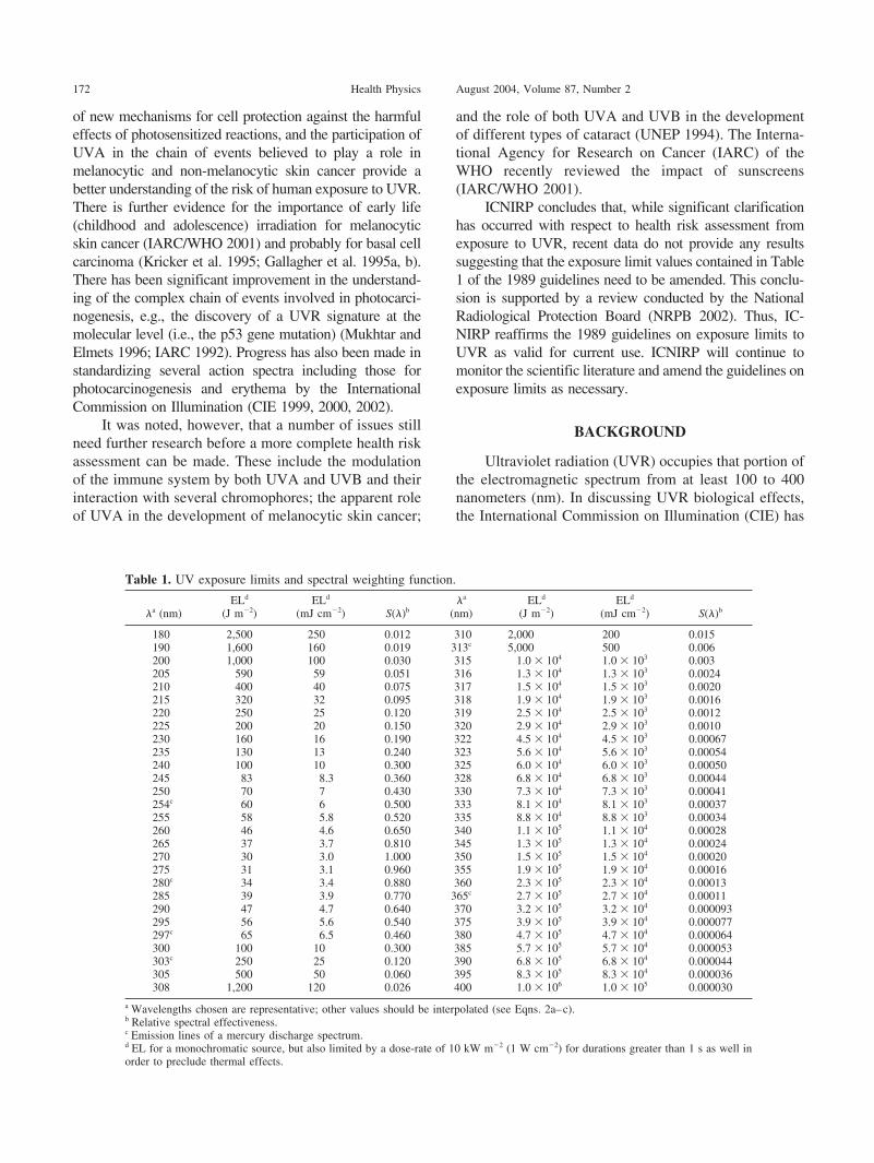
Table 1. UV exposure limits and spectral weighting function.
�a (nm)ELd
(J m�2)ELd
(mJ cm�2) S(�)b�a
(nm)ELd
(J m�2)ELd
(mJ cm�2) S(�)b
180 2,500 250 0.012 310 2,000 200 0.015190 1,600 160 0.019 313c 5,000 500 0.006200 1,000 100 0.030 315 1.0 � 104 1.0 � 103 0.003205 590 59 0.051 316 1.3 � 104 1.3 � 103 0.0024210 400 40 0.075 317 1.5 � 104 1.5 � 103 0.0020215 320 32 0.095 318 1.9 � 104 1.9 � 103 0.0016220 250 25 0.120 319 2.5 � 104 2.5 � 103 0.0012225 200 20 0.150 320 2.9 � 104 2.9 � 103 0.0010230 160 16 0.190 322 4.5 � 104 4.5 � 103 0.00067235 130 13 0.240 323 5.6 � 104 5.6 � 103 0.00054240 100 10 0.300 325 6.0 � 104 6.0 � 103 0.00050245 83 8.3 0.360 328 6.8 � 104 6.8 � 103 0.00044250 70 7 0.430 330 7.3 � 104 7.3 � 103 0.00041254c 60 6 0.500 333 8.1 � 104 8.1 � 103 0.00037255 58 5.8 0.520 335 8.8 � 104 8.8 � 103 0.00034260 46 4.6 0.650 340 1.1 � 105 1.1 � 104 0.00028265 37 3.7 0.810 345 1.3 � 105 1.3 � 104 0.00024270 30 3.0 1.000 350 1.5 � 105 1.5 � 104 0.00020275 31 3.1 0.960 355 1.9 � 105 1.9 � 104 0.00016280c 34 3.4 0.880 360 2.3 � 105 2.3 � 104 0.00013285 39 3.9 0.770 365c 2.7 � 105 2.7 � 104 0.00011290 47 4.7 0.640 370 3.2 � 105 3.2 � 104 0.000093295 56 5.6 0.540 375 3.9 � 105 3.9 � 104 0.000077297c 65 6.5 0.460 380 4.7 � 105 4.7 � 104 0.000064300 100 10 0.300 385 5.7 � 105 5.7 � 104 0.000053303c 250 25 0.120 390 6.8 � 105 6.8 � 104 0.000044305 500 50 0.060 395 8.3 � 105 8.3 � 104 0.000036308 1,200 120 0.026 400 1.0 � 106 1.0 � 105 0.000030
a Wavelengths chosen are representative; other values should be interpolated (see Eqns. 2a–c).b Relative spectral effectiveness.c Emission lines of a mercury discharge spectrum.d EL for a monochromatic source, but also limited by a dose-rate of 10 kW m�2 (1 W cm�2) for durations greater than 1 s as well inorder to preclude thermal effects.
172 Health Physics August 2004, Volume 87, Number 2

divided the UV spectrum into three bands. The band 315to 380–400 nm is designated as UVA, 280 to 315 nm asUVB, and 100 to 280 nm as UVC (CIE 1987, 1999).Wavelengths below 180 nm (vacuum UV) are of littlepractical biologic significance since they are readilyabsorbed in air. Ultraviolet radiation is used in a widevariety of medical and industrial processes and forcosmetic purposes. These include photocuring of inksand plastics (UVA and UVB), photoresist processes (allUV), solar simulation (all UV), cosmetic tanning (UVAand UVB), fade testing (UVA and UVB), dermatology(all UV), and dentistry (UVA). Even though the principaloperating wavelengths for most of these processes are inthe UVA, almost always some shorter wavelength (UVBand UVC) radiation and violet light are emitted as well.Many industrial applications employ arc sources for heator light (e.g., welding), which also produce UVR as anunwanted admixture for which control measures may benecessary. While it is generally agreed that some low-level exposure to UVR benefits health (UNEP 1994;Preece et al. 1975; Clemens et al. 1982; Holick 2000;Webb et al. 1988, 1989; MacLaughlin and Holick 1985),there are adverse effects (de Gruijl 1997; UNEP 1994;ICNIRP/CIE 1998) that necessitate the development anduse of ELs for UVR. However, the development of UVREL poses a real challenge to achieve a realistic balancebetween beneficial and adverse health effects.
Until 1980, it was generally thought that the mostsignificant adverse UVR health effects resulted fromexposures at wavelengths below 315 nm; but today theseeffects are recognized to be produced at longer wave-lengths (UVA) at substantially higher doses. At one time,wavelengths below 315 nm were collectively known as“actinic radiation,” when it was thought that these effectsoccurred only in the UVB and UVC. This guideline hasbeen limited to wavelengths greater than 180 nm whereUVR is transmitted through air. The most restrictivelimits are for exposure to radiation having those wave-lengths less than 315 nm.
PURPOSE AND SCOPE
The purpose of this document is to provide guidanceon maximal limits of exposure to UVR in the spectralregion between 180 nm and 400 nm. The limits representconditions under which it is expected that nearly allindividuals may be repeatedly exposed without acuteadverse effects and, based upon best available evidence,without noticeable risk of delayed effects (see paragraphon Special Considerations). These EL values for expo-sure of the eye or the skin may be used to evaluatepotentially hazardous exposure from UVR; e.g., from
arcs, gas and vapor discharges, fluorescent lamps, incan-descent sources, and solar radiation. The limits do notapply to lasers that emit UVR. Most incoherent UVRsources are broadband, although single emission linescan be produced from low-pressure gas discharges.These values should be used as guides in the control ofexposure to both pulsed and continuous sources wherethe exposure duration is not less than 1 �s. These ELs arebelow levels that would be used for UV exposures ofpatients required as a part of medical treatment or forelective cosmetic purposes. These ELs are exceeded forexposed skin by noonday summer sunlight overhead at0–40° latitude within 5–10 min. The ELs should beconsidered absolute limits for direct exposure of the eyeand “advisory” for skin exposure because of the widerange of susceptibility to skin injury depending on skintype. The ELs should be adequate to protect lightlypigmented individuals.
BASIC CONCEPTS
This document makes use of the spectral banddesignations of the CIE. Unless otherwise stated, UVA isfrom 315 to 400 nm, UVB is from 280 to 315 nm, andUVC is from 100 to 280 nm (CIE 1984, 1987). It shouldbe noted that some specialists follow this general schemebut take the dividing line between UVA and UVB at 320nm. The UVR exposure should be quantified in terms ofan irradiance E (W m�2 or W cm�2) for continuousexposure or in terms of a radiant exposure H (J m�2 or Jcm�2) for time-limited (or pulsed) exposures of the eyeand skin. The geometry of exposure to UVR is veryimportant. For example, the eyes (and to a lesser extentthe skin) are anatomically protected against UVR expo-sure from overhead sources such as the sun overhead(Sliney 1995; UNEP 1994). The limits should be appliedto exposure directed perpendicular to those surfaces ofthe body facing the radiation source, measured with aninstrument having cosine angular response (UNEP1994). For highly non-uniform irradiation the irradianceand radiant exposure need not be averaged over the areaof a circular measurement aperture smaller than 1 mm indiameter for pulsed exposures and 3.5 mm for lengthyexposures.
These ELs should be used as guides in the control ofexposure to UV sources and as such are intended aslimits for non-therapeutic and non-elective exposure. TheELs should be considered as absolute limits for ocularexposure. The ELs were developed by consideringlightly pigmented populations (i.e., white Caucasian)with greatest sensitivity and genetic predisposition forskin cancer. Exposure during sun bathing and tanningunder artificial sources may well exceed these limits but
173Guidelines on limits of exposure to UV radiation ● ICNIRP
exposed individuals should be advised that some healthrisk is incurred from such activity. Eye protection isalways required during therapeutic exposures. Neverthe-less, occasional exposures to conditioned skin may notresult in adverse effects. The rationale for the UVRexposure limits is provided in the Appendix.
EXPOSURE LIMITS
For the EL for both general and occupational expo-sure to UVR incident upon the skin or eye within an 8-hperiod, the following applies.
Exposure of the eyesUltraviolet radiant exposure in the spectral region
180 to 400 nm incident upon the unprotected eye(s)should not exceed 30 J m�2 effective spectrally weightedusing the spectral weighting factors contained in Table 1,and the total (unweighted) ultraviolet radiant exposure inthe spectral region 315 to 400 nm should not exceed 104
J m�2.
Exposure of the skinFor the most sensitive, non-pathologic, skin photo-
types (known as “melano-compromised”), ultravioletradiant exposure in the spectral region 180 to 400 nmupon the unprotected skin should not exceed 30 J m�2
effective spectrally weighted using the spectral weight-ing factors contained in Table 1. This limit should beconsidered a desirable goal for skin exposure to mini-mize the long-term risk, but it must be recognized thatthis limit is difficult to achieve in sunlight and judgmentmust be used in its practical application. It has a verysubstantial safety factor for dark skin phototypes (knownas “melano-competent”) and more generally for individ-uals who have been conditioned by previous, repeatedexposures (known as “melano-adapted,” i.e., tanned).
To determine the effective irradiance of a broadbandsource weighted against the peak of the spectral effec-tiveness curve (270 nm), the following weighting for-mula should be used:
Eeff � � E� � S��� � ��, (1)
where:
Eeff �effective irradiance in �W cm�2 (�J s�1
cm�2) or W m�2 (J s�1 m�2) normalized to amonochromatic source at 270 nm;
E� � spectral irradiance from measurements in �Wcm�2 nm�1 or W m�2 nm�1;
S(�) � relative spectral effectiveness (unitless); and�� �bandwidth in nanometers of the calculation or
measurement intervals.
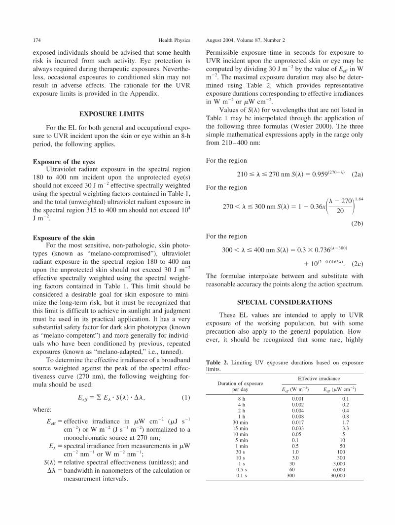
Permissible exposure time in seconds for exposure toUVR incident upon the unprotected skin or eye may becomputed by dividing 30 J m�2 by the value of Eeff in Wm�2. The maximal exposure duration may also be deter-mined using Table 2, which provides representativeexposure durations corresponding to effective irradiancesin W m�2 or �W cm�2.
Values of S(�) for wavelengths that are not listed inTable 1 may be interpolated through the application ofthe following three formulas (Wester 2000). The threesimple mathematical expressions apply in the range onlyfrom 210–400 nm:
For the region
210 � � � 270 nm S��� � 0.959�270��� (2a)
For the region
270 � � � 300 nm S��� � 1 � 0.36x�� � 270
20 �1.64
(2b)
For the region
300 � � � 400 nm S��� � 0.3 � 0.736���300�
� 10�2�0.0163��. (2c)
The formulae interpolate between and substitute withreasonable accuracy the points along the action spectrum.
SPECIAL CONSIDERATIONS
These EL values are intended to apply to UVRexposure of the working population, but with someprecaution also apply to the general population. How-ever, it should be recognized that some rare, highly
Table 2. Limiting UV exposure durations based on exposurelimits.
Duration of exposureper day
Effective irradiance
Eeff (W m�2) Eeff (�W cm�2)
8 h 0.001 0.14 h 0.002 0.22 h 0.004 0.41 h 0.008 0.8
30 min 0.017 1.715 min 0.033 3.310 min 0.05 55 min 0.1 101 min 0.5 5030 s 1.0 10010 s 3.0 3001 s 30 3,000
0.5 s 60 6,0000.1 s 300 30,000
174 Health Physics August 2004, Volume 87, Number 2

photosensitive individuals exist who may react adverselyto exposure at these levels. These individuals are nor-mally aware of their heightened sensitivity. Likewise, ifindividuals are concomitantly exposed to photosensitiz-ing agents (Fitzpatrick et al. 1974; Johnson 1992), aphotosensitizing reaction can take place. It should beemphasized that many individuals who are exposed tophotosensitizing agents (ingested or externally appliedchemicals, e.g., in cosmetics, foods, drugs, industrialchemicals, etc.) probably will not be aware of theirheightened sensitivity. Phototoxic reactions apply to allindividuals and depend upon the quantity of photosensi-tizing chemicals and the UVR exposure, whereas pho-toallergic reactions will be observed for much lowerquantities of the substance in sensitized individuals.Lightly pigmented individuals conditioned by previousUVR exposure (leading to tanning and hyperplasia) andheavily pigmented individuals can tolerate skin exposurein excess of the EL without erythemal effects. However,repeated tanning may increase the risk for those personslater developing signs of accelerated skin aging and evenskin cancer. Such risks should be understood prior to theuse of UVR for medical phototherapy or cosmeticexposures.
PROTECTIVE MEASURES
Protective measures will differ depending uponwhether the UVR exposure results from sunlight orfrom artificial sources. The use of hats, eye protectors,clothing, and sun-shading structures are practical pro-tective measures to reduce sunlight exposure. Whenthese measures are inadequate, topical sunscreensshould be applied to the skin. However, the value ofsunscreens has been questioned, and an IARC Work-ing Group on the Evaluation of Cancer-PreventiveAgents concluded that there was inadequate epidemi-ological evidence in humans for a cancer-preventiveeffect of topical use of sunscreen formulations againstcutaneous malignant melanoma, or basal-cell carci-noma, despite the experimental evidence in animalstudies (IARC/WHO 2001).
When exposure is to artificial sources, as in someindustrial hazard situations, engineering control mea-sures are preferable to protective clothing, goggles, andprocedural safety measures. Glass envelopes for arclamps will filter out most UVB and UVC. Where lengthyexposure to high power glass-envelope lamps and quartzhalogen lamps will occur at close proximity, additionalglass filtration may be necessary (McKinlay et al. 1989).Light-tight cabinets and enclosures and UVR absorbingglass and plastic shielding are the key engineeringcontrol measures used to prevent human exposure to
hazardous UVR produced in many industrial applicationssuch as the fade testing of materials, solar simulation,photoresist applications, and photocuring. For arc weld-ing, cabinets are not practical. Shields, curtains, barriers,and a suitable separation distance are used to protectindividuals against the UVR emitted by open-arc pro-cesses such as arc welding, arc-cutting, and plasmaspraying. Dynamic-filter welding helmets and see-through curtains have improved the safety of weldingoperations in recent decades. There is a need for opera-tional rules to protect potentially exposed individuals.Operators should be trained to follow these general rulesproperly. Ventilation may be required to exhaust ozoneand other airborne contaminants produced by UVCradiation.
MEASUREMENT
UV measurements for health risk evaluation aresometimes of value for indoor exposure assessment.However, they are generally not routinely performed foroutdoor exposure conditions, except with regard to theuse of the Global UV Index (ICNIRP/WHO/WMO/UNEP 2002; Gies et al. 1995).
Although direct-reading UVR radiometers exist,attempts to produce relatively inexpensive field safetysurvey meters that respond directly to UVB and UVCradiation [following the S(�) function] have not beenfully successful. However, relatively expensive instru-ments exist which respond to UVB and UVC radiationaccording to the relative spectral effectiveness, S(�).Spectroradiometric measurements of the source whichcan then be used with the S(�) weighting function tocalculate Eeff are often necessary for measurements moreaccurate than those with simple, direct-reading safetymeters. Whichever measurement technique is applied,the geometry of measurement is important. All thepreceding ELs for UVR apply to exposures that aremeasured with an instrument having a cosine-responsedetector oriented perpendicular to the most directlyexposed surfaces of the body when assessing skin expo-sure. The detector is oriented along (or parallel to) theline(s) of sight of each exposed individual when assess-ing ocular exposure. The use of UV film badges makes itpossible to integrate UV exposure on specific body siteswhich move with respect to the UVR source (Diffey et al.1977; Saunders and Diffey 1995); however, the spectralresponse of such film badges still does not accuratelyfollow S(�).
For outdoor exposure, environmental UVR mea-surements may be of limited use for individual doseassessment because of geometrically changing exposure
175Guidelines on limits of exposure to UV radiation ● ICNIRP
conditions and human behavioral considerations. Per-sonal dosimeters must properly take into considerationthe exposed sites of the individual, time of exposure, sunangle, etc. The Global UV Index can be a useful tool ineducating persons who are outdoors as to the changinglevel of overhead UVR. It is, however, not very predic-tive of ocular exposure since it is a measure of theoverhead UVR incident on a horizontal surface. Ocularexposure is highly dependent upon ground reflectancefactors and the upper lid and brow-ridge block mostoverhead UVR (Sliney 1995).
CONCLUDING REMARKS
Greater attention should be paid to the potentialhazards of UVR exposure. The increasing socially drivensolar exposure as well as the increasing use of artificialUVR sources is a cause for concern. In many popula-tions, skin cancer incidence continues to rise, due in largepart to a poor appreciation of the risk among the generalpopulation. Reduction of risk by avoidance of needlesssunlight exposure and by physical means of protectionshould be an important public health goal. Improvededucational programs are needed for school children, foroutdoor workers and the general public. The presentunderstanding of injury mechanisms and long-term ef-fects of exposure to UVR is incomplete, and awaitsfurther research. The above guidelines will be subject toperiodic review and amendment as appropriate.
Acknowledgments—The support received by ICNIRP from the Interna-tional Radiation Protection Association, the World Health Organization,the International Labor Office, the European Commission, and the GermanGovernment is gratefully acknowledged.
During the preparation of these guidelines, the composition of theInternational Commission on Non-Ionizing Radiation Protection was asfollows:A.F. McKinlay, Chairman (UK)J.H. Bernhardt, Vice-chairman (Germany)A. Ahlbom (Sweden)J-P. Cesarini (France)F. R. de Gruijl (The Netherlands)M. Hietanen (Finland)R. Owen (USA)D.H. Sliney (USA)P. Soderberg (Sweden)A.J. Swerdlow (United Kingdom)M. Taki (Japan)T.S. Tenforde (USA)P. Vecchia (Italy)B. Veyret (France)R. Matthes, Scientific Secretary (Germany)M.H. Repacholi, chairman emeritus (Switzerland)
During the preparation of this document, the composition of the ICNIRPStanding Committee IV and task group was:
D.H. Sliney (USA), ChairmanJ-P. Cesarini (France)F. R. de Gruijl (The Netherlands)B. Diffey (U.K.)M. Hietanen (Finland)M.A. Mainster (USA)T. Okuno (Japan)P. Soderberg (Sweden)B.E. Stuck (USA)
REFERENCES
Anders A, Petry H, Fleming C, Petry K, Brix P, Luke W,Groger H, Schneider E, Kiefer J, Anders F. Increasingmelanoma incidence: Putatively explainable by retrotrans-posons—Experimental contribution of the XiphophorineGordon-Kosswig Melanoma System. Pigment Cell Res7:433–450; 1994.
Anders A, Altheide H, Knalmann M, Tronnier H. Actionspectrum for erythema in humans investigated with dyelasers. Photochem Photobiol 61:200–205; 1995.
Andreassi K, Simoni S, Fiorini P, Fiamiani M. Phenotypiccharacters related to skin type and minimal erythema dose.Photodermatol 4:43–46; 1987.
Armstrong BK, Kricker, A. How much melanoma is caused bysun exposure? Melanoma Res 3:395–401; 1993.
Armstrong BK, Kricker A. Cutaneous melanoma. CancerSurveys 19:219–240; 1994.
Azizi E, Lusky A, Kushelevsky AP, Schewach-Millet M. Skintype, hair colour, and freckles are predictors of decreasedminimal erythema ultraviolet radiation dose. J Am AcadDermatol 19:32–38; 1988.
Bastiaens MT, ter Huuren JA, Kielech C, Gruis NA, Westen-dorp RG, Vermeer BJ, Bavinck JN. Melanocortin-1 recep-tor gene variants determine the risk of non-melanoma skincancer independently of fair skin and red hair. Am J HumGenet 68:884–894; 2001.
Berger D, Urbach F, Davies RE. The action spectrum oferythema induced by ultraviolet radiation (Preliminary Re-port XIII). In: Jadassohn W, Schirren CG, eds. Proceedingsof the Congressus Internationalis Dermatologiae-Munchen1967. New York: Springer-Verlag; 1968: 1112–1117.
Brash DE, Rudolph JA, Simon JA, Lin A, McKenna GJ, BadenHP, Halperin AJ, Ponten J. A role for sunlight in skincancer: UV-induced p53 mutations in squamous cell carci-noma. Proc Natl Acad Sci USA 88:10124–10128; 1991.
Cesarini JP. Ultraviolet radiation: Biological effects and healthconsequences. In: Matthes R, Bernhardt JH, Taki M, eds.Non-ionizing radiation, Proceedings of the 3rd InternationalNon-Ionizing Radiation Workshop, Baden (Austria), April22–26, 1996. Munich: ICNIRP; 1996: 55–76.
CIE. Comptes Rendues de la Commission Internationale del’eclairage. Berlin: CIE; 9:596–625; 1935.
CIE. The spectroradiometric measurement of light sources.Vienna: Commission Internationale de l’Eclairage; Pub. No63; 1984.
CIE. International lighting vocabulary. Vienna: CommissionInternationale de l’Eclairage (International Commission onIllumination); Publication CIE No 17 (E-l.l); 1987.
CIE. Erythema reference action spectrum and standard ery-thema dose. Vienna: CIE; 1998.
CIE. Erythemal reference action spectrum and standard erythe-mal dose. Vienna: CIE; CIE Standard S007–1998; alsoavailable as ISO 17166; 1999a.
CIE. Standardization of the terms UV-A1, UV-A2 and UV-B.Vienna: CIE; Report CIE-134/1; 1999b.
176 Health Physics August 2004, Volume 87, Number 2
CIE. Action spectrum for photocarcinogenesis (non-melanomaskin cancers). Vienna: CIE; CIE 138/2; 2000.
CIE. Action spectroscopy of skin with tunable lasers. Publica-tion Vienna: CIE; CIE 148; 2002.
Clemens TL, Adams JS, Henderson SL, Holick MF. Increasedskin pigment reduces the capacity of skin to synthesizevitamin D3. Lancet i:74–76; 1982.
Coblentz WW, Stair R, Hogue JM. The spectral erythemicreaction of the human skin to ultraviolet radiation. Proc USNat Acad Sci 17:401–403; 1931.
Cooper KD, Oberhelman L, Hamilton TA, Baardsgaard O,Terhune M, LeVee G, Anderson T, Koren H. UV exposurereduces immunization rates and promotes tolerance toepicutaneous antigens in humans: relationship to dose,CD1a- DR� epidermal macrophage induction and Langer-hans cell deletion. Proc Natl Acad Sci USA 89:8497–8501;1992.
Cox NH, Farr PM, Diffey BL. The relationship betweenchronological age and erythemal response to UVB radia-tion. Br J Dermatol 122:272–273; 1990.
Cullen AP, Perera SC. Sunlight and human conjunctival actionspectrum. Proc SPIE, 2134B:24–30; 1994.
Dahaw-Barker P. Ocular photosensitization. Photochem Pho-tobiol 46:1051–1055; 1987.
De Gruijl FR. Health effects from solar UV radiation. RadiatProtect Dosim 72:177–196; 1997.
De Gruijl FR, van der Leun JC. Estimate of the wavelengthdependency of ultraviolet carcinogenesis in humans and itsrelevance to the risk assessment of a stratospheric ozonedepletion. Health Phys 67:319–325; 1994.
Despres S. Effets biologiques des infrarouges et des ultravio-lets. Radioprotection 13:11–21; 1978 (in French).
Diffey BL. Observed and predicted minimal erythema doses: acomparative study. Photochem Photobiol 60:380–382;1994.
Diffey BL. Human exposure to ultraviolet radiation. In: HawkJLM, ed. Photodermatology. London: Chapman and Hall;1998.
Diffey BL, Kerwin M, Davis A. The anatomical distribution ofsunlight. Br J Dermatol 7:407–409; 1977.
Diffey BL, Farr PM, Oakley AM. Quantitative studies on UVAinduced erythema in human skin. Br J Dermatol 117:57–66;1987.
Elwood JM, Jopson J. Melanoma and sun exposure: anoverview of published studies. Int J Cancer 73:196–203;1997.
English DR, Armstrong BK, Kricker A, Winter MG, HeenanPJ, Randell PL. Case-control study of sun exposure andsquamous cell carcinoma of the skin. Int J Cancer 77:347–353; 1996.
Everett MA, Olsen RL, Sayre RM. Ultraviolet erythema. ArchDermatol 92:713–729; 1965.
Ferguson J. Drug and chemical photosensitivity. In: HawkJLN, ed. Photodermatology. London: Chapman and Hall;1998: 155–169.
Fitzpatrick TB, Pathak MA, Harber LC, Seiji M, Kutika A.Sunlight and man. Tokyo: University of Tokyo Press; 1974.
Fitzpatrick TB. Soleil et peau. J Med Esthet 2:33–34; 1975.Freeman RG, Owens DW, Knox JM, Hudson HT. Relative
energy requirements for an erythemal response of skin tomonochromatic wavelengths of ultraviolet present in thesolar spectrum. J Invest Dermatol 47:586–592; 1966.
Gallagher RP, Hill GB, Bajdik CD, Fincham S, Coldman AJ,McLean DI, Threlfall WJ. Sunlight exposure, pigmentaryfactors, and risk of nonmelanocytic skin cancer: I Basal cellcarcinoma. Arch Dermatol 131:157–163; 1995a.
Gallagher RP, Hill GB, Bajdik CD, Fincham S, Coldman AJ,McLean DI, Threlfall WJ. Sunlight exposure, pigmentationfactors, and risk of nonmelanocytic skin cancer: II Squa-mous cell carcinoma. Arch Dermatol 131:164–169; 1995b.
Gange RW, Park YK, Auletta M, Kagetsu N, Blackett AD,Parrish JA. Action spectra for cutaneous responses toultraviolet radiation. In: Urbach F, Gange FW, eds. Thebiological effects of UV-A radiation. New York: Praeger;1986: 57–65.
Gezondheidsraad (Health Council of the Netherlands). Recom-mendations concerning acceptable levels of electromagneticradiation in the wavelength range from 100 nm to 1 mm(micrometre radiation). The Netherlands: Ministry ofHealth and Environmental Protection; Report 65E; 1978.
Gies HP, Roy CR, Toomey S, MacLennan R, Watson M. SolarUVR exposures of three groups of outdoor workers on theSunshine Coast, Queensland. Photochem Photobiol62:1015–1021; 1995.
Green A, Williams G, Neale R, Hart V, Leslie D, Parsons P,Marks GG, Gaffney P, Battistutta D, Frost C, Lang C,Russell A. Daily sunscreen application and beta-carotenesupplementation in prevention of basal-cell and squamous-cell carcinomas of the skin: A randomised controlled trail.The Lancet 354:723–729; 1999.
Ham WT Jr., Mueller HA, Ruffolo JJ Jr., Guerry D III, GuerryRK. Action spectrum for retinal injury from near-ultravioletradiation in the aphakic monkey. Am J Ophthalmol 93:299–306; 1982.
Hamerski W. Studies on the histochemical changes in experi-mental corneal lesions induced with ultraviolet radiationand on prevention of photophthalmia. Klin Oczna 39:537–542; 1969 (in Russian); English translation in Pol Med J8:1469–1476; 1969.
Hausser KW. Influence of wavelength in radiation biology.Strahlentherapie 28:25–44; 1928 (in German).
Hausser KW, Vahle W. Sonnenbrand und Sonnenbraunung.Wissenschaftliche Veroffentlichungen des SiemensKonzern 6:101–120; 1927.
Hawk JLM, Parrish JA. Responses of normal skin to ultravioletradiation. In: Regan JD, Parrish JA, eds. The science ofphotomedicine. New York: Plenum; 1982: 219–260.
Health Council of the Netherlands. UV radiation: Humanexposure to ultraviolet radiation. The Hague: Health Coun-cil of the Netherlands; Report 1986/93; 1986.
Hiller R, Giacometti L, Yuen K. Sunlight and cataract: Anepidemiological investigation. Am J Epidemiol 105:450–459; 1977.
Holick MF. Sunlight and vitamin D: The bone and cancerconnections. Radiat Protect Dosim 91:65–71; 2000.
IARC. Solar and ultraviolet radiation. Monographs on theevaluation of carcinogenic risk to humans. Vol. 55, Solarand UV Radiation. Lyon: International Agency for Re-search on Cancer; 1992.
IARC/WHO. Sunscreens, IARC handbooks of cancer preven-tion. Volume 5. Lyon: IARC; 2001.
ICNIRP. Guidelines on UV radiation exposure limits. HealthPhys 71:978; 1996.
ICNIRP. Guidelines on limits of exposure to optical radiationfrom 0.38 to 3.9 �m. Health Phys 73:539–554; 1997.
ICNIRP. Health issues of ultraviolet tanning appliances usedfor cosmetic purposes. Health Phys 84:119–127; 2003.
ICNIRP/CIE. Measurements of optical radiation hazards. Areference book based on presentations by health and safetyexperts on optical radiation hazards. Matthes R, Sliney D,eds. Gaithersburg MD: ICNIRP/CIE; 1998.
177Guidelines on limits of exposure to UV radiation ● ICNIRP
ICNIRP/WHO/WMO/UNEP. Global solar UV index. A prac-tical guide. Geneva, Switzerland: WHO; 2002.
Jeevan A, Brown E, Kripke ML. UV and infectious diseases.In: Photoimmunology. Krutmann J, Elmets CA, eds. Ox-ford: Blackwell Science; 1995: 153–163.
Johnson BE. Drug and chemical photosensitization. In: Theenvironmental threat to the skin. Marks R, Plewig G, eds.London: Martin Dunitz; 1992: 57–65.
Jose JG, Pitts DG. Wavelength dependency of cataracts inalbino mice following chronic exposure. Exp Eye Res41:545–563; 1985.
Kelly DA, Walker SL, McGregor JM, Potten CS, Young AR.SSR-induced immunosupression in humans has a lowerdose-threshold than erythema. Photochem Photobiol67:46S; 1998.
Kelly DA, Young AR, McGregor JM, Seed PT, Potten CS,Walker SL. Sensitivity to sunburn is associated with sus-ceptibility to ultraviolet radiation-induced suppression ofcutaneous cell-mediated immunity. J Exp Med 191:561–566; 2000.
Kraemer KH. Commentary—Sunlight and skin cancer: An-other link revealed. Proc Natl Acad Sci USA 94:11–14;1997.
Kricker A, Armstrong BK, English DR. Sun exposure andnon-melanocytic skin cancer. Cancer Causes and Control5:367–392; 1994.
Kricker A, Armstrong BK, English DR, Heenan PJ. A dose-response curve for sun exposure and basal cell carcinoma.Int J Cancer 60:482–488; 1995.
Kurtin WE, Zuclich J. Action spectrum for oxygen-dependentnear-ultraviolet induced corneal damage. Photochem Pho-tobiol 27:329–333; 1978.
Luckiesh ML, Holladay L, Tavlor AH. Reaction of untannedhuman skin to ultraviolet radiation. J Opt Soc Am 20:423–432; 1930.
Mackenzie LA. The analysis of the ultraviolet radiation dosesrequired to produce erythemal responses in normal skin.Br J Dermatol 108:1–9; 1983.
MacLaughlin JA, Holick MF. Aging decreases the capacity ofhuman skin to produce vitamin D3. Clin Invest 76:1536–1538; 1985.
Mainster MA. The spectra, classification, and rationale ofultraviolet-protective intraocular lenses. Am J Ophthalmol102:727–732; 1986.
Marrot L, Belaidi JP, Chaubo C, Meunier JR, Perez P,Agapakis-Causse C. An in vivo strategy to evaluate thephototoxicity of solar UV at the molecular and cellularlevel: application to photoprotection assessment. Eur JDermatol 8:403–412; 1998.
McKinlay AF, Diffey BL. A reference action spectrum forultraviolet induced erythema in human skin. CIE J 66:17–22; 1987.
McKinlay AF, Whillock MJ, Meulemans CCE. Ultravioletradiation and blue-light emissions from spotlights incorpo-rating tungsten halogen lamps. Didcot, UK: National Ra-diological Protection Board; Report NRPB-R228; 1989.
Merriam GC, Lofgren S, Michael R, Soderberg PG, Dillon J,Zheng L, Ayala M. An action spectrum for UVB radiationand the rat lens. Invest Ophthalmol Vis Sci 41:2642–2647;2000.
Michael R, Soderberg PG, Chen E. Dose-response function forlens forward light scattering after in vivo exposure toultraviolet radiation. Graefe’s Arch Clin Exp Ophthalmol236:625–629; 1998.
Mukhtar H, Elmets CA. Photocarcinogenesis: Mechanisms,models and human health implications. Photochem Photo-biol 63:355–447; 1996.
Nakasawa H, English D, Randall PL, Nakasawa K, Martell N,Armstrong BK, Yamasaki H. UV and skin cancer: specificp53 gene mutation in normal skin as a biologically relevantexposure measurement. Proc Natl Acad Sci 91:360–364;1994.
Noonan FP, Recio JA, Takayama H, Duray P, Anver MR, RushWL, de Fabo EC, Merlino G. Neonatal sunburn andmelanoma in mice. Nature 413:271–272; 2001.
NRPB. Health Effects from Ultraviolet Radiation. Report of anAdvisory Group on Non-ionizing Radiation. Documents ofthe NRPB. National Radiological Protection Board Vol 13(1). Chilton, Didcot, Oxon: NRPB; 2002.
Olson RL, Sayre RM, Everett MA. Effect of anatomic locationand time on ultraviolet erythema. Arch Dermatol 93:211–215; 1966.
Parkin DM, Whelan SL, Ferlay J, Raymond L, Young J.Cancer incidence in five continents. Lyon: IARC; Vol VII,IARC Sci Publ 143; 1997.
Parrish J, Anderson R, Urbach F, Pitts D. UV-A biologicaleffects of ultraviolet radiation with emphasis on humanresponses to longwave ultraviolet. New York: PlenumPress; 1978.
Parrish JA, Jaenicke KF, Anderson RR. Erythema and mela-nogenesis action spectra of normal human skin. PhotochemPhotobiol 36:187–191; 1982.
Paul B, Parrish J. The interaction of UV-A and UV-B in theproduction of threshold erythema. J Invest Derm 78:371–374; 1982.
Perdiz D, Grof P, Mezzina M, Nikaido O, Moustacchi E, SageE. Distribution and repair of bipyrimidine photoproducts insolar UV-irradiated mammalian cells. Possible role ofDewar photoproducts in solar mutagensis. J BiologicalChemistry 275:26732–26742; 2000.
Pirie A. Formation of N-formylkynurenine in proteins fromlens and other sources by exposure to sunlight. Biochem J125:203–208; 1971.
Pitts D. The ocular ultraviolet action spectrum and protectioncriteria. Health Phys 25:559–566; 1973.
Pitts DG. Ocular effects of radiant energy. In: Pitts DG,Kleinstein RN, eds. Environmental vision. Stoneham, MA:Butterworth-Heinemann; 1993: 151–220.
Pitts DG, Tredici TJ. The effects of ultraviolet on the eye. AmInd Hyg Assoc J 32:235–246; 1971.
Pitts DG, Cullen AP, Hacker PD. Ocular effects of ultravioletradiation from 295 to 365 nm. Invest Ophthalmol Vis Sci16:932–939; 1977.
Ponten F, Berne B, Ren Z-P, Nister M, Ponten J. Ultravioletlight induces expression of p53 and p21 in human skin:Effect of sunscreen and constitutive p21 expression in skinappendages. J Invest Dermatol 105:402–406; 1995.
Preece MA, Tomlinson S, Ribot CA, Pietrek J, Korn HT,Davies DM, Ford JA, Dunnigan MG, O’Riordan JLH.Studies of vitamin D deficiency in man. Quart J Med44:575–589; 1975.
Rees JL. The melanocortin-1 receptor (MC1R): more than justred hair. Pigment Cell Res 13:135–140; 2000.
Ringvold A. In vitro evidence for UV-protection of the eye bythe corneal epithelium mediated by the cytoplasmic protein,RNA, and ascorbate. Acta Ophthalmol Scand 75:496–498;1997.
Ringvold A, Davanger M, Olsen EG, Changes of the corneaendothelium after ultraviolet radiation. Acta Ophthalmo-logica 60:41–53; 1982.
178 Health Physics August 2004, Volume 87, Number 2
Roberts JE. Ocular phototoxicity. J Photochem Photobiol64:136–143; 2001.
Robinson ES, Hill RH, Kripke ML, Setlow RB. The monodel-phis melanoma model: initial report on large ultraviolet Aexposures of suckling young. Photochem Photobiol 71:743–746; 2000.
Sasaki H, Jonasson F, Shui YB, Kojima M, Ono M, Katoh N,Cheng HM, Takahashi N, Sasaki K. High prevalence ofnuclear cataract in the population of tropical subtropicalarea. Dev Ophthalmol 35:60–69; 2002.
Saunders PJ, Diffey BL. Ambulatory monitoring of ultravioleterythema in photosensitive subjects. Photodermatol Photo-immunol Photomed 11:22–24; 1995.
Schmidt K. On the skin erythema effect of UV flashes.Strahlentherapie 124:127–136; 1964.
Setlow RB, Grist E, Thompson K, Woodhead AD. Wave-lengths effective in the induction of malignant melanoma.Proc Natl Acad Sci 90:6666–6671; 1993.
Sherashov SG. Spectral sensitivity of the cornea to ultravioletradiation. Biofizika 15:543–544; 1977 (in Russian).
Sliney DH. The merits of an envelope action spectrum forultraviolet exposure criteria. Am Ind Hyg Assoc J 33:644–653; 1972.
Sliney DH. UV radiation ocular exposure dosimetry. J Photo-chem Photobiol B 31:69–771; 1995.
Sliney DH. Geometrical gradients in the distribution of tem-perature and absorbed ultraviolet radiation in ocular tissues.Dev Ophthalmol 35:40–59; 2002.
Sliney DH, Wolbarsht ML. Safety with lasers and other opticalsources: A comprehensive handbook. New York: PlenumPress; 1980.
Sliney DH, Krueger RR, Trokel SL, Rappaport KD. Photok-eratitis from 193 nm argon-fluoride laser radiation. Photo-chem Photobiol 53:739–744; 1991.
Soderberg PG. Experimental cataract induced by ultravioletradiation. Acta Ophthalmol Suppl 196:1–75; 1990.
Tapaszto I, Vass Z. Alterations in mucopolysaccharide com-pounds of tear and that of corneal epithelium, caused byultraviolet radiation. Ophthalmologica (Additamentum)158:343–347; 1969.
Taylor HR, West SK, Rosenthal FS, Munoz B, Newland HS,Abbey H, Emmett EA. Effect of ultraviolet radiation oncataract formation. New Engl J Med 319:1429–1433; 1988.
UNEP. Ultraviolet radiation. Environmental Health Criteria 14,United Nations Environment Programme, World HealthOrganization, International Commission on Non-IonizingRadiation Protection. Geneva: WHO; 1979.
UNEP. Ultraviolet radiation. Environmental Health Criteria160. United Nations Environment Programme, WorldHealth Organization, International Commission on Non-Ionizing Radiation Protection. Geneva: WHO; 1994.
Urbach F. The ultraviolet action spectrum for erythema—history. In: Matthes R, Sliney D, eds. Measurements ofoptical radiation hazards. Munich: International Commis-sion on Non-Ionizing Radiation Protection; 1998: 51–62.
Urbach F, Epstein JH, Forbes PD. UV carcinogenesis. In:Fitzpatrick TB, Pathak MA, Harber LC, Seiji M, Kutika A,eds. Sunlight and man. Tokyo: University of Tokyo Press;1974: 259–283.
Valverde P, Healy E, Sikkink S, Haldane F, Thody AJ,Carrothers A, Jackson IJ, Rees JL. The Asp84Glu variant ofthe melanocortin-1 receptor (MC1R) is associated withmelanoma. Hum Mol Genet 5:1663–1666; 1996.
Van der Leun JC, Stoop T. In: Urbach F, ed. The biologicaleffects of UV radiation. Oxford: Pergamon Press; 1969:251–254.
Webb AR, Kline LW, Holick MF. Influence of season andlatitude on the cutaneous synthesis of vitamin D3: exposureto winter sunlight in Boston and Edmonton will not promotevitamin D3 synthesis in human skin. J Clin EndocrinolMetab 67:337–338; 1988.
Webb AR, DeCosta BR, Holick MF. Sunlight regulates thecutaneous production of vitamin D3 by causing its photo-degradation. J Clin Endocrinol Metab 68:822–827; 1989.
West SK, Duncan DD, Muoz B, Rubin GS, Fried LP, Bandeen-Roche K, Schein OD. Sunlight exposure and risk of lensopacities in a population-based study: The Salisbury eyeevaluation project. JAMA 280:714–718; 1998.
Wester U. Analytic expressions to represent the hazard ultra-violet action spectrum of ICNIRP and ACGIH. RadiatProtect Dosim 91:231–232; 2000.
Willis I, Kligman A, Epstein J. Effects of long ultraviolet rayson human skin: photoprotective or photoaugmentative. J In-vest Dermatol 59:416–420; 1972.
Young AR, Walker SL. Protection given by sunscreens. RadiatProtect Dosim 91:265–269; 2000.
Young RW. The family of sunlight-related eye diseases.Optometry Visual Sci 71:125–144; 1994.
Ziegler A, Jonason AS, Leffell DJ, Simon JA, Sharma HW,Kimmelman J, Remington L, Jacks T, Brash DE. Sunburnand p53 in the onset of skin cancer. Nature 372:773–776;1994.
Zigman S. Ocular light damage. Photochem and Photobiol57:1060–1068; 1993.
Zuclich JA. Cumulative effects of near-UV induced cornealdamage. Health Phys 38:833–838; 1980.
Zuclich JA. Ultraviolet-induced photochemical damage inocular tissues. Health Phys 56:671–682; 1989.
Zuclich JA, Kurtin WE. Oxygen dependence of near UV-induced corneal damage. Photochem Photobiol 25:133–135; 1977.
Zuclich JA, Taboada J. Ocular hazard from UV laser exhibitingself-mode locking. Appl Opt 17:1482; 1978.
APPENDIX: RATIONALE FOR THE LIMITS OFEXPOSURE TO UVR
BackgroundComprehensive reviews of UVR effects have been
published in conjunction with the United Nations Envi-ronment Program and the World Health Organization(UNEP 1979, 1994), and the interested reader is referredto those documents in particular. The CIE and ICNIRPalso reviewed UVR effects and action spectra in amonograph on optical radiation measurements (ICNIRP/CIE 1998). In addition, the International Agency forCancer Research (IARC) published a monograph onUVR in 1992 (IARC 1992) and published a monographon sunscreens more recently (IARC/WHO 2001). Fur-thermore, the National Radiological Protection Board(NRPB) has recently published a scientific review of thehealth effects of UVR (NRPB 2002). The followingdiscussion is a brief review of those physical andbiological factors used to derive the UVR guidelines.
179Guidelines on limits of exposure to UV radiation ● ICNIRP
General approachLife has evolved under the daily exposure to solar
radiation. Although UVR is only about 5% of the solarspectrum that reaches the earth’s surface, it plays asignificant biological role since individual photon ener-gies are the greatest within the optical spectrum. Theseshorter-wavelength, higher energy photons have suffi-cient energy to produce photochemical alterations thatmay initiate biological effects that are potentially injuri-ous (sometimes referred to as “actinic effects”). Bothbeneficial and unwanted photobiological effects resultfrom UVR exposure. The critical organs for UVR expo-sure are the eye and the skin since they may be readilyexposed.
The approach taken in the development of theseguidelines was to limit exposure to preclude any signif-icant acute photobiologic effects and reduce the risks fordelayed effects from chronic exposure as much as pos-sible, based upon the best available evidence. Thresholdsfor observed bioeffects vary strongly with wavelength.Consequently, various spectral dose-response relation-ships and time-dependent dose-response relations havebeen developed. In photobiology, the term “action spec-trum” refers to the relative spectral effectiveness ofdifferent wavelengths in eliciting a biological effect.Available data on the action spectra and dose responsecurves for each delayed effect were reviewed with thegoal of estimating risk at exposure levels below thoseproducing acute effects. Furthermore, the dose and actionspectra for beneficial effects, such as vitamin-D synthe-sis, were examined to assure that the exposure guidelinesdid not lead to an inadequate level of exposure.
Both the acute skin response (erythema) and long-term risk of skin cancer appear to be related to DNAdamage (de Gruijl and van der Leun 1994; Cesarini1996; Kelly et al. 2000). In theory, only a single UVphoton is required to alter directly a single DNA mole-cule (or indirectly through free-radical production).However, DNA repair mitigates most single-moleculeevents. An enormous number of DNA lesions are pro-duced in one single cell nucleus at the basal epidermallayer in skin irradiated at one minimal erythemal dose.The three major categories of photo-lesions induced insingle cell in culture (hamster ovary mesenchyme) havebeen identified and calculated after exposure to a solarsimulated source. The delivered dose corresponded to thedose received on the skin basal layer from 2 h exposureto a UV-index 6 (Paris mid-summer). The total numberof lesions was around 100,000 photolesions in a singlecell for one standard erythema dose (SED) (Perdiz et al.2000). In practice, laboratory techniques exist to detectUVR-induced changes down to the cellular and molec-ular level (e.g., Marrot et al. 1998). These can detect
DNA lesions and errors in repair (mutations). UVRexposure produces specific types of DNA damage thatresult in “UV signature” mutations, which can be de-tected in skin carcinomas (Brash et al. 1991). Suchmutations in the p53 gene can serve as a biomarker forpast exposure and future carcinoma risk (Nakasawa et al.1994; Ziegler et al. 1994). The commonly experienced“sunburn” (erythema) may be used as a marker for thepresence of substantial UVR-induced DNA damage(Young and Walker 2000). Through DNA damage,erythema is related to skin cancer; and a knowledge oferythema-dose response and action spectra are of valuefor developing guidance that might reduce risk from skincancer.
Acute responses of the skin—ErythemaErythema (i.e., the reddening of the skin as in
sunburn) is the most commonly observed direct effectobserved in the skin following exposure to UVR. UVR-induced erythema results from photochemical damage—principally to DNA—that leads to a cascade of molecularevents resulting in redness due to an increased bloodcontent of the skin by dilatation of the superficial bloodvessels. Unlike the erythema from a thermal insult, theerythema from UVR is delayed by some (1–6) hoursafter the exposure. The duration of the delay is reducedas the exposure dose increases.
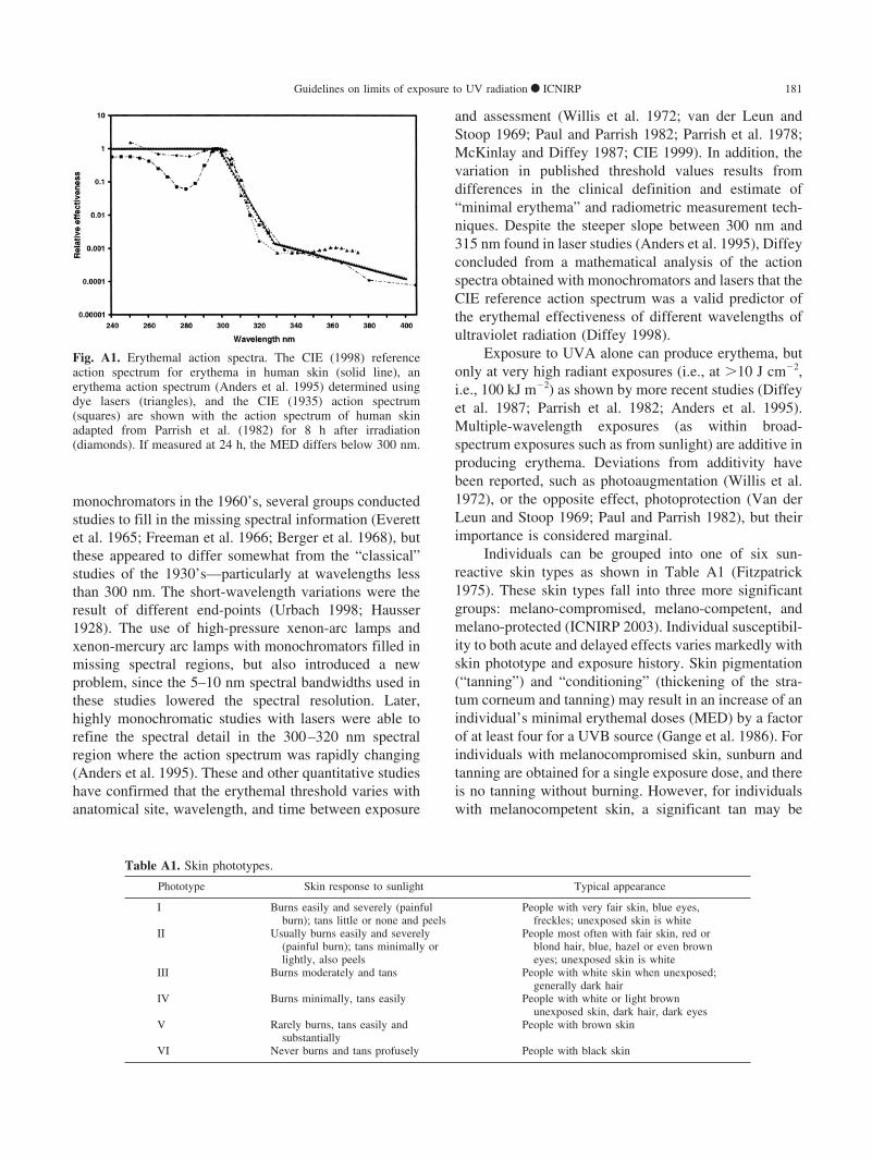
Erythema action spectra have been the subject ofexperimental and theoretical interest for over 70 y(Urbach 1998); and upon first review, there appear to bemany different, and even contradictory, action spectra.The apparent differences relate to different methods ofassessment, types of monochromatic sources, clinicalendpoint, time of assessment, etc. The InternationalCommission on Illumination (CIE) reference actionspectrum E(�) as shown in Fig. A1, is routinely usedtoday to convert absolute UV exposure levels intoerythemally effective irradiance (CIE 1998; McKinlayand Diffey 1987), but this is based upon specific refer-ence conditions. An historical perspective is useful tounderstand the differences in published action spectraand the significance of these differences.
Hausser and Vahle (Hausser 1928; Hausser andVahle 1927) were the first to document quantitativelyerythema as a wavelength-dependent effect in the late1920’s. By 1935, the CIE had recommended an early“standardized” erythemal action spectrum based uponseveral studies using a limited number discrete mono-chromatic emission lines of the mercury lamp (e.g., 254nm, 280 nm, 297 nm, 303 nm, 313 nm, etc.), and thislimited the full spectral detail (Coblentz et al. 1931;Hausser 1928; Luckiesh et al. 1930; Urbach 1998). Withthe development of xenon-arc lamps and their use with
180 Health Physics August 2004, Volume 87, Number 2
monochromators in the 1960’s, several groups conductedstudies to fill in the missing spectral information (Everettet al. 1965; Freeman et al. 1966; Berger et al. 1968), butthese appeared to differ somewhat from the “classical”studies of the 1930’s—particularly at wavelengths lessthan 300 nm. The short-wavelength variations were theresult of different end-points (Urbach 1998; Hausser1928). The use of high-pressure xenon-arc lamps andxenon-mercury arc lamps with monochromators filled inmissing spectral regions, but also introduced a newproblem, since the 5–10 nm spectral bandwidths used inthese studies lowered the spectral resolution. Later,highly monochromatic studies with lasers were able torefine the spectral detail in the 300–320 nm spectralregion where the action spectrum was rapidly changing(Anders et al. 1995). These and other quantitative studieshave confirmed that the erythemal threshold varies withanatomical site, wavelength, and time between exposure
and assessment (Willis et al. 1972; van der Leun andStoop 1969; Paul and Parrish 1982; Parrish et al. 1978;McKinlay and Diffey 1987; CIE 1999). In addition, thevariation in published threshold values results fromdifferences in the clinical definition and estimate of“minimal erythema” and radiometric measurement tech-niques. Despite the steeper slope between 300 nm and315 nm found in laser studies (Anders et al. 1995), Diffeyconcluded from a mathematical analysis of the actionspectra obtained with monochromators and lasers that theCIE reference action spectrum was a valid predictor ofthe erythemal effectiveness of different wavelengths ofultraviolet radiation (Diffey 1998).
Exposure to UVA alone can produce erythema, butonly at very high radiant exposures (i.e., at 10 J cm�2,i.e., 100 kJ m�2) as shown by more recent studies (Diffeyet al. 1987; Parrish et al. 1982; Anders et al. 1995).Multiple-wavelength exposures (as within broad-spectrum exposures such as from sunlight) are additive inproducing erythema. Deviations from additivity havebeen reported, such as photoaugmentation (Willis et al.1972), or the opposite effect, photoprotection (Van derLeun and Stoop 1969; Paul and Parrish 1982), but theirimportance is considered marginal.
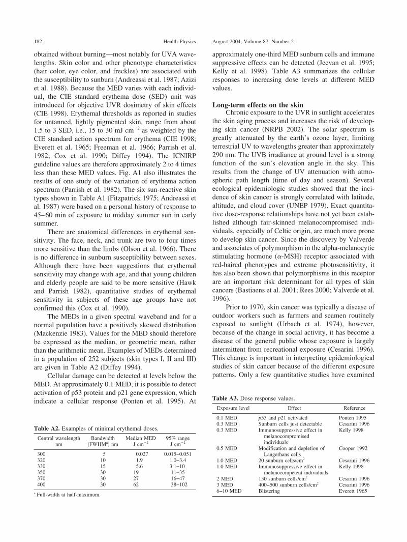
Individuals can be grouped into one of six sun-reactive skin types as shown in Table A1 (Fitzpatrick1975). These skin types fall into three more significantgroups: melano-compromised, melano-competent, andmelano-protected (ICNIRP 2003). Individual susceptibil-ity to both acute and delayed effects varies markedly withskin phototype and exposure history. Skin pigmentation(“tanning”) and “conditioning” (thickening of the stra-tum corneum and tanning) may result in an increase of anindividual’s minimal erythemal doses (MED) by a factorof at least four for a UVB source (Gange et al. 1986). Forindividuals with melanocompromised skin, sunburn andtanning are obtained for a single exposure dose, and thereis no tanning without burning. However, for individualswith melanocompetent skin, a significant tan may be
Table A1. Skin phototypes.
Phototype Skin response to sunlight Typical appearance
I Burns easily and severely (painfulburn); tans little or none and peels
People with very fair skin, blue eyes,freckles; unexposed skin is white
II Usually burns easily and severely(painful burn); tans minimally orlightly, also peels
People most often with fair skin, red orblond hair, blue, hazel or even browneyes; unexposed skin is white
III Burns moderately and tans People with white skin when unexposed;generally dark hair
IV Burns minimally, tans easily People with white or light brownunexposed skin, dark hair, dark eyes
V Rarely burns, tans easily andsubstantially
People with brown skin
VI Never burns and tans profusely People with black skin
Fig. A1. Erythemal action spectra. The CIE (1998) referenceaction spectrum for erythema in human skin (solid line), anerythema action spectrum (Anders et al. 1995) determined usingdye lasers (triangles), and the CIE (1935) action spectrum(squares) are shown with the action spectrum of human skinadapted from Parrish et al. (1982) for 8 h after irradiation(diamonds). If measured at 24 h, the MED differs below 300 nm.
181Guidelines on limits of exposure to UV radiation ● ICNIRP
obtained without burning—most notably for UVA wave-lengths. Skin color and other phenotype characteristics(hair color, eye color, and freckles) are associated withthe susceptibility to sunburn (Andreassi et al. 1987; Aziziet al. 1988). Because the MED varies with each individ-ual, the CIE standard erythema dose (SED) unit wasintroduced for objective UVR dosimetry of skin effects(CIE 1998). Erythemal thresholds as reported in studiesfor untanned, lightly pigmented skin, range from about1.5 to 3 SED, i.e., 15 to 30 mJ cm�2 as weighted by theCIE standard action spectrum for erythema (CIE 1998;Everett et al. 1965; Freeman et al. 1966; Parrish et al.1982; Cox et al. 1990; Diffey 1994). The ICNIRPguideline values are therefore approximately 2 to 4 timesless than these MED values. Fig. A1 also illustrates theresults of one study of the variation of erythema actionspectrum (Parrish et al. 1982). The six sun-reactive skintypes shown in Table A1 (Fitzpatrick 1975; Andreassi etal. 1987) were based on a personal history of response to45–60 min of exposure to midday summer sun in earlysummer.
There are anatomical differences in erythemal sen-sitivity. The face, neck, and trunk are two to four timesmore sensitive than the limbs (Olson et al. 1966). Thereis no difference in sunburn susceptibility between sexes.Although there have been suggestions that erythemalsensitivity may change with age, and that young childrenand elderly people are said to be more sensitive (Hawkand Parrish 1982), quantitative studies of erythemalsensitivity in subjects of these age groups have notconfirmed this (Cox et al. 1990).
The MEDs in a given spectral waveband and for anormal population have a positively skewed distribution(Mackenzie 1983). Values for the MED should thereforebe expressed as the median, or geometric mean, ratherthan the arithmetic mean. Examples of MEDs determinedin a population of 252 subjects (skin types I, II and III)are given in Table A2 (Diffey 1994).
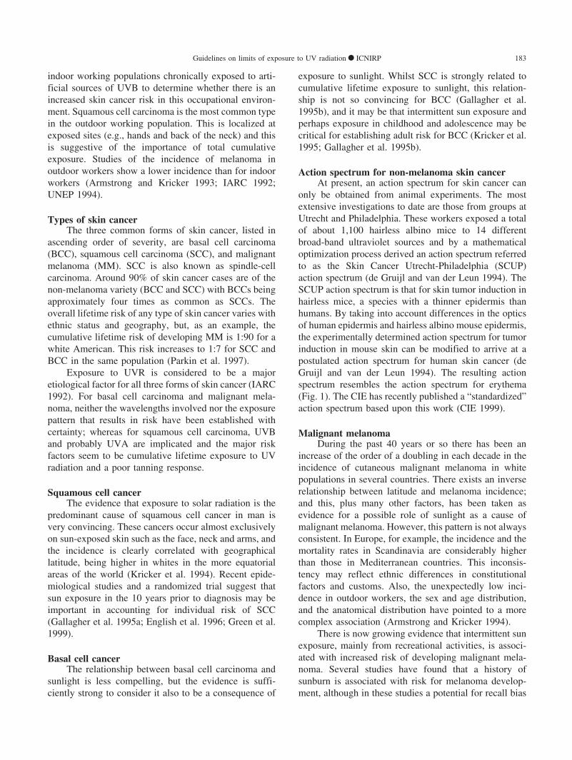
Cellular damage can be detected at levels below theMED. At approximately 0.1 MED, it is possible to detectactivation of p53 protein and p21 gene expression, whichindicate a cellular response (Ponten et al. 1995). At
approximately one-third MED sunburn cells and immunesuppressive effects can be detected (Jeevan et al. 1995;Kelly et al. 1998). Table A3 summarizes the cellularresponses to increasing dose levels at different MEDvalues.
Long-term effects on the skinChronic exposure to the UVR in sunlight accelerates
the skin aging process and increases the risk of develop-ing skin cancer (NRPB 2002). The solar spectrum isgreatly attenuated by the earth’s ozone layer, limitingterrestrial UV to wavelengths greater than approximately290 nm. The UVB irradiance at ground level is a strongfunction of the sun’s elevation angle in the sky. Thisresults from the change of UV attenuation with atmo-spheric path length (time of day and season). Severalecological epidemiologic studies showed that the inci-dence of skin cancer is strongly correlated with latitude,altitude, and cloud cover (UNEP 1979). Exact quantita-tive dose-response relationships have not yet been estab-lished although fair-skinned melanocompromised indi-viduals, especially of Celtic origin, are much more proneto develop skin cancer. Since the discovery by Valverdeand associates of polymorphism in the alpha-melanocyticstimulating hormone (-MSH) receptor associated withred-haired phenotypes and extreme photosensitivity, ithas also been shown that polymorphisms in this receptorare an important risk determinant for all types of skincancers (Bastiaens et al. 2001; Rees 2000; Valverde et al.1996).
Prior to 1970, skin cancer was typically a disease ofoutdoor workers such as farmers and seamen routinelyexposed to sunlight (Urbach et al. 1974), however,because of the change in social activity, it has become adisease of the general public whose exposure is largelyintermittent from recreational exposure (Cesarini 1996).This change is important in interpreting epidemiologicalstudies of skin cancer because of the different exposurepatterns. Only a few quantitative studies have examined
Table A2. Examples of minimal erythemal doses.
Central wavelengthnm
Bandwidth(FWHMa) nm
Median MEDJ cm�2
95% rangeJ cm�2
300 5 0.027 0.015−0.051320 10 1.9 1.0−3.4330 15 5.6 3.1−10350 30 19 11−35370 30 27 16−47400 30 62 38−102
a Full-width at half-maximum.
Table A3. Dose response values.
Exposure level Effect Reference
0.1 MED p53 and p21 activated Ponten 19950.3 MED Sunburn cells just detectable Cesarini 19960.3 MED Immunosuppressive effect in
melanocompromisedindividuals
Kelly 1998
0.5 MED Modification and depletion ofLangerhans cells
Cooper 1992
1.0 MED 20 sunburn cells/cm2 Cesarini 19961.0 MED Immunosuppressive effect in
melanocompetent individualsKelly 1998
2 MED 150 sunburn cells/cm2 Cesarini 19963 MED 400−500 sunburn cells/cm2 Cesarini 19966−10 MED Blistering Everett 1965
182 Health Physics August 2004, Volume 87, Number 2
indoor working populations chronically exposed to arti-ficial sources of UVB to determine whether there is anincreased skin cancer risk in this occupational environ-ment. Squamous cell carcinoma is the most common typein the outdoor working population. This is localized atexposed sites (e.g., hands and back of the neck) and thisis suggestive of the importance of total cumulativeexposure. Studies of the incidence of melanoma inoutdoor workers show a lower incidence than for indoorworkers (Armstrong and Kricker 1993; IARC 1992;UNEP 1994).
Types of skin cancerThe three common forms of skin cancer, listed in
ascending order of severity, are basal cell carcinoma(BCC), squamous cell carcinoma (SCC), and malignantmelanoma (MM). SCC is also known as spindle-cellcarcinoma. Around 90% of skin cancer cases are of thenon-melanoma variety (BCC and SCC) with BCCs beingapproximately four times as common as SCCs. Theoverall lifetime risk of any type of skin cancer varies withethnic status and geography, but, as an example, thecumulative lifetime risk of developing MM is 1:90 for awhite American. This risk increases to 1:7 for SCC andBCC in the same population (Parkin et al. 1997).
Exposure to UVR is considered to be a majoretiological factor for all three forms of skin cancer (IARC1992). For basal cell carcinoma and malignant mela-noma, neither the wavelengths involved nor the exposurepattern that results in risk have been established withcertainty; whereas for squamous cell carcinoma, UVBand probably UVA are implicated and the major riskfactors seem to be cumulative lifetime exposure to UVradiation and a poor tanning response.
Squamous cell cancerThe evidence that exposure to solar radiation is the
predominant cause of squamous cell cancer in man isvery convincing. These cancers occur almost exclusivelyon sun-exposed skin such as the face, neck and arms, andthe incidence is clearly correlated with geographicallatitude, being higher in whites in the more equatorialareas of the world (Kricker et al. 1994). Recent epide-miological studies and a randomized trial suggest thatsun exposure in the 10 years prior to diagnosis may beimportant in accounting for individual risk of SCC(Gallagher et al. 1995a; English et al. 1996; Green et al.1999).
Basal cell cancerThe relationship between basal cell carcinoma and
sunlight is less compelling, but the evidence is suffi-ciently strong to consider it also to be a consequence of
exposure to sunlight. Whilst SCC is strongly related tocumulative lifetime exposure to sunlight, this relation-ship is not so convincing for BCC (Gallagher et al.1995b), and it may be that intermittent sun exposure andperhaps exposure in childhood and adolescence may becritical for establishing adult risk for BCC (Kricker et al.1995; Gallagher et al. 1995b).
Action spectrum for non-melanoma skin cancerAt present, an action spectrum for skin cancer can
only be obtained from animal experiments. The mostextensive investigations to date are those from groups atUtrecht and Philadelphia. These workers exposed a totalof about 1,100 hairless albino mice to 14 differentbroad-band ultraviolet sources and by a mathematicaloptimization process derived an action spectrum referredto as the Skin Cancer Utrecht-Philadelphia (SCUP)action spectrum (de Gruijl and van der Leun 1994). TheSCUP action spectrum is that for skin tumor induction inhairless mice, a species with a thinner epidermis thanhumans. By taking into account differences in the opticsof human epidermis and hairless albino mouse epidermis,the experimentally determined action spectrum for tumorinduction in mouse skin can be modified to arrive at apostulated action spectrum for human skin cancer (deGruijl and van der Leun 1994). The resulting actionspectrum resembles the action spectrum for erythema(Fig. 1). The CIE has recently published a “standardized”action spectrum based upon this work (CIE 1999).
Malignant melanomaDuring the past 40 years or so there has been an
increase of the order of a doubling in each decade in theincidence of cutaneous malignant melanoma in whitepopulations in several countries. There exists an inverserelationship between latitude and melanoma incidence;and this, plus many other factors, has been taken asevidence for a possible role of sunlight as a cause ofmalignant melanoma. However, this pattern is not alwaysconsistent. In Europe, for example, the incidence and themortality rates in Scandinavia are considerably higherthan those in Mediterranean countries. This inconsis-tency may reflect ethnic differences in constitutionalfactors and customs. Also, the unexpectedly low inci-dence in outdoor workers, the sex and age distribution,and the anatomical distribution have pointed to a morecomplex association (Armstrong and Kricker 1994).
There is now growing evidence that intermittent sunexposure, mainly from recreational activities, is associ-ated with increased risk of developing malignant mela-noma. Several studies have found that a history ofsunburn is associated with risk for melanoma develop-ment, although in these studies a potential for recall bias
183Guidelines on limits of exposure to UV radiation ● ICNIRP
exists (Elwood and Jopson 1997) and may be con-founded by skin type. Studies of migrants have led to thesuggestion that sun exposure in childhood and adoles-cence is a particularly critical period in terms of mela-noma risk.
Action spectrum for melanomaAt one time, the only data that existed for an action
spectrum for melanoma induction were those obtainedfrom irradiating hybrids of a small tropical fish withdifferent wavelengths of UVR (Setlow et al. 1993). Thisfish action spectrum suggested that all wavelengths ofUV radiation could be important in melanoma, unlikenon-melanoma skin cancer; however, at least one attemptto replicate this action spectrum was unsuccessful(Anders et al. 1994). More recent studies in transgenicmice (Noonan et al. 2001) and in monodelphis domestica(Robinson et al. 2000) indicate that neonatal UV expo-sure is most significant. In contrast to small UVB doses,Robinson and colleagues also found that large doses ofUVA to neonates could not produce tumors (Robinson etal. 2000). Melanoma incidence is also extremely high inxeroderma pigmentosa (X-P) patients, who lack thecapacity to repair UVB induced damage (Kraemer 1997).The weight of current evidence now suggests that UVBis the primary risk factor for MM.
Ocular effects—PhotokeratoconjunctivitisShort-wavelength UVR (� 300 nm) is strongly
absorbed by the cornea and conjunctiva. Excessive ex-posure of these tissues causes photokeratoconjunctivitis,commonly referred to as “welder’s flash,” “arc-eye,” etc.Several research groups have characterized the course ofordinary clinical photokeratitis (Pitts 1993) and thecellular changes in ocular tissues (Ringvold et al. 1982).The latent period varies inversely with the severity ofexposure ranging from 1⁄2 to 24 h but usually occurswithin 6–12 h. Conjunctivitis tends to develop moreslowly and may be accompanied by erythema of thefacial skin surrounding the eyelids. The individual hasthe sensation of a foreign body or sand in the eyes andmay experience photophobia, lacrimation, and blepharo-spasm to varying degrees. The acute symptoms last from6 to 24 hours and discomfort usually disappears within48 h. Although exposure rarely results in permanentocular injury, the individual is visually incapacitatedduring this 48-h period. Pitts and Tredici (1971) reportedthreshold data for photokeratitis in humans for 10 nmwavebands from 220 to 310 nm (Pitts 1993). Theguideline ELs between 200 nm and 305 nm are about 1.3to 4.6 times less than the threshold for minimal change.The maximum sensitivity of the human eye was found tooccur at 270 nm. The wavelength response (action
spectrum) between 220 and 310 nm does not vary asgreatly as in the case of erythema with the thresholdsvarying from 4–14 mJ cm�2. Sliney and colleagues usedan excimer laser to determine the photokeratitis thresholdat 193 nm (Sliney et al. 1991). Corneal injury from UVAwavelengths requires levels exceeding 10 J cm�2 (Ham-erski 1969; Pitts 1993; Sherashov 1977; Tapaszto andVass 1969; Zuclich and Kurtin 1977; Zuclich 1980;Cullen and Perera 1994).
CataractWavelengths above 295 nm can be transmitted
through the cornea and are absorbed by the lens. Pitts etal. (1977) have shown that both transient and permanentopacities of the lens (cataracts) can be produced inrabbits and primates by exposure to UVR having wave-lengths in the 295–320 nm band. Similar findings werereported for the rat (Soderberg 1990). Thresholds fortransient opacities ranged dramatically with wavelength,from 0.15 to 12.6 J cm�2. Thresholds for permanentopacities were typically twice those for transient opaci-ties (Pitts 1993). Experimental methods cannot readilyshow a threshold, since a measure of increased scatter isdifficult when there is a background level of scattering(Michael et al. 1998). The action spectrum for UVRinduced cataract was recently confirmed in the rat by useof a quantitative criterion for light scattering (Merriam etal. 2000). Opacities from chronic exposure at lowerlevels has been very difficult to show experimentally(Jose and Pitts 1985; Zigman 1993). However, severalepidemiological studies show an association between theincidence of cortical opacities with ambient UVB expo-sure (Hiller et al. 1977; Taylor et al. 1988; West et al.1998). Sasaki has shown a clear correlation of differentforms of cataract with latitude, but does not explicitlylink this with the change of UVR exposure with latitude(Sasaki et al. 2002), although it was speculated that bothtemperature and UVR could be etiologic factors (Sliney2002). A number of biochemical studies of the effects ofUV irradiation of lens proteins have led to the theory thatUVA radiation is a causal factor in cataract (Pirie 1971;Roberts 2001; Young 1994). However, it has beendifficult to link UVA radiation with cataract eitherepidemiologically or experimentally.
Retinal effectsThe cornea and crystalline lens normally sufficiently
shield the retina from acute effects from UVR exposure.Normally, less than 1% of UVA reaches the retina,shorter UVB wavelengths being totally attenuated exceptin neonates (UNEP 1994). Upon removal of the crystal-line lens, Ham and colleagues (Ham et al. 1982) demon-strated acute retinal injury (photoretinitis) at levels of the
184 Health Physics August 2004, Volume 87, Number 2
order of 5 J cm�2 at the retina. Photoretinitis at thesewavelengths is covered by the ICNIRP guidelines forexposure to incoherent optical radiation (ICNIRP 1997).
Envelope action spectrumClearly, the development of UVR exposure limits
for workers and the general population must considertwo risks. These are the risks of acute and chronic injuryto both the eye and skin. The literature indicates thatthresholds for injury vary significantly with wavelengthfor each effect. In the UVB and UVC regions, an actionspectrum curve can be drawn which envelops the thresh-old data for exposure doses (radiant exposures) in therange of reciprocity (Schmidt 1964; Zuclich 1980) foracute effects obtained from recent studies of minimalerythema and keratoconjunctivitis. Reciprocity meansthat irradiance E and exposure duration t have a recipro-cal relation, and a constant product of E and t (i.e.,exposure) results in a given effect. This EL curve doesnot differ significantly from the collective threshold dataconsidering measurement errors and variations in indi-vidual response (Sliney 1972; Sliney and Wolbarsht1980). Although the safety factor is minimal for just-detectable increases in corneal scatter, it is believed torange from 1.5 to 2.0 for acute keratitis. The curve is alsowell below the acute UVB cataractogenic thresholds(Merriam et al. 2000; Pitts 1993). Repeated exposure ofthe eye to potentially hazardous levels of UV is notbelieved to increase significantly the protective capabil-ity of the cornea as does skin tanning and thickening ofthe stratum corneum [although some recent studies showa detectable change in threshold (Ringvold 1997)]. Thus,this EL is more readily applicable to the eye and must beconsidered a limiting value for that organ (Sliney 1972).Any accumulation of UVB and UVC exposures causingphotokeratitis is limited to about 48 h since the outercorneal epithelial layers are replaced in about 48 h by thenormal repair process of this tissue. Some slight additiv-ity of UVA exposures exists beyond 48 hours because ofthe deeper penetration of UVA rays (Zuclich 1980). Theadditivity factors were considered in deriving the mag-nitude of the safety factor built into the guidelines. Onthe basis of acute effects, the safety factor for UVAguidelines is large, varying from about 7 at 320 nm tomore than 100 at 390 nm.
Because of the wide variations in threshold values andexposure history (conditioning) among individuals, theseguidelines should only be used as a starting point forevaluating skin hazards (Despres 1978; Gezondheidsraad1978; NRPB 2002; Sliney and Wolbarsht 1980; UNEP1994). The envelope guideline has some margin of safety toprotect all but the most sensitive individuals. An exact valuefor this margin cannot be given, but for lightly pigmented
persons (i.e., melanocompromised skin phototypes I and II)it varies from about 3 to 20 depending on the spectralcomposition of the radiation. Since there may be more thanone target molecule (chromophore) involved in erythema(and therefore more than one erythemal action spectrum),the effect of radiations of two widely differing wavelengthsin the 180 nm to 315 nm range may not be simply additive.The EL should be used with caution in evaluating sourcessuch as the sun and fluorescent lamps, having a rapidlyincreasing spectral irradiance in the 300–315 nm range.Large errors can arise because of the difficulty in makingaccurate spectral measurements of such sources in thisregion (Sliney and Wolbarsht 1980; ICNIRP/CIE 1998).
The EL may not provide adequate protection forphotosensitive individuals or for normal individuals ex-posed concomitantly to chemical, pharmaceutical, or phyto-photosensitizers, and special precautions must be taken forsuch cases (Dahaw-Barker 1987; Ferguson 1998).
Based upon current knowledge, the EL should preventsignificant acute effects and reduce the magnitude ofchronic skin effects by limiting life-long UV exposure. Theaction spectrum for each type of skin cancer is still debated,although most research suggested that at least squamous-cell carcinoma is probably related (both directly and indi-rectly) to UV-induced molecular damage to DNA, and theaction spectrum is similar to that of the erythemal actionspectrum. Indeed, a Technical Committee of the CIE hasproposed a tentative action spectrum for photocarcinogen-esis (CIE 1999). The Dutch Health Council (Health Councilof the Netherlands 1986) was the first to propose envelopelimits similar to the guidelines developed for acute dailyexposure (up to 10 y duration) and reduced levels for longerperiods to protect against chronic effects. These should havethe same action spectrum in the UVB and UVC. In manycases, occupational exposure to UVB adds to an individu-al’s non-occupational exposure to solar UVB.
It is worthy of note that in addition to the direct hazardof UV exposure, very intense UVC sources (particularly ofwavelengths less than 230 nm) may also produce hazardousconcentrations of ozone and nitrogen oxides from the airand of phosgene gas in the presence of degreasers; thus,many UV germicidal lamps now have quartz-glass enve-lopes that block wavelengths below �230 nm.
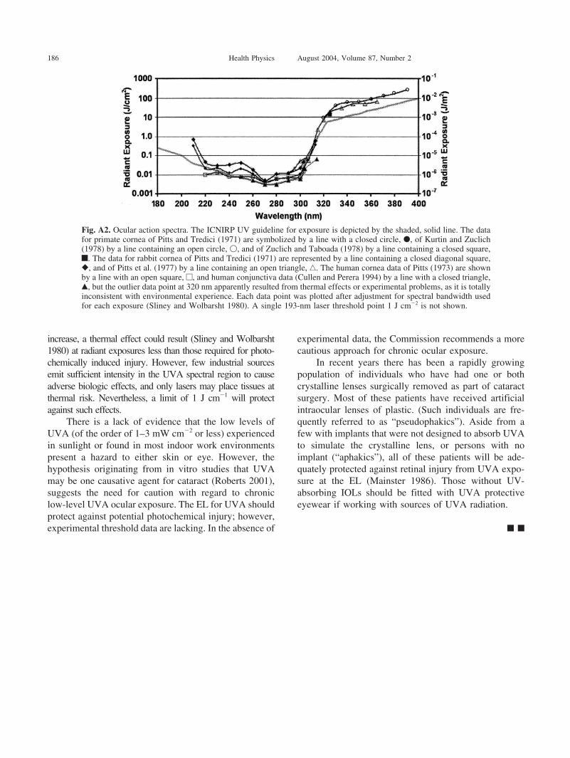
UVA radiation effectsStudies of skin and ocular injury action spectra (Fig. A2)
in the UVA spectral region (315–400 nm) show very similarthresholds for acute injury (Anders et al. 1995; McKinlay andDiffey 1987; Parrish et al. 1982; Pitts et al. 1977; Zuclich1989). These data are sufficient to define the relative spectraleffectiveness, S(�), for exposure guidelines up to 400 nm.However, if radiant energy were to be delivered to the skin orocular tissues sufficiently fast for a substantial temperature
185Guidelines on limits of exposure to UV radiation ● ICNIRP
increase, a thermal effect could result (Sliney and Wolbarsht1980) at radiant exposures less than those required for photo-chemically induced injury. However, few industrial sourcesemit sufficient intensity in the UVA spectral region to causeadverse biologic effects, and only lasers may place tissues atthermal risk. Nevertheless, a limit of 1 J cm�1 will protectagainst such effects.
There is a lack of evidence that the low levels ofUVA (of the order of 1–3 mW cm�2 or less) experiencedin sunlight or found in most indoor work environmentspresent a hazard to either skin or eye. However, thehypothesis originating from in vitro studies that UVAmay be one causative agent for cataract (Roberts 2001),suggests the need for caution with regard to chroniclow-level UVA ocular exposure. The EL for UVA shouldprotect against potential photochemical injury; however,experimental threshold data are lacking. In the absence of
experimental data, the Commission recommends a morecautious approach for chronic ocular exposure.
In recent years there has been a rapidly growingpopulation of individuals who have had one or bothcrystalline lenses surgically removed as part of cataractsurgery. Most of these patients have received artificialintraocular lenses of plastic. (Such individuals are fre-quently referred to as “pseudophakics”). Aside from afew with implants that were not designed to absorb UVAto simulate the crystalline lens, or persons with noimplant (“aphakics”), all of these patients will be ade-quately protected against retinal injury from UVA expo-sure at the EL (Mainster 1986). Those without UV-absorbing IOLs should be fitted with UVA protectiveeyewear if working with sources of UVA radiation.
f f
Fig. A2. Ocular action spectra. The ICNIRP UV guideline for exposure is depicted by the shaded, solid line. The datafor primate cornea of Pitts and Tredici (1971) are symbolized by a line with a closed circle, �, of Kurtin and Zuclich(1978) by a line containing an open circle, �, and of Zuclich and Taboada (1978) by a line containing a closed square,f. The data for rabbit cornea of Pitts and Tredici (1971) are represented by a line containing a closed diagonal square,�, and of Pitts et al. (1977) by a line containing an open triangle, ‚. The human cornea data of Pitts (1973) are shownby a line with an open square, e, and human conjunctiva data (Cullen and Perera 1994) by a line with a closed triangle,Œ, but the outlier data point at 320 nm apparently resulted from thermal effects or experimental problems, as it is totallyinconsistent with environmental experience. Each data point was plotted after adjustment for spectral bandwidth usedfor each exposure (Sliney and Wolbarsht 1980). A single 193-nm laser threshold point 1 J cm�2 is not shown.
186 Health Physics August 2004, Volume 87, Number 2