i. sclerosing lesions of the mediastinum - 01... · walled cyst in continuity with the pericardium...
TRANSCRIPT

I. SCLEROSING LESIONSOF THE MEDIASTINUM
Mark R. Wick, MD
Division of Surgical Pathology
University of Virginia Health System
Charlottesville, VA, USA
SCLEROSING MEDIASTINITIS
• A slowly-evolving tumefactive fibroinflammatory process in the anterior & middle mediastinum that may present as a discrete mass or an infiltrative lesion that entraps great vessels, thymus, and lung tissue
• May be associated with signs & symptoms of superior vena cava syndrome
SCLEROSING MEDIASTINITIS:Other Clinical Data
• Most often seen in Caucasian females (F:M ratio 3:1) below the age of 30 yrs.
• Roughly 40% of patients are asymptomatic and have the condition detected radiographically
• The remainder present with cough, short of breath, chest pain, wheezing, dysphagia, or hemoptysis
• Diffuse effacement of mediastinal architecture may be seen radiographically, or a discrete mass can be present with or without calcifications
• Compression of pulmonary artery branches may cause secondary pulmonary infarction

Potential Etiological Factors for Fibrosing Mediastinitis
Fungal Infections
Histoplasmosis
Aspergillosis
Zygomycosis
Cryptococcosis
Mycobacterial InfectionsTuberculous
Non-tuberculous
Other Bacterial InfectionsNocardiosis
Actinomycosis
Autoimmune Conditions
Behcet syndrome
IgG4-related fibrosclerosing disease
Sarcoidosis
Rheumatic Fever
Prior Trauma
Selected Drugs (Methysergide)
Idiopathic

GMS
ZN
WHAT IS THE RELATIONSHIPBETWEEN SCLEROSING MEDIASTINITIS
& IgG4-RELATED FIBROSCLEROSIS?
• This question is still being examined, but the best hypothesis is that all forms of sclerosing mediastinitis represent a type IV hypersensitivity response that shares similar histologic manifestations, regardless of the inciting factor(s)
• The four principal subsets of the disease are:- Infection-related (20-25%)
- IgG4-related (~30%)
- Autoimmune disease (e.g., Sjogren syndrome, primary sclerosing cholangitis, primary biliary cirrhosis, inflammatory bowel disease)-related (~20%)
- Idiopathic (25-30%)

IgG4
SCLEROSING MEDIASTINITIS:Outcomes
• Most cases pursue a slowly-evolving, self-limited course that may last for several years
• Administration of antifungal or antimycobacterial drugs empirically does not appear to alter outcomes
• Symptomatic patients may benefit from placement of vascular stents, balloon angioplasty, or surgical reconstructive procedures
• Only ~3% of patients die of cardiorespiratory failure
Because of the potential simulation of sclerosing
mediastinitis by other fibrosing conditions, the
former diagnosis is one of ultimate exclusion and close
correlation with clinical findings

FIBROSING NEOPLASMS THAT MAY SIMULATE SCLEROSING MEDIASTINITIS IN SMALL BIOPSIES
• “Obliterative subtotal sclerosis”-type Hodgkin lymphoma
• Sclerosing non-Hodgkin lymphoma (large-cell type)
• Sclerosing seminoma
• Desmoplastic mesothelioma presenting in the mediastinum
• Sclerotic (“ancient”) thymoma
• Sclerosing thymic carcinoid
• Sclerosing paraganglioma
• Calcifying fibrous pseudotumor
• Solitary fibrous tumor
• Peripheral nerve sheath tumors
• Selected metastatic carcinomas
“Obliterative Subtotal Sclerosis”-Type Hodgkin Lymphoma (HL)
• This terminology was used by Rappaport in the 2nd
series AFIP fascicle on hematopoietic tumors in 1966, to refer to a subtype of nodular sclerosis HL cases in which a densely-fibrotic stroma dominated the microscopic image of the lesion
• The OSS variant of HL is not well-recognized by general pathologists, but several publications have described its ability to imitate non-neoplastic fibrosclerosing conditions such as Oulmont’s and Ormond’s diseases in the mediastinum and retroperitoneum

LARGE-CELL NON-HODGKINLYMPHOMA OF THE THYMIC
REGION, SCLEROSING B-CELL TYPE
• Recognized in the late 1970s & early 1980s as a distinctive intrathoracic neoplasm that could be associated with the superior vena cava syndrome
• Now well-characterized as a B-cell proliferation that is centered in the thymus, with singular cytogenetic & molecular characteristics

SCLEROSING SEMINOMA OF THEMEDIASTINUM
• A rare variant of seminoma, previously reported only in the testis; the speaker also has observed 3 such cases in the anterior mediastinum
• Tumor cells are scant in number and obscured by stromal fibrosis and chronic inflammation
• Immunostains for PLAP, CD117, OCT ¾, and SALL4 are usually necessary to document the presence of neoplastic germ cells in such tumors

OCT 3/4
DESMOPLASTIC MESOTHELIOMAOF THE MEDIASTINUM (DMM)
• An uncommon histologic variant of an uncommon neoplasm in an uncommon anatomical location
• DMM may be surprisingly hypocellular, with a predominance of hyalinizing stroma which superficially resembles the histologic appearance of a fibrohyaline (asbestos-related) serosal plaque
• At least modest nuclear atypia and growth into fat are necessary microscopic diagnostic elements
• Neoplastic cells are pankeratin-positive

Pankeratin
“ANCIENT” (SCLEROTIC) THYMOMA
• An unusual thymoma variant reported by Moran & Suster in 2004
• Only 10% of patients had myasthenia gravis; the remainder presented with nondescript signs & symptoms or were entirely asymptomatic
• Densely-fibrotic stroma accounted for 85% to 90% of the tumor masses in each case
• Thorough sampling was necessary to document the presence of epithelial neoplastic cell groups

SCLEROTIC THYMIC CARCINOID
• Extensive sclerosis in neuroendocrine carcinomas of the lung was described by Kalhor et al. in 2010; the speaker has encountered 2 primary thymic neoplasms with similar changes
• Dense stromal fibrosis may obscure the diagnosis in small biopsies and also interfere with grading of the tumors
• Lesional cells are immunoreactive for pankeratin, as well as markers of neuroendocrine differentiation
• Marked sclerosis does not appear to affect behavior or prognosis
Pankeratin
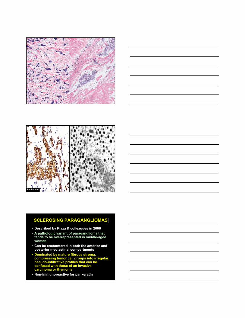
SCLEROSING PARAGANGLIOMAS
• Described by Plaza & colleagues in 2006
• A pathologic variant of paraganglioma that tends to be overrepresented in middle-aged women
• Can be encountered in both the anterior and posterior mediastinal compartments
• Dominated by mature fibrous stroma, compressing tumor cell groups into irregular, pseudo-infiltrative profiles that can be confused with those of an invasive carcinoma or thymoma
• Non-immunoreactive for pankeratin

Chromogranin
CALCIFYING (PSEUDO)TUMOR (CPT)OF THE MEDIASTINUM
• Analogous to other lesions of the soft tissue, serosal surfaces, lungs, esophagus, liver, & spine
• Typically presents as a discrete mass, rather than an infiltrative process such as fibrosing mediastinitis
• Nondescript radiographic & gross appearance, except for the presence of multifocal calcifications
• Paucicellular background fibroblastic proliferation
• Appears to be a singular entity distinct from inflammatory myofibroblastic tumor and solitary fibrous tumor
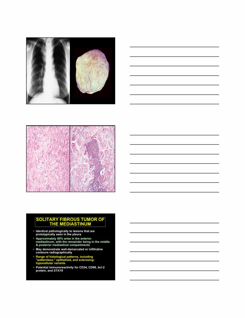
SOLITARY FIBROUS TUMOR OFTHE MEDIASTINUM
• Identical pathologically to lesions that are prototypically seen in the pleura
• Approximately 80% arise in the anterior mediastinum, with the remainder being in the middle & posterior mediastinal compartments
• May demonstrate well-demarcated or infiltrative contours radiographically
• Range of histological patterns, including “patternless,” epithelioid, and sclerosing-hypocellular variants
• Potential immunoreactivity for CD34, CD99, bcl-2 protein, and STAT6
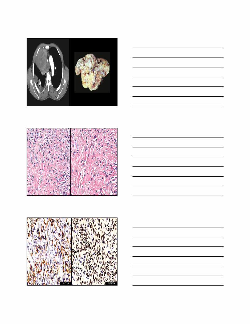
CD34 STAT6

SCLEROTIC PERIPHERAL NERVESHEATH TUMORS
• Typically present as well-delimited masses in the posterior mediastinum
• Often asymptomatic; may cause neural-compressive symptoms & signs or back pain
• Predominantly benign in nature; malignant nerve sheath tumors are rare in the mediastinum
• May be represented histologically by neurofibroma, neurilemmoma (schwannoma) or perineurioma
• Potential immunoreactivity for S100 protein, CD34, CD56, & CD57
S100

SCLEROSING METASTATIC CARCINOMA IN THE MEDIASTINUM
• May or may not be lymph node-based, and can be present in all 3 mediastinal compartments
• Metastatic lobular breast carcinoma and signet ring-cell gastric carcinomas are principally represented
• Linear single-file arrays or small nests of neoplastic cells embedded in a desmoplastic or mature fibrous stromal background
• Pankeratin stains are helpful to delineate the distribution of the tumor cells
Mammaglobin

4/12/2018
1
II. CYSTIC LESIONS OF THE MEDIASTINUM
Mark R. Wick, MD
Division of Surgical PathologyUniversity of Virginia Health System
Charlottesville, VA, USA
Cystic Mediastinal Lesions
•Account for 10-15% of intrathoracic masses found by radiographic imaging
•Several tissue types are represented, including pericardial, thymic, enteric, and bronchogenic elements
•Represent a mixture of developmental and acquired lesions
Developmental (Congenital) Mediastinal
Cysts

4/12/2018
2
Pericardial Cysts
•Usually seen in the basal portion of the mediastinum, abutting the heart shadow, as a rounded mass of variable density on plain films
•CT scans demonstrate a fluid-filled, thin-walled cyst in continuity with the pericardium
•Microscopy shows a mesothelial-lined fibrous cyst– “the hernia sac of the mediastinum”

4/12/2018
3
Calretinin
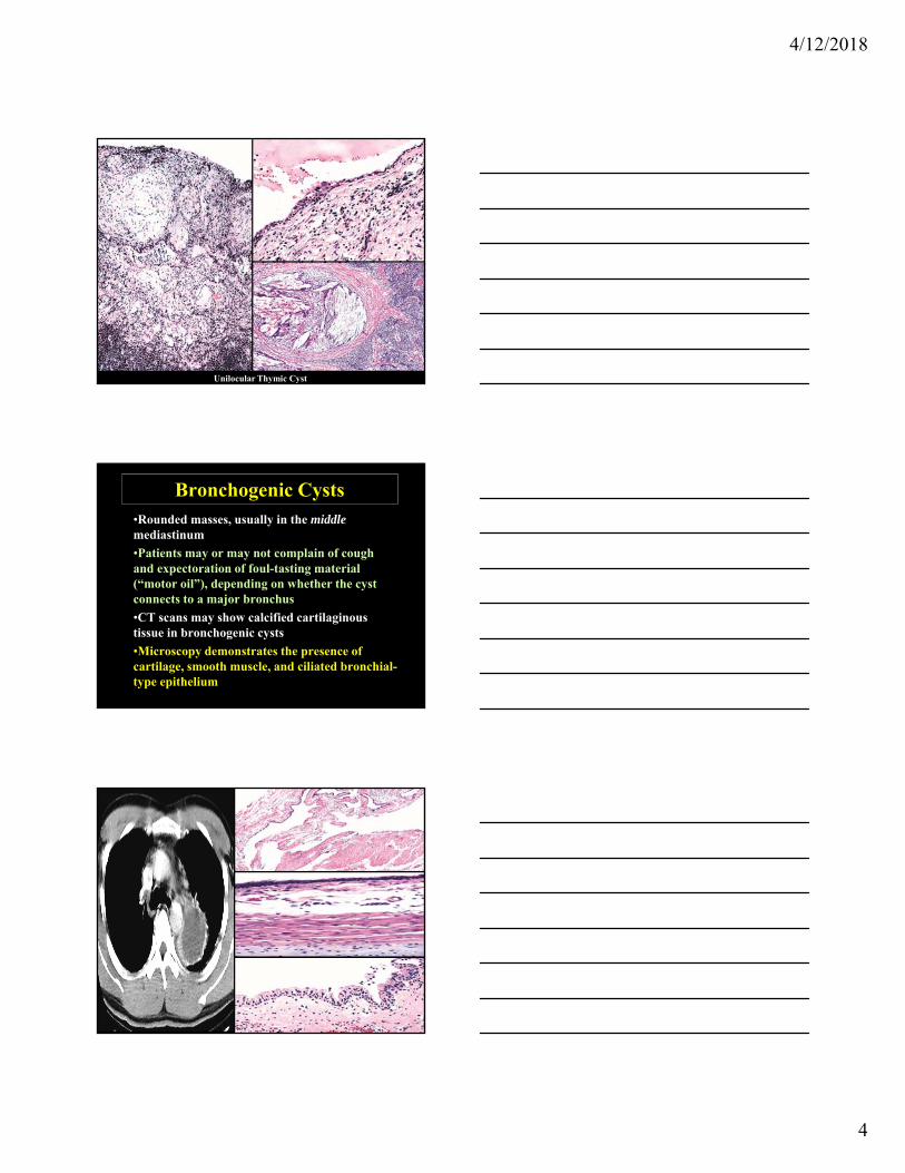
Unilocular Thymic Cysts
•May be present in the anterior or middle mediastinum, as an irregular or rounded density on plain films of the chest
•CT scans demonstrate a cyst with variable dense contents and an irregular wall; multiloculation may be present
•Microscopy shows a squamous lining with thymic tissue sometimes incorporated into the wall of the cyst; cholesterol clefts and calcification are common
4/12/2018
4
Unilocular Thymic Cyst
Bronchogenic Cysts•Rounded masses, usually in the middlemediastinum
•Patients may or may not complain of cough and expectoration of foul-tasting material (“motor oil”), depending on whether the cyst connects to a major bronchus
•CT scans may show calcified cartilaginous tissue in bronchogenic cysts
•Microscopy demonstrates the presence of cartilage, smooth muscle, and ciliated bronchial-type epithelium

4/12/2018
5
Enteric Duplication (Gastroenteric) Mediastinal Cysts
• Probably derived from misplaced foregut rests
• Typically seen in children < 15 years old, who present with dysphagia, cough, or vomiting
• Characteristically present in the posterior mediastinum as spheroid masses that may show internal loculation
• Specialized gastric-mucosal, squamoid, or simple columnar epithelial linings (or mixtures thereof) may be present
Mullerian (Hattori) Cysts of the Posterior Mediastinum
• Paravertebral in location, in women
• Unilocular, with an epithelial lining resembling that of endosalpingiosis
• Immunoreactive for CA-125, ERP, PRP, PAX8, and WT1
• Simple excision is curative
4/12/2018
6
PAX8
ERP
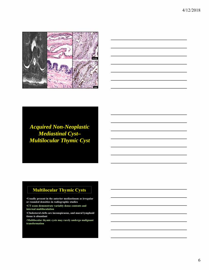
Acquired Non-Neoplastic Mediastinal Cyst–
Multilocular Thymic Cyst
Multilocular Thymic Cysts
•Usually present in the anterior mediastinum as irregular or rounded densities in radiographic studies
•CT scans demonstrate variably dense contents and internal multiloculation
•Cholesterol clefts are inconspicuous, and mural lymphoid tissue is abundant
•Multilocular thymic cysts may rarely undergo malignant transformation

4/12/2018
7
Proliferating Thymic Cysts
•Unusual examples of multilocular thymic cyst in which the squamoid lining
epithelium proliferates irregularly into the cyst wall, yielding an image which simulates that of squamous carcinoma
•Probably represents “pseudoepitheliomatous hyperplasia” of the lining epithelium, with an unknown
cause

4/12/2018
8
Neoplastic & Paraneoplastic Cystic Lesions of the
Mediastinum
Mediastinal Lymphangiomas
• May be seen in all 3 mediastinal compartments, as unilocular or multilocular masses on imaging studies
• Predominate in children
• Interanastomosing vascular channels, associated with infiltrates of lymphocytes, & sometimes containing internal micropapillations

4/12/2018
9
Thymic Cysts in Hodgkin or Non-Hodgkin Lymphoma
•Usually seen after therapy of some kind (radiation; chemotherapy) but may occur as a spontaneous tumor-related phenomenon as well
•A central cystic cavity is surrounded by atypical lymphoid tissue containing diagnostic Reed cells or, alternatively, non-Hodgkin lymphoma
Other Potentially-Cystic Neoplasms of the Anterior Mediastinum
• Teratoma
• Thymoma
• Carcinoma ex thymic cyst
• Cystic de novo thymic carcinoma
• Seminoma
• Thymic carcinoid tumor

4/12/2018
10
Intrathymic Cystic Teratomas• Predominantly seen in children and young
adults, typically presenting with nondescript symptoms & signs or as lesions found incidentally on chest radiographs
• “Eggshell” calcification of the mass is possibly seen in plain-film radiographs
• At least 2 of 3 germ layers must be represented in the lesional tissue
• Immature neuroepithelial components are not prognostically important before the age of 15 years
4/12/2018
11
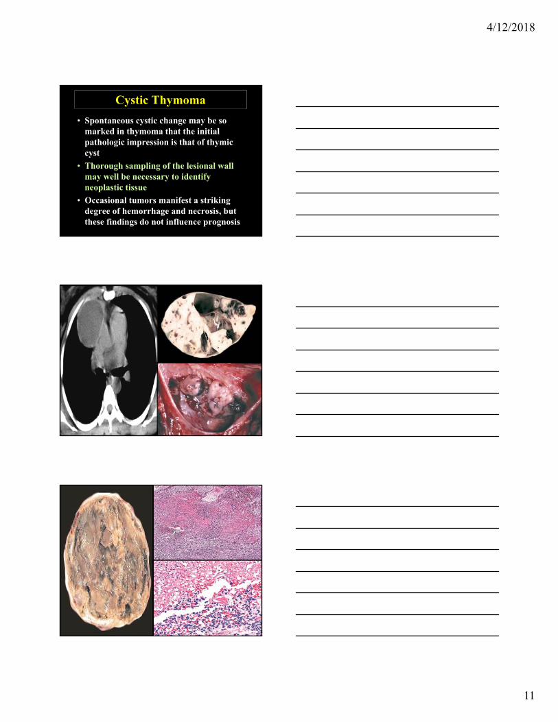
Cystic Thymoma
• Spontaneous cystic change may be so marked in thymoma that the initial pathologic impression is that of thymic cyst
• Thorough sampling of the lesional wall may well be necessary to identify neoplastic tissue
• Occasional tumors manifest a striking degree of hemorrhage and necrosis, but these findings do not influence prognosis
4/12/2018
12
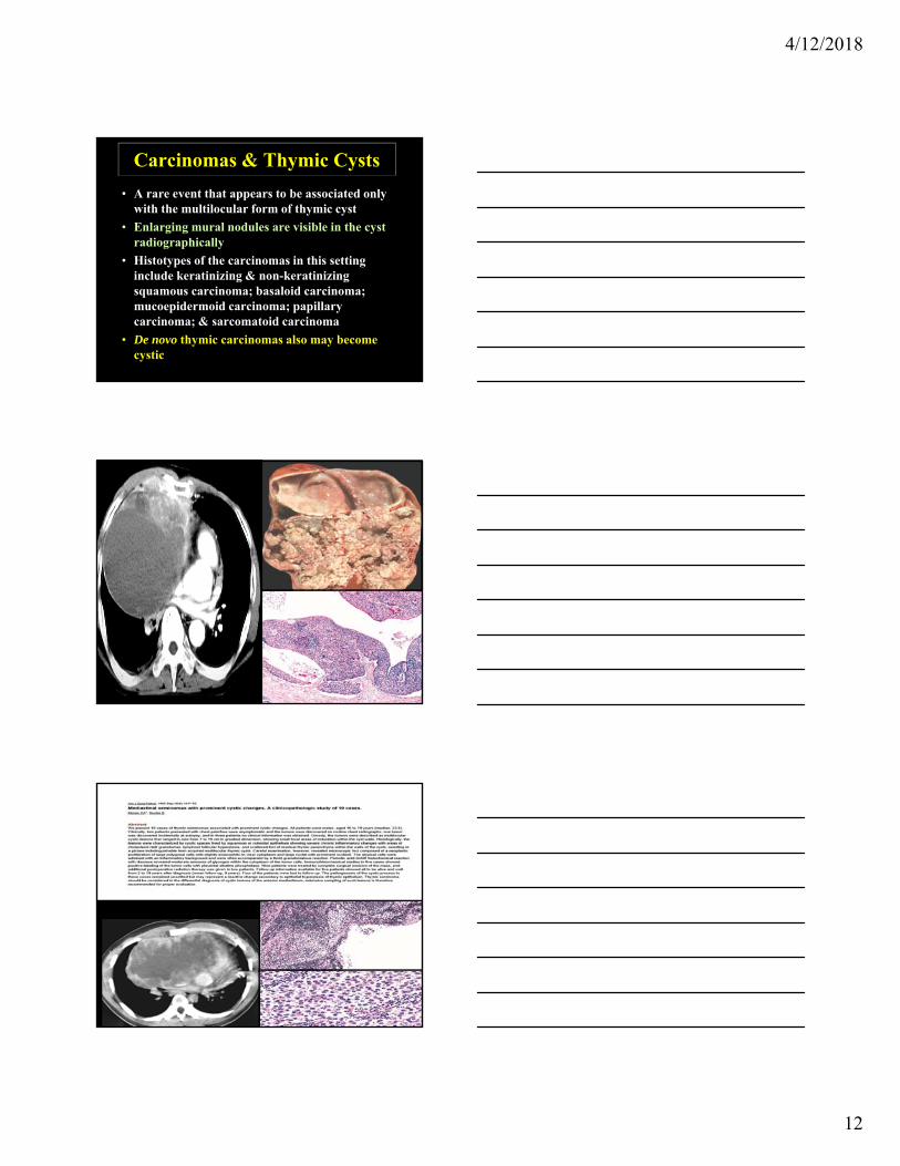
Carcinomas & Thymic Cysts
• A rare event that appears to be associated only with the multilocular form of thymic cyst
• Enlarging mural nodules are visible in the cyst radiographically
• Histotypes of the carcinomas in this setting include keratinizing & non-keratinizing squamous carcinoma; basaloid carcinoma; mucoepidermoid carcinoma; papillary carcinoma; & sarcomatoid carcinoma
• De novo thymic carcinomas also may become cystic

4/12/2018
13