i. project for heavy charged-particle beam multi …. project for heavy charged-particle beam...
TRANSCRIPT

1
CYRIC Annual Report 1998
I. Project for Heavy Charged-particle Beam Multi-purpose Use
Orihara H.
Cyclotron and Radioisotope Center, Tohoku University (http://www.cyric.tohoku.ac.jp)
A brief summary is given for the results of multi-purpose use of a cyclotron and RI in past two decades.This report presents as well a preliminary planning of further development by introducing the facilities,being under construction or consideration, and scientific motivations.
CYRIC (Cyclotron and Radioisotope Center) was established in 1977 as an institution
for carrying out research studies in various fields by the use of a cyclotron and radioisotopes,
and also for training researchers of Tohoku University for safe treatment of radioisotopes and
radiation. The CYRIC cyclotron is a variable energy AVF machine with a K value of 50 MeV;
being capable of acceleration protons up to 40 MeV, deuterons to 25 MeV, α-particles to 50
MeV, and He-3 particles to 65 MeV.
During the past two decades, refereed 501-papers written in English have been
published in scientific journals in the world. Ninety-six dissertations for D.Sc.(37), D.M.(38),
D.Eng.(11), D.Agr.(6), Pharm.D.(2), etc. have been accepted based on the research in CYRIC,
while 154-thesises for master's degree have been presented.
Based on the successful results of the 20 years-long multi-purpose use of Cyclotron
and Radioisotopes, replacement of the present cyclotron with a larger dimension K=130 MeV
one, and construction of experimental facilities have been authorized by Japanese government in
1998 and 1999 financial years.
Since 1979, we have an apparatus for fast neutron time-of-flight analysis equipped
with a 40m long flight path, an electromagnetic isotope separator (EMIS) for on-line and off-line
uses, and an x ray detection system for atomic physics and for element analysis by PIXE method.
Fully automated positron emitter labeled compound synthesis systems is installed for the studies
of biology and medicine. A positron tomograph ECAT-II was installed in 1981. Another
four-rings PET(PT931) and TOF type PET (PT711) scanners were installed in 1986 and 1987,
respectively. Since 1983 school year, these scanners have been extensively used for clinical
researches; for cancer diagnosis and for brain researches, etc., supported by steady operation of
the cyclotron, and by reliable supply of short-lived positron emitter labeled compounds.
Recently, a system of the high-resolution positron-emission tomograph SET 2400W-S has been
installed.
With fast neutron time of flight measurement, we have explored isospin- and spin-
2
isospin excitation in nuclei by (p,n) charge-exchange reactions at 35 MeV. Our interests have
been focused on: (1) Isospin mixing effect in the width of IAS, (2) 0+ to 1+ Gamow-Teller type
transition, (3) Stretched particle-hole excitation, and (4) 0+ to 0- or ∆Jπ = 0- pion-like transition.
These works have established a research field of spin-isospin excitation of nuclei in low energy
(p,n) reaction. In addition, spectroscopic works by the (d,n) reaction have been carried out to
investigate single particle nature of nuclei. Atomic and molecular physics with charged
particles from an AVF cyclotron started at CYRIC exploring inner-shell ionization mechanism,
results have been and extended over applications with the particle induced x-ray emission
(PIXE) method for element analysis.
Researches using an EMIS equipped with a tape-transport and an ion-guide systems
are: (1) Discovery of the heaviest two "mirror-decay" nuclei, 57Cu and 59Zn.(2) Implantation of
radioactive isotopes to make good-quality samples for precision measurement of conversion-
electrons up to the atomic valence shells to derive the M ssbauer isomer-shift scales ∆R/R.
Researches in the field of nuclear spectroscopy, using perturbed angular correlation (PAC) and
perturbed angular distribution (PAD) methods with a magnet, are measurement of fifteen
samples of magnetic-moments of nuclear isomeric-states. In a field of solid-state physics,
PAC measurements, after EMIS for acceleration of Rl, have been carried out to examine the
orientation and magnitude of the electric field gradient created by a vacancy at Rl-probe impact.
Efforts to accelerate light heavy-ion has been continued for further application to scattering
experiments. Heavy ions of 12,13C, 15N and 16O were extracted successfully, and used for
elastic scattering on 28Si at small angles in order to obtain total reaction cross-sections model
independently.
Using 36-MeV α-particles with an energy degrader system to obtain an uniform depth
distribution in the specimen, He implantation effects on mechanical properties of a number of
composites have been studied to apply these composites on structural materials of a fusion
reactor. As a new type of isotope effect in metal acetylacetonates, time-dependent isotope
effect in recoil implantation was studied, and it was found that the decay products 99mTc and 96Tc
tended to form pertechnetate in comparison with the direct nuclear reaction product 95Tc, by
water soluble species of Tc nuclides produced by the (d,xn) reaction on Mo. Further recent
topics in RI-production with 12-MeV protons and 16-MeV deuterons is insertion of radioactive
atoms in C60 and C70 fullerenes. Such endohedral fullerenes 7Be@C60 , 127Xe@C60,70 and 79Kr
@C60,70 and their dimers were detected.
With the neutron facilities in CYRIC, neutron dosimetry and monitoring were studied,
and activation and spallation cross-sections have been measured. Also investigated were
neutron absorption and leakage for purposes of radiation shielding. By combination with
clinical PET studies, absorbed dose in humans due to intravenous administration of positron
emission radiopharmaceuticals were measured.
One of major programs has been instrumental development of positron emission
tomograph and automated labeling system of radiopharmaceuticals with cyclotron-produced
3
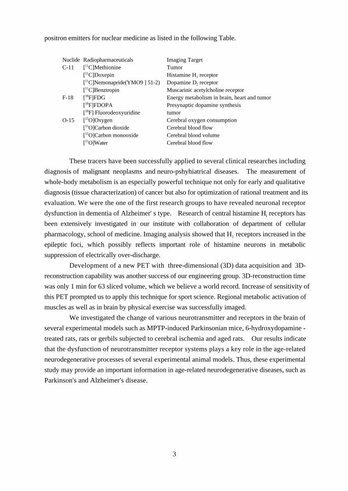
positron emitters for nuclear medicine as listed in the following Table.
Nuclide Radiopharmaceuticals Imaging TargetC-11 [11C]Methionine Tumor
[11C]Doxepin Histamine H1 receptor[11C]Nemonapride(YMO9 ] 51-2) Dopamine D2 receptor[11C]Benztropin Muscarinic acetylcholine receptor
F-18 [18F]FDG Energy metabolism in brain, heart and tumor[18F]FDOPA Presynaptic dopamine synthesis[18F] Fluorodeoxyuridine tumor
O-15 [15O]Oxygen Cerebral oxygen consumption[15O]Carbon dioxide Cerebral blood flow[15O]Carbon monooxide Cerebral blood volume[15O]Water Cerebral blood flow
These tracers have been successfully applied to several clinical researches including
diagnosis of malignant neoplasms and neuro-pshyhiatrical diseases. The measurement of
whole-body metabolism is an especially powerful technique not only for early and qualitative
diagnosis (tissue characterization) of cancer but also for optimization of rational treatment and its
evaluation. We were the one of the first research groups to have revealed neuronal receptor
dysfunction in dementia of Alzheimer' s type. Research of central histamine Hl receptors has
been extensively investigated in our institute with collaboration of department of cellular
pharmacology, school of medicine. Imaging analysis showed that H1 receptors increased in the
epileptic foci, which possibly reflects important role of histamine neurons in metabolic
suppression of electrically over-discharge.
Development of a new PET with three-dimensional (3D) data acquisition and 3D-
reconstruction capability was another success of our engineering group. 3D-reconstruction time
was only 1 min for 63 sliced volume, which we believe a world record. Increase of sensitivity of
this PET prompted us to apply this technique for sport science. Regional metabolic activation of
muscles as well as in brain by physical exercise was successfully imaged.
We investigated the change of various neurotransmitter and receptors in the brain of
several experimental models such as MPTP-induced Parkinsonian mice, 6-hydroxydopamine -
treated rats, rats or gerbils subjected to cerebral ischemia and aged rats. Our results indicate
that the dysfunction of neurotransmitter receptor systems plays a key role in the age-related
neurodegenerative processes of several experimental animal models. Thus, these experimental
study may provide an important information in age-related neurodegenerative diseases, such as
Parkinson's and Alzheimer's disease.
4
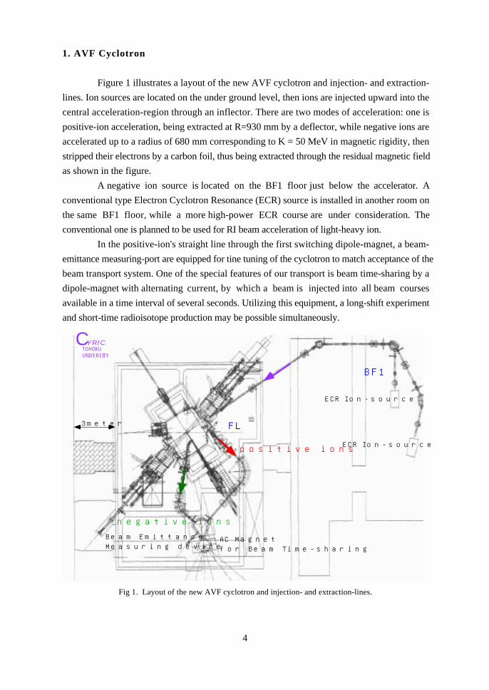
1. AVF Cyclotron
Figure 1 illustrates a layout of the new AVF cyclotron and injection- and extraction-
lines. Ion sources are located on the under ground level, then ions are injected upward into the
central acceleration-region through an inflector. There are two modes of acceleration: one is
positive-ion acceleration, being extracted at R=930 mm by a deflector, while negative ions are
accelerated up to a radius of 680 mm corresponding to K = 50 MeV in magnetic rigidity, then
stripped their electrons by a carbon foil, thus being extracted through the residual magnetic field
as shown in the figure.
A negative ion source is located on the BF1 floor just below the accelerator. A
conventional type Electron Cyclotron Resonance (ECR) source is installed in another room on
the same BF1 floor, while a more high-power ECR course are under consideration. The
conventional one is planned to be used for RI beam acceleration of light-heavy ion.
In the positive-ion's straight line through the first switching dipole-magnet, a beam-
emittance measuring-port are equipped for tine tuning of the cyclotron to match acceptance of the
beam transport system. One of the special features of our transport is beam time-sharing by a
dipole-magnet with alternating current, by which a beam is injected into all beam courses
available in a time interval of several seconds. Utilizing this equipment, a long-shift experiment
and short-time radioisotope production may be possible simultaneously.
BF1
FL
negative ions
positive ions
Beam EmittanceMeasuring device
AC Magnetfor Beam Time-sharing
ECR Ion-source
ECR Ion-source
CYRICTOHOKUUNIVERSITY
3meter
Fig 1. Layout of the new AVF cyclotron and injection- and extraction-lines.

5
Tables 1 and 2 list the specifications and beam characters of the new cyclotron.
Table 1. Specification of the new AVF cyclotron.
Electromagnet System
Weight 200 ton
Extraction Radius 923 mm
Number of Sector 4
Max. Average Induction 19.6 kG (over Hill)
Main Coil Power 230 kW
Number of Trim Coil 12 pairs
Radio-Frequency System
Number of Dee’s 2
Frequency 11-22 MHz
Max. Dee Voltage 50 kV
Max. RF Power 70 kW x 2
External Ion Source
Negative ion Cusp-type
Positive ion ECR, 10GHz
ECR, 14GHz
Table 2. Beam energies of the new AVF cyclotron.
a) Positive ion acceleration.
Accelerated Particle Energy (MeV) Beam intensity(µA)
p 10-90 50
d 10-65 503He 20-170 504He 20-130 5012C 20-397 5p14N 20-463 5p16O 20-530 5p
20Ne 20-662 5p32S 20-698 3p
40Ar 20-744 3p84Kr 20-695 3p
129Xe 20-748 1p
a) Negative ion acceleration.
Accelerated Particle Energy (MeV) Beam intensity(µA)
p 10-50 300
d 10-25 300

6
2. Beam Swinger and Large Solid-angle Neutron Detection System for Time-of-Flight Experiments
As one of the main facilities of the new System for Heavy Charged-particle Multi-purpose Use
in CYRIC, construction the beam swinger, being capable for rotating the beam axis from -5deg.
to 145deg. with K= 130 MeV, is under progress. The other new feature of this system is the
neutron detector matrix, being located at a distance of 44 meter after the neutron flight path, and
consisting of 32 pieces of the counter which contain 50 litter of liquid scintillater in its total
volume.
This system may be a powerful tool to investigate isospin and spin-isospin excitation in
nuclei through the (p,n) reaction in a wide range of incident proton energies. With energetic and
high-intensity monochromatic neutron beams, neutron scattering experiments with high
sensitivity provide a new field to explore charge-symmetry and charge-independent character of
the nuclear forces, and to work with other engineering studies.
Figure 2 shows a lay out of the beam swinger and detector matrix.
IncidentCharged-particle
TripletQuadrupoleManet
Switching Magnet+45 deg(fixed)
Bending Magnet+45 deg(rotated)
Bending Magnet-145 deg(rotated)
NeutronProductionTarget
NeutronScatteringTarget
ScatteringAngle(-5̃145deg)
Neutron Flight Path
Beam Dump
Neutron Detector
Fig. 2. Lay out of the beam swinger and detector matrix.

7
3. On-line Electric and Magnetic Isotope-separator
Study for unstable nuclei provide us with information of such nuclei that have decayed
in the course of history of universe by producing them artificially with an accelerator or reactor.
Energetic and high intensity charged-particle are considered to be the best candidate to explore
such unstable nuclei far from the stability line. Many facilities in the world are oriented to the
accelerated charged-particles, heavy-ions especially, for this purpose. On the other hand,
production of unstable nuclei by neutrons may be more efficient, though it has been limited to a
few cases with nuclear reactors due to the experimental difficulties to combine neutron beams
with an on-line electromagnetic isotope separator.
The new cyclotron at CYRIC provide us with a sufficient high-intensity neutron beam
to investigate unstable nuclei close to the neutron drip-line, together with the on-line
electromagnetic isotope separator (EMIS) equipped with the ion-guide ion-source, and the
high-speed tape-transport system. The additional powerful equipment is a high-resolution and
large solid angle gamma ray detection system consisting of three pairs of four hold clover-type
pure-Ge crystal, each of which is surrounded by 12 pieces of BGO Crystal. Figure 3 shows a
layout of EMIS together with the new Ge-detector ball.
Ion-guide TypeIon Source
Electric and MagneticIsotope-separator
Three pairs of Clover-typeCompton-suppressionGe-detector
Tape-transport
Incident Particle
Fig. 3. Layout of EMIS, tape-transport and Ge-ball .

8
4. High-energy γ-ray Detection System
The system is capable for detecting and analyzing high-energy γ -rays, up to e. g. 500
MeV, and neutral mesons produced by nuclear reactions. It contains 148 pieces of CsI crystal,
the volume of each detector being 1000 cm3, and they are segmentaized into four blocks so as to
cover a half π-radian in solid angle. The minimum internal radius is 55 cm. These four blocks
are mounted on a stem, and each block is separately removable.
Equipped on a beam course of the K=130 MeV, AVF cyclotron facility of CYRIC, this
system is applied for studies of high energy γ -rays production by energetic heavy ion impact up
to the maximum energy kinetically allowed, and η- and π-mesons production energetically
available. Thus, this system is expected to explore interesting phenomena of coherent extreme
in nuclei. It should be noted that the beam transport system with time-sharing AC magnet is
expected to work efficiently for such an experiment with quite rare events. Figure 4 shouws a
schematic view of the CsI crystal high energy γ-ray detector.
Incident Particle
1000 cc CsI-crystal148-pieces of
andPhoto-multipliers
1100 mm
γ
γ
Target
Fig. 4. Schematic view of the present CsI crystal high energy γ -ray detector.

9
5. Neutron-life and Neutron-induced Reaction Analyzing Facility
Energetic and high-intensity charged particle beam, negative ion beam in particular,
provide as well high-intensity white and monochromatic neutron beam, thus enabling us to
carried out a number of neutron induced experiments. The main part of the .spectrometer is the
electromagnet which have been used as that of our old K=50 MeV, AVF cyclotron. Charged
particles emitted from different points along with the plane perpendicular to the incident direction
have a focal plane, where a detector array as calorie meter is located.
A challenging project with this large solid angle magnetic spectrometer and high-
intensity monochromatic neutron beams is measurement of the life-time of neutrons in flight.
Accurate and comprehensive neutron life-time data are of crucial importance for current science
including astrophysics, cosmology, particle and nuclear physics, etc.
Monochromatic 30-MeV neutrons are produced, then they flight through the 20m-long
evacuated tube, reaching to the spectrometer in an average flight-time of ~1 µsec. One neutron
per hour may change to a proton. The neutron flight time, measured in a resolution of 10-3 is
used to identify protons from neutron-decay. The most important point for this experiment is
to measure the total amount of neutrons with sufficient accuracy as high as several 0. 1%.
Figure 5 shows a cross-sectional layout of the facility.
d=1.80 m
Bmax = 1.8 T
TargetWhite andMonochromaticNeutron Beam
Vacuum PumpFocal Plane Calorie Meter
Electromagnet2.17 mH, 1.70 mW, 4.40 mLWeight: 110 t
Pole gap:27 cm
Protons fromNeutron-decay
or
NeutronFlux Meter
Fig. 5. Cross sectional view of magnetic spectrometer and neutron flux meter.

10
6. High Intensity Thermal and Epi-thermal Neutron Source
High intensity proton and deuteron beams by the negative ion acceleration mode
provide a good place for production of thermal and epi-thermal neutrons. Recent development
of high intensity charged-particle accelerator make it possible to use such a neutron beam with
almost equivalent intensity as that by a nuclear reactor.
Figure 6 shows a target and moderator system to produce thermal and epi-thermal
neutrons from high-energy neutrons by a configuration of Al/Pb, Iron, graphite, heavy-water,
and Bi, etc. Of course, there should remain a lot of developing studies to thermalize several tenth
MeV neutrons to thermal ones. Especially, minimization of high-energy neutron background is
the most important point in these studies.
Utility of high-intensity thermal and epi-thermal neutrons may spread over many
research fields, in which activation analysis combined with that by PIXE for medium and heavy
elements, and radiography for light elements may be good candidates. Final goal of application
of these neutrons is that for the study of Boron Neutron-capture Cancer Therapy (BNCT).
Even limited to selective melanoma therapy, collaborations among physicists, pharmacologist,
radiologist, and medical scientists are essential. This project is under consideration, being
discussed in a working-group organized in CYRIC.
IncidentCharged-particle
NeutronProduction-target
Thermal Neutron( nth)
nth
nth
nepi-th
nth
Fig. 6. Sketch of thermal and epi-thermal neutron source.

11
7. Medical Research by Simultaneous Synthesis of Multiple Radiotracer
Energetic beams may open a new field for production of radiotracers through, for
example, (p,xn,yp) reactions on medium and heavy nuclei. With the additional small-size
cyclotron already working, multiple production of radionuclides is possible.
Simultaneous measurement of cerebral blood flow gives an estimate of regional tracer
delivery and thus improves accuracy of the receptor quantification. For quantitative assessment
of neuronal transmitter and receptors, neuronal receptor quantification are made by 11C-labeled
receptor ligand and 15O-labeled water are produced, respectively, by K=130 and K=120MeV
cyclotrons. As such quantitative evaluation of clinical neuro- psychiatricaldiseases such as
dementia, other degenerative disease and psychosis will be carried out. The 45Ti nuclide is
suitable for antibody labeling thanks to a longer half-life and high affinity with proteins.
Intravascular radioactivity of 45Ti can be connected by measurement of tissue vascular fraction
using [15O]carbon monoxide which tightly binds to hemoglobin. Cancer diagnosis using
radiolabeled monoclonal antibody may be performed with 45Ti-monoclonal antibody and [15O]-
CO produced by K=130 and K=12 MeV cyclotrons, respectively. Figure 7 shows a flow-
chrat for nuclear medicine by PET and related facilities.
4
Nuclear Medicineby Positron CT
3
2
18F
γ-線
1
Nuclear Reaction
Charged ParticleBeam
18O(p,n)18F
K= 130MeVAVFCyclotron
Short-livedPositronEmitter
RI Production
Super Computor
γ-線
γ-線
γ-線
γ-線
γ-線
Synthesis Systemof Labeled Compound
LabeledPharmaceutical
3D-PositronEmissionTomography
Diagnostic ofHeart and BrainDiseases, and Cancer
Brain Researchby PositronNuclear Medicine
Raw data
CYRICTOHOKUUNIVERSITY
18F
γ-線
γ-線
11C
γ-線
γ-線
Short-livedPositronEmitter
HM-12AVFCyclotron
Fig. 7. Illustration for nuclear medicine by PET and related facilities.
As a summary, a layout of building and facilities for “Project for Heavy Charged-
particle Beam Multi-purpose Use” are illustrated in the next page.