i occlusion in the digital a of dentistry€¦ · implant occlusion in the digital age of dentistry...
TRANSCRIPT
Implant OcclusIOn In the DIgItal age Of DentIstry
A look at the state of the implant dentistry today with real cases from dentists who utilize technology to manage implant occlusion.
A Tekscan, Inc. Publication
2
Contents
Implant DentIstry toDay 3
occlusIon as you Know It 5
InformatIon transformIng Implant DentIstry 7
managIng Implant occlusIon: a “Dual” wIth nature 8
Implant cases wIth t-scan 11
the prIncess anD the pea 12
the craDle wIll rocK 18
a wrInKle In tIme 23
conclusIon 29
references 30
3
Implant DentIstry toDay
“One can only stop and marvel at man’s ingenuity over the years in this arena of research and scholarship. The materials in which dental implants came into development range from gold ligature wire, shells, ivory to chromium, cobalt, to iridium and platinum. From spiral stainless steel implant designs to double helical creations and endosseous root forms, dental researchers and clinicians worked fast and furiously; they generated many structures to replace the positions that natural teeth once held.
Dental surfaces were also modified to decrease the healing time for osseointegration. Modified surfaces incorporated the use of hydroxyapatite, composites, carbon, glass, ceramic as well as titanium oxide. In order to make the exterior as suitable as possible, implant surfaces have additionally been sandblasted, oxidized, fluoridated, etched, and medicated. The most recent innovative laminin coating is the center of focus in present day implant endeavors. As time marches on in dental implant study, the materials, forms, and surface coatings have been refined and restructured to allow the consumer the very best in tooth replacement choices for their present and future needs.”
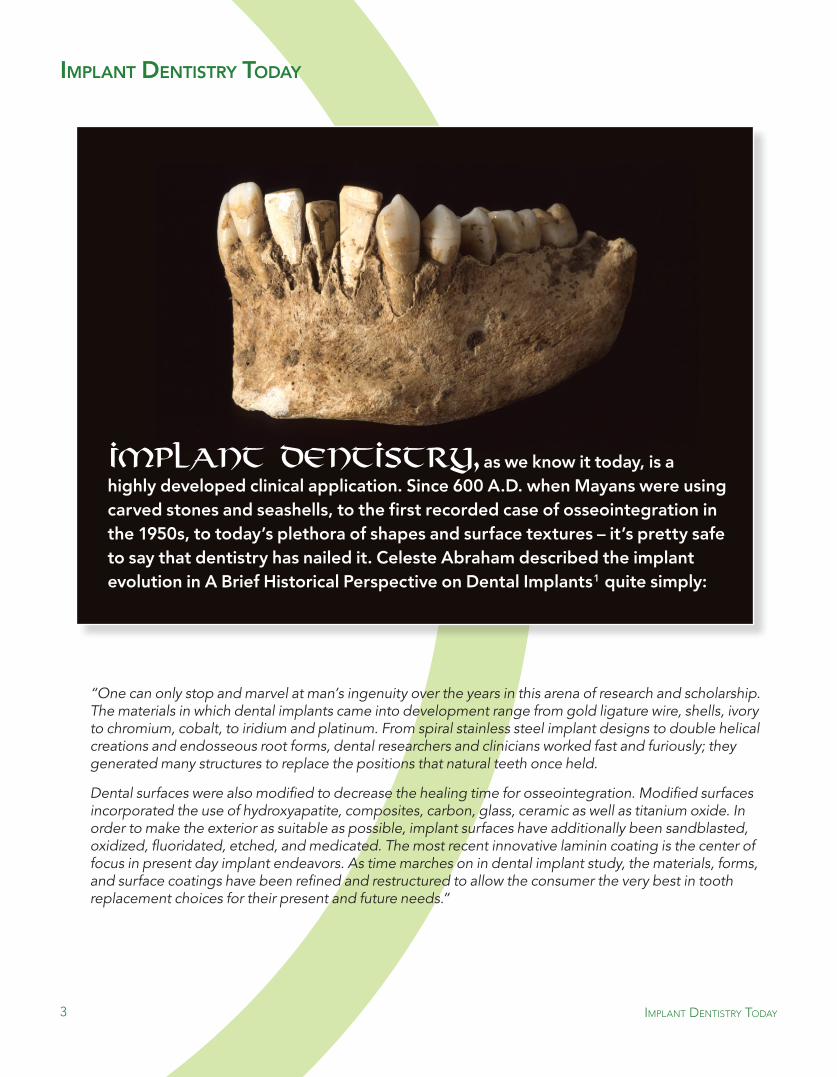
Implant dentistry, as we know it today, is a highly developed clinical application. Since 600 A.D. when Mayans were using carved stones and seashells, to the first recorded case of osseointegration in the 1950s, to today’s plethora of shapes and surface textures – it’s pretty safe to say that dentistry has nailed it. Celeste Abraham described the implant evolution in A Brief Historical Perspective on Dental Implants1 quite simply:
Implant DentIstry toDay
4
In this ever-evolving discipline, there is no doubt that the future is bright. so, what does the landscape look like today in America? Here are some numbers that illustrate implant dentistry’s accelerated growth path:
• More than 30 million Americans are missing all their teeth2 in one or both jaws
• statistics provided by the American Association of oral and Maxillofacial surgeons show that 69% of adults ages 35 to 44 have lost at least one permanent tooth3 to an accident, gum disease, a failed root canal or tooth decay
• It is estimated that roughly 15 million people throughout the United states have a crown or a bridge2 to replace missing teeth
• Industry studies and literature suggest that dental implants have a success rate greater than 90%4.
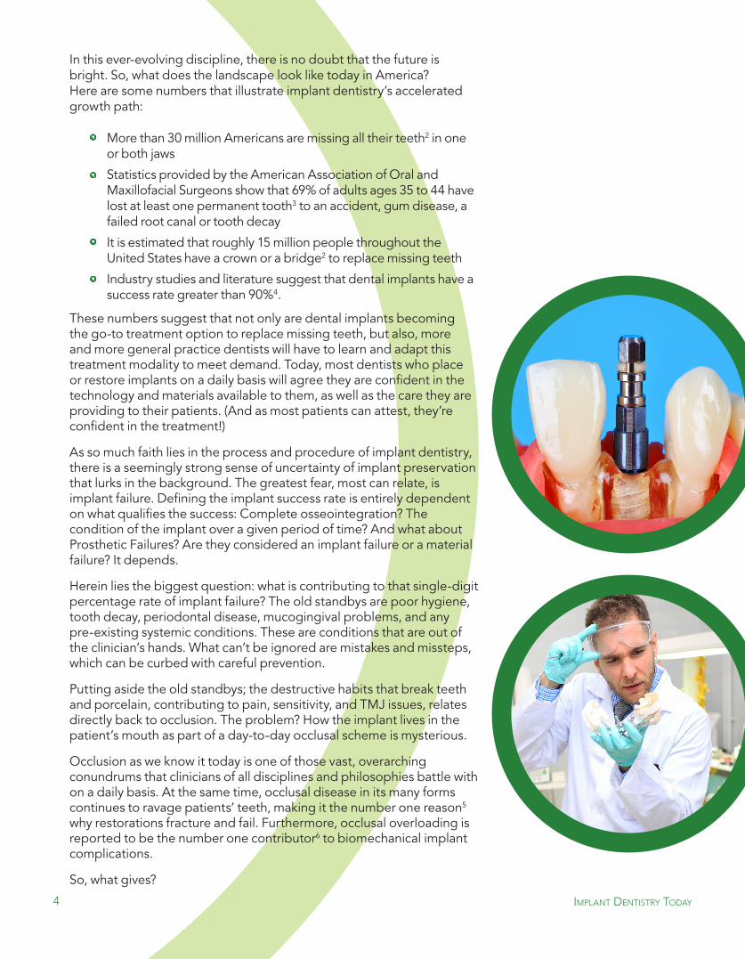
these numbers suggest that not only are dental implants becoming the go-to treatment option to replace missing teeth, but also, more and more general practice dentists will have to learn and adapt this treatment modality to meet demand. today, most dentists who place or restore implants on a daily basis will agree they are confident in the technology and materials available to them, as well as the care they are providing to their patients. (And as most patients can attest, they’re confident in the treatment!)
As so much faith lies in the process and procedure of implant dentistry, there is a seemingly strong sense of uncertainty of implant preservation that lurks in the background. the greatest fear, most can relate, is implant failure. Defining the implant success rate is entirely dependent on what qualifies the success: Complete osseointegration? the condition of the implant over a given period of time? And what about Prosthetic Failures? Are they considered an implant failure or a material failure? It depends.
Herein lies the biggest question: what is contributing to that single-digit percentage rate of implant failure? the old standbys are poor hygiene, tooth decay, periodontal disease, mucogingival problems, and any pre-existing systemic conditions. these are conditions that are out of the clinician’s hands. What can’t be ignored are mistakes and missteps, which can be curbed with careful prevention.
Putting aside the old standbys; the destructive habits that break teeth and porcelain, contributing to pain, sensitivity, and tMJ issues, relates directly back to occlusion. the problem? How the implant lives in the patient’s mouth as part of a day-to-day occlusal scheme is mysterious.
occlusion as we know it today is one of those vast, overarching conundrums that clinicians of all disciplines and philosophies battle with on a daily basis. At the same time, occlusal disease in its many forms continues to ravage patients’ teeth, making it the number one reason5 why restorations fracture and fail. Furthermore, occlusal overloading is reported to be the number one contributor6 to biomechanical implant complications.
so, what gives?
Implant DentIstry toDay

5
occlusIon as you Know ItArguably one of the most controversial subjects in dentistry, occlusion is not fully covered in dental schools. Post-grad and continuing education groups have differing views on treatment options for occlusal disease, plus a number of different treatment modalities may affect occlusion’s role in implant failures. While one might argue a given philosophy, there is a common thread.
no matter what area of dentistry, occlusion is inevitably going to have an effect on the treatment performed. Vice versa, treatments may affect the occlusion. Dentistry Today5 cites three golden rules when it comes to occlusion:
• Bilateral simultaneity
• Posterior disclusion, or anterior/canine guidance
• Unobstructed envelope of function
these principles are scientifically supported to help increase the predictability and quality of dental treatments. the teeth should come together evenly when in a centralized position, the posteriors should immediately disclude during chewing cycles, and protrusive and excursive movements should occur without interference. However, who measures that this is working well? And how?
Dr. sarah Qadeer, International lecturer at Dept. of Prosthodontics, Faculty of Dentistry, thammasat University in thailand, makes note of occlusal indicators, as most clinicians know them today:
“the traditional occlusal indicators used in dental practice are articulation papers, shim-stock foils, elastomeric impression materials, and occlusal wax strips. these static dental materials have been widely believed to have occlusal force descriptive capability. However, modern material studies are challenging the widespread belief that occlusal indicator materials can measure differing occlusal force levels.”
Dr. Qadeer suggests that while static occlusal indicators have some descriptive capability on the occlusion, they don’t paint the total picture. When using static occlusal methods, the concept of “force” is difficult to interpret, because there is one dimension that captures what happens when the teeth occlude: seeing dots and smudges. small mark = less force, Large mark = more force… Right? How much force on one tooth is relative to the forces on all the other teeth? no one wants to do that math.
How is it possible to quantify dynamic occlusal loading based on surface marks?
occlusIon as you Know It
6
“Quantifiable force” is a totally foreign concept to most dental clinicians, but one that would inevitably take treatment paths to a new level. If the clinician is able to see when each tooth comes into contact, they’re able to identify prematurities or occlusal abnormalities. If they’re able to see how much force is applied across the dentition from first contact to MIP, they can identify which teeth are at risk, or see why a patient has a given symptom. Finally, if they’re able to see how teeth fit together as they occlude, they may be able to understand and evaluate potential risks.
this is measureable data that requires technology. t-scan® is the only available tool in dentistry that can show this data.
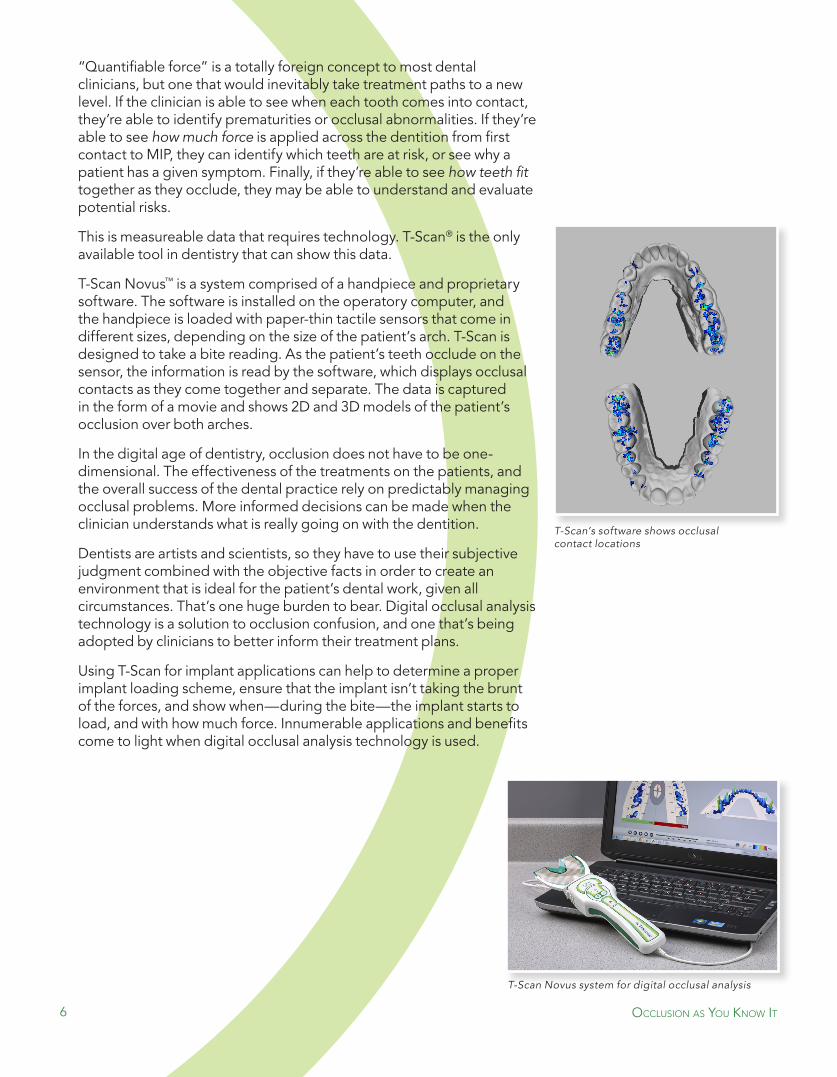
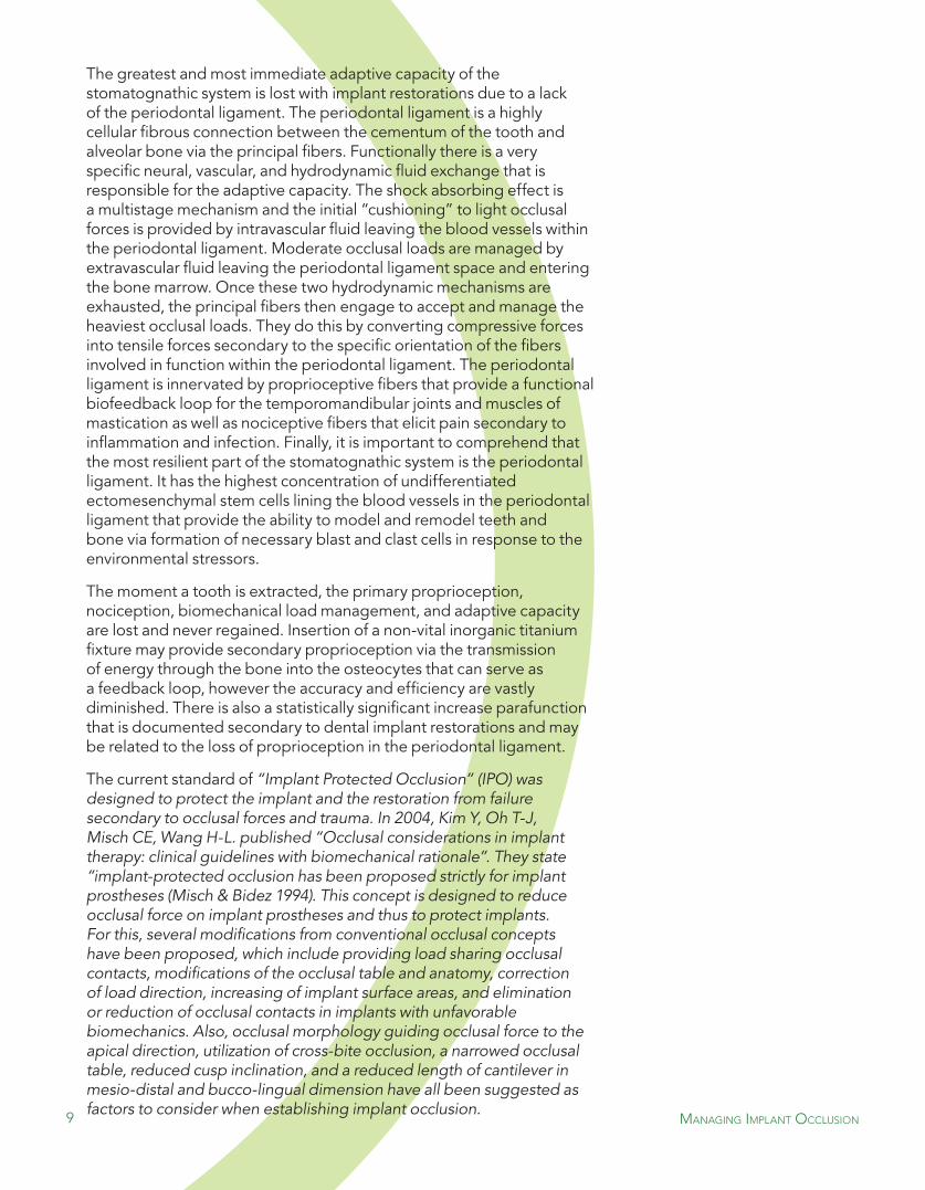
t-scan novus™ is a system comprised of a handpiece and proprietary software. the software is installed on the operatory computer, and the handpiece is loaded with paper-thin tactile sensors that come in different sizes, depending on the size of the patient’s arch. t-scan is designed to take a bite reading. As the patient’s teeth occlude on the sensor, the information is read by the software, which displays occlusal contacts as they come together and separate. the data is captured in the form of a movie and shows 2D and 3D models of the patient’s occlusion over both arches.
In the digital age of dentistry, occlusion does not have to be one-dimensional. the effectiveness of the treatments on the patients, and the overall success of the dental practice rely on predictably managing occlusal problems. More informed decisions can be made when the clinician understands what is really going on with the dentition.
Dentists are artists and scientists, so they have to use their subjective judgment combined with the objective facts in order to create an environment that is ideal for the patient’s dental work, given all circumstances. that’s one huge burden to bear. Digital occlusal analysis technology is a solution to occlusion confusion, and one that’s being adopted by clinicians to better inform their treatment plans.
Using t-scan for implant applications can help to determine a proper implant loading scheme, ensure that the implant isn’t taking the brunt of the forces, and show when—during the bite—the implant starts to load, and with how much force. Innumerable applications and benefits come to light when digital occlusal analysis technology is used.
T-Scan’s software shows occlusal contact locations
T-Scan Novus system for digital occlusal analysis
occlusIon as you Know It

7
InformatIon transformIng Implant DentIstry
the information delivered by technology allows clinicians to see the invisible inside the mouth and the head, even in three dimensions, which helps make treatment more predictable, with more effective results. technology in the dental industry has transformed the way dentists are diagnosing and treating patients. the shift has undeniably changed the modalities of dental artistry for those who adopted it.
Using 3D intraoral data has undisputed success for the foundation of implant placement: identifying the hard and soft tissues of the mandible and adjacent structures, replicating the surface morphology of the teeth and tissues, determining what implant fixtures to use, etc.
But when it comes to the subject of managing occlusal issues in implant patients, t-scan is dentistry’s only digital occlusal analysis technology that can provide a comprehensive view. It reveals bite force dynamics using sensor technology to determine the timing and relative forces between occluding surfaces in the mouth, including implants.
“the relative occlusal force and real-time occlusal contact timing data provided by the t-scan technology can be used to manage the insertion occlusal force design of implant prostheses, as their long-term survivability is tied directly to their installed occlusal function,” says Dr. Jinhwan Kim, contributor to the Handbook of Research on Computerized Occlusal Analysis Technology Applications in Dental Medicine8. “…the clinician eliminates the subjectivity involved in using articulating paper alone, ensuring the occlusal design of newly installed implant prostheses are optimal, and improve prosthesis longevity.”
“I have a simple but strong belief. The most meaningful way to differentiate your company from your competition, the best way to put distance between you and the crowd, is to do an outstanding job with information. How you gather, manage and use information will determine whether you win or lose.”7
–Bill Gates
InformatIon transformIng Implant DentIstry

8
managIng Implant occlusIon: a “Dual” wIth nature
by Dr. sangIv patel A constant in biology is the duality of nature. this is organizationally magnified in the treatment of partially and fully edentulous dental patients. the clinician must understand, plan, and manage the entire stomatognathic system for functional and esthetic prosthetic success and longevity of dental implants and their restorations. the duality with implant occlusion is that the clinician needs to think about managing two environments simultaneously: 1. the implant restoration and, 2. the natural teeth with or without restorations.
there are significant and numerous variables accountable for implant restoration longevity. Dental implants and their restorations are not adaptable, but are surgically placed and restored with the goal of adaptation by the stomatognathic system around the implant and its restoration. once adapted, the longevity is primarily determined by management of materials, mechanics, and bacteria that affect the implant restoration. this is based on the fact that implants and their restorations are inorganic, synthetic, and more rigid in contrast to natural teeth and the stomatognathic system, which is organic and resilient. Among the major influencers, the following 7 observations have the greatest impact:9
1. natural teeth and roots are a modified bone tissue while implant fixtures are made of titanium
2. natural teeth have a periodontal ligament while implants are ankylosed to the bone without a periodontal ligament.
3. natural teeth are protected via an enamel cover with a very specific stress strain curve for adaptation, while implant restorations vary in materials. these materials are usually more rigid and unadaptable.
4. natural teeth have an organic bond between dentin and enamel while implant restorations are cement retained or screw retained.
5. natural dentition is resilient via individual teeth and root systems for each tooth type that allows for resilient energy transfer and vitality, while implant restorations are rigid, non-vital, single rooted solution that is often splinted for full arch restorations.
6. the principles of mandibular flexure is compromised with splinted full arch restorations, especially magnified in implant restorations.
7. there is statistically significant increase in parafunction noted with dental implants.
Managing Implant Occlusion
managIng Implant occlusIon
Ankylosis
Rigid
UnAdAptAble
pdl
Resilient
AdAptAble
9
the greatest and most immediate adaptive capacity of the stomatognathic system is lost with implant restorations due to a lack of the periodontal ligament. the periodontal ligament is a highly cellular fibrous connection between the cementum of the tooth and alveolar bone via the principal fibers. Functionally there is a very specific neural, vascular, and hydrodynamic fluid exchange that is responsible for the adaptive capacity. the shock absorbing effect is a multistage mechanism and the initial “cushioning” to light occlusal forces is provided by intravascular fluid leaving the blood vessels within the periodontal ligament. Moderate occlusal loads are managed by extravascular fluid leaving the periodontal ligament space and entering the bone marrow. once these two hydrodynamic mechanisms are exhausted, the principal fibers then engage to accept and manage the heaviest occlusal loads. they do this by converting compressive forces into tensile forces secondary to the specific orientation of the fibers involved in function within the periodontal ligament. the periodontal ligament is innervated by proprioceptive fibers that provide a functional biofeedback loop for the temporomandibular joints and muscles of mastication as well as nociceptive fibers that elicit pain secondary to inflammation and infection. Finally, it is important to comprehend that the most resilient part of the stomatognathic system is the periodontal ligament. It has the highest concentration of undifferentiated ectomesenchymal stem cells lining the blood vessels in the periodontal ligament that provide the ability to model and remodel teeth and bone via formation of necessary blast and clast cells in response to the environmental stressors.
the moment a tooth is extracted, the primary proprioception, nociception, biomechanical load management, and adaptive capacity are lost and never regained. Insertion of a non-vital inorganic titanium fixture may provide secondary proprioception via the transmission of energy through the bone into the osteocytes that can serve as a feedback loop, however the accuracy and efficiency are vastly diminished. there is also a statistically significant increase parafunction that is documented secondary to dental implant restorations and may be related to the loss of proprioception in the periodontal ligament.
the current standard of “Implant Protected Occlusion” (IPO) was designed to protect the implant and the restoration from failure secondary to occlusal forces and trauma. In 2004, Kim Y, Oh T-J, Misch CE, Wang H-L. published “Occlusal considerations in implant therapy: clinical guidelines with biomechanical rationale”. They state “implant-protected occlusion has been proposed strictly for implant prostheses (Misch & Bidez 1994). This concept is designed to reduce occlusal force on implant prostheses and thus to protect implants. For this, several modifications from conventional occlusal concepts have been proposed, which include providing load sharing occlusal contacts, modifications of the occlusal table and anatomy, correction of load direction, increasing of implant surface areas, and elimination or reduction of occlusal contacts in implants with unfavorable biomechanics. Also, occlusal morphology guiding occlusal force to the apical direction, utilization of cross-bite occlusion, a narrowed occlusal table, reduced cusp inclination, and a reduced length of cantilever in mesio-distal and bucco-lingual dimension have all been suggested as factors to consider when establishing implant occlusion.
managIng Implant occlusIon

10
Basic principles of implant occlusion may include (1) bilateral stability in centric (habitual) occlusion, (2) evenly distributed occlusal contacts and force, (3) no interferences between retruded position and centric (habitual) position, (4) wide freedom in centric (habitual) occlusion, (5) anterior guidance whenever possible, and (6) smooth, even, lateral excursive movements without working/non-working interferences.” the longevity this paradigm and practice of Implant Protected occlusion often yields a clinical result of an under engineered or non-functional occlusion.
the concept of time-delayed loading of dental implants is based on managing this rigid vs resilient duality. the concept in its most simple form can be understood as that natural teeth and dentition should be loaded first. second, implant restorations engaged. third, all teeth and restorations are loaded fully without hyper-occlusion, premature contacts, and excursive interferences with a concurrent acceptable center of force trajectory as possible based on the patient’s physiology. the rationale is based on the data that natural teeth move 56-108 microns laterally and depress 28 microns vertically (Parfitt) while implants only move 10-50 microns laterally and depress 5 microns vertically (sekine). It is based on these statistics that splinting of natural teeth to dental implants has fallen out of favor. time-delayed loading of dental implants enhances the principles of IPo and assures that all dental implant restorations are in function with respect to physiology.
managIng Implant occlusIon

Implant Cases wIth t-sCan
Implant cases wIth t-scan

the prIncess anD the pea
A CAse by
Dr. sCott Keith, DDs, Ms
the prIncess anD the pea
13
Based on the webinar commentary, Improving Implant Surgical and Prosthetic Outcomes with T-Scan10.
A patient was referred to our office for a consultation regarding dental treatment she had rendered in another practice. the patient is an interior designer in her early 60s with very high aesthetic demands. she already had implants placed and was in the middle of a treatment when she presented for a second opinion. Her main concern was that her teeth did not fit together correctly. she reported that she had constant headaches and, in fact, she removed the provisional bridge on her right side so that she can sleep at night, but she would wear it during the day primarily for esthetics.
After the examination and initial interview with the patient, I advised her that her best option was to continue treatment with her current office. I believed that her treatment expectation level was beyond my ability of satisfying her. there was no sign of active infection and her occlusal discomfort, while annoying to her, was not an emergent situation. since her current treating dentist had a plan in place, I recommended that she probably didn’t want me stepping in the middle of her care. she agreed to return to her dentist to complete the existing treatment plan. We thought that would be the end of the story. However, she called the office and requested an appointment a little more than three years later.
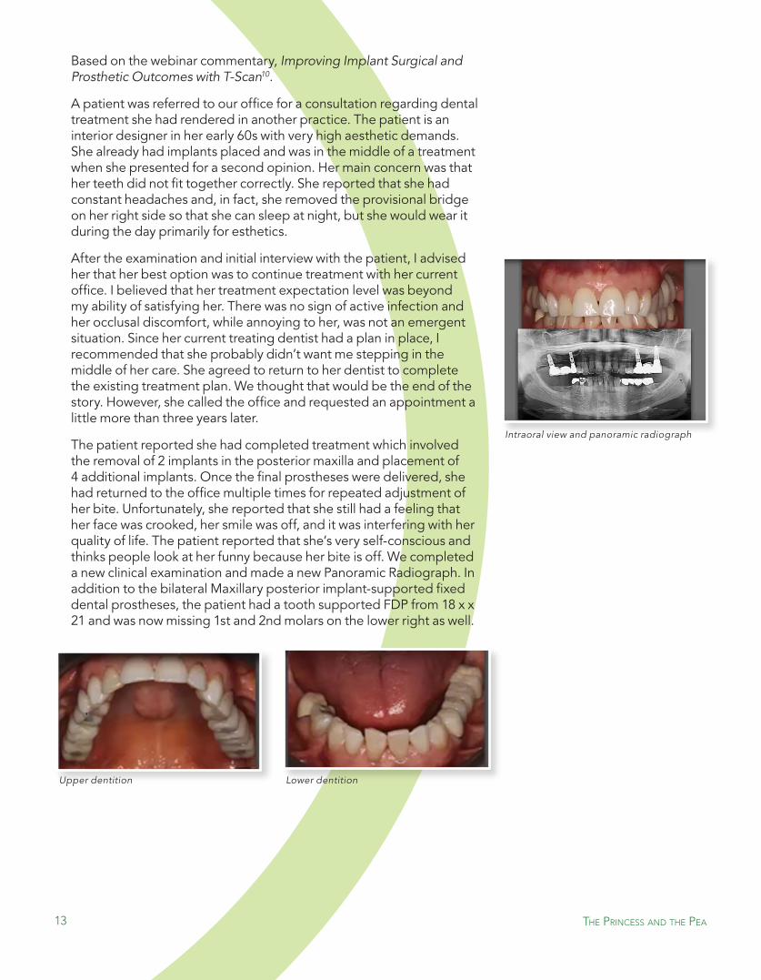
the patient reported she had completed treatment which involved the removal of 2 implants in the posterior maxilla and placement of 4 additional implants. once the final prostheses were delivered, she had returned to the office multiple times for repeated adjustment of her bite. Unfortunately, she reported that she still had a feeling that her face was crooked, her smile was off, and it was interfering with her quality of life. the patient reported that she’s very self-conscious and thinks people look at her funny because her bite is off. We completed a new clinical examination and made a new Panoramic Radiograph. In addition to the bilateral Maxillary posterior implant-supported fixed dental prostheses, the patient had a tooth supported FDP from 18 x x 21 and was now missing 1st and 2nd molars on the lower right as well.
Intraoral view and panoramic radiograph
the prIncess anD the pea
Upper dentition Lower dentition

14
After meeting with the patient and discussing her concerns on a couple of occasions, it becomes clear that she is very occlusally sensitized. In other words, she’s the proverbial Princess and the Pea, from the Hans Christian Anderson literary fairy tale about the princess who can’t sleep, because she can feel a small pea under a stack of 20 mattresses. that’s how sensitive she was to the slightest imbalance or interference in her bite. When asked to indicate which area of her bite is off, she immediately points to her upper right 1st premolar and says, “this tooth is not right! I’m hitting harder. My jaw has to slide to close, and it’s making my jaw crooked.” In this situation, it would not be advisable to start grinding down areas on this patient’s teeth without firm evidence to support those irreversible modifications to her bite. once the first adjustment is made, you become the owner of those occlusal problems.
We use the t-scan novus with the new sensor handle. It’s much more ergonomic than the prior generation t-scan unit. the single red button on the handle allows the operator to open a new scan, as well as start and stop a recording. In addition, based upon what is observed on the bite force reading on-screen, one is able to adjust the bite sensitivity of the sensor right on the handpiece.
After explaining to the patient how the t-scan bite sensor works, we were able to record her first bite force movie. When we then reviewed the data that was captured with the patient, we find that the patient was absolutely correct in identifying her prematurity contact on tooth #5. the software also shows implant warnings, telling us the implants are taking on stronger force, or earlier force than the surrounding teeth. At this point, the patient now recognizes the value of the technol- ogy being used to validate her concerns about the area of her bite that didn’t feel right to her. A look of relief washed over the patient’s face as she was now finally being understood and she was confident her concerns would be addressed and corrected.
T-Scan Novus handpiece, sensor, and sensor support
the prIncess anD the pea
As the treating clinician, it would be unwise to say based on subjective information, “In my opinion, here’s what’s going on and let me grind on your teeth until it starts to feel better to you.” We simply bring out the T-Scan and show the patient the source of her occlusal discrepancy. We can then educate the patient along the lines, “It’s not my opinion. It’s the computer sensor’s objective data that shows us where you’re hitting harder and where your bite might be coming together before the other teeth touch!”
15
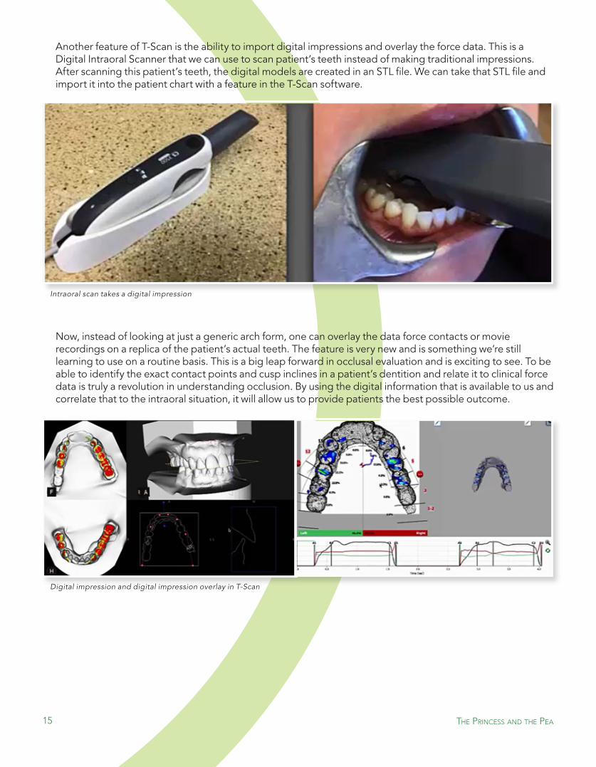
Another feature of t-scan is the ability to import digital impressions and overlay the force data. this is a Digital Intraoral scanner that we can use to scan patient’s teeth instead of making traditional impressions. After scanning this patient’s teeth, the digital models are created in an stL file. We can take that stL file and import it into the patient chart with a feature in the t-scan software.
now, instead of looking at just a generic arch form, one can overlay the data force contacts or movie recordings on a replica of the patient’s actual teeth. the feature is very new and is something we’re still learning to use on a routine basis. this is a big leap forward in occlusal evaluation and is exciting to see. to be able to identify the exact contact points and cusp inclines in a patient’s dentition and relate it to clinical force data is truly a revolution in understanding occlusion. By using the digital information that is available to us and correlate that to the intraoral situation, it will allow us to provide patients the best possible outcome.
Intraoral scan takes a digital impression
Digital impression and digital impression overlay in T-Scan
the prIncess anD the pea
16
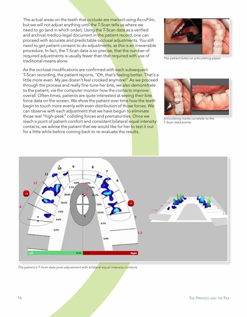
the actual areas on the teeth that occlude are marked using AccuFilm, but we will not adjust anything until the t-scan tells us where we need to go (and in which order). Using the t-scan data as a verified and archival medico-legal document in the patient record, one can proceed with accurate and predictable occlusal adjustments. You still need to get patient consent to do adjustments, as this is an irreversible procedure. In fact, the t-scan data is so precise, that the number of required adjustments is usually fewer than that required with use of traditional means alone.
As the occlusal modifications are confirmed with each subsequent t-scan recording, the patient reports, “oh, that’s feeling better. that’s a little more even. My jaw doesn’t feel crooked anymore!” As we proceed through the process and really fine-tune her bite, we also demonstrate to the patient, via the computer monitor how the contacts improve overall. often times, patients are quite interested at seeing their bite force data on the screen. We show the patient over time how the teeth begin to touch more evenly with even distribution of those forces. We can observe with each adjustment that we have begun to eliminate those real “high-peak” colliding forces and prematurities. once we reach a point of patient comfort and consistent bilateral equal intensity contacts, we advise the patient that we would like for her to test it out for a little while before coming back to re-evaluate the results.
The patient bites on articulating paper
Articulating marks correlate to the T-Scan data points
The patient’s T-Scan data post-adjustment with bilateral equal intensity contacts
the prIncess anD the pea

17
this patient then returned a few weeks later and exclaimed, “You know what? there is a night-and-day difference. I don’t have headaches, my bite isn’t crooked, my jaw has shifted back!” While it is not likely we actually altered anatomically the position of the jaw, the patient reports what she is feeling and it is a significant improvement to the occlusal situation that has dominated her life for the last several years. In addition, it is often prudent to consider the fabrication of an occlusal Guard to be worn at night. For this specific case, a thermoplastic Impac guard was fabricated in the lab and delivered at the follow-up appointment. It is also possible and advisable to use t-scan to adjust the night guard as well.
t-scan is not just for teeth. It is certainly possible to take a bite scan with the night guard in the patient’s mouth to evaluate the forces and loading. the patient is able to clench down on that guard, because it’s a braced position. We make a final adjustment on the guard and we part with the advice, “We want you to protect your dental investment. this guard is like an insurance policy for you now.” We don’t want the prospect of renewing or replacing the work in her mouth anytime soon.
today, we’re doing things with implants that we haven’t done in the past; and it becomes imperative to use a computerized approach beyond 100-plus-year-old carbon paper to mark occlusal forces. steve Jobs told us, “Be a yardstick of quality. some people are not used to an environment where excellence is expected!” expect it in your office.expect it from your team. expect it for your patient care. T-Scan is used to make adjustments for
splint therapy
Dr. scott Keith received his DDs with valedictorian honors from the University of California, san Francisco in 1995. He then completed specialty training in
Prosthodontics at the Baylor College of Dentistry and also earned a Master of science degree in oral Biology. Dr. Keith has published several articles and has presented his research into dental implants internationally. In June of 1998, Dr. Keith’s work won the International team for Implantology (ItI) Research Competition for his presentation at the ItI World symposium in Boston. Dr. Keith was awarded a prestigious surgical implant fellowship by the ItI to further his training at the Harvard school of Dental Medicine where he also maintained a faculty appointment teaching at the pre-doctoral and graduate levels. A board-certified fellow
of the American College of Prosthodontists, Dr. Keith is also a fellow of the ItI, and a member of the Academy of osseointegration, and omicron
Kappa Upsilon. Currently, Dr. Keith maintains a private practice in the Dental Implant Center @ Walnut Creek.
the prIncess anD the pea

the craDle wIll rocK
A CAse by
Dr. Chris stevens, DDs
the craDle wIll rocK
19
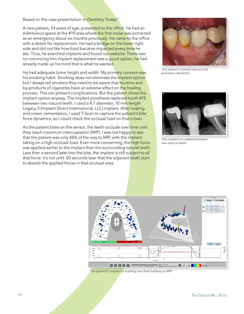
Based on the case presentation in Dentistry Today11.
A new patient, 54 years of age, presented to the office. He had an edentulous space at the #19 area where the first molar was extracted as an emergency about six months previously. He came to the office with a desire for replacement. He had a bridge on the lower right side and did not like how food became impacted every time he ate. thus, he searched implants and found our website. there was no convincing him implant replacement was a good option, he had already made up his mind that is what he wanted.
He had adequate bone height and width. My primary concern was his smoking habit. smoking does not eliminate the implant option but I always tell smokers they need to be aware that nicotine and by-products of cigarettes have an adverse effect on the healing process. this can present complications. But the patient chose the implant option anyway. the implant prosthesis replaced tooth #19, between two natural teeth. I used a 4.7 diameter, 10 mm length Legacy 3 (Implant Direct International, LLC) implant. After healing and crown cementation, I used t-scan to capture the patient’s bite force dynamics, so I could check the occlusal load on that crown.
As the patient bites on the sensor, the teeth occlude over time until they reach maximum intercuspation (MIP). I was not happy to see that the patient was only 46% of the way to MIP, with the implant taking on a high occlusal load. even more concerning, the high force was applied earlier to the implant than the surrounding natural teeth. Less than a second later into the bite, the implant is still subject to all that force. It’s not until .03 seconds later that the adjacent teeth start to absorb the applied forces in that occlusal area.
The patient’s mixed natural and porcelain dentition
The implant is cradled between two natural teeth
the craDle wIll rocK
The patient’s implant is loading less than halfway to MIP
20
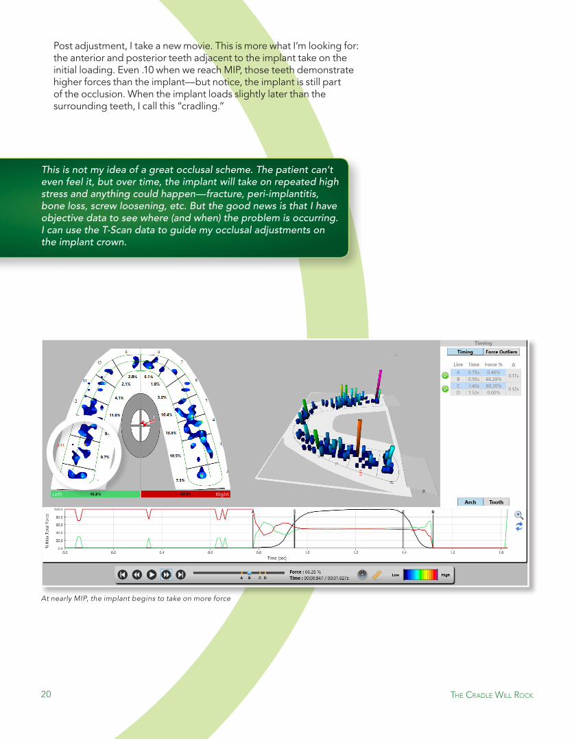
At nearly MIP, the implant begins to take on more force
Post adjustment, I take a new movie. this is more what I’m looking for: the anterior and posterior teeth adjacent to the implant take on the initial loading. even .10 when we reach MIP, those teeth demonstrate higher forces than the implant—but notice, the implant is still part of the occlusion. When the implant loads slightly later than the surrounding teeth, I call this “cradling.”
This is not my idea of a great occlusal scheme. The patient can’t even feel it, but over time, the implant will take on repeated high stress and anything could happen—fracture, peri-implantitis, bone loss, screw loosening, etc. But the good news is that I have objective data to see where (and when) the problem is occurring. I can use the T-Scan data to guide my occlusal adjustments on the implant crown.
the craDle wIll rocK

21
Getting this occlusal scheme to where it needed to be requires that fourth dimension: timing. I was able to time when the implant began to load by identifying how force is distributed over the dentition throughout the course of the bite. I could not do this with the traditional occlusal methods we have today: patient feedback, articulating paper, wax, shim stock foils, imprints, etc. I’m able to control the occlusal situation of the patient by having objective bite force data to complete the equilibration, ensure the long term function of the implant, and manage the prosthesis with measurement going forward.
At recall hygiene appointments, a new movie is taken to insure the occlusal scheme is still favorable for the implant prosthesis. I have found adjustments may need to be made over the life of the implant. However, that quick adjustment saves time, heart muscle, and stomach lining compared to loose or broken implant prostheses.
I find that patients cannot detect an implant prosthesis pre-maturity with any consistency. However, when I have completed the computerized occlusal management, they tend to be surprised how much better the bite feels than before I adjusted.
the craDle wIll rocK
Post-adjustment, Dr. Stevens can see the implant starting to load later than the natural surrounding teeth

22
T-Scan data post-adjustment revealing occlusal load changes on the dentition
Dr. Chris stevens, DDs has a dental practice in sun Prairie, Wisconsin that focuses on Family Dentistry and Cosmetic Dentistry. He graduated from Marquette school of Dentistry in 1981 and began practicing in sun Prairie, Wisconsin in 1982. Dr. stevens is recognized as an international expert in Cosmetic Dentistry and Full Mouth Reconstruction. He is committed to staying in the forefront of the latest techniques by attending high-level continuing education. He frequently teaches other dentists across the country and abroad in areas such as Cosmetic Dentistry and Full Mouth Reconstruction, principles of occlusion and diagnosis and treatment of temporomandibular (tMJ) disorders. He
has been an active lecturer since 1989 and has spoken to thousands of care providers including dentists, physicians, chiropractors and physical
therapists across the country and abroad.
the craDle wIll rocK

a wrInKle In tIme
A CAse by
Dr. sAngiv PAtel, DDs
a wrInKle In tIme

24
In June of 2008, a 56-year-old male patient came to me with a recent history of a lower right first molar that had fractured and was deemed unrestorable. the patient was advised by his periodontist to do a dental implant (and not another bridge) in order to regain function. the molar was consequentially removed and the site received a bone graft. A dental implant was placed by the periodontist after the bone graft healed.
the patient knew about our facility and advanced dental technology including CeReC CADCAM and t-scan technology, because at the time I was providing care to his elderly in-laws for many years. He decided to travel about 350 miles from tallahassee, Florida to our practice in Melbourne for his implant restoration. (our care in conjunction with the application of these technologies was the primary reason for selecting our practice for his restorative care.) the patient requested restoration of the lower right posterior quadrant. Upon examination of the area and occlusion, a limited treatment plan was generated, reviewed and consented for restorative care with CeReC CAD/CAM restorations in conjunction with t-scan equilibration and occlusal load management.
the t-scan software records how each tooth comes into contact with the opposing teeth and defines the location, intensity, and duration of occlusal loads graphically. the Center of Force trajectory (CoF) demonstrates the net location of occlusal loads in one-tenth of a second increments once the maximum loads exceeds 10% during the occluding cycle. the CoF exhibits where in the mouth the forces begin, travel, and end, as the loads are distributed throughout the occluding cycle. A simple analogy to aid in patient education is to relate the CoF to a hurricane tracker. the Circle of Mannes (gray and white oval in the center) can be referred to as a safe bunker where the energy is well distributed, most functional, and least harmful.
a wrInKle In tIme
Pre-restoration MIP T-Scan recording (Highlighted)

25
the initial t-scan test was recorded in MIP. the first thing that caught my eye was the CoF that begins over the first right maxillary and mandibular molars (teeth #3 and #30), and proceeds distally to the left side as the occluding cycle leads to MIP. the second thing I noticed is obviously the premature hyper-intense contact on the second molars with a load distribution of 10%. Based on the pre-restorative CoF, it is very possible this patient would have never lost that particular molar if the CoF was managed with an equilibration prior to the molar fracturing. there is a direct cause-and-effect relationship incriminating the unrestorable molar fracture to the excessive occlusal loads in the area. the data reveals that the second molars on the right side are also at risk of failure due to occlusal trauma. this negligence in monitoring the occlusion to the extent and detail required resulted in the patient becoming a tooth amputee, requiring multiple surgeries and a prosthetic replacement.
the second variable that can only be predictably diagnosed and managed with the t-scan is Bilateral simultaneity. It accounts for all teeth and restorations meeting at the same time and hence accounts for occlusion time and CoF.
this was the patient’s first dental implant with an occlusal scheme of crowns, bridges, and a few natural teeth. A pre-restoration full mouth equilibration was provided to achieve anterior guidance with bilateral simultaneity. the post equilibration CoF demonstrates the anterior guidance with elimination of the pre-mature contacts on the second molars with concurrent increase in the MIP load from 10% to 13%. this is now an atraumatic stable occlusal scheme that is better engineered to tolerate parafunctional loads. these are the benefits of a digitally managed equilibration. the Implant at site #30 is now ready to be restored.
This case emphasizes the need to monitor the occlusion digitally with a T-Scan vs any other traditional occlusal analysis method, as only the T-Scan technology can expose the time and force vectors that can impact dental longevity.
Pre-restoration Equilibration completed
a wrInKle In tIme

26
the adjacent teeth were restored with CeReC restorations—an onlay on tooth #29 and a crown on tooth #31. the integrated straumann implant was uncovered and a prefabricated abutment was placed and torqued to 35 ncm.
the final restoration at site #30 was a full coverage all ceramic crown. the material we used to restore sites #29, #30 and #31 was VItA Mark II, which is bit “softer” on the stress strain curve in comparison to enamel. the energy transmission of the cement retained restorations is going to be much more favorable against the opposing dentition. If there is occlusal trauma, the material will fracture and no subsequent energy will travel to the abutment screw or implant body. this inherently mitigates against crestal bone loss. the potential crown failure will be easily visible and readily repaired.
After cementation and x-ray verification of cement removal, time Delayed Loading of implant was implemented. the goal is to sequentially load the natural and restored teeth, then the implant restoration, then finally achieve MIP without excursive interferences. this will achieve proper CoF, Bilateral simultaneity, occlusion, and disclusion times. the pre-restoration equilibration had established anterior guidance and MIP without excursive interferences. Utilizing the t-scan for guidance and verification, minimal reductions in the intensity of the centric stops on the abutment-supported crown was initiated. this realized the goal of time Delay Loading.
Pre-restoration Straumann implant
Abutment torqued to 35 Ncm
CEREC digital rendering of restoration
Final restoration using VITA MARK II
Using x-ray to verify cement removal
a wrInKle In tIme
27 a wrInKle In tIme
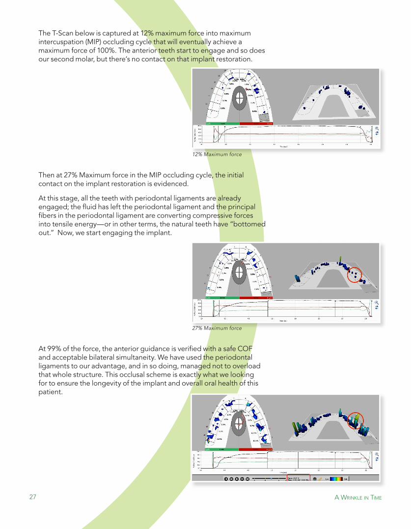
12% Maximum force
27% Maximum force
the t-scan below is captured at 12% maximum force into maximum intercuspation (MIP) occluding cycle that will eventually achieve a maximum force of 100%. the anterior teeth start to engage and so does our second molar, but there’s no contact on that implant restoration.
then at 27% Maximum force in the MIP occluding cycle, the initial contact on the implant restoration is evidenced.
At this stage, all the teeth with periodontal ligaments are already engaged; the fluid has left the periodontal ligament and the principal fibers in the periodontal ligament are converting compressive forces into tensile energy—or in other terms, the natural teeth have “bottomed out.” now, we start engaging the implant.
At 99% of the force, the anterior guidance is verified with a safe CoF and acceptable bilateral simultaneity. We have used the periodontal ligaments to our advantage, and in so doing, managed not to overload that whole structure. this occlusal scheme is exactly what we looking for to ensure the longevity of the implant and overall oral health of this patient.

28
Dr. sangiv I. Patel, DDs is the founder and developer of the Innovative smile. He is a general dentist in private practice since 1993, has served as faculty at the Advanced Dental Implant Institute’s AAID Maxicourse in Puerto Rico, and formerly served on the faculty at Loyola University of Chicago- school of Dentistry and Brevard Community College. He is among 30 clinicians worldwide to have received Mastership in Dental Biometrics. He is a published author, international lecturer. He has served as a beta tester for BioReseARCH Inc., tekscan Inc., and CeReC 3D by sirona and collaborated with Carestream the manufacturers of CBCt technology. His experience in cutting-edge dentistry runs long and deep. Dr. Patel offers physics based model
on the principles of rigid vs. resilient dynamics in the stomatognathic system that paves a road for logical, predictable and evidenced based
diagnostics and restorative single visit dentistry.
this particular patient travelled about six hours each way to achieve these predictable results. If his dentist had the technology to recognize and address the occlusal trauma, he would have never needed implant surgery and restoration procedures. When he learned that he could have actually avoided the implant, it broke his heart.
I say this all the time to people in our industry, including patients, “It’s your ethical duty to inform the patient on the state of their occlusion.” When I walk into my doctor’s office, they don’t ask me how my blood pressure feels today. they slap a cuff on my arm and take the data. the cardiologist doesn’t live without blood pressure, but why does dentistry live without teeth pressure measurements and hope to achieve longevity. Is that okay?
occlusion is not a static thing, it’s like blood pressure: it fluctuates, and our job isn’t to perform supervised negligence but rather control and manage its energy so it does not lead to negative, destructive effects; hypertension leads to a heart attack or stroke, occlusal trauma leads to tooth attack(s). the t-scan is incredibly powerful as a diagnostic and treatment instrument required for comprehensive management of dental occlusion.
a wrInKle In tIme
29
conclusIon
While traditional occlusal indicators have some descriptive capability, they don’t paint the whole picture.
occlusal data is multi-dimensional, which allows the clinician to see the force, timing, and location of occlusal contacts. In three clinical cases, t-scan data was used to help describe the occlusal condition of each patient. this allowed the doctors to understand the occlusal environment that the implant lives in. In these cases, it was essential to understanding how the implant restorations will survive in the mouth over the long term.
Information is the cornerstone of any diagnosis. Using this information, these doctors were able to quickly and effectively balance the occlusion, protect the implant, and send the patient home confident in their treatment.
occlusion management is key for implants, but also for other dental applications, whether it’s cosmetics, tMD, hygiene, splint therapy, etc. Using technology will not only support diagnosis and treatment plans with solid, objective data, but also differentiates the doctor and practice.
conclusIon
30
references
[1] Abraham, C. (2014). A Brief Historical Perspective on Dental Implants, their surface Coatings and treatments. the open Dentistry Journal, Volume 9, 2015(1874-2106), 50-55. doi:10.2174/1874210601408010050
[2] Dental Implants Facts and Figures. (2016). Retrieved from http://www.aaid.com/about/press_room/dental_implants_faq.html
[3] Gaviria L, salcido JP, Guda t, ong JL. Current trends in dental implants. J Korean Assoc oral Maxillofac surg. 2014 Apr;40(2):50-60. http://dx.doi.org/10.5125/jkaoms.2014.40.2.50
[4] How Are Implants Placed? (2014). Retrieved from http://www.osseo.org/neWhowAreImplantsPlaced.html
[5] Ruiz, J. (2010, october 6). three Golden Rules of occlusion. Retrieved from http://www.dentistrytoday.com/occlusion/3807-the-three-golden-rules-of-occlusion
[6] Vasquez, R. (2013, April 9). Management of occlusion over Implants, Part 1: three 10-Year Case Follow-ups and evaluations. Retrieved from http://www.dentistrytoday.com/restorative/8867-management-of-occlusion-over-implants-part-1-three-10-year-case-follow-ups-and-evaluations
[7] Gates, B., & Hemingway, C. (1999). Business @ the speed of thought: Using a digital nervous system. new York, nY: Warner Books.
[8] Kim, DDs, Ms, PhD, J. (2015). Digitalized Implant occlusion with the t-scan system. In R. Kerstein, DMD (ed.), Handbook of Research on Computerized occlusal Analysis technology Applications in Dental Medicine (pp. 562-601). Hershey, PA: . doi:10.4018/978-1-4666-6587-3.ch012
[9] Kaptein, M., De Putter, C., De Lange, G., & Blijdorp, P. (1999). A clinical evaluation of 76 implant-supported superstructures in the composite grafted maxilla. Journal of oral Rehabilitation, (August), 619-623. Retrieved from http://www.ncbi.nlm.nih.gov/pubmed/10447813
[10] Keith, s. (Writer). (2016, February 25). Improving Implant surgical and Prosthetic outcomes with t-scan [Video file]. Retrieved from https://www.tekscan.com/events/improving-implant-surgical-and-prosthetic-outcomes-t-scan
[11] stevens, C., DDs. (2016, January). technology to Control excessive occlusal Contact Force in enhancing Implant Longevity. Dentistry today, 112-117.
DIsCLAIMeR: the contents of this publication may be of interest to medical professionals or other health care providers. such persons should exercise their own judgment in determining whether a particular product, treat-ment, therapy option, procedure, program or service is appropriate or legal for their practice or their patients.
references


+1 . 617. 4 6 4 . 4 2 8 0 1 . 8 0 0 . 2 4 8 . 3 6 6 9 | i n f o @ t e k s c a n . c o m | w w w. t e k s c a n . c o m /d e n t a l
ContaCt today for a demonstration!
© 2016 tekscan, Inc.