i european annals of allergy and clinical immunology
TRANSCRIPT
4/2014
The perception of allergen-specific immunotherapy among chest physicians: an Italian survey
Tetranychus urticae allergy in a population without occupational exposure
In patients with LTP syndrome food-specific IgE show a predictable hierarchical order
Middle lobe syndrome: a rare presentation of allergic bronchopulmonary aspergillosis
Prurigo simplex subacuta or prurigo simplex acuta?
Diagnosis of eosinophilic esophagitis in an infant undergoing milk oral immunotherapy - a case report
An unusual complication of late onset allergic contact dermatitis to povidone iodine in Oral & Maxillofacial Surgery – A report of 2 cases
European Annalsof Allergy and
Clinical Immunology
Issn 1764-1489 Volume 46 n. 4/2014 – July 2014
THE OFFICIAL JOURNAL OF AAITO | ASSOCIAZIONE ITALIANA ALLERGOLOGI IMMUNOLOGI TERRITORIALI E OSPEDALIERI
THE OFFICIAL JOURNAL OF SPAIC | SOCIEDADE PORTUGUESA DE ALERGOLOGIA E IMUNOLOGIA CLINICA
AbbAAITO210x270.indd 1 02/07/14 14.21

informazionicosti
le più esaurienti e affidabili informazioni sui farmaci da oggi e per tutto il 2014a un prezzo speciale
Le offerte
• volume Parafarmaceutici• volume Indirizzi
a soli 99 euro
• volume Medicinali• volume Indirizzi
a soli 99 euro
• volume Medicinali• volume Parafarmaceutici• volume Indirizzi
a soli 149 euro
© EDRA LSWR SpA
via Spadolini, 7 - 20141 MilanoTel.: 02.88184.317 - Fax: 02.93664.151e-mail: [email protected] - www.edizioniedra.it
DIS
3201
4
Senza titolo-1 1 02/04/14 17.53
INFORMAZIONEPolitica Sanitaria, clinica,
farmaci e terapie e la cronaca quotidiana
dei fatti di attualità
CANALI TEMATICICardiologia, Pediatria,
Ginecologia, Diabetologia, Medicina interna
CAsI CLINICIOltre 120.000 medici coinvolti nei casi clinici messi a disposizione
dei colleghi per un consulto
COMMUNITYL’area Social con i servizi
“L’Esperto risponde”, “Doctor Alert”, “Forum”,
“Preferiti”
DOWNLOAD CENTER
Contenuti di formazione e aggiornamento gratuiti tratti
dalle pubblicazioni Edra Masson
ECM La formazione di qualità
di Doctor33
BACHECAANNUNCI
La più ricca sezione di annunci sul web per
i professionisti della medicina
Il portale dei protagonisti della salute
www.doctor33.it
NOvITà

EDITORS IN CHIEFR. Asero (Milano – Italy)
M.Morais - Almeida (Lisbon – Portugal)
HONORARY EDITORA. Sabbah (Angers – France)
ASSOCIATE EDITORSS. Bonini (Roma – Italy), A. Tedeschi (Milano – Italy)
EDITORIAL BOARDM.B. Bilò (Ancona – Italy)
F. Bonifazi (Ancona – Italy)L. Cecchi (Firenze – Italy)
L. Delgado (Oporto – Portugal)P. Demoly (Montpellier – France)
G. D’Amato (Napoli – Italy)M. Drouet (Angers – France)
M. Fernandez-Rivas (Madrid – Spain)A. Fiocchi (Milano – Italy)
D. Macchia (Firenze – Italy)F. Mastrandrea (Taranto – Italy)
D.A. Moneret-Vautrin (Nancy – France)M. Morais-Almeida (Lisbon – Portugal)
G. Moscato (Pavia – Italy)C. Nunes (Portimao – Portugal)
M. Olivieri (Verona – Italy)P. Parronchi (Firenze – Italy)
G. Passalacqua (Genova – Italy)G. Pauli (Strasbourg – France)
A. Perino (Torino – Italy)O. Quercia (Faenza – Italy)A. Romano (Roma – Italy)G. Senna (Verona – Italy)
A. Todo Bom (Coimbra – Portugal)S. Voltolini (Genova – Italy)
SCIENTIFIC COMMITTEEL. Antonicelli (Italy)
A. Bener (Qatar)H. Bazin (Belgium)
J. Bellanti (USA)C. Geller-Bernstein (Israel)
S. Bonini (Italy)G.W. Canonica (Italy)
M. Cugno (Italy)B. David (France)
S. Durham (London)R. de Marco (Italy)
G.P. Girolomoni (Italy)R. Jarish (Austria)
S.G.O. Johansson (Sweden)F. Levi-Shaffer (Israel)
C. Lombardi (Italy)P. Lowenstein (Denmark)
J.L. Malo (Canada)A.G. Palma-Carlos (Portugal)
G. Scadding (London)G. Scadding (LondonE. Stevens (Belgium)
R. van Ree (Amsterdam)
FOUNDER AND CORRESPONDING MEMBERG.M. Halpern (USA)
Editors in ChiefRiccardo AseroMário Morais-Almeida
Publishing DirectorNicola Miglino
Publishing EditorChiara [email protected]. 02 88184.257
Production ManagerWalter [email protected]. 02 88184.222
Sales & MarketingLudovico [email protected]. 02 88184.354
TrafficDonatella [email protected]. 02 88184.292
[email protected] - Tel. 02 88184.317 - Fax 02 88184.151Italy subscription: 60 euroWorld subscription: 85 eurowww.eurannallergyimm.com
PrintingProntoStampa SrlVia Praga, 1 - 24040 Verdellino (BG)
EDRA LSWR SpAVia G. Spadolini, 720141 Milano - ItalyTel. 0039 (0)2-88184.1Fax 0039 (0)2-88184.301www.edizioniedra.it
© 2014 Associazione Italiana Allergologi Immunologi Territoriali e Ospedalieri - AAITO. Published by EDRA LSWR SpA.All rights reserved.
The contents of this Journal are indexed in PubMed and SCOPUS®
European Annalsof Allergy and
Clinical ImmunologyTHE OFFICIAL JOURNAL OF AAITO
ASSOCIAZIONE ITALIANA ALLERGOLOGI IMMUNOLOGI TERRITORIALI E OSPEDALIERI
THE OFFICIAL JOURNAL OF SPAICSOCIEDADE PORTUGUESA DE ALERGOLOGIA E IMUNOLOGIA CLINICA
AAITOAssociazione Italiana Allergologi Immunologi Territoriali e Ospedalieri
Directory BoarD
PresidentMaria Beatrice Bilò
Designate PresidentAntonino Musarra
Vice PresidentsRiccardo AseroFrancesco Murzilli
TreasurerOliviero Quercia
Honorary PresidentFloriano Bonifazi
MembersLorenzo CecchiDomenico GarganoGiuseppina ManzottiLionello Muratore Susanna Voltolini Marcello Zambito

130 Author Guidelines
European Annals of Allergy and Clinical Immunology will accept for publication suitable manuscripts dealing with the ae-tiology, diagnosis, and treatment of allergic and immunologic diseases. These might include the study of methods of con-trolling immunologic and allergic reactions, human and ani-mal models of hypersensitivity and other aspects of basic and applied clinical allergy in its broadest sense.We encourage case reports that focus on topic(s) of extreme contemporary interest. Paper reporting the results of drug trials will be considered.European Annals of Allergy and Clinical Immunology also publishes solicited and usolicited review articles on subjects of topical interest to clinical and experimental allergy.
Manuscript
We request that all manuscripts should be submitted online through our web-based peer review system.Submitted contributions are accepted for publication on the ba-sis of scientific interest and relevance, at the final discretion of the Editors in Chief, who will have blinded written evaluations from at least two anonymous reviewers. Once a manuscript has been accepted for publication, Authors will receive an electronic page proof for review and approval, following which the manuscript is published in the print jour-nal and on the journal website.Following acceptance, Authors are also requested to return both completed and signed Journal Publishing Agreement and Con-flict of interest disclosure forms by e-mail to: [email protected]
Full Authors Guidelines, online Submission System link, Jour-nal Publishing Agreement and Conflict of interest forms are available on Journal website: www.eurannallergyimm.comTyped manuscripts at 30 lines per page: maximum lenght 10 pages, around 300 lines.Manuscripts should be typewritten (double spacing) on one side of the paper; on a separate sheet, should bear the title of the paper, name, postal and e-mail address of the Author, together with the name of institution where the work was done.Generally, papers should be divided into the following parts and in the order indicated:1. Summary and key words: english, limited to 15 lines.2. Introduction: containing the reasons for doing the work.3. Materials and methods.4. Results: these should be given concisely; the use of tables
and figures to illustrate the same results will only rarely be allowed.
5. Discussion: the presentation of results should be separated from a discussion of their significance.
6. References.
Units and Abbreviations
European Annals of Allergy and Clinical Immunology rec-ognizes the adoption of the International Systems of Units (SI-Units). Abbreviations to be put in a glossary at the foot of page 1 on the text.
References
References should be in the order:• the order number corresponding with that of appearance in
the text;• the author’s name(s), followed by initial or first name;• the title of the work, in the original language;• for journals: usual title abbreviations according to interna-
tional nomenclature and in the order: year, volume number, issue number (in parenthesis), first and last page numbers of the work.
For example:Bodtger U, Linnegerg A. Remission of allergic rhinitis: An 8-year observational study. J Allergy Clin Immunol 2004; 114(6): 1384-8.• for books: name of the author/editor, title, publisher/institu-
tion, town where published, year of publication, first and last page numbers of the work.
For example:Paupe J, Scheinman P (Eds.). Allergologie Pédiatrique. Flam-marion, Paris, 1988: 324-42.
Illustrations
• Figures always on separate numbered sheets and legends on the back in pencil
• Figures always saved on separate numbered files• Figures, diagrams: JPG, 300 dpi minimum• Radiographs: JPG, 300 dpi minimum
All tables, figures, radiographs, etc. must be referenced in the text.Legends should be put on a separate sheet, saved on a separate file and have the same numbers as the figures.
The “pdf ” of the article will be sent to the author by e-mail.
EDRA LSWR SpAVia Spadolini, 720141 Milano - ItalyTel. 0039 (0)2-88184.1Fax 0039 (0)2-88184.301www.eurannallergyimm.com
Original ArticlesThe perception of allergen-specific immunotherapy among chest physicians: an Italian survey . . . 132c. LomBarDi, G.W. canonica, G. PassaLacqua
Tetranychus urticae allergy in a population without occupational exposure . . . . . . . . . . . . . 137n. santos, V. iraoLa, J.L. PLáciDo
In patients with LTP syndrome food-specific IgE show a predictable hierarchical order . . . . . . . 142r. asero
Case ReportsMiddle lobe syndrome: a rare presentation of allergic bronchopulmonary aspergillosis . . . . . . . 147a. shah, s. Behera, c. PanJaBi
Prurigo simplex subacuta or prurigo simplex acuta? . . . . . . . . . . . . . . . . . . . . . . . . 152h. h. akar, F. tahan, s. BaLkanLi, s. saDet Özcan
Diagnosis of eosinophilic esophagitis in an infant undergoing milk oral immunotherapy - a case report . . . . . . . . . . . . . . . . . . . . . . . . . . . . . . . . . . . 154P. morais siLVa, J. antunes, m. chamBeL, s. Prates, P. Leiria Pinto
An unusual complication of late onset allergic contact dermatitis to povidone iodine in Oral & Maxillofacial Surgery – A report of 2 cases . . . . . . . . . . . . . . . . . . . . . . . 157m.a. reyazuLLa, a.L. GoPinath, n. VaiBhaV, r.P. raut
ANNOUNCEMENT . . . . . . . . . . . . . . . . . . . . . . . . . . . . . . . . . . . . . . . 160
tAble of Contents

O R I G I N A L A R T I C L E S Eur Ann AllErgy Clin immunol VoL 46, n 4, 132-136, 2014
IntroductionAllergy still remains a major public health concern which has pandemic proportions, affecting more than 150 million people in Europe. Taking into account the epidemiological trends, it is hypothesized that within 15 years more than 50% of the Eu-ropean population will suffer from some type of allergy (1,2). Allergic patients suffer from a debilitating disease, with a major impact on their quality of life (QoL) and work/school perfor-mance, and constitute a significant burden on health econom-ics, due to lost productivity and absenteeism (3). Given that allergy triggers such as urbanization, pollution and climate
change are not expected to change significantly, the only ways to afford this burden are strengthening and optimizing the pre-ventive and treatment strategies. Nonetheless, it has been re-peatedly shown that the available pharmacological treatments are neither capable to achieve a long-term effect once stopped, nor to induce significant immunological changes. On the con-trary, allergen-specific immunotherapy (SIT), which is based on the subcutaneous (SCIT) or sublingual (SLIT) administration of high allergen doses, was proven able to reduce asthma/rhi-nitis symptoms and to achieve a long-lasting effect. However, SIT is currently used only as a second-line treatment, and often
SummaryBackground and Aim. This questionnaire-based study evaluated the overall level of knowl-edge about allergen-specific immunotherapy (SIT) among chest physicians, who are frequent-ly involved in the management of respiratory allergies. This represents an interesting aspect, because chest physicians intercept many of the patients with allergic rhinitis w/wo asthma, in which SIT could be potentially indicated. Methods. A panel of experts prepared a ques-tionnaire, involving 16 main points of interest/questions concerning the knowledge and use of SIT. Questionnaires were e-mailed (September-October, 2011) to randomly-selected special-ists, and were returned anonymously. Results. 81 questionnaires from specialists using SIT were eligible. The respondent population had a mean age of 51 years (range 33-63 years, 74% male). The general knowledge on SIT is overall satisfactory among pulmonologists, and they are well aware that SIT is recommended in the available guidelines. Nevertheless, about 50% of physicians still believe that SIT has to be used as a last-choice adjunct when pharmacother-apy fails. Chest physicians are well aware that SIT has a disease-modifying effect, in addition to its short-term clinical efficacy. Finally, the majority of interviewed specialists agree on the need of getting more information and education on SIT. Conclusions. This survey about the perception of SIT among chest physicians in Italy highlighted a satisfactory overall knowledge of SIT and only few weak points. These results would allow to take appropriate educational actions and this questionnaire could be used to monitor the possible effects of divulgation and educational initiatives over time.
Corresponding authorGiovanni PassalacquaAllergy and Respiratory Diseases IRCCS S. Martino-University Hospital-IST Pad. MaraglianoGenoa, Italy Phone: +39 010 555 84 59 Fax: +39 010 353 89 04E-mail: [email protected]
Key words
Allergen-specific immunotherapy; chest physicians; asthma; allergic rhinitis
1Respiratory Allergy Unit, Dept of Internal Medicine and Geriatry, Poliambulanza Hospital, Brescia, Italy2Allergy & Respiratory Diseases, IRCCS S. Martino-University Hospital-IST, Genoa, Italy
The perception of allergen-specific immunotherapy among chest physicians: an Italian survey
c. LomBarDi1, G.W. canonica2, G. PassaLacqua2

133The perception of allergen-specific immunotherapy among chest physicians: an Italian survey
Methods
A panel of experts, based on guidelines and literature analysis prepared a questionnaire of 16 points of interest/questions spe-cifically dedicated to chest physician specialists. The question-naire (Y/N or multiple-choice answers) was subdivided into five main sections (clinical/general aspects, efficacy perception, pharmacoeconomic aspects, SLIT versus SCIT, awareness of guidelines) (see table 1). The study, since cross-sectional and observational, did not need an official approval by the Eth-ic Committee to whom it was simply notified to warrant the privacy of the recorded data. The questionnaires were emailed to 115 specialists in Respiratory Medicine, randomly selected from the databases of the Italian societies of respiratory medi-cine, and had to be returned anonymously. Only the returned questionnaires of those physicians currently using SIT were taken into consideration. The selection of the chest physicians also took into account the harmonic distribution across the Italian Country, in order to avoid any “bias” connected to dif-ferent attitudes about SIT. Questionnaires were sent to pul-monologists from the beginning of September to the end of October 2011.
suggested as a last-choice. Indeed, in the more recent guidelines and academic position statements, the use of SIT has been ad-vocated for those patients with milder disease, also in order to prevent the progression of allergic respiratory diseases. In fact, the clinical value of SIT has been confirmed in multiple clini-cal trials and meta-analyses, also improving the patient report-ed outcomes, such as Quality of Life (QoL), long-term costs, burden of allergies, and effect on the course of the disease (4-6). Despite the aforementioned experimental evidence, SIT is still not receiving an adequate attention from Medical Institu-tions, as the general underuse of this treatment clearly demon-strates (7). In this context, the partnership and cooperation of different medical subjects (i.e. general practitioners, allergists, pediatricians, chest physicians) would be crucial. In a previous questionnaire-based study we observed that, at least among the Italian GPs, the perception of SIT as a valuable treatment was near to optimal (8). It is true that GPs are primarily responsible for education and information about respiratory allergy and its treatment, but it is also true that pulmonologists are often in-volved in the primary diagnosis and care of this disease (9). This survey was specifically designed for chest physicians, intended to assess their knowledge on SIT. This was done by a question-naire-based survey.
Table 1 - Number and % of responses to the items in the SIT - pulmonologist questionnaire.
ITEM N %1. In subjects with respiratory allergies (rhinitis and asthma), do you believe that SIT allergenic extracts may be a valid therapeutic option?Yes No
738
9010
2. Do you give the SIT directly to your patients? YesNoI follow patients when SIT was initiated by another specialist (e.g. allergist)
531612
652015
3. If so, how many patients come in a year?More than 10 patients Between 5 and 10 patients Between 1 and 5 patients
362025
44.524.531
4. In patients dealing with SIT, the decision was taken:Not answering After consulting with allergist In total autonomy
151650
18.52061.5
5. If using the SIT, which is its main indication?AsthmaRhinitis plus asthmaRhinitis
105021
1261.526.5
continues...

134 C. Lombardi, G.W. Canonica, G. Passalacqua
ITEM N %6. If administering the SIT, which allergenic extracts do you use? PerennialSeasonalBoth in equal measure
201744
24.52154.5
7. Which schedule does the patient usually follow (pre-coseasonal, continuous, continuous for seasonal forms also)?Pre-coseasonal ContinuousContinuous for seasonal forms also
253323
314128
8. In your opinion, SIT is: An alternative treatment with respect to pharmacologic therapyA limited efficacy treatmentA treatment that can only be used in a small proportion of patients The only treatment availableA complementary treatment to drug therapy
24102441
2.551229.551
9. Which route of administration do you use primarily?SublingualSubcutaneousBoth
501120
6213.524.5
10. Which route of administration do you consider clinically superior?Effective in equal measureSublingualSubcutaneous
292131
362638
11. How long do you continue the SIT treatment?5 or more years3 years1 year
16605
20746
12. Can SIT modify the natural history of the respiratory allergic disease?YesNo
6516
8020
13. Which of the following statements do you believe is the most significant? SIT may reduce the risk of new sensitizationsSIT is effective in reducing the use of medications SIT may reduce the risk of asthmaSIT is effective in reducing allergic symptoms
8162136
10202644
14. In a symptomatic asthmatic patient, do you continue to administer SIT? NoYesOnly after having checked the symptoms with anti-asthma drugs
72153
8.52665.5
15. Would you like to receive more information about SIT (meetings/respiratory medicine journals)?YesNo
747
91.58.5
16. Is SIT mentioned in asthma/rhinitis guidelines, as GINA and ARIA?Yes, in bothNoOnly in ARIA guidelinesOnly in GINA guidelines
645 93
796114
...continues
135The perception of allergen-specific immunotherapy among chest physicians: an Italian survey
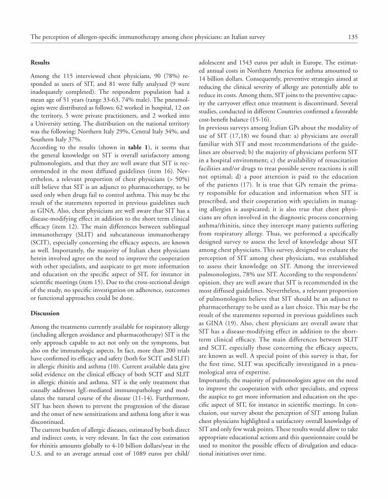
adolescent and 1543 euros per adult in Europe. The estimat-ed annual costs in Northern America for asthma amounted to 14 billion dollars. Consequently, preventive strategies aimed at reducing the clinical severity of allergy are potentially able to reduce its costs. Among them, SIT joins to the preventive capac-ity the carryover effect once treatment is discontinued. Several studies, conducted in different Countries confirmed a favorable cost-benefit balance (15-16). In previous surveys among Italian GPs about the modality of use of SIT (17,18) we found that: a) physicians are overall familiar with SIT and most recommendations of the guide-lines are observed; b) the majority of physicians perform SIT in a hospital environment; c) the availability of resuscitation facilities and/or drugs to treat possible severe reactions is still not optimal; d) a poor attention is paid to the education of the patients (17). It is true that GPs remain the prima-ry responsible for education and information when SIT is prescribed, and their cooperation with specialists in manag-ing allergies is auspicated; it is also true that chest physi-cians are often involved in the diagnostic process concerning asthma/rhinitis, since they intercept many patients suffering from respiratory allergy. Thus, we performed a specifically designed survey to assess the level of knowledge about SIT among chest physicians. This survey, designed to evaluate the perception of SIT among chest physicians, was established to assess their knowledge on SIT. Among the interviewed pulmonologists, 78% use SIT. According to the respondents’ opinion, they are well aware that SIT is recommended in the most diffused guidelines. Nevertheless, a relevant proportion of pulmonologists believe that SIT should be an adjunct to pharmacotherapy to be used as a last choice. This may be the result of the statements reported in previous guidelines such as GINA (19). Also, chest physicians are overall aware that SIT has a disease-modifying effect in addition to the short-term clinical efficacy. The main differences between SLIT and SCIT, especially those concerning the efficacy aspects, are known as well. A special point of this survey is that, for the first time, SLIT was specifically investigated in a pneu-mological area of expertise.Importantly, the majority of pulmonologists agree on the need to improve the cooperation with other specialists, and express the auspice to get more information and education on the spe-cific aspect of SIT, for instance in scientific meetings. In con-clusion, our survey about the perception of SIT among Italian chest physicians highlighted a satisfactory overall knowledge of SIT and only few weak points. These results would allow to take appropriate educational actions and this questionnaire could be used to monitor the possible effects of divulgation and educa-tional initiatives over time.
Results
Among the 115 interviewed chest physicians, 90 (78%) re-sponded as users of SIT, and 81 were fully analyzed (9 were inadequately completed). The respondent population had a mean age of 51 years (range 33-63, 74% male). The pneumol-ogists were distributed as follows: 62 worked in hospital, 12 on the territory, 5 were private practitioners, and 2 worked into a University setting. The distribution on the national territory was the following: Northern Italy 29%, Central Italy 34%, and Southern Italy 37%.According to the results (shown in table 1), it seems that the general knowledge on SIT is overall satisfactory among pulmonologists, and that they are well aware that SIT is rec-ommended in the most diffused guidelines (item 16). Nev-ertheless, a relevant proportion of chest physicians (> 50%) still believe that SIT is an adjunct to pharmacotherapy, to be used only when drugs fail to control asthma. This may be the result of the statements reported in previous guidelines such as GINA. Also, chest physicians are well aware that SIT has a disease-modifying effect in addition to the short term clinical efficacy (item 12). The main differences between sublingual immunotherapy (SLIT) and subcutaneous immunotherapy (SCIT), especially concerning the efficacy aspects, are known as well. Importantly, the majority of Italian chest physicians herein involved agree on the need to improve the cooperation with other specialists, and auspicate to get more information and education on the specific aspect of SIT, for instance in scientific meetings (item 15). Due to the cross-sectional design of the study, no specific investigation on adherence, outcomes or functional approaches could be done.
Discussion
Among the treatments currently available for respiratory allergy (including allergen avoidance and pharmacotherapy) SIT is the only approach capable to act not only on the symptoms, but also on the immunologic aspects. In fact, more than 200 trials have confirmed its efficacy and safety (both for SCIT and SLIT) in allergic rhinitis and asthma (10). Current available data give solid evidence on the clinical efficacy of both SCIT and SLIT in allergic rhinitis and asthma. SIT is the only treatment that causally addresses IgE-mediated immunopathology and mod-ulates the natural course of the disease (11-14). Furthermore, SIT has been shown to prevent the progression of the disease and the onset of new sensitizations and asthma long after it was discontinued. The current burden of allergic diseases, estimated by both direct and indirect costs, is very relevant. In fact the cost estimation for rhinitis amounts globally to 4-10 billion dollars/year in the U.S. and to an average annual cost of 1089 euros per child/
136 C. Lombardi, G.W. Canonica, G. Passalacqua
10. Passalacqua G, Durham SR; Global Allergy and Asthma European Network: Allergic rhinitis and its impact on asthma update: allergen immunotherapy. J Allergy Clin Immunol. 2007;119(4):881-891.
11. Di Rienzo V, Canonica GW, Passalacqua G. Long-lasting effect of sublingual immunotherapy in children with asthma due to house dust mite: a 10 year prospective study. Clin Exp Allergy. 2003;33:206-10.
12. Jacobsen L, Niggermann B, Dreborg S and the PAT Investigator Group. Specific immunotherapy has long-term preventive effect on seasonal and perennial asthma: 10-year follow-up on the PAT study. Allergy. 2007;62:943-8.
13. Novembre E, Galli E, Landi F, Caffarelli C, Pifferi M, De Marco E, Burastero SE, Calori G, Benetti L, Bonazza P, Puccinelli P, Parmiani S, Bernardini R, Vierucci A. Coseasonal sublingual immunother-apy reduces the development of asthma in children with allergic rhinoconjunctivitis. J Allergy Clin Immunol. 2004;114(4):851-7.
14. Tripodi S, Di Rienzo Businco A, Benincori N, Scala G, Pingitore G. Safety and tolerability of ultra-rush induction, less than one hour, of sublingual immunotherapy in children. Int Arch Allergy Immunol. 2005;139(2):149-52.
15. Berto P, Passalacqua G, Crimi N, Frati F, Ortolani C, Senna G, Ca-nonica GW. Economic evaluation of sublingual immunotherapy vs symptomatic treatment in adults with pollen-induced respiratory allergy: the Sublingual Immunotherapy Pollen Allergy Italy (SPAI) study. Ann Allergy Asthma Immunol. 2006;97:615-21.
16. Berto P, Frati F, Incorvaia C. Economic studies of immunotherapy: a review. Curr Opin Allergy Clin Immunol. 2008;8:585-9.
17. Lombardi C, Bettoncelli G, Passalacqua G, Canonica GW. The perception of allergen-specific immunotherapy among general practitioners. Eur Ann Allergy Clin Immunol, in press.
18. Lombardi C, Passalacqua G. Specific Immunotherapy among Ital-ian specialists. Allergy. 2006;61:898-9.
19. Global Initiative for Asthma. GINA Guidelines, last accessed Dec 2012. www.ginasthma.org
References
1. Asher MI, Montefort S, Björkstén B, Lai CK, Strachan DP, Wei-land SK, Williams H. ISAAC Phase Three Study Group. World-wide time trends in the prevalence of symptoms of asthma, allergic rhinoconjunctivitis, and eczema in childhood: ISAAC Phases One and Three repeat multicountry cross-sectional surveys. Lancet. 2006;368(9537):733-43.
2. Bousquet J, Khaltaev N, Cruz AA, et al; World Health Organi-zation; GA(2)LEN; AllerGen. Allergic Rhinitis and its Impact on Asthma (ARIA). 2008 update (in collaboration with the World Health Organization, GA(2)LEN and AllerGen). Allergy. 2008;63Suppl86:8-160.
3. Pawankar R, Canonica GW, Holgate ST, Lockey RF. World Aller-gy Organization White Book on Allergy. 2011.
4. Passalacqua G. Specific immunotherapy: beyond the clinical scores. Ann Allergy Asthma Immunol. 2011;107:401-6.
5. Durham SR, Emminger W, Kapp A, de Monchy JG, Rak S, Scadding GK et al. SQ-standardized sublingual grass immu-notherapy: confirmation of disease modification 2 years after 3 years of treatment in a randomized trial. J Allergy Clin Immunol. 2012;129:717-725.
6. Marogna M, Spadolini I, Massolo A, Canonica GW, Passalacqua G. Long-lasting effects of sublingual immunotherapy according to its duration: a 15-year prospective study. J Allergy Clin Immunol. 2010;126:969-75.
7. Canonica GW, Baena-Cagnani CE, Compalati E, Bohle B, Bonini S, Bousquet J et al. 100 Years of Immunotherapy: The Monaco Charter. Under the High Patronage of His Serene Highness Prince Albert II of Monaco. Int Arch Allergy Immunol. 2012;160:346-9.
8. Calderon M, Alves B, Jacobson M, Hurwitz B, Sheikh A, Durham S. Allergen injection immunotherapy for seasonal allergic rhinitis. Cochrane Database Syst Rev. 2007;(1):CD001936.
9. Canonica GW, Bousquet J, Casale T, et al. Sub-lingual immu-notherapy: World Allergy Organization Position Paper Allergy. 2009Dec;64 Suppl91:1-59.
O R I G I N A L A R T I C L E S Eur Ann AllErgy Clin immunol
SummaryBackground. Tetranychus urticae is a phytophagus mite found in the leaves of numerous plants. High sensitization rates have been demonstrated, however, provocation tests have only been performed in an occupational setting. Objective. To assess accuracy of skin prick tests and clinical relevance of T. urticae sensitization by means of conjunctival provocation tests (CPT) in a population without occupational exposure and to evaluate possible environmental risk factors for T. urticae allergy. Methods. Patients ≥ 18 years old sensitized to T. urticae (n = 12) and a non-sensitized control group (n = 12) were invited to perform CPT with T. urticae and fulfill a questionnaire including demographic data, questions on environmental exposure to T. urticae and allergy symptoms/diagnosis. A single-blinded placebo-controlled CPT with T. urticae (Leti®) was performed with increasing concentrations (0.002, 0.02, 0.2 and 2 mg/mL) and considered positive if conjunctival hyperemia, palpebral edema or lacri-mation were observed in the tested eye. Results. Of T. urticae sensitized patients (mean wheal 4.4±1.5 mm), 9 had a positive CPT, including 3 monosensitized. A good diagnostic accuracy was found for skin prick tests: AUC = 0.952, sensitivity = 100%, specificity = 80%, positive likelihood ratio = 5 and negative likelihood ratio = 0 for a 3 mm wheal. No differences were found between allergic and non-allergic subjects regarding atopy, allergic disease or farming activities. Conclusions. A high prevalence of allergy to Tetranychus urticae was found in the north of Portugal. Future studies with a larger number of patients are needed to evaluate its relation to clinical symptoms and the impact of environmental factors.
Corresponding authorNatacha [email protected]ço de Imunoalergologia Centro Hospitalar São João, E.P.E.Alameda Prof. Hernâni Monteiro 4300 Porto, Portugal
Key words
Tetranychus urticae; spider mite; conjunctival provocation test; allergy
1Serviço de Imunoalergologia, Centro Hospitalar São João, E.P.E., Porto, Portugal2Departamento de Investigación y Desarrollo, Unidad de Alergia, Laboratorios Leti
Tetranychus urticae allergy in a population without occupational exposure
n. santos1, V. iraoLa2, J.L. PLáciDo1
Introduction
Tetranychus urticae, commonly known as red spider mite or two-spotted spider mite, is a macroscopic phytophagus mite of the Acari subclass and Trombidiformes order, found in the leaves of numerous plants and an important plague in agricul-ture, especially in association with sulfur pesticides utilization, to which it is specially resistant (1). A high sensitization rate to this mite has been shown both in occupational (2) and non-occupational exposure (3), and both
species specific and cross-reactive allergens with house dust mites have been identified (4,5). In Portugal, predominantly in the North of the country, viti-culture, farming and greenhouses are important economic ac-tivities, and we have previously reported that almost 40% of patients followed in the Immunoallergy Department of our University Hospital were sensitized to T. urticae (6). However, the clinical relevance of this sensitization was not assessed. Allergy to T. urticae has been associated with symptoms of rhini-tis in the summer and autumn, asthma, contact dermatitis and
VoL 46, n 4, 137-141, 2014

138 N. Santos, V. Iraola, J.L. Plácido
E.P.E., consisting of atopic and non-atopic subjects not sensi-tized to T. urticae was also recruited.24 patients were included, 20 (83.3%) female, mean (SD) age 32.8 (10.9) years old, ranging from 19 to 54 years old.This study was approved by the ethics committee of Centro Hospitalar São João, E.P.E. Written informed consent was ob-tained from all subjects included.
Skin prick tests
Skin prick tests were performed with an extract of T. urticae bodies (Leti©) 2 mg/mL, histamine 10 mg/mL and a saline solution as negative control. The procedure was performed by the investigator. Extracts were applied in the volar surface of the forearm and pricked with an allergen prick lancet (Heinz Herenz Medizinalbedarf GmbH©), according to international recommendations (11). The mean wheal size was measured and registered. Subjects were considered sensitized if a mean wheal diameter ≥ 3mm for T. urticae was observed after 15 minutes.Other sensitizations, as for house dust mites (Dermatophagoides pteronyssinus and D. farinae), storage mites (Lepidoglyphus de-structor, Tyrophagus putrescentiae and Acarus siro), cat and dog dander, pollen (Platanus acerifola, Betula verrucosa, Olea europea, grass mix, Parietaria judaica, Plantago lanceolata and Artemisia vulgaris) and fungi (Alternaria, Cladiosporum, Aspergillus and Penicillium) were evaluated according to the results obtained in our previous study (6), using the same methodology.
Conjunctival provocation test (CPT)
A single-blinded placebo-controlled CPT with T. urticae was performed, according to the Allergy Department’s protocol (12). Exclusion criteria were ocular infection, dry eye syndrome, autoimmune disorders and allergic conjunctivitis exacerbation. Subjects were instructed to stop any topical eye drugs 2 weeks before, oral or nasal corticosteroids 2 weeks before, oral antihis-tamines 1 week before and not to wear contact lenses on CPT day. Subjects who could not stop medication were also excluded. Four concentrations of T. urticae were prepared from the orig-inal lyophilized 2 mg/mL extract (0.002, 0.02, 0.2 and 2 mg/mL) according to manufacturer’s instructions and used in the 24 hours following preparation.One single drop of increasing concentration was applied in the conjunctival eye sac, while a control solution of diluent was ap-plied in the opposite eye. The eyes were evaluated after 10 min-utes of each concentration. The CPT was considered positive if objective signs of conjunc-tival hyperemia, palpebral edema or lacrimation were observed in the tested eye. Patients with positive tests did not proceed to the next concentration and were treated with a topical an-tihistamine (opatanol 1 mg/mL) and eventually topical corti-
urticaria (3,7,8), and was previously evaluated with bronchial (9,10), and environmental provocation (8), but not with oth-er safer and simpler target organ provocations, namely nasal or conjunctival provocation tests. Our aims were to assess accuracy of skin prick tests and clinical relevance of T. urticae sensitization by means of conjunctival provocation test (CPT) in a population without known occu-pational exposure to this mite and to evaluate possible environ-mental risk factors for T. urticae allergy.
Methods
Study design and participants
Subjects aged 18 years or older who participated in a previous study to determine sensitization frequency to T. urticae (6) and had a positive skin prick test, were eligible (figure 1).
Figure 1 - Study protocol and patients excluded.
Subjects were contacted by telephone and invited to participate in the study, consisting on a skin prick test to T. urticae to assess wheal size, a CPT and a questionnaire on allergy symptoms and risk factors for T. urticae exposure. Those to whom the phone number was no longer attributed, who did not answer the telephone after 3 attempts, who could not commute to the hospital in one of the four days set to perform the study, who were under current or previous aller-gen specific immunotherapy with mites, who were pregnant, breastfeeding or presented any contra-indication for CPT, were excluded.A control group of medical, nursing and technical personal of the Immunoallergy Department of Centro Hospitalar São João,
139Tetranychus urticae allergy in a population without occupational exposure
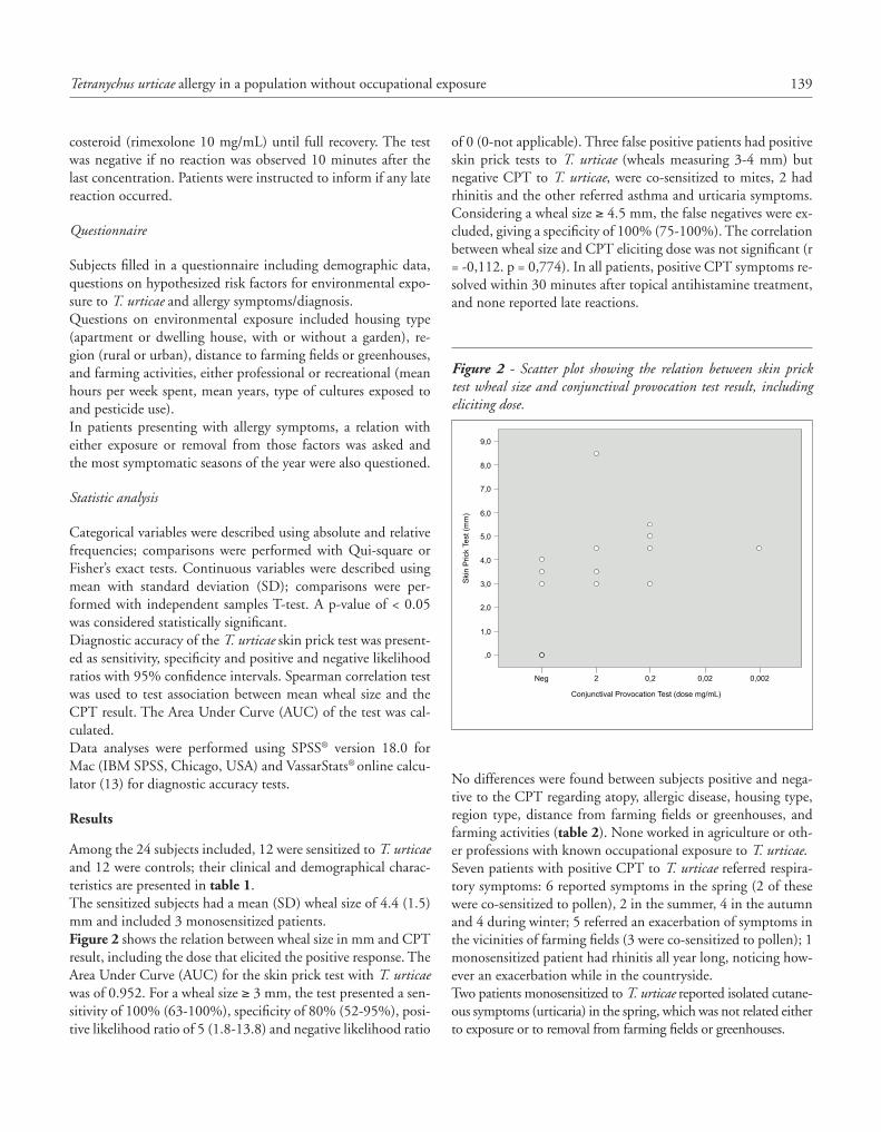
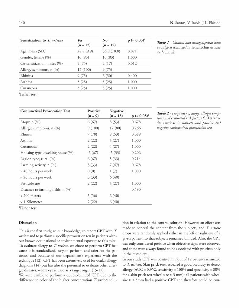
of 0 (0-not applicable). Three false positive patients had positive skin prick tests to T. urticae (wheals measuring 3-4 mm) but negative CPT to T. urticae, were co-sensitized to mites, 2 had rhinitis and the other referred asthma and urticaria symptoms. Considering a wheal size ≥ 4.5 mm, the false negatives were ex-cluded, giving a specificity of 100% (75-100%). The correlation between wheal size and CPT eliciting dose was not significant (r = -0,112. p = 0,774). In all patients, positive CPT symptoms re-solved within 30 minutes after topical antihistamine treatment, and none reported late reactions.
Figure 2 - Scatter plot showing the relation between skin prick test wheal size and conjunctival provocation test result, including eliciting dose.
Neg
,0
1,0
2,0
3,0
4,0
5,0
6,0
7,0
8,0
9,0
2 0,2 0,02 0,002
Conjunctival Provocation Test (dose mg/mL)
Ski
n P
rick
Test
(mm
)
No differences were found between subjects positive and nega-tive to the CPT regarding atopy, allergic disease, housing type, region type, distance from farming fields or greenhouses, and farming activities (table 2). None worked in agriculture or oth-er professions with known occupational exposure to T. urticae. Seven patients with positive CPT to T. urticae referred respira-tory symptoms: 6 reported symptoms in the spring (2 of these were co-sensitized to pollen), 2 in the summer, 4 in the autumn and 4 during winter; 5 referred an exacerbation of symptoms in the vicinities of farming fields (3 were co-sensitized to pollen); 1 monosensitized patient had rhinitis all year long, noticing how-ever an exacerbation while in the countryside. Two patients monosensitized to T. urticae reported isolated cutane-ous symptoms (urticaria) in the spring, which was not related either to exposure or to removal from farming fields or greenhouses.
costeroid (rimexolone 10 mg/mL) until full recovery. The test was negative if no reaction was observed 10 minutes after the last concentration. Patients were instructed to inform if any late reaction occurred.
Questionnaire
Subjects filled in a questionnaire including demographic data, questions on hypothesized risk factors for environmental expo-sure to T. urticae and allergy symptoms/diagnosis. Questions on environmental exposure included housing type (apartment or dwelling house, with or without a garden), re-gion (rural or urban), distance to farming fields or greenhouses, and farming activities, either professional or recreational (mean hours per week spent, mean years, type of cultures exposed to and pesticide use). In patients presenting with allergy symptoms, a relation with either exposure or removal from those factors was asked and the most symptomatic seasons of the year were also questioned.
Statistic analysis
Categorical variables were described using absolute and relative frequencies; comparisons were performed with Qui-square or Fisher’s exact tests. Continuous variables were described using mean with standard deviation (SD); comparisons were per-formed with independent samples T-test. A p-value of < 0.05 was considered statistically significant.Diagnostic accuracy of the T. urticae skin prick test was present-ed as sensitivity, specificity and positive and negative likelihood ratios with 95% confidence intervals. Spearman correlation test was used to test association between mean wheal size and the CPT result. The Area Under Curve (AUC) of the test was cal-culated. Data analyses were performed using SPSS® version 18.0 for Mac (IBM SPSS, Chicago, USA) and VassarStats® online calcu-lator (13) for diagnostic accuracy tests.
Results
Among the 24 subjects included, 12 were sensitized to T. urticae and 12 were controls; their clinical and demographical charac-teristics are presented in table 1. The sensitized subjects had a mean (SD) wheal size of 4.4 (1.5) mm and included 3 monosensitized patients. Figure 2 shows the relation between wheal size in mm and CPT result, including the dose that elicited the positive response. The Area Under Curve (AUC) for the skin prick test with T. urticae was of 0.952. For a wheal size ≥ 3 mm, the test presented a sen-sitivity of 100% (63-100%), specificity of 80% (52-95%), posi-tive likelihood ratio of 5 (1.8-13.8) and negative likelihood ratio
140 N. Santos, V. Iraola, J.L. Plácido
tion in relation to the control solution. However, an effort was made to conceal the content from the subjects, and T. urticae drops were randomly applied either in the left or right eye of a given patient, so that subjects remained blinded. Also, the CPT was only considered positive when objective signs were observed and these were always found to be associated with pruritus only in the tested eye.In our study CPT was positive in 9 out of 12 patients sensitized to T. urticae. Skin prick tests revealed a good accuracy to detect allergy (AUC = 0.952, sensitivity = 100% and specificity = 80% for a skin prick test wheal size ≥ 3 mm); all patients with wheal size ≥ 4.5mm had a positive CPT and therefore could be con-
Discussion
This is the first study, to our knowledge, to report CPT with T. urticae and to perform a specific provocation test in patients with-out known occupational or environmental exposure to this mite. To evaluate allergy to T. urticae, we chose to perform CPT be-cause it is standardized, easy to perform and safer for the pa-tients, and because of our department’s experience with the technique (12). CPT has been extensively used for ocular allergy diagnosis (14) but has also the potential to evaluate other aller-gic diseases, where eye is used as a target organ (15-17).We were unable to perform a double-blinded CPT due to the difference in color of the higher concentration T. urticae solu-
Sensitization to T. urticae Yes(n = 12)
No(n = 12)
p (< 0.05)1
Age, mean (SD) 28.8 (9.9) 36.8 (10.8) 0.071
Gender, female (%) 10 (83) 10 (83) 1.000
Co-sensitization, mites (%) 9 (75) 2 (17) 0.012
Allergy symptoms, n (%) 12 (100) 9 (75)
Rhinitis 9 (75) 6 (50) 0.400
Asthma 3 (25) 3 (25) 1.000
Cutaneous 3 (25) 3 (25) 1.0001Fisher test
Table 1 - Clinical and demographical data on subjects sensitized to Tetranychus urticae and controls.
Table 2 - Frequency of atopy, allergic symp-toms and evaluated risk factors for Tetrany-chus urticae in subjects with positive and negative conjunctival provocation test.
Conjunctival Provocation Test Positive(n = 9)
Negative(n = 15) p (< 0.05)1
Atopy, n (%) 6 (67) 8 (53) 0.678
Allergic symptoms, n (%) 9 (100) 12 (80) 0.266
Rhinits 7 (78) 8 (53) 0.389
Asthma 2 (22) 4 (27) 1.000
Cutaneous 2 (22) 4 (27) 1.000
Housing type, dwelling house (%) 6 (67) 5 (33) 0.206
Region type, rural (%) 6 (67) 5 (33) 0.214
Farming activity, n (%) 3 (33) 7 (47) 0.678
> 40 hours per week 0 (0) 1 (7) 1.000
< 20 hours per week 3 (33) 6 (40)
Pesticide use 2 (22) 4 (27) 1.000
Distance to farming fields, n (%) 0.590
< 200 meters 5 (56) 6 (40)
> 1 Kilometer 2 (22) 6 (40)1Fisher test

141Tetranychus urticae allergy in a population without occupational exposure
2. Navarro AM, Delgado J, Sanchez MC, Orta JC, Martinez A, Pala-cios R, et al. Prevalence of sensitization to Tetranychus urticae in greenhouse workers. Clin Exp Allergy. 2000;30:863-6.
3. Millán C, Iraola V, Pinto H, Carnés J, Fatou R, Reguera V. Sensibi-lización a araña roja Tetranychus urticae en pacientes sin exposición ocupacional. J Invest Allergol Clin Immunol. 2008;18Suppl3:179.
4. Park HS, Jee YK, Kim YK, Lee SK, Lee MH, Kim YY. Identifica-tion of immunoglobulin E binding components of the two-spot-ted spider mite Tetranychus urticae: allergenic relationships with the citrus red mite and house-dust mite. Allergy Asthma Proc. 2002;23:199-204.
5. Kim YK, Oh SY, Jung JW, Min KU, Kim YY, Cho SH. IgE bind-ing components in Tetranychus urticae and Panonychus ulmi-de-rived crude extracts and their cross-reactivity with domestic mites. Clin Exp Allergy. 2001;31:1457-63.
6. Santos N, Plácido JL. Sensitization to Tetranychus urticae in the North of Portugal. Rev Port Imunoalergologia (in press).
7. Astarita C, Di Martino P, Scala G, Franzese A, Sproviero S. Con-tact allergy: another occupational risk to Tetranychus urticae. J Al-lergy Clin Immunol. 1996;98:732-8.
8. Astarita C, Gargano D, Manguso F, Romano C, Montanaro D, Pezzuto F, et al. Epidemiology of allergic occupational diseases in-duced by Tetranychus urticae in greenhouse and open-field farmers living in a temperate climate area. Allergy. 2001;56:1157-63.
9. Delgado J, Orta JC, Navarro AM, Conde J, Martinez A, Martinez J, et al. Occupational allergy in greenhouse workers: sensitization to Tetranychus urticae. Clin Exp Allergy. 1997;27:640-5.
10. Jee YK, Park HS, Kim HY, Park JS, Lee KY, Kim KY, et al. Two-spotted spider mite (Tetranychus urticae): an important al-lergen in asthmatic non-farmers symtomatic in summer and fall months. Ann Allergy Asthma Immunol. 2000;84:543-8.
11. Bousquet J, Heinzerling L, Bachert C, Papadopoulos NG, Bousquet PJ, Burney PG, et al. Practical guide to skin prick tests in allergy to aeroallergens. Allergy. 2012;67:18-24.
12. Couto M, Silva D, Moreira A, Plácido JL. Utility and safety of conjunctival challenges with Dermatophagoides pteronyssinus: the experience of a center. Allergy. 2012;67:482.
13. Lowry, R. “Diagnostic accuracy tests”. Retrieved February 17th, 2013, from www.vassarstats.net/clin1.html
14. 14. Mortemousque B, Fauquert JL, Chiambaretta F, Demoly P, Helleboid L, Creuzot-Garcher C, et al. [Conjunctival provocation test: recommendations]. J Fr Ophtalmol. 2006; 29:837-46.
15. Tabar AI, Anda M, Bonifazi F, Bilo MB, Leynadier F, Fuchs T, et al. Specific immunotherapy with standardized latex extract versus placebo in latex-allergic patients. International Archives of Allergy and Immunology. 2006;141:369-76.
16. Kopac P, Rudin M, Gentinetta T, Gerber R, Pichler C, Hausmann O, et al. Continuous apple consumption induces oral tolerance in birch-pollen-associated apple allergy. Allergy. 2012;67:280-5.
17. Olaguibel JM, Tabar AI, Garcia Figueroa BE, Cortes C. Immu-notherapy with standardized extract of Dermatophagoides pteron-yssinus in bronchial asthma: a dose-titration study. Allergy. 1997; 52:168-78.
18. Kim DS, LEE JH. Oviposition model of overwintered adult Tetranychus urticae (Acari: Tetranychidae) and mite phenol-ogy on the ground cover in apple orchards. Entomol Exp Appl. 2003;31:191-208.
sidered allergic without performing a specific provocation test. In relation to other studies where a provocation test was per-formed, Astarita et al. (8) evaluated T. urticae allergy through a single-blinded specific exposure test, with symptoms and peak expiratory flow rate monitorization, in a pesticide-free oleander greenhouse specifically infested by T. urticae; it was positive in all 28 farmers sensitized to T. urticae who reported occupation-al symptoms, inducing the referred respiratory or cutaneous symptoms until 1 hour after exposure. Delgado et al. (9) and Jee et al. (10) evaluated suspected T. urticae induced asthma with a specific bronchial provocation test, which was positive in 12 out of 13 carnation greenhouse workers and 10 out of 16 asthmat-ics living near pear orchards, respectively. Almost half of them presented a late asthmatic response. Most of our T. urticae allergic patients, even without pol-len sensitization, reported nasal symptoms in the spring. Al-though T. urticae levels in different cultures peak between July and August and that Astarita et al. (8) reported that recurrent summer-autumn occupational rhinitis showed the best clinical correlation with T. urticae in Italian farmers, it has been described that T. urticae populations increase in spring in adventitious plants and ground cover vegetation, moving to the cultures in summer (18), which might explain the occurrence of spring symptoms even in patients without pollen sensitization.Regarding housing type, region type, distance to farming fields or greenhouses, and farming activities, no differences were found between those with and without T. urticae allergy. How-ever, we point out that allergy symptoms were self-reported and retrospective, and that assessed risk factors were not confirmed through objective measures of T. urticae exposure. To conclude, a high prevalence of sensitization and conjuncti-val provocation test confirmed allergy to Tetranychus urticae was found in a non-occupational population from the North of Por-tugal. Future studies with a larger number of patients are needed to evaluate its relation to clinical symptoms and the impact of environmental factors.
The data in this article has been briefly presented in poster form in the EAACI-WAO Congress 2013: Santos N, Plácido JL. Sensitization to Tetranychus urticae: prevalence and clinical relevance. Allergy 2013; 68 (Suppl.97):463.
Funding sources: Tetranychus urticae extract for skin prick tests and conjunctival porvocation tests have been provided by Leti®.
References
1. Grbic M, Van Leeuwen T, Clark RM, Rombauts S, Rouze P, Grbic V, et al. The genome of Tetranychus urticae reveals herbivorous pest adaptations. Nature. 2011;479:487-92.
O R I G I N A L A R T I C L E S Eur Ann AllErgy Clin immunol VoL 46, n 4, 142-146, 2014
Introduction
Lipid transfer protein (LTP), the major food allergen in the Mediterranean basin, is widely distributed throughout the plant kingdom. LTPs from distinct plant-derived foods show a moderate to high degree of cross-reactivity, due to a sequence homology ranging between 35% and 95% (1). Most allergists agree that the peach is the primary source of sensitization to this protein, and dominates the immune response to this allergen (2,3), although a possible influence by sensitization to LTP present in some pollens, particularly
those of plane tree and mugwort has been suggested (3-5). The clinical spectrum of LTP allergy ranges from patients allergic uniquely to the peach to patients allergic to a large array of fruits and vegetables. Previous studies found that the level of IgE specific for peach LTP, Pru p 3, is critical for the occurrence of cross-reactivity to botanically related (Ro-saceae) and unrelated plant foods (6,7), although subsequent investigations were unable to detect sharp cutoff levels able to discriminate allergic patients from subjects with clinically irrelevant sensitization (8). However, it has to be considered that the reactivity of the patients varies with the amount of
SummaryBackground. Lipid transfer protein (LTP) is a widely cross-reacting allergen in plant foods. Objective. To assess whether IgE to vegetable foods show predictable trends in LTP allergic patients. Methods. Clinical allergy to plant foods other than peach was sought in 15 con-secutive peach-allergic adults monosensitized to LTP. IgE specific for peach, apple, hazelnut, walnut, peanut, lentil, maize, soybean, tomato, sesame, mustard melon, kiwi, and celery as well as to mugwort pollen was measured. Results. Peach-specific IgE levels exceeded IgE to all other study foods. The higher were peach-specific IgE levels, the higher was the probability that other plant-derived foods scored positive. Mean IgE levels specific for all study foods were strongly correlated to peach specific IgE. Food-specific IgE followed a rather precise hierarchy, both in terms of number of positive in-vitro tests and of IgE levels, with apple at the second place after peach, followed by walnut, hazelnut, peanut, lentil, maize, soybean, tomato, kiwi, sesame, mustard, melon, and celery. Such hierarchy was not necessarily paralleled by clinical allergy as lentil, maize, and soybean scored positive in the majority of patients, but induced allergy in 0, 1, and 0 patients, respectively. IgE levels were not necessarily correlated with the severity of clinical allergy. Little or no IgE reactivity to mugwort pollen was found. Conclu-sion. In LTP syndrome, IgE reactivity to foods other than peach is in most cases predictable and follows a regular sequence that probably depends on the degree of homology with Pru p 3. The reasons why some foods are tolerated by most patients despite elevated IgE reactivity remains to be elucidated.
Corresponding authorRiccardo AseroAmbulatorio di AllergologiaClinica San CarloVia Ospedale 2120037 Paderno Dugnano (MI), ItalyPhone: +39 02 990 38 470 Fax: +39 02 990 38 223 E-mail: [email protected]
Key words
Food allergy; lipid transfer proteins; specific IgE
Ambulatorio di Allergologia, Clinica San Carlo, Paderno Dugnano (MI), Italy
In patients with LTP syndrome food-specific IgE show a predictable hierarchical order
r. asero
143In patients with LTP syndrome food-specific IgE show a predictable hierarchical order
using Pearson’s coefficient method. A probability value < 5% was considered statistically significant.Since the study was retrospective and based on routine investi-gations performed on patients spontaneously presenting at the clinic for suspect food allergy, no institutional ethical permis-sion was needed.
Results
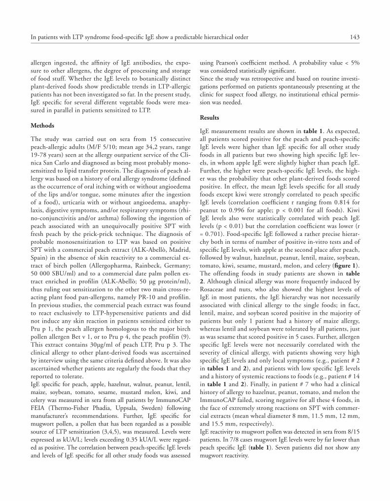
IgE measurement results are shown in table 1. As expected, all patients scored positive for the peach and peach-specific IgE levels were higher than IgE specific for all other study foods in all patients but two showing high specific IgE lev-els, in whom apple IgE were slightly higher than peach IgE. Further, the higher were peach-specific IgE levels, the high-er was the probability that other plant-derived foods scored positive. In effect, the mean IgE levels specific for all study foods except kiwi were strongly correlated to peach specific IgE levels (correlation coefficient r ranging from 0.814 for peanut to 0.996 for apple; p < 0.001 for all foods). Kiwi IgE levels also were statistically correlated with peach IgE levels (p < 0.01) but the correlation coefficient was lower (r = 0.701). Food-specific IgE followed a rather precise hierar-chy both in terms of number of positive in-vitro tests and of specific IgE levels, with apple at the second place after peach, followed by walnut, hazelnut, peanut, lentil, maize, soybean, tomato, kiwi, sesame, mustard, melon, and celery (figure 1). The offending foods in study patients are shown in table 2. Although clinical allergy was more frequently induced by Rosaceae and nuts, who also showed the highest levels of IgE in most patients, the IgE hierarchy was not necessarily associated with clinical allergy to the single foods; in fact, lentil, maize, and soybean scored positive in the majority of patients but only 1 patient had a history of maize allergy, whereas lentil and soybean were tolerated by all patients, just as was sesame that scored positive in 5 cases. Further, allergen specific IgE levels were not necessarily correlated with the severity of clinical allergy, with patients showing very high specific IgE levels and only local symptoms (e.g., patient # 2 in tables 1 and 2), and patients with low specific IgE levels and a history of systemic reactions to foods (e.g., patient # 14 in table 1 and 2). Finally, in patient # 7 who had a clinical history of allergy to hazelnut, peanut, tomato, and melon the ImmunoCAP failed, scoring negative for all these 4 foods, in the face of extremely strong reactions on SPT with commer-cial extracts (mean wheal diameter 8 mm, 11.5 mm, 12 mm, and 15.5 mm, respectively).IgE reactivity to mugwort pollen was detected in sera from 8/15 patients. In 7/8 cases mugwort IgE levels were by far lower than peach specific IgE (table 1). Seven patients did not show any mugwort reactivity.
allergen ingested, the affinity of IgE antibodies, the expo-sure to other allergens, the degree of processing and storage of food stuff. Whether the IgE levels to botanically distinct plant-derived foods show predictable trends in LTP-allergic patients has not been investigated so far. In the present study, IgE specific for several different vegetable foods were mea-sured in parallel in patients sensitized to LTP.
Methods
The study was carried out on sera from 15 consecutive peach-allergic adults (M/F 5/10; mean age 34,2 years, range 19-78 years) seen at the allergy outpatient service of the Cli-nica San Carlo and diagnosed as being most probably mono-sensitized to lipid transfer protein. The diagnosis of peach al-lergy was based on a history of oral allergy syndrome (defined as the occurrence of oral itching with or without angioedema of the lips and/or tongue, some minutes after the ingestion of a food), urticaria with or without angioedema, anaphy-laxis, digestive symptoms, and/or respiratory symptoms (rhi-no-conjunctivitis and/or asthma) following the ingestion of peach associated with an unequivocally positive SPT with fresh peach by the prick-prick technique. The diagnosis of probable monosensitization to LTP was based on positive SPT with a commercial peach extract (ALK-Abellò, Madrid, Spain) in the absence of skin reactivity to a commercial ex-tract of birch pollen (Allergopharma, Rainbeck, Germany; 50 000 SBU/ml) and to a commercial date palm pollen ex-tract enriched in profilin (ALK-Abellò; 50 µg protein/ml), thus ruling out sensitization to the other two main cross-re-acting plant food pan-allergens, namely PR-10 and profilin. In previous studies, the commercial peach extract was found to react exclusively to LTP-hypersensitive patients and did not induce any skin reaction in patients sensitized either to Pru p 1, the peach allergen homologous to the major birch pollen allergen Bet v 1, or to Pru p 4, the peach profilin (9). This extract contains 30µg/ml of peach LTP, Pru p 3. The clinical allergy to other plant-derived foods was ascertained by interview using the same criteria defined above. It was also ascertained whether patients ate regularly the foods that they reported to tolerate. IgE specific for peach, apple, hazelnut, walnut, peanut, lentil, maize, soybean, tomato, sesame, mustard melon, kiwi, and celery was measured in sera from all patients by ImmunoCAP FEIA (Thermo-Fisher Phadia, Uppsala, Sweden) following manufacturer’s recommendations. Further, IgE specific for mugwort pollen, a pollen that has been regarded as a possible source of LTP sensitization (3,4,5), was measured. Levels were expressed as kUA/L; levels exceeding 0.35 kUA/L were regard-ed as positive. The correlation between peach-specific IgE levels and levels of IgE specific for all other study foods was assessed

144 R. Asero
Table 1 - Level of IgE to the study foods measured in 15 patients monosensitized to LTP.
Patients
Peach
Apple
Walnut
Hazelnut
Peanut
Lentil
Maize
Soybean
Tomato
Kiw
i
Sesame
Mustard
Melon
Celery
Mugw
ort pollen
1 58,1 59,2 43,3 7,66 20,9 23 21,9 12,9 7,37 1,92 7,21 2,54 1,07 2 0,61
2 16,6 12 11,1 1,17 0,65 0,82 0 0,39 0 1,18 0 0 0 0 1,03
3 12,1 9,1 4,93 1,12 1,9 0,83 0,79 0,58 0,47 0,68 0 0,49 0 0 1
4 11,6 9,25 4,52 3,05 3,75 2,94 2,23 1,37 1,1 1,1 0 0,42 0 0 1,77
5 11,4 11,9 6,61 6,29 2,05 1,77 3,85 0,75 0,67 2,01 0,71 0 0 0,36 1,81
6 7,04 5,34 4,39 1,86 2,14 0,61 3,92 1,14 2,8 0,76 0,67 0 0 1,08 0
7 4,58 2,57 2,05 0 0 0,37 1,53 0 0 0,41 0 0 0 0 1,05
8 3,81 2,33 1,32 0,49 1,86 1,18 1,73 0,79 1,02 0 0,55 0 0 0 0
9 3,12 1,44 0,38 0 0 0 0 0 0 0 0 0 0 0 0,74
10 2,75 1,92 1,59 1,18 0,86 0,75 0,43 0,47 0 0,74 0,56 0 0 0 0
11 1,75 0,74 0 0 0 0 0 0 0 0 0 0 0 0 0
12 1,44 0,77 0 0 0 0,42 0 0 0 0 0 0 0 0 0
13 1,11 0,84 0 0 0 0 0 0 0 0,83 0 0 0 0 0
14 1,08 0,39 0 0 0 0 0 0 0 0 0 0 0 0 0
15 0,41 0 0 0 0 0 0 0 0 0 0 0 0 0 0,61
Specific IgE values are expressed as kU/L (positive if > 0.35 kU/l). Patients have been ordered on the basis of peach-specific IgE levels.
Figure 1 - Specific IgE levels in patients 2-15. Numbers under the horizontal line indicate the study foods (1: Peach; 2: Apple; 3: Walnut; 4: Hazelnut; 5: Peanut; 6: Lentil; 7: Maize; 8: Soybean; 9: Tomato; 10: Kiwi; 11: Sesame: 12: Mustard; 13: Melon; 14: Celery). Num-bers along the vertical line are specific IgE levels in Ku/l. Patient # 1 data were exclud-ed due to the excess of specific IgE vs all other patients (see table 1).
145In patients with LTP syndrome food-specific IgE show a predictable hierarchical order
ing reduces somehow the allergenicity of this protein in these foods. Both lentil and soybean belong to the Leguminosae fam-ily, and the good tolerance to legumes by LTP-allergic subjects was already observed several years ago (10). Further, some data suggest that LTP-containing foods may be more harmful if they are ingested alone (11). Finally, although LTP is known as an ex-tremely heat-stable protein (12,13), some data seem to suggest that prolonged heat-treatments at high temperatures may sig-nificantly reduce its allergenicity (14). The case of kiwi is some-how special. Recent studies showed that kiwi-allergic patients living in southern Europe are mostly sensitized to profilin or to Act d 10, the kiwi lipid transfer proteins (15). In this study 9/15 patients showed IgE to kiwi, but only 3 patients reported aller-gic reaction to this fruit and, notably, one of them did not show any IgE reactivity to kiwi both on ImmunoCAP and on SPT, whereas in the remaining 2 specific IgE levels were low. Further, kiwi-specific IgE levels showed a significantly lower correlation with peach-specific IgE than all other study foods, suggesting that either patients were sensitized also to kiwi allergens other Act d 10, or that kiwi LTP shows a rather low homology to Pru p 3 causing a trend in specific IgE that differs from that of all
Discussion
With the obvious limitations of being retrospective and based on a limited number of patients due to cost problems, this study shows that in patients with lipid transfer protein aller-gy syndrome, specific IgE levels follow a rather precise hierar-chical order. Although this could theoretically be related to the amount of LTP present in the various extract for immunoassay, the possibility that it reflects the degree of homology between the proteins from different botanical sources and the LTP from the primary sensitizer, the peach, seems a more likely explana-tion. As observed before (3), the higher was the level of IgE to peach LTP, the higher was the likelihood of cross-sensitization to a large number of botanically unrelated plant-derived foods. However, hypersensitivity did not always reflect into clinical cross-reactivity; in fact, for several foods including lentil, soy-bean, maize and sesame the large majority of patients did not report any clinical reaction despite specific IgE levels that were frequently quite high. This might suggest either that in these foods the protein is less abundant than in the peach, or that it is absorbed more slowly in the gut, or that the thermal process-
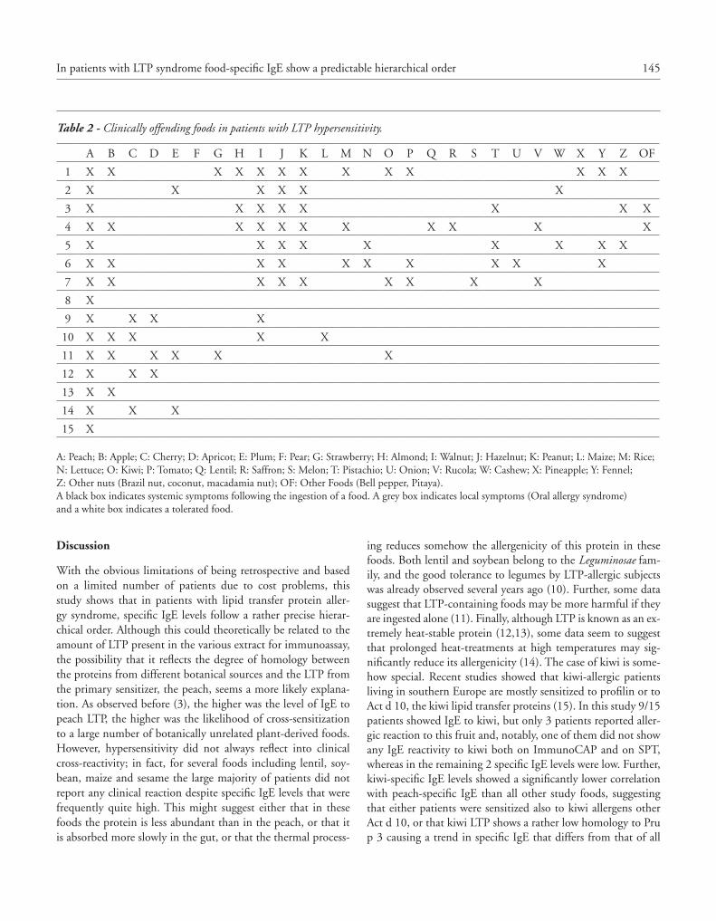
Table 2 - Clinically offending foods in patients with LTP hypersensitivity.
A B C D E F G H I J K L M N O P Q R S T U V W X Y Z OF
1 X X X X X X X X X X X X X
2 X X X X X X
3 X X X X X X X X
4 X X X X X X X X X X X
5 X X X X X X X X X
6 X X X X X X X X X X
7 X X X X X X X X X
8 X
9 X X X X
10 X X X X X
11 X X X X X X
12 X X X
13 X X
14 X X X
15 X
A: Peach; B: Apple; C: Cherry; D: Apricot; E: Plum; F: Pear; G: Strawberry; H: Almond; I: Walnut; J: Hazelnut; K: Peanut; L: Maize; M: Rice; N: Lettuce; O: Kiwi; P: Tomato; Q: Lentil; R: Saffron; S: Melon; T: Pistachio; U: Onion; V: Rucola; W: Cashew; X: Pineapple; Y: Fennel; Z: Other nuts (Brazil nut, coconut, macadamia nut); OF: Other Foods (Bell pepper, Pitaya).A black box indicates systemic symptoms following the ingestion of a food. A grey box indicates local symptoms (Oral allergy syndrome) and a white box indicates a tolerated food.
146 R. Asero
6. Asero R, Arena A, Cecchi L, et al. Are IgE levels to foods other than Rosaceae predictive of allergy in lipid transfer protein-hypersensi-tive patients? Int Arch Allergy Immunol. 2011;155:149-54.
7. Garcia-Selles FJ, Díaz-Perales A, Sánchez-Monge R, Alcántara M, Lombardero M, Barber D, Salcedo G, Fernández-Rivas M. Pattern of reactivity to lipid transfer proteins of plant foods and Artemisia pollen: an in-vivo study. Int Arch Allergy Immunol. 2002Jun;128(2):115-22.
8. Lombardero M, García-Sellés FJ, Polo F, Jimeno L, Chamorro MJ, García-Casado G, Sánchez-Monge R, Díaz-Perales A, Salcedo G, Barber D. Prevalence of sensitization to Artemisia allergens Art v 1, Art v 3 and Art v 60 kDa. Cross-reactivity among Art v 3 and other relevant lipid-transfer protein allergens. Clin Exp Allergy. 2004Sep;34(9):1415-21.
9. Asero R. Plant food allergies: a suggested approach to allergen-re-solved diagnosis in the clinical practice by identifying easily available sensitization markers. Int Arch Allergy Immunol. 2005;138:1-11.
10. Asero R, Mistrello G, Roncarolo D, Amato S, Falagiani P. Why do lipid transfer protein-hypersensitive patients tolerate bean (and other legumes)? Int Arch Allergy Immunol. 2005;137:236-40.
11. Arena A. Anaphylaxis to apple: is fasting a risk factor for LTP-al-lergic patients? Eur Ann Allergy Clin Immunol. 2010;42:155-8.
12. Pastorello EA, Pompei C, Pravettoni V, Farioli L, Calamari AM, Scibilia J, Robino AM, Conti A, Iametti S, Fortunato D, Bonomi S, Ortolani C. Lipid-transfer protein is the major maize allergen maintaining IgE-binding activity after cooking at 100 degrees C, as demonstrated in anaphylactic patients and patients with positive double-blind, placebo-controlled food challenge results. J Allergy Clin Immunol. 2003;112:775-83.
13. Asero R, Mistrello G, Roncarolo D, Amato S, Falagiani P. Analysis of the heat stability of lipid transfer protein from apple. J Allergy Clin Immunol. 2003;112:1009-11.
14. Sancho AI, Rigby NM, Zuidmeer L, Asero R, Mistrello G, Ama-to S, González-Mancebo E, Fernández-Rivas M, van Ree R, Mills EN. The effect of thermal processing on the IgE reactivity of the non-specific lipid transfer protein from apple, Mal d 3. Allergy. 2005;60:1262-8.
15. Le TM, Bublin M, Breiteneder H, Fernández-Rivas M, Asero R, Ballmer-Weber B, et al. Kiwifruit allergy across Europe: clinical manifestation and IgE recognition patterns to kiwifruit allergens. J Allergy Clin Immunol. 2013;131:164-71.
other plant-derived foods. Further studies are needed to eluci-date this point. Regarding the hypothesis that mugwort pollen may represent the primary allergen source for LTP sensitization, the results of the present study seem to rule out this possibility. In fact 7/15 patients did not show any IgE reactivity to this pollen, and in 7 out of the 8 remaining patients mugwort IgE were so much low-er than peach IgE to make the hypothesis of a primary airborne, mugwort pollen-driven LTP sensitization unrealistic. Of course, it is possible that these findings are geographically specific for the population studied.In conclusion, in patients with lipid transfer protein syndrome, IgE reactivity to foods other than peach is in most cases predict-able and follows a regular sequence that depends on the degree of homology with Pru p 3. The reasons why some foods are tol-erated by most patients despite elevated IgE reactivity remains to be elucidated.
References
1. Asero R, Mistrello G, Roncarolo D, et al. Immunological cross-re-activity between lipid transfer proteins from botanically unrelated plant-derived foods: a clinical study. Allergy. 2002;57:900-6.
2. Schulten V, Nagl B, Scala E, et al. Pru p 3, the nonspecific lipid transfer protein from peach, dominates the immune response to its homolog in hazelnut. Allergy. 2011;66:1005-13.
3. Palacin A, Gomez-Casado C, Rivas LA, Aguirre J, Tordesillas L, Bartra J, et al. Graph based study of allergen cross-reactivity of plant lipid transfer proteins (LTPs) using microarray in a multi-center study. PLOS ONE. 2012;7:e50799.
4. Asero R, Mistrello G, Roncarolo D, Amato S. Relationship be-tween peach lipid transfer protein specific IgE levels and hypersen-sitivity to non-Rosaceae vegetable foods in patients allergic to lipid transfer protein. Ann Allergy Asthma Immunol. 2004;92:268-72.
5. Hartz C, Lauer I, Del Mar San Miguel Moncin M, et al. Compari-son of IgE-binding capacity, cross-reactivity and biological potency of allergenic nonspecific lipid transfer proteins from peach, cherry and hazelnut. Int Arch Allergy Immunol. 2010;153:335-46.
C A S E R E P O R T S Eur Ann AllErgy Clin immunol VoL 46, n 4, 147-151, 2014
SummaryAllergic bronchopulmonary aspergillosis (ABPA) is a disease predominantly seen in susceptible asthmatic subjects, due to a hypersensitivity phenomenon caused by colonisation of the airways by Aspergillus species. Although collapse, both lobar and segmental due to mucoid impaction, is not uncommon in ABPA, a middle lobe syndrome (MLS) secondary to ABPA is rather an uncommon association. We report this rare and unusual clinical presentation in a 36-year-old male, who presented for evaluation of a “non resolving pneumonia”. Imaging suggested the presence of a MLS and central bronchiectasis. Further investigations revealed that the patient met 6/8 of the essential diagnostic criteria for ABPA. Appropriate therapy with oral corticoste-roids resulted in remarkable symptomatic improvement.
Corresponding authorProfessor Ashok ShahDepartment of Respiratory MedicineVallabhbhai Patel Chest InstituteUniversity of DelhiDelhi 110 007 P.O. Box 2101, IndiaPhone: + 91 11 2543 3783Fax: +91 11 2766 6549Email: [email protected]
Key words
Allergic bronchopulmonary aspergillosis; Aspergillus; bronchial asthma; central bronchiectasis; middle lobe syndrome
Department of Respiratory Medicine, Vallabhbhai Patel Chest Institute, University of Delhi, Delhi 110 007, India1Current affiliation: Department of Respiratory Medicine, Mata Chanan Devi Hospital, New Delhi, India
Middle lobe syndrome: a rare presentation of allergic bronchopulmonary aspergillosis
a. shah, s. Behera, c. PanJaBi1
Introduction
Allergic bronchopulmonary aspergillosis (ABPA) is an immuno-logically-mediated lung disease occurring in susceptible patients with asthma and cystic fibrosis who develop hypersensitivity to the colonised Aspergillus species in the airways, especially A. fumigatus. This potentially destructive lung disease has a world-wide distribution and affects approximately 2% of patients with asthma (1). A middle lobe syndrome (MLS) is a clinical entity characterised by chronic or recurrent collapse of the right mid-dle lobe. This term was coined by Graham et al. (2) in 1948, when they described 12 patients with middle lobe atelectasis due to enlarged lymph nodes of non-tuberculous origin. This description followed the original report by Brock and colleagues (3) in 1937, who described eight patients with recurrent atel-ectasis of the right middle lobe due to extrinsic compression by enlarged tuberculous lymph nodes. Even today, MLS caused
by tuberculous lymph nodes is popularly called a “Brock’s syn-drome”. Radiologically, ABPA is a very “picturesque” disease and has protean manifestations (4). Collapse, both lobar and segmental (5), caused by proximal occlusion of the bronchi by mucoid impaction is not uncommon in ABPA, but a MLS caused by this clinical entity is rather rare and to our knowledge has been documented only twice before (6,7). We report a young man with ABPA who presented with a MLS.
Case Report
A 36-year-old man, a never smoker, was referred for evaluation of a “non-resolving pneumonia”. He had a childhood history of episodic wheezing dyspnoea and productive cough, which was associated with recurrent sneezing along with rhinorrhoea. In spite of stains and cultures being negative for Mycobacterium tu-

148 A. Shah, S. Behera, C. Panjabi
as a MLS was made, and the patient was initiated on oral prednisolone in the dosage of 0.5 mg/kg daily, which was further tapered at the rate of 5 mg per month over the next 4 months, as the patient improved steadily. In addition, for the management of asthma and rhinosinusitis, he received combination of inhaled budesonide and formoterol, along with intranasal mometasone. Within a fortnight he was, to a large extent, relieved of his symptoms. His complaints of cough and breathlessness had decreased significantly, while wheezing was abolished. Spirometry after 4 months of initi-ation of therapy with oral corticosteroids for ABPA showed an improvement of 300 mL in FEV
1, and the total IgE levels
had reduced by 43% to 976 kU/L. However, the chest radio-graph continued to depict the middle lobe opacity despite the patient being asymptomatic on tapering doses of oral prednisolone.
Figure 1A - Chest radiograph PA view showing an ill-defined opacity abutting the right cardiac border, with loss of cardiac sil-houette suggestive of a middle lobe syndrome.
Figure 1B - Lateral view showing wedge shaped antero-inferior opacity confirming a middle lobe syndrome.
Figure 2A - Computed tomography of the thorax (mediastinal window) showing collapse of the right middle lobe.
Figure 2B - High resolution computed tomography of the thorax, showing central bronchiectasis.
berculosis, the patient had received two complete courses of an-tituberculous therapy based on his clinical profile. Despite this, there was no resolution of either the symptoms or the opacity for which he was referred. On presentation, he complained of chest pain for the last 15 days along with aggravation of other symp-toms. No co-morbidities were reported by the patient and there was no significant family history. Physical examination revealed a young man in no acute distress with no cyanosis or clubbing. Bilateral polyphonic rhonchi with bibasilar coarse crepitations were audible on auscultation. The haemoglobin level was 15.8 gm/dl, along with a total leucocyte count of 11,200 cells/mm3, with a differential count of neutrophils 70%, lymphocytes 17%, monocytes 1% and eosinophils 1.5%. The absolute eosinophil count was 200 cells/mm3. On pulmonary function testing, FVC was 3.34 L (76% predicted), FEV
1 was 1.44 L (39% predicted)
and FEV1/FVC ratio was 43% (51% predicted). The total lung
capacity was 5.84 L (99% of predicted), residual volume was 2.42 L (157% of predicted) and RV/TLC was 41% (164% of predicted). After inhalation of 400 µg of salbutamol, the FVC was 3.49 L (80% of predicted) and FEV
1 was 1.66 L (45% of
predicted). This was indicative of severe airflow limitation with moderate air trapping, and significant reversibility was observed with bronchodilators. Chest radiograph showed an ill-defined opacity abutting the right cardiac border with loss of cardiac silhouette, which appeared as a MLS on the lateral view (figure 1A and 1B). Computed tomography (CT) of the thorax with high resolution cuts (HRCT) confirmed the MLS and, in ad-dition, revealed central bronchiectasis characterised by a ‘string of pearls’ appearance (figure 2A and 2B), a feature pathog-nomonic of ABPA (8) prompting further investigations. Skin prick test with antigens of A. fumigatus and A. flavus elicited a strong type I reaction, whilst strong bands of serum precipitins were detected against the same antigens. Total serum IgE levels were elevated (1,708 kU/L) while specific IgE and IgG were positive for A. fumigatus. However, sputum for pathogenic fun-gi, M. tuberculosis and other aerobic organisms were negative. CT of the paranasal sinuses showed left sided anterior, posterior ethmoidal, maxillary and sphenoidal polypoidal sinusitis, but allergic Aspergillus sinusitis (AAS) could not be confirmed as the patient refused to undergo functional endoscopic sinus surgery for the diagnosis. The diagnosis of ABPA was based on: a) presence of asthma; b) type I hypersensitivity to extracts of A. fumigatus and A. flavus as evidenced by a positive skin prick test; c) elevat-ed total serum IgE levels; d) presence of serum precipitins against A. fumigatus and A. flavus; e) presence of specific IgE against A. fumigatus; f ) presence of specific IgG against A. fumigatus; and g) central/proximal bronchiectasis on the HRCT scan of the thorax. Our patient met 6/8 of the major diagnostic criteria (table 1). A diagnosis of ABPA presenting
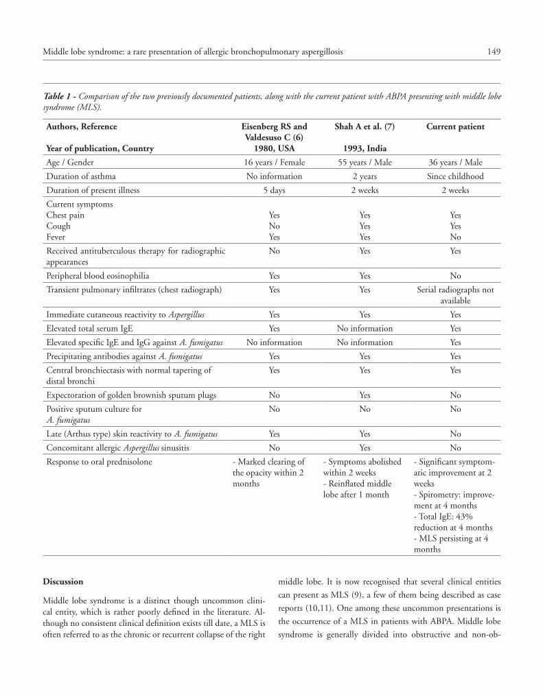
149Middle lobe syndrome: a rare presentation of allergic bronchopulmonary aspergillosis
middle lobe. It is now recognised that several clinical entities
can present as MLS (9), a few of them being described as case
reports (10,11). One among these uncommon presentations is
the occurrence of a MLS in patients with ABPA. Middle lobe
syndrome is generally divided into obstructive and non-ob-
Discussion
Middle lobe syndrome is a distinct though uncommon clini-cal entity, which is rather poorly defined in the literature. Al-though no consistent clinical definition exists till date, a MLS is often referred to as the chronic or recurrent collapse of the right
Table 1 - Comparison of the two previously documented patients, along with the current patient with ABPA presenting with middle lobe syndrome (MLS).
Authors, Reference
Year of publication, Country
Eisenberg RS and Valdesuso C (6)
1980, USA
Shah A et al. (7)
1993, India
Current patient
Age / Gender 16 years / Female 55 years / Male 36 years / Male
Duration of asthma No information 2 years Since childhood
Duration of present illness 5 days 2 weeks 2 weeks
Current symptomsChest painCoughFever
YesNoYes
YesYesYes
YesYesNo
Received antituberculous therapy for radiographic appearances
No Yes Yes
Peripheral blood eosinophilia Yes Yes No
Transient pulmonary infiltrates (chest radiograph) Yes Yes Serial radiographs not available
Immediate cutaneous reactivity to Aspergillus Yes Yes Yes
Elevated total serum IgE Yes No information Yes
Elevated specific IgE and IgG against A. fumigatus No information No information Yes
Precipitating antibodies against A. fumigatus Yes Yes Yes
Central bronchiectasis with normal tapering of distal bronchi
Yes Yes Yes
Expectoration of golden brownish sputum plugs No Yes No
Positive sputum culture for A. fumigatus
No No No
Late (Arthus type) skin reactivity to A. fumigatus Yes Yes No
Concomitant allergic Aspergillus sinusitis No Yes No
Response to oral prednisolone - Marked clearing of the opacity within 2 months
- Symptoms abolished within 2 weeks- Reinflated middle lobe after 1 month
- Significant symptom-atic improvement at 2 weeks- Spirometry: improve-ment at 4 months- Total IgE: 43% reduction at 4 months- MLS persisting at 4 months

150 A. Shah, S. Behera, C. Panjabi
tribute to the development of chronic infection and atelectasis. Due to the vicious cycle of recurrent inflammation and obstruc-tion, leading to a greater impairment of the cough mechanism and expectoration of secretions, these patients are at a greater risk for recurrent collapse of the middle lobe bronchus. While reviewing 1340 chest radiographs in 113 patients with ABPA, lobar/segmental collapse was observed in 17% patients (17).The comparative features of the two published reports of MLS in ABPA along with the current patient are summarised in table 1. The duration of the presenting illness was brief in all the three patients. Symptomatic improvement with oral corticosteroids was observed in all. Although radiological clearance was found in the first two patients, the chest radiograph of the current pa-tient continued to show MLS. Both our patients (cases 2 and 3) had received antituberculous therapy prior to being referred to us for evaluation. In high tuberculous prevalence countries, patients with ABPA are often erroneously diagnosed as pulmo-nary tuberculosis due to the strikingly similar chest radiograph appearances. This often results in initiation of antituberculous drugs while lung damage continues to progress (16).The middle lobe has a propensity to collapse in isolation while in ABPA, segmental or lobar collapse is not uncommon. It is rather surprising that a search of the literature revealed only two previous reports of ABPA presenting MLS (6,7). Since the MLS is first detected on imaging, a high index of suspicion would obviate the need for invasive diagnostic procedures like fibre-optic bronchoscopy, that these patients often undergo. ABPA should be considered among the differentials while evaluating a MLS, especially in patients with history suggestive of bronchial asthma.
References
1. Shah A., Panjabi C. Allergic bronchopulmonary aspergillosis: a review of a disease with a worldwide distribution. J Asthma. 2002;39:273-89.
2. Graham EA, Burford TH, Mayer JH. Middle lobe syndrome. Post-grad Med. 1948;4:29-34.
3. Brock RC, Cann RJ, Dickinson JR: Tuberculous mediastinal lymphadenitis in childhood: secondary effects on the lungs. Guy’s Hosp Rep. 1937;87:295.
4. Shah A. Aspergillus associated hypersensitivity respiratory disor-ders. Indian J Chest Dis Allied Sci. 2008;50:117-28.
5. Shah A, Panchal N, Agarwal AK. Allergic bronchopulmonary as-pergillosis: the spectrum of roentgenologic appearances. Indian J Radiol Imag. 1999;9:107-12.
6. Eisenberg RS, Valdesuso C. Middle lobe syndrome secondary to al-lergic bronchopulmonary aspergillosis. Ann Allergy. 1980;44:217-9.
7. Shah A, Bhagat R, Panchal N, Jaggi OP, Khan ZU. Allergic bron-chopulmonary aspergillosis with middle lobe syndrome and aller-gic Aspergillus sinusitis. Eur Respir J. 1993;6:917-8.
8. Panchal N, Bhagat R, Pant C, Shah A. Allergic bronchopulmonary aspergillosis: the spectrum of computed tomography appearances. Respir Med. 1997;91:213-9.
structive types. Obstruction of the middle lobe bronchus may be due to an intraluminal or extraluminal obstruction, and is the characteristic feature of MLS (9). There are several causative factors, which include both benign and malignant conditions. Tumours, including primary carcinoma of the lung, account for 24% of patients with MLS and 8 to 10% are secondary to tu-berculosis (Brock’s syndrome) (12). The non-obstructive form is typically caused by inflammation, commonly as the result of infection in majority of patients with MLS. Benign inflamma-tory disease accounts for around 62% of cases and has been identified as the most common cause of MLS (12). It can occur in adults and children with recurrent pneumonia, and is often associated with asthma, bronchiectasis and cystic fibrosis (9).It has been postulated that the middle lobe has a greater tenden-cy to collapse, as the middle lobe bronchus has a narrow origin with an acute angle, which can easily be obstructed. Further-more, if the surrounding lymph nodes are enlarged either due to inflammation or a tumour, extrinsic compression can occur. In addition, the anatomical separation of the middle lobe from the right upper and lower lobes by fissures can result in poor collateral ventilation from the surrounding areas (13). Imaging plays a key role in the diagnosis of MLS. It is now recognised that the atelectatic middle lobe is classically seen on a lateral chest roentgenogram as a wedge-shaped density extend-ing from the hilum, anteriorly and inferiorly (14). Though dif-ficult to detect on a posteroanterior radiograph, the volume loss within the right middle lobe is often seen as obscuring the right cardiac border (Silhouette sign) due to the loss of contact of the right middle lobe with the lateral wall of the right atrium (9). However, when the signs of middle lobe collapse are equivocal, a lordotic view could be of help. In this view, the MLS results in a wedge shaped density in the basal central zone of the right lower lung field, due to parenchymal involvement of the middle lobe (15). The advent of HRCT has helped with the diagnos-tic confirmation of MLS. It presents as a trapezoidal or broad triangular opacity, which has its base towards the hilum and is contiguous with the right cardiac border. CT imaging can also evaluate endobronchial or parenchymal abnormalities (8), as well as demonstrate bronchiectasis, as was seen in our patient. Bronchial patency, lymph node enlargement and calcifications or other causes of extrinsic compression of the right middle lobe airway too can be visualised (9). Our patient met 6 of the 8 essential diagnostic criteria for ABPA, including central bronchiectasis, a sine qua non (16) for the diagnosis. This was detected on HRCT, done to evaluate MLS. Mucous hypersecretion, commonly seen in patients with asthma, is a well recognised feature of ABPA. Secretions and vis-cid sputum result in the mucoid impaction and consequent col-lapse. Ineffective clearance of this impacted mucous along with anatomically poor collateral ventilation of the middle lobe con-
151Middle lobe syndrome: a rare presentation of allergic bronchopulmonary aspergillosis
13. Robinson DA, Lee PC. Middle lobe syndrome: case report and review of literature. Clin Pulm Med. 2007;14:244-8.
14. Saha SP, Mayo P, Long GA, McElvein RB. Middle lobe syndrome: diagnosis and management. Ann Thorac Surg. 1982;33:28-31.
15. Kopstein GG. Right middle lobe syndrome in children. JAMA. 1966;197:73.
16. Shah A. Allergic bronchopulmonary aspergillosis: an Indian per-spective. Curr Opin Pulm Med. 2007;13:72-80.
17. Shah A, Panchal N, Panjabi C. Allergic bronchopulmonary asper-gillosis: a review from India [abstract]. Allergy Clin Immunol Int. J World Allergy Org. 2003;(Suppl 1):104.
9. Gudbjartsson T, Gudmundsson G. Middle lobe syndrome: a re-view of clinicopathological features, diagnosis and treatment. Res-piration. 2012;84:80-6.
10. Kala J, Sahay S, Shah A. Bronchial anthracofibrosis and tubercu-losis presenting as a middle lobe syndrome. Prim Care Respir J. 2008;17:51-5.
11. Blaivasa AJ, Straussa W. Middle lobe syndrome in the left lower lobe in chronic obstructive pulmonary disease. Prim Care Respir J. 2009;18:331-3.
12. Wagner RB, Johnston MR: Middle lobe syndrome. Ann Thorac Surg. 1983;35:679-86.

C A S E R E P O R T S Eur Ann AllErgy Clin immunol VoL 46, n 4, 152-153, 2014
Introduction
The lesions of prurigo simplex subacuta may be disseminat-ed, or they may be acneiform, or localised on the extremities only (1). Prurigo denotes of a papular and⁄or nodular condition that itches intensely. Clinically, it appears as excoriated papules mostly in a symmetrical distribution on the extensor surfaces of the extremities, neck, lower trunk, and buttocks. It predomi-nantly affects middle-aged women, and may be associated with internal disorders such as diabetes mellitus and atophy, and re-nal as well as liver insufficiency appears to exist (2-6).
Case report
A 4-year-old boy presented with a 2-month history of intense pruritus and excoriated papules on trunk and extremities. On physical examination, some of the excoriating erythematous le-
sions, crusted papules, and small nodules were found predomi-nantly on the abdomen and on lower extremity, especially below the knee (figure 1). There was no difference between day and night symptoms, no connection to foods, drugs, bite of insects, and other family members had no similar symptoms. In medical history he had no endogenous (e.g. diabetes mellitus, hyperten-sion) or skin diseases (e.g. atopic dermatitis). Laboratory blood investigation revealed normal level of IgE; 28.8 (0-90 iu/mL) and eosinophilia (5.7%, 490 eosinophils/mm3). 23 parameters skin prick test was negative. A skin punch biopsy was performed from the abdominal lesions, and histopathologic examination showed hyperkeratosis and parakeratosis covered with stratified squamous epithelium skin tissue fragment, perivascular mono-nuclear cells infiltrates, proliferation of vascular endothelial, and also extravasated erythrocytes in the upper dermis. Toluidine blue staining showed that number of the mast cells was with-in the normal histological range (figure 2). The diagnosis was
SummaryPrurigo is a condition of nodular cutaneous lesions that itch intensely. Prurigo lesions are divided into acute, subacute and chronic forms that itch intensely. Subacute prurigo (SP) clinically presents as excoriated papules mostly in a symmetrical distribution on the extensor surfaces of the extremities, neck, lower trunk, and buttocks. It tends to occur in middle-aged patients, especially in women. Herein, we described prurigo simplex subacuta in a 4-year-old boy. It was histopathologically documented.
Corresponding author H. Haluk Akar Erciyes University School of Medicine Pediatric Asthma and Allergy Unit Kayseri, TurkeyPhone: + 90 352 20 76 666 / 25 125Fax: + 90 352 437 58 25E-mail: [email protected]
Key words
Prurigo simplex subacuta; prurigo simplex acuta; childhood
1Erciyes University School of Medicine, Pediatric Asthma and Allergy Unit, Kayseri, Turkey2Erciyes University School of Medicine, Department of Pathology, Kayseri, Turkey
Prurigo simplex subacuta or prurigo simplex acuta?
h. h. akar1, F. tahan1, s. BaLkanLi2, s. saDet Özcan2
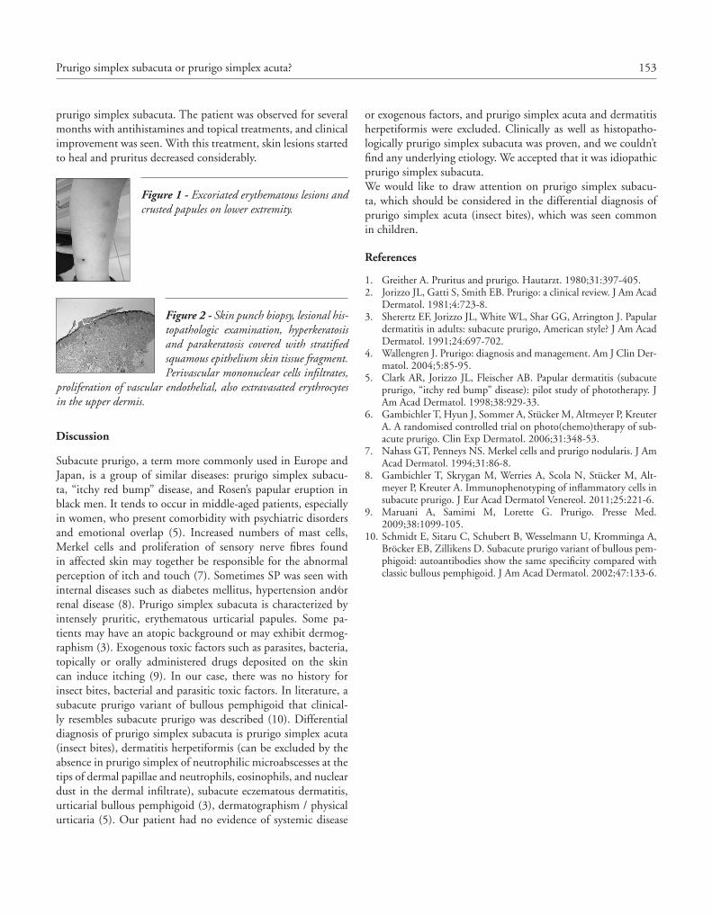
153Prurigo simplex subacuta or prurigo simplex acuta?
or exogenous factors, and prurigo simplex acuta and dermatitis herpetiformis were excluded. Clinically as well as histopatho-logically prurigo simplex subacuta was proven, and we couldn’t find any underlying etiology. We accepted that it was idiopathic prurigo simplex subacuta. We would like to draw attention on prurigo simplex subacu-ta, which should be considered in the differential diagnosis of prurigo simplex acuta (insect bites), which was seen common in children.
References
1. Greither A. Pruritus and prurigo. Hautarzt. 1980;31:397-405.2. Jorizzo JL, Gatti S, Smith EB. Prurigo: a clinical review. J Am Acad
Dermatol. 1981;4:723-8. 3. Sherertz EF, Jorizzo JL, White WL, Shar GG, Arrington J. Papular
dermatitis in adults: subacute prurigo, American style? J Am Acad Dermatol. 1991;24:697-702.
4. Wallengren J. Prurigo: diagnosis and management. Am J Clin Der-matol. 2004;5:85-95.
5. Clark AR, Jorizzo JL, Fleischer AB. Papular dermatitis (subacute prurigo, “itchy red bump” disease): pilot study of phototherapy. J Am Acad Dermatol. 1998;38:929-33.
6. Gambichler T, Hyun J, Sommer A, Stücker M, Altmeyer P, Kreuter A. A randomised controlled trial on photo(chemo)therapy of sub-acute prurigo. Clin Exp Dermatol. 2006;31:348-53.
7. Nahass GT, Penneys NS. Merkel cells and prurigo nodularis. J Am Acad Dermatol. 1994;31:86-8.
8. Gambichler T, Skrygan M, Werries A, Scola N, Stücker M, Alt-meyer P, Kreuter A. Immunophenotyping of inflammatory cells in subacute prurigo. J Eur Acad Dermatol Venereol. 2011;25:221-6.
9. Maruani A, Samimi M, Lorette G. Prurigo. Presse Med. 2009;38:1099-105.
10. Schmidt E, Sitaru C, Schubert B, Wesselmann U, Kromminga A, Bröcker EB, Zillikens D. Subacute prurigo variant of bullous pem-phigoid: autoantibodies show the same specificity compared with classic bullous pemphigoid. J Am Acad Dermatol. 2002;47:133-6.
prurigo simplex subacuta. The patient was observed for several months with antihistamines and topical treatments, and clinical improvement was seen. With this treatment, skin lesions started to heal and pruritus decreased considerably.
Figure 1 - Excoriated erythematous lesions and crusted papules on lower extremity.
Figure 2 - Skin punch biopsy, lesional his-topathologic examination, hyperkeratosis and parakeratosis covered with stratified squamous epithelium skin tissue fragment. Perivascular mononuclear cells infiltrates,
proliferation of vascular endothelial, also extravasated erythrocytes in the upper dermis.
Discussion
Subacute prurigo, a term more commonly used in Europe and Japan, is a group of similar diseases: prurigo simplex subacu-ta, “itchy red bump” disease, and Rosen’s papular eruption in black men. It tends to occur in middle-aged patients, especially in women, who present comorbidity with psychiatric disorders and emotional overlap (5). Increased numbers of mast cells, Merkel cells and proliferation of sensory nerve fibres found in affected skin may together be responsible for the abnormal perception of itch and touch (7). Sometimes SP was seen with internal diseases such as diabetes mellitus, hypertension and⁄or renal disease (8). Prurigo simplex subacuta is characterized by intensely pruritic, erythematous urticarial papules. Some pa-tients may have an atopic background or may exhibit dermog-raphism (3). Exogenous toxic factors such as parasites, bacteria, topically or orally administered drugs deposited on the skin can induce itching (9). In our case, there was no history for insect bites, bacterial and parasitic toxic factors. In literature, a subacute prurigo variant of bullous pemphigoid that clinical-ly resembles subacute prurigo was described (10). Differential diagnosis of prurigo simplex subacuta is prurigo simplex acuta (insect bites), dermatitis herpetiformis (can be excluded by the absence in prurigo simplex of neutrophilic microabscesses at the tips of dermal papillae and neutrophils, eosinophils, and nuclear dust in the dermal infiltrate), subacute eczematous dermatitis, urticarial bullous pemphigoid (3), dermatographism / physical urticaria (5). Our patient had no evidence of systemic disease

C A S E R E P O R T S Eur Ann AllErgy Clin immunol VoL 46, n 4, 154-156, 2014
SummaryAlthough the standard of care for cow’s milk (CM) allergy is strict food avoidance, oral immu-notherapy (OIT) is being widely investigated as an alternative management option in certain cases. Immediate adverse reactions to OIT have been described, but its long-term effects are much less often reported.We present the case of a girl diagnosed with IgE-mediated CM allergy that was proposed for our CM OIT protocol at the age of 3 years. The first sessions (dose escalation up to 5 ml) were well tolerated, however eight hours after her daily morning dose of 5ml CM the child developed late episodes of vomiting. No other symptoms, particularly immediately after CM ingestion, were reported. These episodes became progressively worse and on the third day she presented mild dehydration and blood eosinophilia. After OIT interruption, a progressive clinical improvement was observed. An esophageal endoscopy was performed, showing signs of eosinophilic esophagitis (EoE) with peak 20 eosinophils/hpf. After treatment with topical swallowed fluticasone (500 mcg bid) and a CM-free diet for 4 months, the child was asymp-tomatic and endoscopy and biopsy findings were normal.The long-term effects of milk OIT are still in part unknown. We hypothesize that eosinophilic esophagitis may have been a consequence of OIT in this case. The findings seem to indicate that food allergy may play a role in the pathogenesis of esophageal eosinophilia and stress the impor-tance of a well programmed long-term follow-up of patients that have undergone milk OIT.
Corresponding author Pedro Morais SilvaServiço de Imunoalergologia Hospital de Santa MariaCentro Hospitalar Lisboa Norte EPEAv. Prof. Egas Moniz, 1649-028 Lisboa, PortugalPhone: +351 917 374 928 E-mail: [email protected]
Key words
Eosinophilic esophagitis; oral immunotherapy; cow’s milk hypersensitivity; food hypersensitivity; eosinophilia; late-onset hypersensitivity
1Department of Immunoallergology, Hospital de Santa Maria, Centro Hospitalar Lisboa Norte EPE, Lisbon, Portugal2Department of Immunoallergology, Hospital Dona Estefânia, Centro Hospitalar Lisboa Centro EPE, Lisbon, Portugal
Diagnosis of eosinophilic esophagitis in an infant undergoing milk oral immunotherapy - a case report
Case Report
Cow’s milk (CM) allergy is a common disorder for which the standard of care is strict food avoidance. In recent years, CM oral immunotherapy (OIT), achieved by oral exposure to in-creasing doses of the food, has received particular attention as an alternative in cases where spontaneous tolerance acquisition is unlikely (1), increasing both quality of life and safety in the event of accidental ingestion of the offending food. Immedi-ate adverse reactions during OIT have often been reported and
seem to occur in 50 to 100% of cases (2-5). Delayed reactions, however, are much less often reported (3,5).We present the case of a 3-year old girl with atopic eczema and CM allergy. This condition manifested at 2 months of age in association with several episodes of urticaria, angioedema and vomiting following CM ingestion. IgE-mediated allergy was confirmed by skin prick tests, positive serum specific IgE (sIgE) and a positive oral food challenge. A complete eviction of CM from her diet resulted in resolution of all symptoms.
P. morais siLVa1, J. antunes2, m. chamBeL2, s. Prates2, P. Leiria Pinto2
155Diagnosis of eosinophilic esophagitis in an infant undergoing milk oral immunotherapy - a case report
reactions associated with CM ingestion developed into late gas-trointestinal symptoms at the third week of OIT protocol.EoE is an immune-mediated disorder in which the esophagus is infiltrated by eosinophils, presumably through a TH
2-driv-
en process (7). In this case, diagnosis was confirmed by en-doscopy and biopsy findings. Recovery following withdrawal of CM suggests a probable causal relation with CM ingestion during OIT. As no endoscopy was performed prior to OIT, we can only speculate that EoE developed during the treatment. However, symptoms of recurrent vomiting and malaise were absent previously, and blood eosinophilia also developed only after initiation of the protocol. According to recent guidelines (7) proton-pump inhibitor therapy was attempted, in order to discard gastroesophageal reflux disease or proton-pump inhibi-tor-responsive esophageal eosinophilia. Unfortunately, it had to be interrupted due to an apparent adverse reaction and a deci-sion was made not to delay swallowed fluticasone therapy. It was therefore not entirely possible to exclude those diseases. Howev-er, we consider that the fact that the child remained asymptom-atic and free of histologic abnormalities long after interruption of pharmacological treatment, strongly supports the diagnosis of CM induced EoE.It has been hypothesized that exposure of the esophagus of predisposed individuals to high antigen loads may be the core mechanism responsible for the development of EoE (8). In animal models, mice with respiratory, oral or epicutaneous sensitization that are rechallenged with intranasal exposure to allergens, develop esophageal eosinophilic infiltrates which mimic EoE (9,10). In humans, cases of EoE have been attributed to large volume allergen exposures (8), aeroallergen sublingual immunotherapy (11) and OIT (12-14).Our review has found reports of 4 patients who developed EoE following CM OIT protocols (12,13). Furthermore, OIT pro-tocols with peanut (3) and egg (13,14) have also been implied. Although this condition is probably underdiagnosed, the scarci-ty of reports suggest that EoE associated with OIT is likely rare. This is the first suggestive case in over 30 CM OIT performed in our center.
Although a favorable outcome seems to have occurred in this case, long-term evolution of esophageal eosinophilia and EoE is still uncertain. We agree with authors (12) who suggest that in the presence of this reaction, OIT should be interrupted.The long-term effects of CM OIT are still in part unknown and adverse reactions may be more common than previously expect-ed. The findings in this case seem to indicate that food allergy may play a role in the pathogenesis of esophageal eosinophilia and possibly of EoE and stress the importance of a well pro-grammed long-term follow-up of patients that have undergone CM OIT.
At the age of 3 years and 9 months, after several immediate reactions with cutaneous and gastrointestinal involvement asso-ciated with unintentional CM ingestion, and since sIgE levels were persistently elevated (milk: 60 KUA/L; casein: 29 KUA/L), the child was proposed to undergo a OIT protocol (6). Consent from our institution’s review board and from her parents was obtained prior to protocol start.It consisted of administration of increasing doses of CM in our outpatient allergy center at 2 to 3 week intervals followed by daily consumption of the tolerated dose at home. In the first session, a dose of 1 ml was reached and mild immediate reac-tions (facial urticaria, oral itching) were observed. No reactions occurred during the maintenance period at home, thus a new dose escalation was done from 1 to 5 ml after 3 weeks. Thereaf-ter, parents were instructed to maintain a daily morning dose of 5 ml at home. On the following day however, the child devel-oped vomiting episodes approximately eight hours after CM in-gestion. No other symptoms, particularly immediately after the dose, were reported. These episodes became progressively worse and on the third day she returned to the hospital. She looked ill and mildly dehydrated. A complete blood cell count revealed 1020 eosinophils/mm3 (prior levels were 340 eosinophils/mm3). At this time point, OIT was interrupted and progressive clinical improvement was observed on the course of the following days.Five days later an esophageal endoscopy was performed, show-ing rings and white mucosal exudates with a peak of 20 eosino-phils/hpf in mucosal biopsies from the upper, middle and lower esophagus. Gastric and duodenal biopsies had no alterations and Helicobacter pylori (HP) and Giardia lamblia were absent. Markers for celiac disease and HP were negative. A pH-moni-toring test was attempted but not tolerated by the child. Sub-sequent skin tests with other foods commonly associated with eosinophilic esophagitis (EoE) were negative.Treatment was started with lanzoprazol but there was an epi-sode of vomiting and urticaria immediately after the first dose, and therefore a decision was made to initiate topical swal-lowed fluticasone therapy (500 mcg bid) immediately. After a 4 month treatment with fluticasone and a CM-free diet, the child was asymptomatic and a new endoscopy with multiple biopsies found no alterations. Fluticasone was stopped and CM avoidance was maintained until now. At present, the child re-mains symptom-free 28 months after fluticasone discontinua-tion. Control endoscopies and biopsies performed at 12 and 27 months showed no sign of esophagitis. Specific IgE measured 14 months after the interruption of OIT was slightly higher (milk: 70 KUA/L; casein: 41 KUA/L) and presently a spontaneous de-crease is observed (milk: 34 KUA/L; casein: 27 KUA/L).We describe a patient with a typical history of IgE-mediated CM allergy, who developed a different clinical profile after the beginning of OIT: initial immediate skin and gastrointestinal

156 P. Morais Silva, J. Antunes, M. Chambel, S. Prates, P. Leiria Pinto
ommendations for children and adults. J Allergy Clin Immunol. 2011;128:3-20
8. Wolf WA, Jerath MR, Dellon ES. De-novo onset of eosinophilic esophagitis after large volume allergen exposures. J Gastrointestin Liver Dis. 2013;22:205-8.
9. Mishra A, Hogan SP, Brandt EB, Rothenberg ME. An etiological role for aeroallergens and eosinophils in experimental esophagitis. J Clin Invest. 2001;107:83-90.
10. Akei HS, Mishra A, Blanchard C, Rothenberg ME. Epicutaneous antigen exposure primes for experimental eosinophilic esophagitis in mice. Gastroenterology. 2005;129:985-94.
11. Miehlke S, Alpan O, Schröder S, Straumann A. Induction of Eo-sinophilic Esophagitis by Sublingual Pollen Immunotherapy. Case Rep Gastroenterol. 2013;7:363-8.
12. Sanchez-Garcia S, Del Rio PR, Escudero C , Martinez-Gomez MJ, Ibañez MD. Possible eosinophilic esophagitis induced by milk oral immunotherapy. J Allergy Clin Immunol. 2012;129:1155-7.
13. Perez TM, Astudillo JK, Herrero SI, Aparicio VF, Remón IZ, Lebrero EA. Esofagitis eosinofílica en relación com inducción de tolerância oral a leche y huevo. A propósito de dos casos. J Invest Allergol Clin Immunol. 2011;21Suppl4:142(abstract).
14. Ridolo E, De Angelis GL, Dallaglio P. Eosinophilic esophagitis af-ter specific oral tolerance induction for egg protein. Ann Allergy Asthma Immunol. 2011;106:73-4.
References
1. Nowak-Wegrzyn A, Sampson HA. Future therapies for food aller-gies. J Allergy Clin Immunol. 2011;127:558-73.
2. Skripak JM, Nash SD, Rowley H, Brereton NH, Oh S, Hamilton RG, et al. A randomized, double-blind, placebo-controlled study of milk oral immunotherapy for cow’s milk allergy. J Allergy Clin Immunol. 2008;122:1154-60.
3. Hofmann AM, Scurlock AM, Jones SM, Palmer KP, Lokhnygina Y, Steele PH et al. Safety of a peanut oral immunotherapy pro-tocol in children with peanut allergy. J Allergy Clin Immunol. 2009;124(2):286-91.
4. Martorell A, De la Hoz B, Ibañez MD, Bone J, Terrados MS, Michavila A, et al. Oral desensitization as a useful treatment in 2-year-old children with cow’s milk allergy. Clin Exp Allergy. 2011;41:1297-304.
5. Narisety SD, Skripak JM, Steele P, Hamilton RG, Matsui EC, Burks AW, et al. Open-label maintenance after milk oral immuno-therapy for IgE-mediated cow’s milk allergy. J Allergy Clin Immu-nol. 2009;124:610-2.
6. Morais-Almeida M, Piedade S, Couto M, Sampaio G, Santa-Marta C, Gaspar A. Inovação na indução de tolerância oral em crianças com anafilaxia às proteínas do leite de vaca. Rev Port Imunoalergo-logia. 2011;19(3):161-9.
7. Liacouras CA, Furuta GT, Hirano I, Atkins D, Attwood SE, Bo-nis PA, et al. Eosinophilic esophagitis. Updated consensus rec-

C A S E R E P O R T S Eur Ann AllErgy Clin immunol VoL 46, n 4, 157-159, 2014
Corresponding authorVaibhav N.Department of Oral and Maxillofacial Surgery Vokkaligara Sangha Dental College & Hospital and Research CentreKr Road, VvpuramBangalore, [email protected] phone: + 91 9844 258 076
SummaryBackground. Povidone iodine (PVP-I) is a chemical complex of polyvinylpyrrolidone (also known as povidone or PVP) and elemental iodine. Iodine containing compounds are widely and commonly used as antiseptics and disinfectants. They are available in various forms like aqueous solution, tincture, aerosol, ointment, or foam. Skin irritation to them is due to the oxidative effects of iodine. Because of the low free iodine concentration in povidone-iodine, skin irritation is less frequent from short contact (1). Case. We report two cases, both with no history of allergy to any drugs, who developed an allergic reaction 24 hours after the usage of povidone iodine as a pre-operative antiseptic to prepare (scrub) the lower third of face before surgical removal of third molars. This case report was granted an exemption by the ethical committee of our institution review board. The Helsinki declaration doesn’t apply to this case presentation. Conclusion. PVP-1 is the most commonly used antiseptic scrubbing solution prior to most surgeries. However, allergic contact dermatitis due to PVP-I has not been ex-tensively documented or reported and hence under evaluated, compared to other commonly encountered allergens. There is almost general agreement to the proposition that PVP-I is a very rare sensitizer (2,3) though there are some conflicting reports on the matter. However, sometimes when prolonged skin contact with PVP-I occurs when used as a pre-operative anti-septic agent, it can cause allergic dermatitis (4,5). Does this finding make pre-operative testing for allergies to PVP-1 necessary in all patients? A point to ponder.
1Assoc. Professor, Department Of Oral And Maxillofacial Surgery, Vokkaligara Sangha Dental College & Hospital And Research Centre, Kr Road, Vvpuram, Bangalore, India 2Junior Resident, Department Of Oral And Maxillofacial Surgery, Vokkaligara Sangha Dental College & Hospital And Research Centre, Kr Road, Vvpuram, Bangalore, India
Key words
Allergic dermatitis; povidone iodine (PVP-I)
An unusual complication of late onset allergic contact dermatitis to povidone iodine in Oral & Maxillofacial Surgery – A report of 2 cases
m.a. reyazuLLa1, a.L. GoPinath1, n. VaiBhaV2, r.P. raut2
Introduction
Povidone iodine preparations were introduced in the 1960s and are now the most common iodophor in clinical use because of their broad antimicrobial spectrum of activity, and unrivalled antiseptic properties. They are available in different formula-tions including solutions, creams, ointments, sprays and wound dressings. Povidone is a polymer similar to dextran and it acts as a carrier that delivers complexed diatomic iodine, which is bac-tericidal. It affects the structure and functions of enzymes and cell proteins. It also damages bacterial cell function by blocking
hydrogen bonding and altering the membrane structure. These multiple modes of action ensure the rapid death of microbes, and also importantly help to prevent the development of bacte-rial resistance (6). Acute or late onset allergic contact or irritant dermatitis due to povidone-iodine is a rare complication, espe-cially keeping in mind the extensive usage of the same. Systemic reactions to povidone-iodine are rare, but there are case reports of generalised urticaria and even anaphylactic shock (7,8). These cases have the characteristics of IgE-mediated reactions and in one case specific IgE against povidone was found.

158 M.A. Reyazulla, A.L. Gopinath, N. Vaibhav, R.P. Raut
complete regression of the symptoms, whereas only 1 week was sufficient for the symptoms to subside in case 2.
Figure 1 - Case 1.
Figure 2 - Case 2.
Discussion
Povidone-iodine 5% is a compound of iodine (0.5% releasable iodine but only 0.001% free iodine) and povidone, with addi-tives of glycerin, nonoxynol-9, disodiumphosphate, citric acid, and polyoxyethylene nonylphenyl ether. It is commonly used as an antibacterial agent and antiseptic. Povidone itself is also used in products such as foods, medicaments, hair cosmetics, and toothpastes (9). Povidone is a synthetic polymer used as a suspending agent for many pharmaceutical products, including iodine. In this form, povidone-iodine is a commonly used anti-septic in a variety of surgical cases, providing a broad spectrum of germicidal properties against a wide range of bacteria, viruses, fungi, protozoa, and spores. Systemic absorption after topical
Cases
Two patients reported to the Department of Oral & Maxillofa-cial Surgery with a chief complaint of pain in lower back tooth region of jaw. A detailed case history of both the patients was re-corded and it did not reveal any history of systemic disorders in-cluding asthma or allergy to any medication. Routine blood in-vestigations were carried out, which were within normal limits. Preoperative orthopantomogram was ordered, which showed partially erupted lower third molars with inflamed pericoronal flap. Surgical removal of third molars was decided as a treatment plan for both the patients. Patients were positioned on dental chair in semi-reclined po-sition and face was first scrubbed with savlon. The savlon was then wiped using sterile gauze. The face was then painted with 5% povidone iodine solution and draped with a sterile cloth, as a part of the standard protocol followed preoperatively before surgical removal of third molars. The procedure in both cases was carried out as planned (based on the evidence provided by the orthopantomogram regarding the position of the offending tooth) without any unforeseen complication. The average dura-tion for the procedure was approximately 45 minutes. Standard post-operative antibiotics and analgesics (Amoxicillin 500 mil-ligrams and Diclofenac sodium 50 mg, paracetamol 325 mg) were prescribed. Both the patients were not given pre-operative or post-operative IV steroid because, as per the evidence provid-ed by the orthopatomogram, excessive bone guttering or soft tissue retraction wasn’t needed during the procedure. Both the patients reported 24 hrs after the procedure with com-plaint of rashes only over the face, in the region painted with the PVP1 solution prior to the surgical removal. The patient first noted a stinging or burning sensation, with the onset of erythematous plaques that spread on both sides of the face over cheek region. On further clinical examination, a diffuse erythematous patch over the face, which was slightly oedem-atous with follicular elevations looking like tiny papules, were observed in both the patients. The erythematous patches were confined only to the areas which were painted with 5% povi-done iodine. Although intra-oral irrigation was done using the same solution (a diluted solution 1:10 with normal saline) there were no inflammatory or allergic reactions seen intra-oral-ly. Both the patients were referred to a dermatologist, and the diagnosis arrived at in both the instances was contact allergic dermatitis due to povidone iodine, for which both the patients were advised to use a desquamating soap; topical and systemic steroids as well as systemic antihistamines were prescribed (Sys-temic antihistamines Tab Cetrizine 10 mg bid, Tab Dazit 5 mg once daily, Inj Dexona 4 mg and vitamin E ointment (Evion cream) for local application). Both the patients were advised to avoid exposure to sunlight to prevent aggravation of the symp-toms. With this treatment protocol, case 1 took 10 days to show
159An unusual complication of late onset allergic contact dermatitis to povidone iodine in Oral & Maxillofacial Surgery
References
1. Chambers HF. Miscellaneous antimicrobial agents; disinfectants, antiseptics and sterilants. In: Basic and Clinical Pharmacology, 8th edition, Katzung B G(ed.): New York, NY, Lange Medical Books/McGraw-Hill,2001:852.
2. Brandao F M, Goossens A, Tosti A. Topical drugs. In: Textbook of Contact Dermatitis, 3rd edition, Rycroft R J G et al. (eds), Berlin, Springer, 2001:687-723.
3. Marks J G Jr, Elsner P, De Leon V. Contact and Occupational Dermatology, 3rd edition. St. Louis, Mosby, 2002.
4. Velázquez D, Zamberk P, Suárez R, Lázaro P. Allergic contact der-matitis to povidone-iodine. Contact Dermatitis. 2009;60:348-9.
5. Allergic contact dermatitis from povidone-iodine: a re-evaluation study. Contact Dermatitis. 2005;52:9-10.
6. Schreier H, Erdos G, Reimer, K et al. Molecular effects of povi-done-iodine on relevant micro-organisms: an electron-microscopic and biochemical study. Dermatology. 1997;195Suppl:111-6.
7. Adachi A, Fukunaga A, Hayashi K, Kunisada M, Horikawa T. Ana-phylaxis to polyvinylpyrrolidone after vaginal application of povi-done-iodine. Contact Dermatitis. 2003;48:133-6.
8. Waran KD, Munsick RA. Anaphylaxis from povidone-iodine. Lan-cet. 1995; 345:1506.
9. Adachi A, Fukunaga A, Hayashi K et al. Anaphylaxis to polyvin-ylpyrrolidone after vaginal application of povidone-iodine. Con-tact Dermatitis. 2003:48:133-6.
10. Ancona A, Suarez de la Torre R, Macotela E. Allergic contact der-matitis from povidone-iodine. Contact Dermatitis. 1985;13:66-8.
11. Lopez Saez MP, de Barrio M, Zubeldia JH, Priet A, Olalde S, Bae-za ML. Acute IgE-mediated generalized urticaria-angioedema after topical application of povidone-iodine. Allergol Immunopathol. 1998;26:23-6.
12. Rahimi S, Lazarou G. Late-onset allergic reaction to povidone-io-dine resulting in vulvar edema and urinary retention. Obstet Gy-necol. 2010;116:562-4.
application can potentially lead to immunoglobulin E (IgE) mediated allergic reactions. In its most benign form, local skin pruritus and irritation occur and resolve shortly after remov-al of povidone-iodine from skin (10). More severe reactions to povidone-iodine leading to anaphylaxis have also been seen in patients with an allergy to components of povidone-iodine. Less commonly reported are cases of angioedema immediately after contact with povidone-iodine (11). Regardless of the severity of symptoms, most reported reactions have developed immedi-ately after exposure to povidone-iodine, representing the early phase of an IgE-mediated allergic reaction. Fewer cases of late phase allergic reactions have been reported with symptoms pre-senting 6-8 hours after exposure (12). Unique in our cases were that our patient did not have any history of allergic reaction and exhibited no immediate signs of a reaction after exposure to the agent. Their symptoms became apparent almost 24 hours after exposure to povidone-iodine. This is the most commonly used antiseptic scrubbing solution used prior to surgeries. Every clinician should be aware of the potential allergic reaction that the solution may cause (and in some cases due to prolonged exposure). Most clinical setups have a protocol to test for safety of the local anesthetic solution for a particular patient by giving the so called ‘test dose’ subcutaneously prior to the procedure. The question is whether reports of allergic reaction to PVP1 solution warrant a similar pre-operative testing protocol, to avoid such complications.

ANNOUNCEMENT
The new online submission system
European Annals of Allergy and Clinical Immunology is proud to announce that a fully online submission and review system for all papers has been recently implemented. Electronic submission will allow a more efficient processing of manuscripts and will offer authors the option to track the progress of the review process whenever they need to. The link to this system has been made available on the new journal website, that has been launched simultaneously with the submission system at: www.eurannallergyimm.com.
We encouraged all Authors to submit manuscripts through our dedicated online peer review system; manuscripts will no longer be accepted for review by email or in paper form. We advise all the authors to carefully read the instructions for authors before starting the submissions.During submission, Authors will be first asked to select the article type, enter the manuscript title and provide author information. Through a menu, a general topic area should be selected: these will help us match manuscripts to the best available editors and reviewers. Reviewers will access papers via the system website and will be directed to it when requested by email to evaluate submissions. For full information on the manuscript preparation Authors are invited to read our updated Guide for Authors available on the Journal website.
Full Authors Guidelines, online Submission System link, Journal Publishing Agreement and Conflict of interest forms are available on new Journal website: www.eurannallergyimm.com
European Annalsof Allergy and
Clinical Immunology

informazionicosti
le più esaurienti e affidabili informazioni sui farmaci da oggi e per tutto il 2014a un prezzo speciale
Le offerte
• volume Parafarmaceutici• volume Indirizzi
a soli 99 euro
• volume Medicinali• volume Indirizzi
a soli 99 euro
• volume Medicinali• volume Parafarmaceutici• volume Indirizzi
a soli 149 euro
© EDRA LSWR SpA
via Spadolini, 7 - 20141 MilanoTel.: 02.88184.317 - Fax: 02.93664.151e-mail: [email protected] - www.edizioniedra.it
DIS
3201
4
Senza titolo-1 1 02/04/14 17.53
INFORMAZIONEPolitica Sanitaria, clinica,
farmaci e terapie e la cronaca quotidiana
dei fatti di attualità
CANALI TEMATICICardiologia, Pediatria,
Ginecologia, Diabetologia, Medicina interna
CAsI CLINICIOltre 120.000 medici coinvolti nei casi clinici messi a disposizione
dei colleghi per un consulto
COMMUNITYL’area Social con i servizi
“L’Esperto risponde”, “Doctor Alert”, “Forum”,
“Preferiti”
DOWNLOAD CENTER
Contenuti di formazione e aggiornamento gratuiti tratti
dalle pubblicazioni Edra Masson
ECM La formazione di qualità
di Doctor33
BACHECA ANNUNCI
La più ricca sezione di annunci sul web per
i professionisti della medicina
Il portale dei protagonisti della salute
www.doctor33.it
NOvITà

4/2014
The perception of allergen-specific immunotherapy among chest physicians: an Italian survey
Tetranychus urticae allergy in a population without occupational exposure
In patients with LTP syndromefood-specific IgE show a predictable hierarchical order
Middle lobe syndrome: a rare presentation of allergic bronchopulmonary aspergillosis
Prurigo simplex subacuta or prurigo simplex acuta?
Diagnosis of eosinophilic esophagitis in an infant undergoing milk oral immunotherapy - a case report
An unusual complication of late onset allergic contact dermatitis to povidone iodine in Oral & Maxillofacial Surgery – A report of 2 cases
European Annalsof Allergy and
Clinical Immunology
Issn 1764-1489 Volume 46 n. 4/2014 – July 2014
THE OFFICIAL JOURNAL OF AAITO | ASSOCIAZIONE ITALIANA ALLERGOLOGI IMMUNOLOGI TERRITORIALI E OSPEDALIERI
THE OFFICIAL JOURNAL OF SPAIC | SOCIEDADE PORTUGUESA DE ALERGOLOGIA E IMUNOLOGIA CLINICA
SUBSCRIBE NOW! www.eurannallergyimm.com• 6 print issues per year• full access to www.eurannallergyimm.com,
featuring all current and archived issues
European Annals of Allergy and Clinical Immunology is a bimonthly peer-reviewed publication• The official Journal of the “Associazione Italiana Allergologi Immunologi Territoriali
e Ospedalieri” (Italian Association of Hospital Allergists and Immunologists - AAITO)and the “Sociedade Portuguesa de Alergologia e Imnunologia Clinica” (Portuguese Societyof Allergology and Clinical Immunology - SPAIC)
• indexed in PubMed and Scopus
• collects reviews, original works and case reports concerning etiology,diagnosis and treatment of allergic and immunological disorders
• includes a section of information comingfrom the main international health associations and authorities.
Italy SubScrIptIon only 60,00 Euro
InternatIonal SubScrIptIon
only 85,00 Euro
To submit your paper go to http://eaaci.edmgr.com
AbbAAITO210x270.indd 1 02/07/14 14.21