hypoxic effects on corneal morphology and function. computer with an enhanced graphics adapter (ega)...
TRANSCRIPT

Investigative Ophthalmology & Visual Science. Vol. 31, No. 8. August 1990Copyright © Association for Research in Vision and Ophthalmology
Hypoxic Effects on Corneal Morphology and FunctionKenneth A. Poise,* Richard J. Brand.t Stephen R. Cohen,* and Michel Guillon^:
Normal corneal metabolism depends on a critical level of oxygen, below which a series of acute cornealresponses occur, including an increase in stromal lactate, a reduction in intercellular pi I, and anincrease in corneal hydration. These acute responses arc reversible when normal oxygen is restored;however, it has been shown that chronic exposure to low oxygen levels can result in permanentmorphologic changes in the corneal endothelium. Clinicians have expressed concern that these ob-served structural changes may also be accompanied by alterations in corneal physiology. Whethersuch effects occur is not known, since it has been difficult to assess human corneal function accurately.Recently, we have developed an in vivo test, able to measure overall corneal hydration control, that canbe used to study the effects of hypoxia on corneal function. This test provides information on severalcharacteristics of hydration control, one of which is the percent corneal thickness recovery per hour(PRPH) after inducing corneal swelling. In this study, we assumed that corneal hypoxia accompaniesboth extended and polymethylmethacrylatc (PMMA) contact lens wear and that the dose received isrelated to the years of past lens wear. Using this paradigm, we explored the relationship of hypoxicdose to an endothelial polymegcthism index (EPI), endothclial cell density (ECD), and PRPH in 36subjects with varying contact lens wearing histories. Based on multiple regression analysis, the rela-tive change (expressed as percent per year) associated with hypoxic dose (adjusted for age and gender)was found to be dose-dependent and corresponded to estimated changes of 1.70%/yr, -0.25%/yr, and-1.26%/yr, with 95% confidence limits of (-0.3,3.7), (-1.4,0.9), and (-2.6,0.06) for EPI, ECD, andPRPH, respectively. These preliminary data suggest that hypoxic exposure alters endothelial mor-phology and reduces corneal function; however, it is important to indicate that this was a exploratoryinvestigation with several limitations, and that therefore these results should be viewed as preliminaryuntil more definitive studies are completed. Invest Ophthalmol Vis Sci 31:1542-1554, 1990
Normal corneal metabolism depends on a criticallevel of oxygen, below which a series of acute meta-bolic events occur, including an increase in stromallactate, a reduction in intercellular pH, and an in-crease in corneal hydration.1"3 These acute responsesappear to be reversible when normal oxygen levels arerestored.3 However, chronic exposure to low oxygenlevels apparently can cause structural changes thatmay take several months to reverse (epithelial micro-cysts) or that may be permanent (endothelial poly-megethism).4"9
Although the currently available information islimited, many clinicians have expressed concerns
From the *School of Optometry and the tSchool of PublicHealth, the University of California, Berkeley. California, and the^Contact Lens Research Associates. London, United Kingdom.
tMajor. United States Army. The views expressed in this articlearc those of the author and do not reflect the official policy of theDepartment of Defense of the United States Government.
Supported by grant R01 EY-04930 from the National Eye Insti-tute, Bcthesda, Maryland.
Submitted for publication: September 21. 1989; accepted Febru-ary I. 1990.
Reprint requests: Kenneth A. Poise. OD, School of Optometry,University of California. Berkeley, CA 94720.
that chronic hypoxic exposure results in endothelialpolymegethism, which might be connected with al-tered corneal physiology. Evidence that polymegeth-ism may be accompanied by a functional impairmentin corneal function comes primarily from the work ofRao and co-workers, who found that cataract patientswith preoperative polymegethism had a longer post-operative degree of corneal edema compared to cata-ract patients who did not demonstrate such morpho-logic changes.10 More recently, Lass and colleagues"and Dutt and colleagues12 used fluorophotometry todemonstrate that extended- and long-term poly-methylmethacrylate (PMMA) contact lens wearerswith polymegethism had a decrease in endothelialbarrier function and an increase in endothelial pumprate. Also, other studies, in which animal corneaswere exposed to hypoxia, found a reduction in endo-thelial pump site density.13 If this loss also occurs inhumans after hypoxic exposure, impairment to cor-neal hydration control may be expected.
Although these studies suggest that contact lenswear may result in a functional deficit to the cornea,there is also evidence that indicates that the low oxy-gen levels are not accompanied by adverse effects oncorneal physiology. For example, in experiments sim-
Downloaded From: https://iovs.arvojournals.org/pdfaccess.ashx?url=/data/journals/iovs/933156/ on 09/16/2018

No. 8 HYPOXIC EFFECTS ON CORNEAL MORPHOLOGY AND FUNCTION / Poise er ol 1543
ilar to those conducted by Lass et al," Carlson andco-workers did not find differences in endothelial per-meability between contact and non-contact lenswearers.14 Other studies have shown that in individ-uals who had worn extended-wear (EW) lenses forseveral years, corneal thickness returned to normallevels about 2 days after lens wear was discontinued—a result that would not be expected if there were afunctional deficit in the endothelium.7
Conclusions about the effects of long-term hypoxicexposure to corneal structure and function remainunclear, and important questions remain unan-swered. For example, is there a measurable structuraland physiologic response from chronic exposure tolow oxygen levels, and if so, how marked are thesechanges compared to those that occur in subjects whohave not had chronic hypoxic exposure? If chronicexposure has an effect, does it depend on the degree,duration, and timing of the low oxygen dose?
These and related issues need to be explored in partbecause there seems to be an evolving interest in EWcontact lenses among patients, and among cliniciansand manufacturers in prescribing and marketingthem. However, since most currently available EWmaterials do not meet required oxygen levels duringeye closure, the cornea will typically be exposed tohypoxic levels for a number of hours every day thelenses are worn.1516
It has been possible to study structural changes thatresult from contact lens wear through morphometricanalysis of endothelial photomicrographs; however,in vivo measurements of corneal function have notbeen readily available for assessing the physiologiceffects of hypoxic exposure.681 l l 4 Recently, we de-scribed a method for in vivo assessment of overallcorneal hydration control that provides one approachfor studying the effects of contact lens wear on cor-neal physiology.17 In the current report we presentresults from an exploratory study, the purpose ofwhich was to provide preliminary estimates of therelationships among various quantitative measures oflens wear, several measures of corneal morphology,and this new measure of corneal hydration control.This exploratory study was conducted to obtain in-formation to determine whether or not further inves-tigation seems warranted and to guide the design ofstudies that can yield more definitive assessments ofthe possible impact of contact lens wear on cornealstructure and function.
Materials and Methods
Subjects
Subjects were recruited from the campus commu-nity of the University of California at Berkeley. All
candidates received a complete eye examination, andonly those subjects who were free of ocular diseasewere accepted into the study. The emphasis in re-cruiting was to obtain a subgroup of patients with nohistory of contact lens wear and a subgroup who hadsome experience with a lens-wearing mode that pre-sumably would have caused some level of cornealhypoxia. No volunteers whose past lens wear con-sisted entirely of soft contact lens daily wear(SCLDW) or rigid gas-permeable daily wear(RGPDW) were accepted. Recruitment efforts pro-duced 36 subjects including 7 with no history of lenswear. Before beginning any of the testing procedures,each subject was given a description of the study, andinformed consent was obtained.
Measurement Procedures
Endothelial morphology: Endothelial photomicro-graphs were taken with a Nikon AS-1 EnhancedGraphics Adapter (EGA) noncontact specular micro-scope. The 35-mm slides were rear-projected onto a24 X 24-inch screen, and a Bio-Optics (Bio-OpticsInc., Arlington, MA) digitizer coupled to an IBM-XTcomputer with an enhanced graphics adapter (EGA)board was used to trace each cell within a definedarea of the endothelium (approximately 100 X 500nxn). The analyzed cell parameters were mean celldensity (cells/mm2) and mean and standard deviationof cell areas (mm2). These last two measures wereused to compute the coefficient of variation of cellarea (standard deviation of cell size divided by meancell area), which provides an endothelial poly-megelhism index (EPI).
Corneal thickness: Optical pachometry was doneon each subject with a modified slit lamp to provideimproved corneal thickness measurements. A moredetailed description of this instrument is providedelsewhere.18
Corneal Hydration Control
Assessment of corneal hydration control was basedon two different data sets obtained on separate days.One test consisted of two repeat measurements ofcorneal thickness every 15 min over a period of 1-2hr to assess the open-eye steady-state (OESS) thick-ness. These OESS measurements were made in thelate afternoon after the subject had been awake for 6or more hr.
The second procedure required that the subject re-port to the laboratory in the morning for a "stress"test that involves wearing a 400-/*m-thick 40% waterhydrogel contact lens for 2 hr with the eyes closed.The estimated oxygen tension under this lens duringeye closure was at or near 0 mmHg.15 Before inserting
Downloaded From: https://iovs.arvojournals.org/pdfaccess.ashx?url=/data/journals/iovs/933156/ on 09/16/2018

1544 INVESTIGATIVE OPHTHALMOLOGY & VISUAL SCIENCE / Augusr 1990 Vol. 01
the contact lens, baseline pachometry measurementswere taken. After the 2-hr stress period, the contactlenses were removed, and two repeat corneal thick-ness measurements were made at approximately 30-min intervals for 4-5 hr. For 23 of 36 subjects, theOESS assessment was made before the stress test. Forthose 13 subjects who had the stress test done first,two had discontinued their lens wear for 3 days whilethe other 11 had their lenses out 6 or more days.
Endothelial photographs were taken usually on theday the stress test was given, approximately 3-5 hrafter the removal of the stress lens. Specular micro-graphy was timed in this manner to minimize possi-ble short-term effects of the subjects' own contactlenses on corneal clarity or endothelial morphology.Although it is possible that the cell density or EPImay have changed over the 3-day period after contactlens discontinuance, most current information sug-gests that such changes would be unlikely. No at-tempt was made to quantify these effects.5-6-8-9"1214
It also has been shown that wearing soft lenses canhave a small transient effect (eg, blebs) on some indi-viduals; however, since this transient change lasts lessthan 30 min, it is not likely to have had any affect onthe determination of endothelial cell morphologydone in this study.19
These two data sets were then combined by meansof a composite exponential model that provides esti-mates of several aspects of an individual's overallcorneal hydration control. The key characteristic thatwas analyzed for this study measures the rate at whichthe cornea returned to OESS thickness (deswelled)from an induced level of edema. This quantity, calledthe percent recovery per hour (PR.PH), describes theamount of recovery (by percentage) that the corneaundergoes in any 1-hrtime period during exponentialrecovery. A more complete description of the com-posite experimental model and its use, along withsample deswellingcurves, are provided elsewhere.1720
Contact Lens Wear
Obtaining complete and accurate informationabout lens wear history is an important element in astudy of this type, which depends on establishing oneor more operational definitions of lens wear to pro-vide variables for statistical analysis. Choosing one ormore lens wear measures involves a working assump-tion that other aspects of lens wear can safely be ig-nored when the chosen measures are studied relativeto corneal structure or function. For example, ananalysis that ignores daily wear (DW) would be validonly if DW is unrelated to the corneal propertiesunder study or is not associated with the measure oflens wear that is being studied.
A patient's contact lens history is often a fairly
complex sequence that may involve periods of lenswear with different types of lenses or modes of wear,and possibly interspersed periods of no contact lenswear. Also, the impact of lens wear may differ de-pending on the number of hours of lens wear eachday, the nature of the lens fit, the companion solu-tions used, and other behavior connected with wear-ing and maintaining the lenses. Consequently, a con-tact lens wear history can be complex and multidi-mensional.
For this preliminary analysis we have made theworking assumptions that the historic time when par-ticular types of lenses were worn is immaterial, andthat the sequencing of types of lens wear also is im-material in cases with histories of more than one typeof lens wear. We have chosen to examine only thetotal amount of previous lens wear in each of severalbroad categories. We considered this to be the kind ofinformation that could be obtained most reliably inan exploratory study based on self-reported lens wear.Since we were attempting to investigate long-termeffects, it seemed appropriate to focus on the amountof wear without regard to the timing of wear.
Because we were interested in studying hypoxic ef-fects, it was important to establish an operational def-inition of the types of contact lens wear that involvehypoxia. For the purposes of this study, we consid-ered either PMMA, soft contact lens extended wear(SCLEW), or rigid gas-permeable extended wear(RGPEW) to expose the subject to some level of hyp-oxia, since there is considerable evidence to show thatnone of these wearing modalities supplies sufficientoxygen for normal corneal metabolism in at leastsome part of the wearing cycle.716-21 No attempt wasmade to determine the oxygen level under the contactlens; rather, we presumed that corneal hypoxia oc-curred to some extent during some period of the daywhen these lenses were worn. The hypoxic dosagewas defined simply by the number of years that thesubject wore any of these lens-materials/wearing-mode combinations.
It is also possible that SCLDW or RGPDW mayresult in a modest level of hypoxia, since many DWhydrogel materials have only modest oxygen trans-missibilities (Dk/L).15 Several of the rigid gas-perme-able (RGP) materials that currently are worn or thathave been worn in the past also have low enoughDk/L values to possibly cause some oxygen depriva-tion.15 22 The period of use (years) of SCLDW orRGPDW also was studied for its influence on cornealproperties.
Lens Wearing Before Test Measurements
The principal characteristics evaluated in this studywere determined from information gathered from the
Downloaded From: https://iovs.arvojournals.org/pdfaccess.ashx?url=/data/journals/iovs/933156/ on 09/16/2018

No. 8 HYPOXIC EFFECTS ON CORNEAL MORPHOLOGY AND FUNCTION / Poise er ol 1545
endothelial photomicroscopy and optical pachome-try measurements. Conceivably, acute effects of cur-rent contact lens use by wearers who had not re-moved their contact lenses for sufficiently long pe-riods before testing could have influenced either ofthese measurements. Unfortunately, there is little in-formation to indicate how long contact lens wearmust be discontinued to eliminate any possible acuteeffects that lens wear may have on test outcomes. Weattempted to eliminate the acute effects of contactiens wear by having subjects who were current con-tact lens wearers discontinue any hypoxic wear for 3days before testing. Three days was selected based onprevious studies indicating that corneal thicknesschanges stabilize after discontinuing lens wear for 2days or more and that transient morphologic changestypically clear within 2 hr after lens removal.716 Atthe time the protocol was designed, we were less con-cerned about SCLDW or RGPDW, so lens removalfor 1 or more days before testing was planned for thistype of wear.
The actual experience with lens removal beforetesting differed for the OESS and stress tests becausethey occurred on different days. The five subjectswhose OESS tests were performed the soonest afterdiscontinuation of hypoxic lens wear had been with-out lenses for 2, 3, 6, 30, and 64 days. Most subjectshad not worn hypoxic lenses for a considerable lengthof time, and the average time without hypoxic lenseswas 793 days among the 29 subjects with past hyp-oxic wear. The five shortest durations between re-moval of hypoxic lenses and the stress test were 0, 3,3, 31, and 78 days, and the average was 794 days.After checking that the two subjects with protocolviolations did not dominate the findings from thisstudy, they were used for data analysis.
SCLDW or RGPDW tended to occur in muchcloser proximity to the OESS and stress test times.The number of days without lens wear prior to theOESS test for the 21 subjects with past SCLDW orRGPDW were 0 or 1 (2 subjects), 2 (6 subjects), 3 (5subjects), 4 (2 subjects), 7 or 9 (2 subjects), and 370 ormore days (4 subjects). The corresponding number ofdays in connection with the stress test were 1 (12subjects), 2 or 3 (2 subjects), 4 (2 subjects), 14 (1subject), and 364 or more days (4 subjects).
For the 29 subjects with contact-lens-wearing his-tory, 24 had endothelial photomicroscopy done onthe day of the stress test (after the lenses had beenremoved for 4 or more hr). Of the remaining 5 sub-jects, 1 had photos taken on the day of the OESS, 3had photos taken within 8 weeks of completing thestress test, and 1 was photographed 2 months beforeother testing was done. Since data from previousstudies have shown that endothelial cell coefficient ofvariation and cell density change relatively slowly,
the photos of these 5 subjects most likely are validindicators of the subjects' endothelial morphology atthe time of the stress test.5-6-8-9"-1214 In general, thetiming of measurements related to last lens wear is anaspect of study design that needs to be carefully re-considered before confirmatory studies are con-ducted.
Statistical Analysis Methods
The relationships between corneal properties andmeasures of lens wear were analyzed by standardmethods of multiple regression analysis that were im-plemented with a statistical package on an MS-DOSpersonal computer.23 In these analyses, the average ofright and left eye measurements of corneal propertieson each subject provided the dependent variablesused in the analysis. In this way errors in the measure-ment of corneal properties were diminished and sta-tistically independent data were obtained for analysis.In all cases, the lens wear measurements, which pro-vided the predictor variables for analysis, were thesame for both eyes of all the subjects. For purposes ofthis exploratory study and analysis, estimates of themagnitudes of specified relationships of interest alongwith associated confidence intervals have been em-phasized for summarizing results.
Results
The presentation of the results is organized in threeparts. First, the lens wear experience in the studygroup is described. Then differences in lens wear byage and gender are examined. Finally, multiple linearregression methods are used to investigate the rela-tionship between lens wear variables and three cor-neal properties; EPI, ECD, and corneal hydrationcontrol as measured by the PRPH.
Lens Wear Experience
The study of past lens wear was based on five com-ponent measures extracted from the self report pro-vided by the subjects. These measures are SCLDW,SCLEW, RGPDW, RGPEW, and PMMA.
Twenty-nine of the 36 subjects had a history of lenswear that included either extended or PMMA wear.Many of these subjects also had a history of DW. Asummary of the amount of past lens wear for each ofthe five lens-wear categories is provided in the upperportion of Table 1. Subjects who had more than onetype or mode of lens wear contributed data to morethan one category.
EW is one important type of past wear that pre-sumably led to some degree of corneal hypoxia. Inthis category, only a small amount of past wear in-volved RGP lenses. Except for two subjects who re-
Downloaded From: https://iovs.arvojournals.org/pdfaccess.ashx?url=/data/journals/iovs/933156/ on 09/16/2018

1546 INVESTIGATIVE OPHTHALMOLOGY & VISUAL SCIENCE / Augusr 1990 Vol. 31
Table 1. Past lens experience among the 29 study subjects with history of lens wear
Type of lens wear
Component measures*SCLDWSCLEWRGPDWRGPEWPMMA
Combined measures!
EWJEW + PMMA
Number ofsubjects
914122
20
191629
Mean
2.53.42.40.97.3
2.73.16.8
Years of lens
SD
1.71.61.30.22.6
1.81.73.3
wear
Min.
0.30.50.30.72.0
0.30.52.0
Max.
5.25.24.61.0
12.0
7.05.2
15.1
* Subjects with multiple types of past lens wear contribute to more thanone category.
t DW = SCLDW + RGPDW; EW = SCLEW + RGPEW.
t Two subjects had both SCLDW and RGPDW episodes. No subject hadboth SCLEW and RGPEW episodes.
ported RGPEW of 9 and 12 months as their onlyepisodes of EW. all subjects with a history of EW hadonly SCLEW episodes. The more commonly en-countered SCLEW was reported by 14 (38.9%) of the36 study subjects. The total duration of SCLEWranged from 0.5 to 5.2 yr, and the mean and standarddeviation of the length of wear were 3.4 and 1.6 yr.respectively. Because there is so little RGPEW experi-ence, for subsequent analyses the 16 subjects withsome history of EW all were assigned a value for totalyears of EW without regard to the type of lens mate-rial. In the interpretation of results, it must thereforebe remembered that the EW variable is heavily domi-nated by SCLEW.
As mentioned above, we have assumed that bothPMMA and EW would lead to some degree of cor-neal hypoxia. In order to examine the contribution ofboth PMMA and EW as components of hypoxicwear, the joint distribution of these measures of pastlens wear is presented in Figure 1. Most subjects hadeither PMMA or EW episodes, but not both. Theexceptions are four subjects who had predominantlyPMMA wear along with 1 yr or less of SCLEW andthree additional subjects who had a substantialamount of both PMMA and SCLEW. The singlepoint in Figure 1 at the origin where SCLEW= PMMA = 0 represents the subgroup of seven sub-jects who had no history of prior lens wear.
Under the assumption that EW and PMMA wearoperate similarly in their impact on corneal proper-ties, it would be appropriate to sum these two mea-sures to give the combined total years of hypoxicwear. Graphically, the sum would be represented bythe projection of the data points in Figure 1 onto the45° line shown there. For each of the corneal proper-ties studied, this summary measure of hypoxic wearhas been used in one of the regression models used tostudy the effect of past lens wear. Use of this sum-
mary measure of hypoxic wear is motivated by thepotential gain in statistical precision that can be ex-pected from entering the combined informationabout past hypoxic wear into the analysis in an effi-cient way. However, if the assumption that EW andPMMA wear operate similarly is false, then analysiswith combined hypoxic wear may be misleading, be-cause it will give only a weighted average of the indi-vidual impacts of the two hypoxic wear components.Thus, the relationship of one component measurewill be understated and the other will be overstated.Consequently, in connection with each corneal prop-erty we also have used a second regression model inwhich each of these component measures is studiedas a separate predictor. This provides some indicationof the independent relationship between each compo-nent and the corneal property under study, but someof these estimated component relationships have re-duced precision.
^^l_COOi
PM
MA
12 -
9 -
6 -
3 -
O
O
OO OD
Ooooo
oo
0 - <n> on> o QD
EW (yrs)
Fig. 1. Scattcrplot of the years of PMMA vs EW among 36subjects. The 0.0 point represents 7 subjects who had no prior lenswear.
Downloaded From: https://iovs.arvojournals.org/pdfaccess.ashx?url=/data/journals/iovs/933156/ on 09/16/2018

No. 8 HYPOXIC EFFECTS ON CORNEAL. MORPHOLOGY AND FUNCTION / Poise er ol 1547
As shown in Figure 2, many subjects also had ahistory of SCLDW or RGPDW. Here again, subjectstended to wear one or the other type of lens, exceptfor two subjects who had worn both types of lenses.As with the hypoxic wear components, the SCLDWand RGPDW components could be treated sepa-rately or combined to give a single measure. Giventhe small size of this exploratory study and the ex-pectation that these types of DW probably are notrelated to corneal properties, it was decided to workwith a combined SCLDW and RGPDW measure.Consequently, subsequent analysis with this com-bined variable (SCLDW + RGPDW) will give onlythe weighted average of the two component effects.
The description of the amount of lens wear in thecombined categories (EW = SCLEW + RGPEW;DW = SCLDW + RGPDW, and hypoxic = EW+ PMMA) is provided in the lower part of Table 1.Considering all hypoxic components, 29 subjects hadan average of 6.8 yr of wear. Also, there were 19subjects with an average of 2.7 yr SCLDW andRGPDW. In Figure 3, the scatter diagram of DW vshypoxic wear shows there is not a strong associationbetween those two aspects of lens wear history. Thisconfiguration is fortunate since it facilitates assess-ment of the separate relationship between each ofthese measures and the corneal properties understudy.
These descriptions of the lens wear history provideinformation about the data that form the basis forstudying the effects of lens wear on corneal proper-ties. Clearly, they apply only to the subjects in thisparticular study; however, they probably are indica-tive of the general kind of lens wear patterns that arelikely to arise in other studies.
e -
co 4 -
Q
£> 2 -I
0 -OO O O O O O O QD O
RGP DW (yrs)
Fig. 2. Scattcrplot of the years of SCLDW vs RGPDW among 36subjects. The 0,0 point represents 7 subjects who had no prior lens
Q
8 -
6 -
4 -
2 -
0 -\
0 4 8 12
Combined PMMA & EW (yrs)
Fig. 3. Scattcrplot of the years of combined SCLDW andRGPDW vs combined PMMA and EW among 36 subjects. The 0.0point represents 7 subjects who had no prior lens wear.
Age and Gender Differences in Past Lens Wear
It is also important to consider the possibility thatthe nature and length of time of past lens wear islikely to be associated with two co-variables: age andgender. Moreover, these co-variables may be relatedto corneal properties by way of mechanisms that donot involve any of the lens wear history variables. Forexample, evidence indicates that both endothelialmorphology and corneal hydration control showchanges with age, in the absence of lens wear.17-24-25
Since one would expect lens wear history to be age-related, it is necessary to make statistical adjustmentsfor age in an effort to obtain a more direct assessmentof possible lens wear effects that are not confoundedby age differences. Similar possibilities can be imag-ined in connection with gender.1
A full assessment of the relationship between age,gender, and the various measures of past contact lenswear would require numerous tables and graphs.However, to illustrate the potential of these co-vari-ables to confound the relationship between lens wearmeasures and corneal properties, we can examine theimportant lens wear variable—years of hypoxic wear—in relationship to age and gender. Figure 4 shows apair of scatter diagrams that plot separately hypoxicwear (years) vs age for the subgroups consisting of 11males and 25 females (of the 36 points, 4 are coinci-dent). Not surprisingly, in both the male and femalegroups, there is a tendency for older subjects to havehad a longer duration of hypoxic wear. Consequently,any effects of age will tend to be confounded withlength of hypoxic wear. This illustrates the impor-tance of adjusting for age and gender when attempt-ing to isolate the direct association between lens wearand corneal properties.
Downloaded From: https://iovs.arvojournals.org/pdfaccess.ashx?url=/data/journals/iovs/933156/ on 09/16/2018

1548 INVESTIGATIVE OPHTHALMOLOGY G VISUAL SCIENCE / Augusr 1990 Vol 31
/rs)
VIM
A &
EW
(n
ed
PC
omb
15 -
10 -
5 -
o -
<c
o
o
o oo
oo o
o o0
o o °5 o
o o
8
o
15 20 25 30
Age (yrs)
35 40 15 20 25 30 35 40
Age (yrs)
Fig. 4. Scattcrplot of years of combined PMMA and EW vs age for subgroups of (A) 11 male and (B) 25 female subjects.
Corneal Properties
A brief description of the corneal characteristics ofthe study group is provided in Table 2. Age and cor-neal properties arc described for the two subgroupsconsisting of 7 subjects with no history of lens wearand 29 subjects with past lens wear. These results givesome indication of the overall differences amongsubjects that depend on the absence or presence oflens wear without specific consideration to type oramount of lens wear or adjustment for age andgender. Also, the mean values for the 7 subjects withno past lens wear will be used in subsequent tables inthis paper as a reference value in the construction ofmeasures of association that describe the relation-ships between several corneal properties and lenswear variables.
Lens Wear Measures in Relationto Corneal Properties
Endolhelialpolymegethism: The first corneal prop-erty studied in relation to past lens wear is the coeffi-
cient of variation of the endothelial cell area, whichwe refer to as the EPI. In Table 3, the results from twomultiple linear regression analyses examine measuresof past lens wear as predictors of EPI with adjustmentfor age and gender. In the first regression analysis(model I), there are only two measures of lens wear—hypoxic wear and DW—in addition to age andgender. The traditional estimated regression coeffi-cients and their estimated standard errors are pro-vided in the third and fourth columns. However,these basic results are not easy to interpret directly, soin column 5 a related but more interpretable measureis given. This is the estimated percentage change inthe EPI per year relative to a reference level that isprovided by the average EPI observed for the 7 studysubjects who had no history of contact lens wear.These estimates are accompanied by 95% confidenceintervals (column 6). In the lower half of Table 3corresponding results are provided for the analysis inwhich the two components of hypoxic wear, namelyEW and PMMA, arc both entered into the regressionanalysis in place of the combined measure.
Table 2. Personal and corneal characteristics of 7 subjects without and 29 subjects with past contact lens wear
VariablePast
lens wear Mean SD Min. Max.
Age (yr)
EPI*
Endothelial cell densityt
PRPH
NoYesNoYesNoYesNoYes
729
729
729
729
24.427.9
0.2590.300
3I92.13037.8
57.353.7
3.65.9
0.0340.057477.7340.7
5.08.2
1819
0.2110.192
2564.52569.5
51.538.6
2938
0.3070.504
4106.53754.5
65.266.3
• Coefficient of variation of sizes of endothelial cells in an area sample. t Endothelial cell density in a specified area sample (cell/mm2).
Downloaded From: https://iovs.arvojournals.org/pdfaccess.ashx?url=/data/journals/iovs/933156/ on 09/16/2018
No 8 HYPOXIC EFFECTS ON CORNEAL MORPHOLOGY AND FUNCTION / Poise er ol 1549
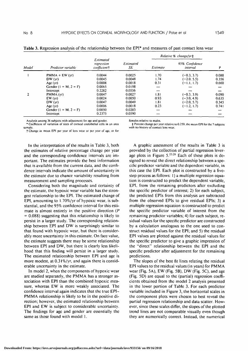
Table 3. Regression analysis of the relationship between the EPI* and measures of past contact lens wear
Model
1
2
Predictor variable
PMMA + EW (yr)DW (yr)Age (yr)Gender (1 = M, 2 = F)InterceptPMMA(vr)EW (yr)DW (yr)Age (yr)Gender (1 = M. 2 = F)Intercept
Estimatedregression
coefficient}
0.00440.00450.00080.00650.22820.00470.00240.00470.00060.00500.2375
EstimatedSE
0.00250.00480.00180.01980.05560.00270.00500.00490.00180.02030.0590
Relative °A
Estimate
1.701.740.31——1.810.931.810.23——
b change/yr%
95% Confidenceinterval
(-0.3, 3.7)(-2.0. 5.5)(-1.1. 1.7)
——
(-0.3, 3.9)(-3.0, 4.9)(-2.0. 5.7)(-1.2, 1.7)
——
P
0.0880.3560.660
——
0.0900.6350.3450.741
—
Analysis among 36 subjects with adjustment for age and gender.• Coefficient of variation of sizes of corneal cndotheliul cells in an area
sample.t Change in mean EPI per year of lens wear or per year of age. or for
females relative to males.t Percent change per year relative to 0.259. the mean EPI for the 7 subjects
with no historv of contact lens wear.
In the interpretation of the results in Table 3, boththe estimates of relative percentage change per yearand the corresponding confidence intervals are im-portant. The estimates provide the best informationthat is available from the current data, and the confi-dence intervals indicate the amount of uncertainty inthe estimate due to chance variability resulting frommeasurement and sampling processes.
Considering both the magnitude and certainty ofthe estimate, the hypoxic wear variable has the stron-gest relationship to the EPI. The estimated change ofEPI, amounting to 1.70%/yr of hypoxic wear, is sub-stantial, and the 95% confidence interval for this esti-mate is almost entirely in the positive direction (P= 0.088) suggesting that this relationship is likely topersist in a larger study. The corresponding relation-ship between EPI and DW is surprisingly similar tothat found with hypoxic wear, but there is consider-ably more uncertainty in this estimate. On face value,the estimate suggests there may be some relationshipbetween EPI and DW, but there is clearly less likeli-hood that this finding will persist in a larger study.The estimated relationship between EPI and age ismore modest, at 0.31%/yr, and again there is consid-erable uncertainty in the estimate.
In model 2, when the components of hypoxic wearare studied separately, the PMMA has a stronger as-sociation with EPI than the combined hypoxic mea-sure, whereas EW is more weakly associated. Theconfidence interval again indicates that the true EPI-PMMA relationship is likely to be in the positive di-rection; however, the estimated relationship betweenEPI and EW is subject to considerable uncertainty.The findings for age and gender are essentially thesame as those found with model 1.
A graphic assessment of the results in Table 3 isprovided by the collection of partial regression lever-age plots in Figure 5.23-26 Each of these plots is de-signed to reveal the direct relationship between a spe-cific predictor variable and the dependent variable, inthis case the EPI. Each plot is constructed by a five-step process as follows: 1) a multiple regression equa-tion is constructed to predict the dependent variable,EPI. from the remaining predictors after excludingthe specific predictor of interest; 2) for each subject,the predicted EPIs from this analysis are subtractedfrom the observed EPIs to give residual EPIs; 3) amultiple regression equation is constructed to predictthe specific predictor variable of interest from theremaining predictor variables; 4) for each subject, re-sidual values for the specific predictor are constructedby a calculation analogous to the one used to con-struct residual values for the EPI; and 5) the residualEPI values are plotted against the residual values forthe specific predictor to give a graphic impression ofthe "direct" relationship between the EPI and thespecific predictor after adjustment for the remainingpredictions.
The slopes of the best fit lines relating the residualEPI values to the residual values (in years) for PMMAwear (Fig. 5A), EW (Fig. 5B), DW (Fig. 5C), and age(Fig. 5D) are equal to the (partial) regression coeffi-cients obtained from the model 2 analysis presentedin the lower portion of Table 3. For each predictorvariable included in Figure 3, the horizontal scales inthe component plots were chosen to best reveal thepartial regression relationship and data scatter. How-ever, since these scales differ, the slopes of the plottedtrend lines are not comparable visually even thoughthey are numerically correct. Instead, the numerical
Downloaded From: https://iovs.arvojournals.org/pdfaccess.ashx?url=/data/journals/iovs/933156/ on 09/16/2018

1550 INVESTIGATIVE OPHTHALMOLOGY 6 VISUAL SCIENCE / Augusr 1990 Vol. 31
-0.1 -
-10 -5 0 5 10
Residual PMMA Wear (yrs)
0 2 4
Residual EW (yrs)
CO
p'55CD
CC
0.2 -
0.1 -
0 -
-0.1 - -0.1 -
- 1 0
Residual DW (yrs)
o io
Residual Age (yrs)
20
Fig. 5. Partial regression leverage plots that show the direct relationship between the EPI and (A) PMMA wear, (B) combined EW. (C)combined SCLDW and RGPDW. or (D) age, among 36 subjects after adjustment for the other variables in model 2.
values given for percentage change per year in Tables3, 4, and 5 should be used to compare the predictivestrength of the lens wear variables and age.
In general, these plots show the amount of trend inthe data relative to the scatter. They also show thatthe scatter is fairly evenly distributed on either side ofthe trend line, and indicate that the linear multipleregression model is adequate for purposes of thisanalysis.
Endothelial Cell Density
The estimates for the relationship between ECDand measures of past lens wear are shown in Table 4,based on an analysis approach identical to that usedfor endothelial polymegethism. In this case, the esti-mated relationships all are in a plausible directioncorresponding to a small decrease in cell density withage or lens wear. However, none of these estimateshas confidence intervals that give a very strong indi-
cation of whether positive or negative relationshipswill occur in confirmatory studies (P > 0.269).
Corneal Hydration Control
With PRPH as the operational measure, cornealhydration control was studied in relationship to pastlens wear and two co-variables, age and gender, andthe results are given in Table 5. Combined hypoxicwear is substantially related to PRPH, with an esti-mated 1.26% reduction per year (P = 0.060). Thecorresponding 95% confidence interval is located pri-marily in the negative direction, giving fairly goodcertainty that this finding can be replicated. When thecomponents of hypoxic wear are analyzed separately,in model 2, both have estimated relationships of thesame order of magnitude as the combined measure.However, the estimate for the PMMA component ismore convincingly positive because of the location ofits confidence interval. The corresponding partial re-
Downloaded From: https://iovs.arvojournals.org/pdfaccess.ashx?url=/data/journals/iovs/933156/ on 09/16/2018

No. 8 HYPOXIC EFFECTS ON CORNEAL MORPHOLOGY AND FUNCTION / Poise er ol 1551
Table 4. Regression analysis of the relationship between endothelial cell density and measuresof past contact lens wear
Model
1
2
Predictor variable
PMMA + EW(yr)DW (yr)Age (yr)Gender (1 = M, 2 = F)InterceptPMMA(yr)EW (yr)DW (yr)Age (yr)Gender (1 = M. 2 = F)Intercept
Estimatedregression
coefficients*
-8.03-6.29
-11.16132.92
3215.39-2.72
-41.06-3.28
-14.65108.86
3373.56
EstimatedSE
18.2734.7712.65
142.14399.48
18.8535.2434.7713.00
143.68423.62
Relative %
Estimate
-0.25-0.20-0.35
——
-0.09-1.29-0.10-0.46
——
change/yrf
95% Confidenceinterval
(-1.4.0.9)(-2.4. 2.0)(-1.2,0.5)
——
(-1.3, 1.1)(-3.5, 1.0)(-2.3,2.1)(-1.3,0.4)
——
P
0.6630.8580.384
——
0.8860.2530.9250.269
——
Analysis among 36 subjects with adjustment for age and gender.* Change in mean endothelial cell density per year of lens wear or per year
of age. or for females relative to males.
t Percent change per year relative to 3192.1 cells/mm2, the mean cndoihc-lial cell density for the 7 subjects with no history of contact lens wear.
gression leverage plots for the prediction of PRPH inmodel 2 are provided in Figure 6.
In this analysis the DW results go in a directionthat is inconsistent with the EP1 findings and alsoopposed to effects, if any, that might be anticipatedfrom this type of lens wear. However, since the confi-dence interval for this estimate is quite large, thesefindings well may reverse in a confirmatory study.
The estimated relationship of PRPH with age isquite low and the confidence intervals are not exces-sively large. This suggests that for subjects in the agerange of 18-35 yr, like those in this study, there is nota major decline of PRPH with age. Since previouswork has indicated that, by an average age of 70 yr,people show a substantial decline in PRPH,17 there
may be some nonlinearity in the change of PRPHwith age in which there is little decline before age 40and an accelerated decline sometime thereafter.
Discussion
In this report we have provided results from anexploratory study that was conducted to obtain a pre-liminary assessment of the impact of contact lenswear on corneal properties, with particular emphasison corneal hydration control. On the basis of theresults that have been obtained so far, there seems tobe a real possibility that chronic corneal hypoxiacauses permanent changes in corneal structure andfunction. Although the magnitude of these effects de-
Table 5. Regression analysis of the relationship between corneal hydration control (PRPH) and measuresof past contact lens wear
Model
1
2
Predictor variable
PMMA + EW (vr)DW (yr)Age (yr)Gender (1 = M, 2 = F)InterceptPMMA (vr)EW (yr)DW (yr)Age (yr)GenderIntercept
Estimatedregression
coefficients*
-0.7240.2150.065
-0.72057.472-0.720-0.748
0.2170.063
-0.73957.588
EstimatedSE
0.3710.7060.2572.8848.1060.3900.7290.7190.2692.9738.766
Relative %
Estimate
-1.260.380.11——
-1.26-1.31
0.380.11——
change/yr^
95% Confidenceinterval
(-2.6. 0.06)(-2.1.2.9)(-0.8, 1.0)
——
(-2.6.0.1)(-3.9. 1.3)(-2.2, 2.9)(-0.8, 1.1)
—
P
0.0600.7630.802
——
0.0750.3130.7650.816
—
Analysis among 36 subjects with adjustment for age and gender.• Change in mean percent recovery per hour per year of lens wear or per
year of age. or for females relative to males.
t Percent change per year relative to 57.3%/hr. the mean PRPH for the 7subjects with no history of contact lens wear.
Downloaded From: https://iovs.arvojournals.org/pdfaccess.ashx?url=/data/journals/iovs/933156/ on 09/16/2018

1552 INVESTIGATIVE OPHTHALMOLOGY & VISUAL SCIENCE / Augusr 1990 Vol. 31
20 -
I 1 0 •Q .OCQ ."55 o -
$C -10 H
B
-20 -
20 -
10 •
a 0 -
-10 •
-20 •
oo
<
o°0
o•jS
o0
o
0
o—— .
°o<o
o
oo
o
—'
0o
o
Q
• .
o
o
-10 - S O S
Residual PMMA (yrs)
10 - 2 0 2 4
Residual EW (yrs)
IQ_OCCL15•p'55<D
OC
20 -
10 -
o -
-10 -
-20 -
o °
o °°
o
o o
o
o
o
o°o
o
o
o
o
oo
IQ.OCQ.—
Resi
c20 -
io -
o •
-10 -
-20 -
° 0
° oo o
0o o
o
o o
° ° o oo o
a o
°°o° °o
o
o
o
o
-5 - 1 0
Residual DW (yrs)
o io
Residual Age (yrs)
20
Fig. 6. Partial regression leverage plots that show the direct relationship between the PRPH and (A) PMMA wear, (B) combined EW, (C)combined SCLDW and RGPDW, or (D) age, among 36 subjects after adjustment for the other variables in model 2.
pends to a certain degree on the type of lens wear, theanalysis provided by the partial regression leverageplots suggests that the observed effects are dose-de-pendent. This finding is of clinical importance sincemany of the newer EW soft lens regimens (eg, dis-posal and frequent replacement) do not meet cornealoxygen requirements during periods of eye closure.The long-term effects of such hypoxic exposure needto be carefully explored.
Our estimated associations between non-PMMADW and the EPI values, and between non-PMMADW and PRPH values, were mixed in their indica-tions. Non-PMMA DW was associated with an in-crease in the EPI, which indicates a deterioration ofendothelial structure; however, it was conversely as-sociated with a small increase in the PRPH value,which indicates improved function with lens wear.Both of these estimated relationships lacked preci-sion, so future studies with substantially more sub-jects will be needed for clarification.
It is important to point out that there are severallimitations to the precision and conclusiveness of thecurrent findings. We cannot be sure that all residualacute effects of lens wear were restored prior to themeasurements of corneal structure and function. Itremains to be established how much time must passfor all acute effects to reverse after lens wear has beendiscontinued. To ensure that the OESS thickness andrecovery measurements were not influenced by con-tact lens wear, we attempted to have all subjects dis-continue hypoxic lens wear for 3 days before testing;however, not all subjects met this requirement (seeMaterials and Methods). Whether this amount oftime is required to reach corneal stability is notknown, and to our knowledge, there have been onlytwo studies that have monitored corneal thicknessafter discontinuation of extended contact lens wear.One study measured thickness recovery of unilateralEW subjects and showed that the cornea thinned byapproximately 7 and 11 /urn after 2 and 7 days and
Downloaded From: https://iovs.arvojournals.org/pdfaccess.ashx?url=/data/journals/iovs/933156/ on 09/16/2018

No. 8 HYPOXIC EFFECTS ON CORNEAL MORPHOLOGY AND FUNCTION / Poise er ol 1553
then remained stable.5 In another study, in whichcorneal thickness change was monitored on EW sub-jects, the cornea returned to baseline thickness 2 daysafter discontinuing lens wear.7 Although more infor-mation is needed to provide convincing evidence thatthe corneal thickness is stable after 2-3 days of dis-continuing lens wear, for this current exploratorystudy, we assumed that the cornea was stable for thefunction test assessments.
A second limitation of the current study is the sub-stantial uncertainty in some of the estimated rela-tionships, due to the relatively small number of sub-jects. A clear limitation of the data set is the absenceof a solid basis for assessing the impact of any sub-stantial past history of a mixture of PMMA andSCLEW, since there were not enough subjects whohad experience with both modes of wear.
A third concern is that the subjects in this studywere a convenient sample of volunteers, so there is noguarantee that they provided valid estimates of corre-sponding relationships in the general population ofcontact lens wearers, for which we would like to makeinferences. Thus, the possibility of selection bias thatmay be an influence in the findings cannot be dis-counted. It should be noted, however, that in thistype of study it is not necessary to have a randomsample of individuals from the target population. In-stead, a valid estimate of the relationships betweenthe predictor and corneal variables depends only onhaving a fair sample from the distributions of out-come variable values conditional on the predictorvariables, for the subjects used. In practice, studies ofthe type involved here always depend on volunteersamples, and it is virtually impossible to rule out thepossibility that a particular study is subject to somebias. Consequently, it is essential to do replicate stud-ies under diverse conditions to see if consistent resultscan be obtained.
Finally, the measures based on self reports of lenswear used in the study were fairly crude. We had nospecific data on their validity compared to that ofclinical records. Also, we did not consider the averageamount of wear per day or any other aspects of lenswear behavior. Intuitively, however, it seems morelikely that weakened rather than strengthened associ-ations would result from these measurement prob-lems, which probably would tend to obscure the truerelationships.
Throughout the analysis and interpretation of re-sults, we took the position that this is a preliminarystudy, in which estimated strengths of relationshipsplay the primary role. Use of these estimates was thentempered by considering their uncertainty as indi-cated by confidence intervals. Possible sources of bias
were also considered. Although the results of this ex-ploratory study cannot be considered definitive, theyare, in our opinion, sufficient to indicate the need forfurther investigation of the structural and functionalchanges that occur as a result of wearing contactlenses that cause corneal hypoxia.
Key words: corneal function, corneal cndothclial morphol-ogy, hypoxia, contact lens, corneal edema
References
1. Maurice D: The cornea and sclcra. /// The Eye. Davson H,editor. New York. Academic Press. 1984. pp. 1-158.
2. Klycc S: Stromal lactate accumulation can account for conicaloedema osmotically following epithelial hypoxia in the rabbit.J Physiol 321:49. 1981.
3. Bonanno J and Poise K: Corneal acidosis during contact lenswear: Effects of hypoxia and CO2. Invest Ophthalmol Vis Sci28:1514. 1987.
4. Schoesslcr J and Woloschak M: Corneal cndothelium in vet-eran PMMA contact lens wearers. International Contact LensClinic 8:19. 1981.
5. Holdcn B, Sweeney D. Vannas A. Nilsson K. and Efron N:Effects of long-term extended contact lens wear on the humancornea. Invest Ophthalmol Vis Sci 26:35. 1985.
6. MacRac S, Matsuda M, Shellans S. and Rich L: The effects ofhard and soft contact lenses on the conical cndothelium. Am JOphthalmol 102:50. 1986.
7. Kenyon E. Poise K. and Scgcr R: Influence of wearing scheduleon extended-wear complications. Ophthalmology 92:231,1986.
8. MacRac S. Matsuda M. and Shellans S: Corneal cndothclialchanges associated with contact lens wear. CLAO J I5(l):82,1989.
9. Sibug M. Datiles M. McCain L. Kashima K. Edwards P.Kaiser-Kupfer M. and Krachcr G: Specular microscopy stud-ies on the corneal cndothelium after cessation of contact lenswear. ARVO Abstracts. Invest Ophthalmol Vis Sci29(Suppl):257, 1989.
10. Rao G, Shaw E, Arthur E. and Aquavclla J: Endothclial cellmorphology and corneal dcturgescencc. Ann Ophthalmol11:885. 1979.
11. Lass J. Dutt R, Spurvcy R. Stockcr E, WolffC. and Glavan 1:Morphologic and fluoromctric analysis of the conical endothc-lium in long-term hard and soft contact lens wearers. CLAO J14(2): 105. 1988.
12. Dutt R, Stockcr E, Wolff C. Glavan I, and Lass J: A morpho-logic and fluorometric analysis of the corneal cndothelium inlong-term extended wear soft contact lens wearers. CLAO JI5(2):121. 1989.
13. MacDonald J and McCarey B: Hypoxic stress of contact lenswear on the pump site density of the conical cndothelium.ARVO Abstracts. Invest Ophthalmol Vis Sci 29(Suppl):4l9,1988.
14. Carlson K, Bourne W, and Brubaker R: Effect of long-termcontact lens wear on corneal cndothelial cell morphology andfunction. Invest Ophthalmol Vis Sci 29:185. 1988.
15. O'Neal M. Poise K, and Sarvcr M: Corneal response to rigidand hydrogel lenses during eye closure. Invest Ophthalmol VisSci 25:837. 1984.
Downloaded From: https://iovs.arvojournals.org/pdfaccess.ashx?url=/data/journals/iovs/933156/ on 09/16/2018

1554 INVESTIGATIVE OPHTHALMOLOGY & VISUAL SCIENCE / Augusr 1990 Vol. 31
16. Holdcn B, Mcrtz G, and McNally J: Corneal swelling responseto contact lenses worn under extended wear conditions. InvestOphthalmol Vis Sci 24:218, 1983.
17. Poise K, Brand R, Mandcll R. Vastine D, Dcmartini D, andFlom R: Age differences in corneal hydration control. InvestOphthalmol Vis Sci 30:392. 1989.
18. Mandcll R, Poise K, and Bonanno J: Reassessment of opticalpachomctry. In The Cornea: Transactions of the World Con-gress on the Cornea III, Cavanagh H, editor. New York.Raven, 1988, p. 201.
19. Holden B, Williams L, and Zantos S: The etiology of transientendothelial changes in the human cornea. Invest OphthalmolVis Sci 26:1354, 1985.
20. Mandcll R, Poise K, Brand R, Mandell R, Vastine D, Dcmar-tini D, and Flom R: Corneal hydration control in Fuchs' dys-trophy. Invest Ophthalmol Vis Sci 30:845. 1989.
21. Mandell R, Poise K, and Fatt I: Corneal swelling caused bycontact lens wear. Arch Ophthalmol 83:3. 1970.
22. Sarver M, Poise K. and Harris M: Patient responses to gas-pcr-mcablc hard (Polycon) contact lenses. Am J Optom 54:195,1977.
23. Computing Resource Center: Stata Reference Manual, Ver-sion 2.05. Los Angles, Computing Resources Center. 1989 p.387.
24. Laing R, Sandstrom M. Berrospi A. and Leibowitz H: Changesin the corneal endothclium as a function of age. Exp Eye Res22:587, 1976.
25. Ycc R, Matsuda M, Schultz R, and Edclhauscr H: Changes inthe normal corneal endothelial cellular pattern as a function ofage. Curr Eye Res 4:671. 1985.
26. Myers R: Classical and modern regression with applications.Boston. Duxbury Press. 1986 p. 155.
Downloaded From: https://iovs.arvojournals.org/pdfaccess.ashx?url=/data/journals/iovs/933156/ on 09/16/2018