hypothalamic and brainstem neuronal circuits controlling
TRANSCRIPT

JournalofEndocrinology
Thematic ReviewM SCHNEEBERGER and others Neuronal circuits and energy
balance220 :2 T25–T46
Hypothalamic and brainstemneuronal circuits controllinghomeostatic energy balance
Marc Schneeberger1,2,3, Ramon Gomis1,2,3 and Marc Claret1,3
1Diabetes and Obesity Research Laboratory, Institut d’Investigacions Biomediques August Pi i Sunyer (IDIBAPS),
08036 Barcelona, Spain2Department of Endocrinology and Nutrition, School of Medicine, Hospital Clınic, University of Barcelona, 08036
Barcelona, Spain3Centro de Investigacion Biomedica en Red de Diabetes y Enfermedades Metabolicas Asociadas (CIBERDEM), 08036
Barcelona, Spain
http://joe.endocrinology-journals.orgDOI: 10.1530/JOE-13-0398
� 2014 Society for EndocrinologyPrinted in Great Britain
Published by Bioscientifica Ltd.This paper is one of four papers that form pEnergy, Insulin Resistance and Metabolic Sywas Shaodong Guo, Texas A&M University,
Downloa
Correspondence
should be addressed
to M Claret
Abstract
Alterations in adequate energy balance maintenance result in serious metabolic
disturbances such as obesity. In mammals, this complex process is orchestrated by multiple
and distributed neuronal circuits. Hypothalamic and brainstem neuronal circuits are critically
involved in the sensing of circulating and local factors conveying information about the
energy status of the organism. The integration of these signals culminates in the generation
of specific and coordinated physiological responses aimed at regulating energy balance
through the modulation of appetite and energy expenditure. In this article, we review
current knowledge on the homeostatic regulation of energy balance, emphasizing recent
advances in mouse genetics, electrophysiology, and optogenetic techniques that have
greatly contributed to improving our understanding of this central process.
Key Words
" CNS
" ghrelin
" leptin
" neuroendocrinology
" obesity
artndrTX,ded
Journal of Endocrinology
(2014) 220, T25–T46
Introduction
The regulation of appetite and body weight is an intricate
process controlled by redundant and distributed neural
systems that integrate a myriad of cognitive, hedonic,
emotional, and homeostatic cues to precisely regulate
systemic energy balance through behavioral, autonomic,
and endocrine outputs. These sophisticated biological
programs are influenced by multiple factors, including
environmental, genetic, and epigenetic mechanisms. The
immense complexity of these systems illustrates the
biological importance of adequate nutrient and energy
balance, a process that has been evolutionarily conserved
and refined to guarantee appropriate adiposity levels.
Despite the precision of these systems in matching energy
demand with energy expenditure, contemporary, and
lifestyle factors are themaincausesof theprevailingobesity
epidemics. The present review attempts to summarize
current understanding of the anatomy, neurochemistry,
functions, and interactions of relevant neural circuits
involved in the homeostatic regulation of energy balance.
The homeostatic system: hypothalamus andbrainstem
The hypothalamus: neuronal anatomy, nuclei, and
neuropeptides
Seminal lesioning studies conducted in rodents during
the 1940s and 1950s highlighted the importance of the
hypothalamus in the regulation of body weight. Since
then, extensive experimental evidence and extraordinary
of a thematic review section onome. The Guest Editor for this sectionUSA from Bioscientifica.com at 03/01/2022 01:09:52AM
via free access

JournalofEndocrinology
Thematic Review M SCHNEEBERGER and others Neuronal circuits and energybalance
220 :2 T26
progress in understanding the neurobiology of obesity
have firmly established the mediobasal hypothalamus as a
fundamental nexus in the neuronal hierarchy controlling
whole-body energy balance. The hypothalamus is con-
stituted by distinct hypothalamic nuclei including the
arcuate nucleus (ARC), the paraventricular nucleus (PVN),
the lateral hypothalamic area (LHA), the dorsomedial
nucleus (DMN), and the ventromedial nucleus (VMN).
Arcuate nucleus The ARC is a very important area
of the CNS involved in the control of energy homeostasis. It
is located below the VMN, on both sides of the third
ventricle, and immediately adjacent to the median emi-
nence (ME). This area has a semi-permeable blood–brain
barrier (BBB; Broadwell & Brightman 1976), and thus it is
strategically positioned to sense hormonal and nutrient
fluctuations in the bloodstream. In the ARC, there are
at least two major populations of neurons controlling
appetite and energy expenditure: i) a subset of neurons
that coexpress orexigenic neuropeptideY (NPY) andagouti-
related peptide (AGRP) and ii) a population of neurons
that coexpress the anorexigenicneuropeptides cocaine- and
amphetamine-regulated transcript (CART (CARTPT)) and
a-melanocyte-stimulating hormone (a-MSH, a product of
proopiomelanocortin (POMC) processing). These two
populations of neurons (hereafter referred to as AgRP and
POMC respectively), together with downstream target
neurons expressing the melanocortin receptor 4 (MC4R)
and MC3R, constitute the central melanocortin system.
This neuronal circuit is crucial for sensing and integrating a
number of peripheral signals allowing for a precise control
of food intake and energy expenditure (see section ‘ARC
neuronal circuits: POMC, AgRP, and RIPCre neurons’).
NPY is widely expressed throughout the CNS, but it is
most densely localized in the ARC in the hypothalamus
(Gehlert et al. 1987). The expression and release of ARC
NPY respond to changes in energy status, being reduced
under feeding conditions and increased under fasting
conditions (Beck et al. 1990, Kalra et al. 1991). Increasing
NPY tone pharmacologically results in hyperphagia and
reduced thermogenesis of brown adipose tissue (BAT),
associated with diminished activity of the thyroid axis
(Clark et al. 1984, Stanley et al. 1986, Egawa et al. 1991).
Although NPY acts at five different receptors (Y1, Y2, Y3,
Y4, and y6), genetic and pharmacological studies suggest
that postsynaptic Y1 and Y5 receptors mediate the effects
of NPY on positive energy balance (Nguyen et al. 2012,
Sohn et al. 2013).
AGRP is also an orexigenic neuropeptide that is
exclusively expressed in the ARC, where it colocalizes
http://joe.endocrinology-journals.orgDOI: 10.1530/JOE-13-0398
� 2014 Society for EndocrinologyPrinted in Great Britain
with NPY and the neurotransmitter g-aminobutyric acid
(GABA; Broberger et al. 1998, Cowley et al. 2001). The
central administration of AGRP or its genetic over-
expression stimulates food intake, reduces energy
expenditure, and causes obesity (Graham et al. 1997,
Ollmann et al. 1997, Small et al. 2003). Interestingly,
lasting orexigenic effects (over days) after AGRP delivery
have been reported (Hagan et al. 2000).
AgRP neurons express receptors for peripheral
hormonal signals such as insulin (Marks et al. 1990),
leptin (Elmquist et al. 1998), and ghrelin (Willesen et al.
1999). These neurons send projections mainly into the
PVN, DMN, and LHA. Despite the well-documented effects
of NPY and AGRP as positive modulators of energy
balance, genetic studies have yielded conflicting results.
For example, Agrp- and Npy-knockout (KO) mice failed to
exhibit alterations in body weight or feeding behavior
(Palmiter et al. 1998, Qian et al. 2002, Corander et al.
2011). However, the ablation of AgRP neurons in adults
leads to uncontrolled anorexia but is well tolerated in
neonates, indicating the existence of developmental
compensations (Bewick et al. 2005, Gropp et al. 2005,
Luquet et al. 2005).
CART is widely expressed in the brain, but it is
particularly abundant in the hypothalamus, and it coloca-
lizes (O95%) with POMC in the ARC (Elias et al. 1998). Its
expression is enhanced under feeding conditions and
reduced under fasting conditions (Kristensen et al. 1998),
and it has been shown that i.c.v. infusion of CART inhibits
food intake, while antibodies against CART reverse this
effect (Kristensen et al. 1998). Furthermore, CART also
stimulates the thermogenesis of BAT (Kotz et al. 2000).
However, Cartpt-deficient mice exhibit no alterations in
food intake or body weight when fed with a standard diet,
but develop obesity after being fed with a high-fat diet
(HFD;Asnicar et al. 2001). Interestingly, andcontrary to the
prevailing anorexigenic view, other studies have shown
that under certain experimental conditions CART may
stimulate food intake (Abbott et al. 2003, Kong et al. 2003).
Collectively, results regarding the effects of CART on
feeding behavior are inconclusive and indicate anatomi-
cally divergent roles for this neuropeptide.
POMC is a prohormone precursor that is cleaved into
several bioactive peptides in the hypothalamus, including
a-MSH, which exerts potent anorexigenic effects by
binding to MC3R and MC4R (Mercer et al. 2013). The
levels of Pomc transcripts and a-MSH are increased under
feeding conditions and decreased under fasting conditions
(Schwartz et al. 1997). The i.c.v. administration of a-MSH
or its delivery into the PVN suppresses food intake and
Published by Bioscientifica Ltd
Downloaded from Bioscientifica.com at 03/01/2022 01:09:52AMvia free access

JournalofEndocrinology
Thematic Review M SCHNEEBERGER and others Neuronal circuits and energybalance
220 :2 T27
reduces body weight (Poggioli et al. 1986, Wirth et al.
2001). Genetic manipulation of the Pomc gene leading to
the overexpression of a-MSH has been shown to cause
anti-obesity effects in genetic and diet-induced obesity
(DIO) models (Mizuno et al. 2003, Savontaus et al. 2004,
Lee et al. 2007). A key role for POMC in whole-body energy
homeostasis is evident, as mice lacking Pomc, melano-
cortin peptides, or POMC neurons develop obesity
(Yaswen et al. 1999, Gropp et al. 2005, Xu et al. 2005a,
Smart et al. 2006). Furthermore, mutations in the POMC
gene have been reported to be associated with morbid
obesity in humans (Krude et al. 1998, Lee et al. 2006).
GABAergic and glutamatergic subpopulations of POMC
neurons have been described, although their functional
roles are unclear (Mercer et al. 2013).
Paraventricular nucleus The PVN is located in the
anterior hypothalamus, just above the third ventricle, and
expresses high levels of MC3R/MC4R. It receives inner-
vation not only from the AgRP and POMC neurons of the
ARC but also from extrahypothalamic regions such as
the nucleus of the tractus solitarius (NTS). The PVN is
an important integration site involved in whole-body
energy homeostasis, as shown by the diverse afferent
inputs and its high sensitivity to the administration of
endogenous neuropeptides involved in the regulation
of food intake such as NPY, AGRP, and a-MSH, among
others (Stanley et al. 1986, Kim et al. 2000). Part of these
effects are mediated by a subset of neurons that express
thyrotropin-releasing hormone (TRH), which are activated
by a-MSH and inhibited by AGRP (Fekete et al. 2000, 2004).
Another relevant subset of neurons express corticotrophin-
releasing hormone (CRH), which are directly involved
in the control of energy balance throughAGRP innervation
or indirectly through the regulation of adrenal gluco-
corticoids controlling the expression of POMC (Richard
& Baraboi 2004).
Lateral hypothalamus area The LHA plays a critical
role in the mediation of orexigenic responses, a function
that can be significantly attributed to orexin andmelanin-
concentrating hormone (MCH) neurons. Orexin neurons
produce orexin A and orexin B from prepro-orexin, the
expression of which is increased under fasting conditions
(Sakurai et al. 1998). The central administration of orexins
not only increases food intake (Sakurai et al. 1998, Dube
et al. 1999), but also promotes behavioral responses to
food reward and increases arousal (Cason et al. 2010).
Orexin neurons project not only within the LHA, ARC,
PVN, and NTS, but also into other regions involved
http://joe.endocrinology-journals.orgDOI: 10.1530/JOE-13-0398
� 2014 Society for EndocrinologyPrinted in Great Britain
in additional physiological functions such as body
temperature and wakefulness control, among others
(Peyron et al. 1998). Similarly, fasting enhances the
expression of Mch (Pmch) mRNA and its i.c.v. adminis-
tration or genetic overexpression causes an orexigenic
output (Qu et al. 1996, Ludwig et al. 2001). Conversely,
mice with reduced MCH tone or disruption of the MCH1
receptor are lean (Marsh et al. 2002).
Dorsomedial nucleus The DMN is involved in a
range of physiological processes, including appetite,
thermoregulation, stress, and circadian rhythms. It
receives projections from most of the hypothalamic
nuclei, especially the ARC, and sends projections into
the PVN and LHA. A number of neuropeptides (such as
NPY and CRH) as well as receptors for peptides involved in
the control of appetite and energy balance are expressed
within the DMN. Increased expression of NPY in the
DMN has been reported in several rodent models of
obesity (Guan et al. 1998, Bi et al. 2001), and it may play
a significant role in the regulation of thermogenesis and
the development of DIO (Chao et al. 2011).
Ventromedial nucleus The AgRP and POMCneurons
of the ARC project into the VMN. In turn, VMN neurons
project into hypothalamic and extrahypothalamic areas
such as the brainstem (Cheung et al. 2013). Laser-
microdissection studies have identified a number of
VMN-enriched genes (Segal et al. 2005), including ster-
oidogenic factor 1 (Sf1 (Nr5a1)), which has been directly
implicated in the development of the VMN (Parker et al.
2002, Davis et al. 2004). SF1-expressing neurons play
significant roles in the control of energy balance, as
demonstrated by themetabolic phenotypes of conditional
KOmice (Bingham et al. 2008, Zhang et al. 2008, Kim et al.
2011). Another abundantly expressed protein in the VMN
is the brain-derived neurotrophic factor (BDNF). The lack
of BDNF or its receptor (TRKB (NTRK2)) leads to hyper-
phagia and obesity in humans andmice (Lyons et al. 1999,
Yeo et al. 2004). In contrast, the central or peripheral
administration of BDNF results in the loss of body weight
and reduction in food intake through MC4R signaling
(Xu et al. 2003). The VMN also plays a key role in the
regulation of thermogenesis (Lopez et al. 2010, Kim et al.
2011,Martinez deMorentin et al. 2012,Whittle et al. 2012).
The brainstem
Brainstem neurons make key contributions to the control
of energy balance by processing energy status information
Published by Bioscientifica Ltd
Downloaded from Bioscientifica.com at 03/01/2022 01:09:52AMvia free access

JournalofEndocrinology
Thematic Review M SCHNEEBERGER and others Neuronal circuits and energybalance
220 :2 T28
at four different levels: i) by sensing circulating meta-
bolites and hormones released by peripheral organs; ii) by
receiving vagal inputs from the gastrointestinal (GI) tract;
iii) by receiving neuronal inputs from midbrain and
forebrain nuclei that also detect and integrate energy-
related signals; and iv) by projecting into local brainstem
circuits and other regions of the brain to provide
information that will be integrated by these neurons to
control energy balance. Within the brainstem, the dorsal
vagal complex (DVC) is a keymodule for the integration of
energy-related cues by relying peripheral signals through
vagal afferents and projecting into the hypothalamus and
other relevant areas. The DVC comprises the dorsal motor
nucleus of the vagus, the NTS, and the area postrema (AP),
which has an incomplete BBB and therefore it is accessible
to peripheral signals.
The brainstem is constituted by heterogeneous popu-
lations of neurons, with distinct biophysical and neuro-
chemical properties, that express appetite-modulatory
neuropeptides such as tyrosine hydroxylase (TH), pro-
glucagon, CART, GABA, NPY, BDNF, and POMC, among
others. These neurons also express a variety of receptors
mediating the effects of some of the aforementioned
neuropeptides, indicating the existence of local circuits
that contribute to the regulation of ingestive behaviors.
In addition, receptors for a number of circulating hormones
such as leptin, ghrelin, glucagon-like peptide 1 (GLP1), and
cholecystokinin (CCK) have been described in brainstem
neurons or in vagal afferent projections to brainstem areas.
Vagal signaling from the GI tract is an important
afferent to the NTS, conveying information about luminal
distension, nutritional content, and locally produced
peptides via glutamate neurotransmission (Travagli et al.
2006). This vagal sensory and hormonal information will
be assimilated by second-order NTS neurons that project
into the hypothalamus and other basal forebrain areas to
elaborate precise outputs. The significance of the vagus
nerve transmission has been demonstrated through a
number of manipulations to eliminate or enhance its
activity. For example, chronic or acute vagus nerve
stimulation in rats leads to a reduction in body weight
and food intake, indicating that direct vagal afferent
interventions influence feeding behavior (Krolczyk et al.
2001, Gil et al. 2011). Vagal signaling also plays important
roles in the regulation of meal size and duration (Schwartz
et al. 1999).
The NTS receives inputs from descending projections
from the hypothalamus. In particular, ARC POMC
neurons project into the NTS, where high expression
levels of MC4R have been reported (Kishi et al. 2003).
http://joe.endocrinology-journals.orgDOI: 10.1530/JOE-13-0398
� 2014 Society for EndocrinologyPrinted in Great Britain
In addition to the release of a-MSH from ARC POMC
neurons, the NTS also receives melanocortin agonist
signals from a local population of w300 POMC neurons
(around 10% of the total number of POMC neurons;
Palkovits & Eskay 1987). Recent pharmacogenetic studies
have shown different functions and time scale effects of
ARC and NTS POMC neurons on food intake and
metabolism (Zhan et al. 2013). The importance of this
neuronal circuit is further demonstrated by hindbrain
MC4R agonist delivery, which leads to a reduction in food
intake and an increase in energy expenditure, whereas
MC4R antagonism drives the opposite effect (Williams
et al. 2000, Skibicka & Grill 2009b). MC4Rs in the NTS
seem to mediate not only the satiation effects of CCK
(Fan et al. 2004), but also the anorexigenic effects of
hypothalamic and brainstem leptin signaling (Skibicka &
Grill 2009a, Zheng et al. 2010).
The NTS also receives descending projections from
orexin andMCHneurons located in the LHA (Ciriello et al.
2003), and the delivery of orexin A into the hindbrain
increases food intake (Parise et al. 2011). The orexigenic
nature of the LHA and the anatomical connection with
the NTS indicated that this system may serve as a
mechanism to limit the satiety signals from the GI tract.
Another hypothalamic nucleus sending projections
into the NTS is the PVN (Sawchenko & Swanson 1982,
Luiten et al. 1985). The PVN–brainstem pathway plays a
significant role in the regulation of energy balance, as
contralateral disruption of PVN output and NTS input
causes hyperphagic obesity (Kirchgessner & Sclafani
1988). Different areas of the brainstem show TRH-positive
fibers, and evidence indicates that TRH is involved in the
brainstem regulation of energy homeostasis by integrating
endocrine and vagal–sympathetic responses (Ao et al.
2006, Zhao et al. 2013).
Hormonal signals involved in energyhomeostasis control
Peripheral adiposity signals: leptin and insulin
The discovery of leptin, the product of the Ob gene, in
1994 (Zhang et al. 1994) opened a new dimension in the
field of the central regulation of energy balance. Leptin is
an anorexigenic adipose tissue-derived hormone that
circulates in proportion to fat mass (Considine et al.
1996). It reaches the CNS through a saturable transport
system and conveys information about the energy status
of the organism. There are multiple leptin receptor (LEPR)
isoforms, with the long form (LEPRb) being essential for
Published by Bioscientifica Ltd
Downloaded from Bioscientifica.com at 03/01/2022 01:09:52AMvia free access
JournalofEndocrinology
Thematic Review M SCHNEEBERGER and others Neuronal circuits and energybalance
220 :2 T29
the effects of leptin. The lack of leptin or LEPRb in both
rodents and humans causes a phenotype characterized by
hyperphagia, reduced energy expenditure, and severe
obesity (Halaas et al. 1995, Chen et al. 1996, Montague
et al. 1997, Clement et al. 1998). Most obese patients
exhibit a state of leptin resistance, which is the inability of
high circulating leptin levels to exert central anorexigenic
actions, which precludes the use of leptin as a thera-
peutical approach.
LEPRb is highly expressed in different hypothalamic
nuclei and other CNS regions involved in the control of
energy balance (Elmquist et al. 1998). In the ARC, the
POMC, and AgRP neurons are the direct targets of leptin
(Cheung et al. 1997, Elias et al. 1999, Cowley et al. 2001).
The ablation of LEPRb in POMC neurons, AgRP neurons,
or both populations of neurons causes increased body
weight, emphasizing the importance of leptin signaling
(Table 1). However, the magnitude of these changes is
smaller than that observed in mice globally lacking Lepr,
indicating the existence of additional subsets of neurons
mediating the effects of leptin on food intake and body
weight. Leptin binds to LEPRb and activates JAK2, which,
in turn, phosphorylates several tyrosine residues on the
intracellular domain of the LEPRb. This results in the
activation, dimerization, and nuclear translocation of
STAT3 (Robertson et al. 2008). In the nucleus, STAT3
enhances Pomc gene expression and inhibits Agrp gene
expression (Munzberg et al. 2003, Kitamura et al. 2006).
Accordingly, Stat3 deficiency in POMC neurons results in
overweight and Pomc gene transcriptional defects in
females (Table 1). This signaling cascade is negatively
regulated by the suppressor of cytokine signaling
3 (SOCS3), the expression of which is also regulated by
STAT3 and protein tyrosine phosphatase 1B (PTP1B)
(Robertson et al. 2008). Consistent with this, deletion of
either Socs3 or Ptp1b (Ptpn1) in POMC neurons leads to
reduced adiposity, improved leptin sensitivity, and
increased energy expenditure under HFD conditions
(Table 1). In addition, leptin also activates the phospha-
tidylinositol-3-kinase (PI3K) pathway. A variety of genetic
mouse models targeting the catalytic or regulatory
subunits of PI3K in specific subsets of neurons have been
reported with divergent results (Table 1). Overall, these
studies indicate that PI3K is required for leptin-mediated
regulation of energy balance and that, contrary to the
prevailing view, the catalytic p110b subunit in ARC
neurons may play a more prominent role than p110a.
PI3K generates phosphatidylinositol-3,4,5-triphosphate
(PIP3) and activates downstream targets such as
phosphoinositide-dependent kinase 1 (PDK1) and AKT
http://joe.endocrinology-journals.orgDOI: 10.1530/JOE-13-0398
� 2014 Society for EndocrinologyPrinted in Great Britain
(also known as protein kinase B), which consecutively
phosphorylates the transcription factor forkhead box
protein O1 (FOXO1). Upon phosphorylation, FOXO1 is
excluded from the nucleus, allowing STAT3 to bind to
Pomc and Agrp promoters, thereby stimulating and
inhibiting respectively the expression of these neuro-
peptides (Kitamura et al. 2006). These findings are
consistent with the effects of genetic manipulations
in vivo (Table 1). PI3K signaling is counterbalanced by
phosphatase and tensin homolog (PTEN), which speci-
fically dephosphorylates PIP3. The loss of Pten in POMC
neurons results in increased PIP3 signaling and diet-
sensitive obesity via KATP channel modulation, suggesting
a role for the PI3K pathway in the regulation of the activity
of this channel (Table 1). Overall, leptin stimulates Pomc
transcription, depolarizes POMC neurons, and also
increases a-MSH processing and secretion (Cowley et al.
2001, Munzberg et al. 2003, Guo et al. 2004) while
attenuating the expression and release of orexigenic NPY
and AGRP neuropeptides (Stephens et al. 1995, Mizuno &
Mobbs 1999).
Insulin, produced by pancreatic b-cells, has tradition-
ally been associated with glucose metabolism, but
compelling evidence indicates that insulin also acts as an
anorectic signal within the CNS. Glucose-induced insulin
is secreted into the bloodstream in proportion to fat stores
(Bagdade et al. 1967) and enters the brain through a
saturable transport mechanism (Baura et al. 1993). The
i.c.v. or intrahypothalamic administration of insulin to
primates and rodents reduces food intake (Woods et al.
1979, McGowan et al. 1993, Air et al. 2002). Insulin
receptor (IR (INSR)), as well as its downstream signaling
machinery, is expressed in hypothalamic areas involved in
the control of appetite (Havrankova et al. 1978, Corp et al.
1986) and colocalizes with AgRP and POMC neurons
(Benoit et al. 2002). Surprisingly, the loss of Insr in either
POMC or AgRP neurons does not lead to alterations in
energy balance (Table 1), although hepatic glucose
production defects have been observed in mice lacking Ir
in AgRP neurons (Konner et al. 2007). Neuron-specific IR
reconstitution in L1 mice (which haveO90% reduction of
IR levels in the ARC) confirmed that insulin signaling in
AgRP and POMC neurons controls glucose metabolism
and energy expenditure respectively (Table 1). Insulin
binding to IR leads to the autophosphorylation of the
receptor and the consequent recruitment of IRS proteins,
which converge with the leptin pathway at the PI3K level
(Xu et al. 2005b). Negative regulators of the LEPR, such as
SOCS3 and PTP1B, also directly inhibit the IR and its
signaling cascade acting on IRS1. The activation of the
Published by Bioscientifica Ltd
Downloaded from Bioscientifica.com at 03/01/2022 01:09:52AMvia free access

Table
1Su
mm
ary
of
rele
van
tg
en
eti
cm
ou
sem
od
els
use
din
the
an
aly
sis
of
lep
tin
an
din
suli
nsi
gn
ali
ng
path
ways
inPO
MC
an
dA
gR
Pn
eu
ron
s
Geneticm
anipulation
Neuro
nalce
llty
pe
BW
Adiposity
Foodinta
ke
Energ
y
expenditure
Diet
Oth
erfe
atu
res
Refe
rence
s
Lepr
dele
tio
nPO
MC
CC
ZZ
Ch
ow
Alt
ere
dn
eu
rop
ep
tid
eexp
ress
ion
Balt
hasa
retal.
(2004)
Lepr
dele
tio
nA
gR
PC
CZ
ZC
ho
wR
ed
uce
dlo
com
oto
ract
ivit
yva
nd
eW
alletal.
(2008)
Lepr
dele
tio
nPO
MC
an
dA
gR
PC
CTr
an
sien
tC
KC
ho
wIn
crease
dre
spir
ato
ryexc
han
ge
rati
ova
nd
eW
alletal.
(2008)
Ird
ele
tio
nPO
MC
ZZ
ZN
DC
ho
wan
dH
FD–
Ko
nn
eretal.
(2007)
Ird
ele
tio
nA
gR
PZ
ZZ
ND
Ch
ow
an
dH
FDEn
han
ced
hep
ati
cg
luco
sep
rod
uct
ion
Ko
nn
eretal.
(2007)
Irre
-exp
ress
ion
inL1
mic
ePO
MC
KZ
CC
Ch
ow
Insu
lin
resi
stan
ceLi
netal.
(2010)
Irre
-exp
ress
ion
inL1
mic
eA
gR
PK
ZZ
CC
ho
wR
esc
ued
hep
ati
cg
luco
sep
rod
uct
ion
Linetal.
(2010)
Lepr
an
dIr
dele
tio
nPO
MC
CZ
ZK
Ch
ow
Insu
lin
resi
stan
cean
dre
du
ced
fert
ilit
yin
fem
ale
s
Hil
letal.
(2010)
Irs2
dele
tio
nPO
MC
ZZ
ZZ
Ch
ow
No
rmal
insu
lin
an
dle
pti
nle
vels
Ch
ou
dh
ury
etal.
(2005)
Ptp1b
dele
tio
nPO
MC
KK
ZC
HFD
Imp
rove
dle
pti
nse
nsi
tivi
tyB
an
noetal.
(2010)
Stat3
dele
tio
nPO
MC
CC
CN
DC
ho
wN
orm
al
ph
en
oty
pe
inm
ale
mic
eX
uetal.
(2007)
Stat3
dele
tio
nA
gR
PC
CC
ND
Ch
ow
Hyp
ore
spo
nsi
veto
lep
tin
Go
ngetal.
(2008)
Stat3
con
stit
uti
veact
ive
form
PO
MC
CC
CN
DC
ho
wN
oad
dit
ion
al
eff
ect
on
HFD
ad
min
istr
ati
on
Ern
stetal.
(2009)
Stat3
con
stit
uti
veact
ive
form
Ag
RP
KK
ZC
Ch
ow
an
dH
FDIn
crease
dlo
com
oto
ract
ivit
yM
esa
rosetal.
(2008)
Pdk1
dele
tio
nPO
MC
CC
CZ
Ch
ow
Decr
ease
dPomc
gen
eexp
ress
ion
Iskan
daretal.
(2010)
Pdk1
dele
tio
nA
gR
PK
KK
ZC
ho
wR
esc
ued
by
do
min
an
tn
eg
ati
veFo
xo1
Caoetal.
(2011)
Pdk1
dele
tio
nPO
MC
Tran
sien
tC
Tran
sien
tC
Tran
sien
tC
ND
Ch
ow
an
dH
FDR
esc
ued
by
do
min
an
tn
eg
ati
veFo
xo1
Belg
ard
tetal.
(2008)
Foxo
1d
ele
tio
nPO
MC
KK
KZ
Ch
ow
Incr
ease
dCpe
exp
ress
ion
an
da
-MSH
leve
lsPlu
metal.
(2009)
Foxo
1co
nst
itu
tive
act
ive
form
PO
MC
C(F
em
ale
s)C
(Fem
ale
s)C
(Fem
ale
s)Z
Ch
ow
Decr
ease
dPomc
gen
eexp
ress
ion
Iskan
daretal.
(2010)
Foxo
1d
ele
tio
nA
gR
PZ
KK
ZC
ho
wR
esi
stan
tto
HFD
Renetal.
(2012)
Socs3
dele
tio
nPO
MC
KN
DZ
CH
FDN
ob
od
yw
eig
ht
ph
en
o-
typ
eo
nch
ow
die
tK
ievi
tetal.
(2006)
Socs3
ove
rexp
ress
ion
PO
MC
CC
ZK
Ch
ow
Lep
tin
resi
stan
ceR
eedetal.
(2010)
Socs3
ove
rexp
ress
ion
Ag
RP
ZZ
CC
Ch
ow
Alt
ere
dg
luco
sem
eta
b-
oli
smO
lofs
sonetal.
(2013)
Pten
dele
tio
nPO
MC
CC
CZ
Ch
ow
Gen
der
dim
orp
his
mo
nH
FDad
min
istr
ati
on
Plu
metal.
(2009)
JournalofEndocrinology
Thematic Review M SCHNEEBERGER and others Neuronal circuits and energybalance
220 :2 T30
http://joe.endocrinology-journals.orgDOI: 10.1530/JOE-13-0398
� 2014 Society for EndocrinologyPrinted in Great Britain
Published by Bioscientifica Ltd
Downloaded from Bioscientifica.com at 03/01/2022 01:09:52AMvia free access
Table
1Continued
Geneticm
anipulation
Neuro
nalce
llty
pe
BW
Adiposity
Foodinta
ke
Energ
y
expenditure
Diet
Oth
erfe
atu
res
Refe
rence
s
p85
(Pik3r1
)d
ele
tio
nPO
MC
ZN
DN
DN
DC
ho
wG
en
der
dim
orp
his
mo
nH
FDad
min
istr
ati
on
Hil
letal.
(2009)
p110a
(Pik3ca
)d
ele
tio
nPO
MC
CC
ZK
(Fem
ale
s)C
ho
wSe
nsi
tive
toH
FDH
illetal.
(2009)
P110a
dele
tio
nPO
MC
ZZ
ZZ
Ch
ow
Sen
siti
veto
HFD
Al-
Qass
abetal.
(2009)
p110a
dele
tio
nA
gR
PZ
ZZ
ZC
ho
wan
dH
FDB
lun
ted
insu
lin
-in
du
ced
dep
ola
riza
tio
nA
l-Q
ass
abetal.
(2009)
p110b
(Pik3cb
)d
ele
tio
nPO
MC
ZC
CZ
Ch
ow
Sen
siti
veto
HFD
Al-
Qass
abetal.
(2009)
p110b
dele
tio
nA
gR
PK
KK
ZC
ho
wan
dH
FDB
lun
ted
insu
lin
-in
du
ced
dep
ola
riza
tio
nA
l-Q
ass
abetal.
(2009)
Ampka2
(Prkaa1)
dele
tio
nPO
MC
CC
CA
fter
fast
KC
ho
wan
dH
FDN
eu
ron
sin
sen
siti
veto
glu
cose
chan
ges
Cla
retetal.
(2007)
Ampka2
dele
tio
nA
gR
PK
ZZ
ZC
ho
wN
eu
ron
sin
sen
siti
veto
glu
cose
chan
ges
Cla
retetal.
(2007)
ND
,n
ot
dete
rmin
ed
.
JournalofEndocrinology
Thematic Review M SCHNEEBERGER and others Neuronal circuits and energybalance
220 :2 T31
http://joe.endocrinology-journals.orgDOI: 10.1530/JOE-13-0398
� 2014 Society for EndocrinologyPrinted in Great Britain
IR signaling pathway results in reduced expression of NPY
and increased levels of POMC in the ARC, thus stimulating
an anorexigenic effect (Schwartz et al. 1992, Sipols et al.
1995, Benoit et al. 2002).
Leptin and insulin also regulate the activity of AMPK,
an evolutionarily conserved cellular and organismal
energy sensor that plays a central role in the hypothalamic
regulation of energy homeostasis (Minokoshi et al. 2004,
Claret et al. 2007). In particular, both hormones inhibit
AMPK and its downstream targets in the hypothalamus
(Minokoshi et al. 2004). A recent study has reported that
leptin-mediated inhibition of AMPK is achieved through
phosphorylation on serine491 by mTOR/p70S6K, an event
that is necessary for the action of leptin on food intake and
body weight (Dagon et al. 2012).
The molecular significance and detailed mechanisms
of the different components of the aforementioned
signaling pathways have become better understood,
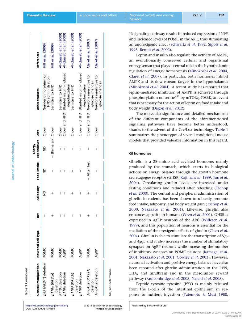
thanks to the advent of the Cre/Lox technology. Table 1
summarizes the phenotypes of several conditional mouse
models that provided valuable information in this regard.
GI hormones
Ghrelin is a 28-amino acid acylated hormone, mainly
produced by the stomach, which exerts its biological
actions on energy balance through the growth hormone
secretagogue receptor (GHSR; Kojima et al. 1999, Sun et al.
2004). Circulating ghrelin levels are increased under
fasting conditions and reduced after refeeding (Tschop
et al. 2000). The central and peripheral administration of
ghrelin in rodents has been shown to robustly promote
feed intake, adiposity, and body weight gain (Tschop et al.
2000, Nakazato et al. 2001). Likewise, ghrelin also
enhances appetite in humans (Wren et al. 2001). GHSR is
expressed in AgRP neurons of the ARC (Willesen et al.
1999), and this population of neurons is essential for the
mediation of the orexigenic effects of ghrelin (Chen et al.
2004). Ghrelin is able to stimulate the transcription of Npy
and Agrp, and it also increases the number of stimulatory
synapses on AgRP neurons while increasing the number
of inhibitory synapses on POMC neurons (Kamegai et al.
2001, Nakazato et al. 2001, Cowley et al. 2003). However,
neuronal activation and positive energy balance have also
been reported after ghrelin administration in the PVN,
LHA, and hindbrain and in the mesolimbic reward
pathway (Faulconbridge et al. 2003, Naleid et al. 2005).
Peptide tyrosine tyrosine (PYY) is mainly released
from the L-cells of the intestinal epithelium in res-
ponse to nutrient ingestion (Tatemoto & Mutt 1980,
Published by Bioscientifica Ltd
Downloaded from Bioscientifica.com at 03/01/2022 01:09:52AMvia free access

JournalofEndocrinology
Thematic Review M SCHNEEBERGER and others Neuronal circuits and energybalance
220 :2 T32
Adrian et al. 1985). Circulating PYY levels are proportional
to calorie intake and are reduced under fasting conditions
(Adrian et al. 1985). Two endogenous forms, PYY1–36 and
PYY3–36, are synthesized and secreted. The latter form is
the most abundant in the bloodstream and exerts a direct
action in the ARC. This has been demonstrated by
peripheral and intra-ARC administration of PYY3–36,
which increases neuronal activity in this region and
reduces appetite and body weight in a dose-dependent
manner (Batterham et al. 2002, Challis et al. 2003). These
anorexigenic effects are mediated via the inhibition of
ARC Y2 receptors, as demonstrated by pharmacological
(Abbott et al. 2005, Scott et al. 2005) and genetic
(Batterham et al. 2002) studies, which eventually leads to
increased a-MSH and reduced NPY release (Batterham et al.
2002). The effects of PYY3–36 in the brainstem and the
vagal–brainstem circuit have also been confirmed, as the
peripheral delivery of this peptide has been shown to
increase neuronal activity in NTS and AP neurons and
stimulate vagal afferent firing (Koda et al. 2005, Blevins
et al. 2008). Consistent with a role for PYY in the
regulation of appetite and body weight, transgenic mice
globally lacking or overexpressing Pyy exhibit opposite
alterations in energy balance control (Batterham et al.
2006, Boey et al. 2008).
GLP1, the cleavage product of proglucagon in the
intestine and brain, is mainly secreted from intestinal
L-cells. Similar to PYY, circulating GLP1 levels are high
following a meal and are low under fasting conditions.
This hormone exerts a strong incretin effect, via the GLP1
receptor (GLP1R) expressed in pancreatic islets, enhancing
insulin secretion after carbohydrate ingestion (Kreymann
et al. 1987). GLP1R is also expressed in key CNS areas
involved in the control of energy balance, such as the
hypothalamus and brainstem (Merchenthaler et al. 1999).
A number of studies have shown that the central or site-
specific administration of GLP1 or GLP1 analogs inhibits
food intake in rodents (Tang-Christensen et al. 1996,
Turton et al. 1996,McMahon&Wellman 1998, Hayes et al.
2008). Interestingly, neurons expressing the proglucagon
gene are present in the NTS, suggesting the existence of a
local circuit involved in the control of appetite (Merch-
enthaler et al. 1999). In fact, recent studies have provided
evidence for a dual (peripheral and central) role of GLP1 in
the suppression of appetite mediated by local vagal
afferents and a gut–brain feedback mechanism (Barrera
et al. 2011).
CCK is postprandially secreted from I-cells from
the small intestine and its systemic delivery suppresses
food intake in both animal models and humans
http://joe.endocrinology-journals.orgDOI: 10.1530/JOE-13-0398
� 2014 Society for EndocrinologyPrinted in Great Britain
(Gibbs et al. 1973, Gibbs & Smith 1977, Kissileff et al.
1981). CCK1 and CCK2 receptors are expressed in the
brainstem and hypothalamus, but the anorectic effects of
CCK are critically mediated by vagal sensory neurons that
project into the NTS (Moran et al. 1997). Interestingly, NTS
POMC neurons are activated by CCK and brainstemMC4R
signaling is required for CCK-induced suppression of
appetite (Fan et al. 2004). It has also been reported that
ghrelin attenuates and leptin synergistically potentiates
the effects of CCK on appetite (Barrachina et al. 1997,
Lee et al. 2011).
Neural circuits regulating homeostatic energybalance
Certain physiological conditions, such as the prandial
state, are associated with notable changes in the
circulating concentration of metabolites and hormones
involved in the regulation of whole-body energy homeo-
stasis. For example, in a postabsorptive situation, circulat-
ing cues of energetic surfeit (leptin, insulin, GLP1, PYY,
and glucose) are elevated, while cues of energetic deficit
(ghrelin) are reduced. The opposite is true under fasting
conditions. These hormones act in concert to engage
specific neuronal circuits in different brain regions,
including the hypothalamus and brainstem, establishing
reciprocal and dynamic interactions to restore systemic
energy balance. In this section, we summarize the main
circuits and the neuronal responses engaged by leptin and
ghrelin, as prototypical examples of anorexigenic and
orexigenic signals respectively.
ARC neuronal circuits: POMC, AgRP, and RIPCre neurons
Melanocortin peptides and NPY are two basic components
of a critical hypothalamic circuit involved in the conver-
gence and integration of nutritional and hormonal cues
aimed at regulating organismal energy balance. In the
ARC, the POMC, and AgRP neurons are located in
proximity to each other and project in parallel into similar
brain areas expressing MCRs. Both POMC and AgRP
neurons are able to sense a number of peripheral (leptin,
insulin, and ghrelin) and central (NPY, GABA, serotonin,
and melanocortin) signals, which are able to acutely
modulate their electrical activity influencing the release
of neuropeptides and neurotransmitters to ultimately
regulate appetite, energy expenditure, and metabolism.
In general terms, POMC (anorexigenic) and AgRP
(orexigenic) neurons have opposite physiological func-
tions, which are largely the consequence of the
Published by Bioscientifica Ltd
Downloaded from Bioscientifica.com at 03/01/2022 01:09:52AMvia free access

JournalofEndocrinology
Thematic Review M SCHNEEBERGER and others Neuronal circuits and energybalance
220 :2 T33
contrasting actions of a-MSH and AGRP peptides on
MCRs: while a-MSH is an endogenous MCR agonist,
AGRP is an inverse agonist (Haskell-Luevano & Monck
2001, Nijenhuis et al. 2001, Tolle & Low 2008). Indeed,
substantial experimental evidence indicates that the
agonism of MCRs attenuates appetite and enhances
energy expenditure, whereas their antagonism has essen-
tially the opposite effects (Fan et al. 1997, Harrold et al.
1999, Hwa et al. 2001). This is consistent with data
showing that the loss of or mutations in MC3R and
MC4R genes cause obesity both in rodents and in humans
(Huszar et al. 1997, Butler et al. 2000, Farooqi 2008). In
addition to the inhibition of MCR signaling, the orexi-
genic actions of AgRP neurons are also mediated by the
release of NPY and GABA.
The anorexigenic effects of leptin are basically
achieved by repressing AgRP neurons and activating
POMC neurons (Fig. 1A). Leptin enhances Pomc gene
expression and processing into a-MSH (Schwartz et al.
1997, Thornton et al. 1997, Mizuno et al. 1998).
Electrophysiological studies have demonstrated that
locally applied leptin is able to depolarize (excite) POMC
neurons (Cowley et al. 2001, Claret et al. 2007, 2011, Hill
et al. 2008, Al-Qassab et al. 2009, Qiu et al. 2010) probably
through TRPC channels (Qiu et al. 2010). In contrast,
leptin inhibits the transcription of Npy and Agrp genes in
the hypothalamus (Stephens et al. 1995, Schwartz et al.
1996, Mizuno & Mobbs 1999). Electrophysiological
recordings have shown that leptin decreases the GABA-
ergic-mediated tone induced by AgRP neurons onto
neighboring POMCneurons, resulting in the disinhibition
of POMC neuron activity (Cowley et al. 2001). The ability
of leptin to directly hyperpolarize (inhibit) AgRP neurons
is controversial (Cowley et al. 2001, Claret et al. 2007,
Al-Qassab et al. 2009), but studies in rats have reported
leptin-mediated inhibition of identified NPY neurons
(van den Top et al. 2004). In addition, leptin also acts
directly on presynaptic GABAergic neurons that do not
express AGRP, reducing the inhibitory input to post-
synaptic POMC neurons, thus further contributing to the
maintenance of the anorexigenic actions mediated by this
hormone (Fig. 1A; Vong et al. 2011).
On the other hand, under conditions of negative
energy balance, circulating ghrelin levels are increased.
The actions of ghrelin on food intake and energy balance
are mediated by AgRP neurons, as mice lacking Agrp and
Npy are insensitive to the orexigenic effects of external
ghrelin (Chen et al. 2004, Luquet et al. 2007). In line with
this, ghrelin increases the expression of Npy and Agrp
transcripts (Kamegai et al. 2001, Nakazato et al. 2001)
http://joe.endocrinology-journals.orgDOI: 10.1530/JOE-13-0398
� 2014 Society for EndocrinologyPrinted in Great Britain
and depolarizes AgRP neurons while increasing the
number of GABAergic inhibitory synapses on POMC
neurons (Fig. 1B) (Cowley et al. 2003, van den Pol et al.
2009, Yang et al. 2011, Atasoy et al. 2012). The importance
of these GABAergic stimuli in the control of energy
balance has been substantially demonstrated (Horvath
et al. 1997,Wu et al. 2009, 2012,Wu& Palmiter 2011), and
conditional deletion of the vesicular GABA transporter in
AgRP neurons blunts the inhibitory tone onto
postsynaptic POMC neurons, leading to an enhanced
melanocortigenic output and a lean phenotype (Tong
et al. 2008). Moreover, AGRP and NPY directly hyper-
polarize POMC neurons and decrease the production and
release of a-MSH, further inhibiting the activity of this
population of neurons (Roseberry et al. 2004, Smith et al.
2007, Cyr et al. 2013). Thus, AgRP neurons are able to
negatively modulate the anorexigenic effects of POMC
neurons by direct (GABAergic synapsis) and indirect (MCR
antagonism) mechanisms (Fig. 1B).
In addition to changes in neuropeptide release, leptin
and ghrelin also exert rapid and reversible effects on
synaptic connections onto POMC and AgRP neurons.
Seminal studies carried out at the Horvath laboratory have
provided the first evidence for synaptic plasticity in
hypothalamic energy balance circuits and established the
basis for a new mechanism by which these hormones
dynamically regulate circuit responsiveness to control
energy homeostasis (Pinto et al. 2004). The role of synaptic
remodeling in neuronal circuits regulating metabolism
has recently been reviewed in detail (Zeltser et al. 2012,
Dietrich & Horvath 2013).
A novel subpopulation of ARC neurons involved in
the control of energy balance (defined by virtue of
Cre-mediated expression of rat insulin II promoter-Cre
transgene and called RIPCre neurons) has recently been
described. Comparative electrophysiological and histo-
logical studies indicate that RIPCre neurons constitute a
distinct population from POMC or AgRP neurons
(Choudhury et al. 2005). However, close apposition of
these neuronal subsets suggests that RIPCre neurons may
be the targets of POMC and/or AgRP neurons. Indeed, bath
application of a melanocortin agonist has been found to
cause direct long-lasting depolarization and increased
firing in ARC RIPCre neurons (Choudhury et al. 2005).
Interestingly, insulin has also been found to depolarize
these neurons, while leptin has been found to not cause
any electrophysiological effect (Choudhury et al. 2005).
Although a number of mouse genetic studies indicate
that ARC RIPCre neurons regulate systemic energy balance
(Cui et al. 2004, Choudhury et al. 2005), this interpretation
Published by Bioscientifica Ltd
Downloaded from Bioscientifica.com at 03/01/2022 01:09:52AMvia free access
Anorectic output
NPYR1
NPYR5
F.I.
E.E
MC4R
MC4R
MC4R
LEPR
MC4R
AGRP
AGRP
NPY
α-MSH
α-MSH
α-MSH
NPYPOMC
POMC
LEPR
Leptin
WAT
A B
Preganglionicneuron
GABAergicpresynaptic
neuron
GABAergic
PVNneuron
Second-orderneurons
Arcuatenucleus
NPYR1
NPYR5MC4R
MC4R
GHSR
GHSRMC4R
MC4R
E.E.
F.I.
AGRP
α-MSH
NPYPOMC
Ghrelin
Orexigenicoutput
GABAergic
GABAergic
GABAergic
Second-orderneurons
Arcuatenucleus
AGRP
NPY
α-MSH
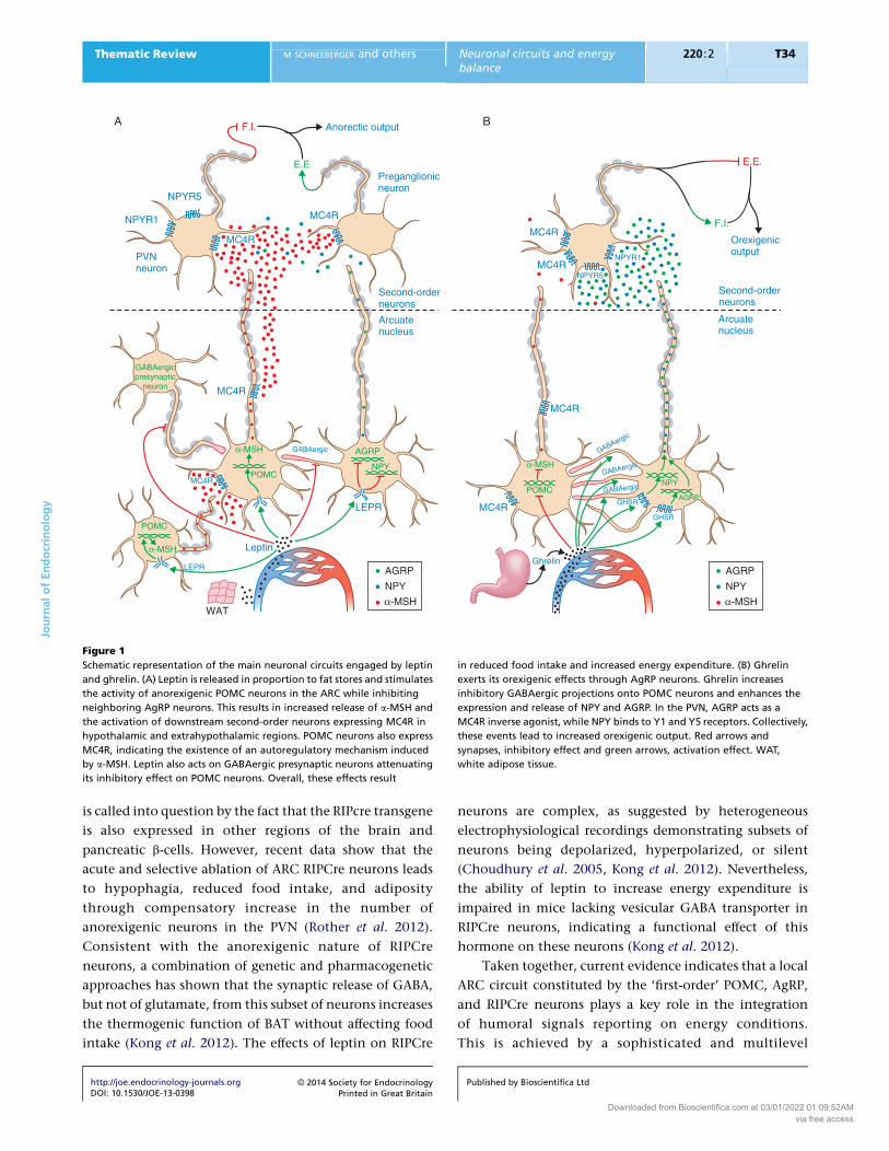
Figure 1
Schematic representation of the main neuronal circuits engaged by leptin
and ghrelin. (A) Leptin is released in proportion to fat stores and stimulates
the activity of anorexigenic POMC neurons in the ARC while inhibiting
neighboring AgRP neurons. This results in increased release of a-MSH and
the activation of downstream second-order neurons expressing MC4R in
hypothalamic and extrahypothalamic regions. POMC neurons also express
MC4R, indicating the existence of an autoregulatory mechanism induced
by a-MSH. Leptin also acts on GABAergic presynaptic neurons attenuating
its inhibitory effect on POMC neurons. Overall, these effects result
in reduced food intake and increased energy expenditure. (B) Ghrelin
exerts its orexigenic effects through AgRP neurons. Ghrelin increases
inhibitory GABAergic projections onto POMC neurons and enhances the
expression and release of NPY and AGRP. In the PVN, AGRP acts as a
MC4R inverse agonist, while NPY binds to Y1 and Y5 receptors. Collectively,
these events lead to increased orexigenic output. Red arrows and
synapses, inhibitory effect and green arrows, activation effect. WAT,
white adipose tissue.
JournalofEndocrinology
Thematic Review M SCHNEEBERGER and others Neuronal circuits and energybalance
220 :2 T34
is called into question by the fact that the RIPcre transgene
is also expressed in other regions of the brain and
pancreatic b-cells. However, recent data show that the
acute and selective ablation of ARC RIPCre neurons leads
to hypophagia, reduced food intake, and adiposity
through compensatory increase in the number of
anorexigenic neurons in the PVN (Rother et al. 2012).
Consistent with the anorexigenic nature of RIPCre
neurons, a combination of genetic and pharmacogenetic
approaches has shown that the synaptic release of GABA,
but not of glutamate, from this subset of neurons increases
the thermogenic function of BAT without affecting food
intake (Kong et al. 2012). The effects of leptin on RIPCre
http://joe.endocrinology-journals.orgDOI: 10.1530/JOE-13-0398
� 2014 Society for EndocrinologyPrinted in Great Britain
neurons are complex, as suggested by heterogeneous
electrophysiological recordings demonstrating subsets of
neurons being depolarized, hyperpolarized, or silent
(Choudhury et al. 2005, Kong et al. 2012). Nevertheless,
the ability of leptin to increase energy expenditure is
impaired in mice lacking vesicular GABA transporter in
RIPCre neurons, indicating a functional effect of this
hormone on these neurons (Kong et al. 2012).
Taken together, current evidence indicates that a local
ARC circuit constituted by the ‘first-order’ POMC, AgRP,
and RIPCre neurons plays a key role in the integration
of humoral signals reporting on energy conditions.
This is achieved by a sophisticated and multilevel
Published by Bioscientifica Ltd
Downloaded from Bioscientifica.com at 03/01/2022 01:09:52AMvia free access
JournalofEndocrinology
Thematic Review M SCHNEEBERGER and others Neuronal circuits and energybalance
220 :2 T35
organizational structure that allows the accurate
regulation of orexigenic and anorexigenic outputs
through direct and indirect mechanisms.
Downstream neurocircuitry engaged by hypothalamic
neuron activity
Given that POMC and AgRP neurons are the sole source of
MCRligands in thebrain,afinebalancebetweena-MSHand
AGRP is necessary to precisely regulate their mediated
physiological outputs on MC4Rs in target areas. These
receptors are localized in many nuclei involved in the
regulation of energy balance where POMC and AgRP
neurons send axon projections. MC4Rs are G-protein-
coupled receptors that stimulate adenylyl cyclase, thereby
increasing intracellular cAMP levels (Florijn et al. 1993).
A series of elegant studies using a cell-specific MC4R
re-expression strategy indicate that MC4Rs in the PVN are
mainly involved in the control of food intake (Balthasar
et al. 2005), while MC4Rs in autonomic preganglionic
neurons regulate energy expenditure and hepatic glucose
production (Rossi et al. 2011) (Fig. 1A). Furthermore, and
contrary to the prevailing view, a recent report has shown
that POMC neurons also express MC4Rs that contribute to
the regulation of body weight and composition through
changes in both feeding behavior and energy expenditure
(do Carmo et al. 2013). This autoregulatory mechanism,
induced by a-MSH released from the same cell and/or
neighboring POMCneurons, could represent an additional
layer of regulation within a widely segregated network of
melanocortin receptors involved in the regulation of
homeostatic (appetite) and autonomic (thermogenesis,
hepaticmetabolism,and insulin release) functions (Fig. 1A).
NPY receptors are Gi/o-protein-coupled receptors that
reduce cAMP production, leading to the activation of
G-protein-gated inwardly rectifying KC (GIRK) channels
and inhibition of voltage-dependent Ca2C channels (Sohn
et al. 2013). The precise roles of NPY receptors and their
contribution to the mediation of the orexigenic effects of
NPY have been difficult to delineate due to the paradoxical
phenotypes of receptor KOmousemodels. This is probably
the consequence of receptor redundancies and compensa-
tory mechanisms exhibited after the application of germ-
line deletion strategies. Despite these limitations,
pharmacological and genetic studies indicate that the
orexigenic actions of NPY are mediated by postsynaptic Y1
and Y5 within the PVN (Nguyen et al. 2012, Sohn et al.
2013; Fig. 1B). Notably NPY from ARC neurons acts
through PVN Y1, resulting in a functional inhibition of
TH tonus and BAT thermogenesis (Shi et al. 2013).
http://joe.endocrinology-journals.orgDOI: 10.1530/JOE-13-0398
� 2014 Society for EndocrinologyPrinted in Great Britain
Furthermore, NPY may also decrease pro-TRH transcrip-
tion and proconvertase 2-mediated pro-TRH processing in
the PVN through Y1/Y5 receptors (Cyr et al. 2013). Taken
together, abundant amounts of evidence suggest that the
effects of ARC NPY on energy balance are principally
mediated by the PVN. However, it is important to note
that other sources of NPY may also play a role in the
regulation of energy balance.
Correlating neuronal circuit activity with behavioral
responses by pharmacogenetic and optogenetic
techniques
Most of the experimental findings that have allowed
researchers to outline the models suggested so far are
largely the result of circumstantial evidence. However, the
recent development of pharmacogenetic and optogenetic
techniques has provided a way to exert temporally and
spatially precise control over the activity of defined circuit
elements. This permits the establishment of causal
connections between circuit activity and behavioral
responses (Sternson 2013).
Using an elegant combination of optogenetics and
mouse genetic approaches, Aponte et al. (2011) have
confirmed that the selective activation of AgRP neurons is
sufficient to evoke voracious feeding behavior in mice,
withoutprevious trainingand independentofmelanocortin
signaling.The levelofneuronal activationhasbeen found to
correlatewith themagnitude,dynamics, anddurationof the
induced behavioral response. Furthermore, continuous
photostimulation is required to maintain evoked feeding
behaviour, indicating that the activation of AgRP neurons
doesnot initiate a sustainedpropagating effect (Aponte et al.
2011). In contrast, prolonged (but not brief) optogenetic
stimulation of POMC neurons has been shown to result in
reduced food intake and body weight gain, which requires
downstreamMC4R activity (Aponte et al. 2011).
The behavioral effects on food intake caused by AgRP
or POMC neuron activation have been further supported
by studies using pharmacogenetic (designer receptors
exclusively activated by designer drugs (DREADDs))
technology. Pharmacogenetic activation of AgRP neurons
rapidly induces feeding and food-seeking behaviors
associated with decreased energy expenditure and
enhanced adiposity (Krashes et al. 2011). Consistent with
the optogenetic data (Aponte et al. 2011), long-term
stimulation of ARC POMC neurons is necessary to reduce
appetite. Interestingly, the acute stimulation of NTS
POMC neurons has been shown to generate an immediate
suppression of food intake (Zhan et al. 2013).
Published by Bioscientifica Ltd
Downloaded from Bioscientifica.com at 03/01/2022 01:09:52AMvia free access

JournalofEndocrinology
Thematic Review M SCHNEEBERGER and others Neuronal circuits and energybalance
220 :2 T36
In a subsequent study, the Sternson group performed
a series of experiments to determine which brain regions
and cell types mediate evoked feeding behavior triggered
by activated AgRP neurons. The authors used optogenetic
approaches to map synaptic connections downstream of
AgRP neurons and assessed their role in terms of ingestive
behavior by perturbing electrical activity in presynaptic
and postsynaptic neuronal types (Atasoy et al. 2012).
Notably the authors found that ARC AgRP neurons induce
evoked feeding behavior through inhibitory input onto
oxytocin neurons in the PVN, while ARC POMC neurons
are involved in the long-term control of appetite and
energy balance (Atasoy et al. 2012).
Collectively, these results emphasize the previously
unrecognized importance of the temporal and spatial
activation of POMC and AgRP neurons. Thus, ARC AgRP
and NTS POMC neurons could be involved in the
regulation of acute feeding behavior while ARC POMC
neurons may be involved in long-term responses. This
demonstrates the existence of multiple, distinct behavioral
and anatomical modules that act in synchrony to regulate
whole-body energy balance. The use of these tools in the
field of central control of energy balance has providednovel
valuable information and has confirmed previous findings.
However, it has also generated some controversial obser-
vations. Further research needs to be conducted to precisely
define the importance of these factors and to reconcile
these observations with previous evidence (Mercer et al.
2013). Nevertheless, these reports demonstrate that opto-
genetics and pharmacogenetics are exceptionally useful
tools to study the interrelationships between synaptology,
neuronal circuit activity, and behavioral outputs.
New players in energy balance control
Non-neuronal cell types: macroglia and microglia
Glial cells have traditionally been considered satellite
neuronal partners with supportive and structural roles.
However, in recent years, glial cells have acquired a new
rank and are now regarded as active players in many
physiological functions including energy balance control.
Astrocytes are star-shaped cells that are involved in a
number of functions, such as metabolic support to
neurons and transmitter uptake and release as well as
synaptic remodeling (Sofroniew & Vinters 2010).
Astrocytes express LEPR (Cheunsuang & Morris 2005,
Hsuchou et al. 2009b), and modifications in circulating
leptin levels alter hypothalamic astrocyte expression of
structural proteins as well as glutamate and glucose
http://joe.endocrinology-journals.orgDOI: 10.1530/JOE-13-0398
� 2014 Society for EndocrinologyPrinted in Great Britain
transporters (Garcia-Caceres et al. 2011, Fuente-Martin
et al. 2012). This may cause changes in the synaptic
plasticity and excitability of surrounding neurons, leading
to metabolic adaptations. In fact, HFD administration in
rodents is associated with increased glial coverage of
POMC neuron perikarya (Horvath et al. 2010). It has also
been reported that DIO mice exhibit increased expression
of functional astrocytic LEPR in the hypothalamic region,
an effect that may play a role in the development of leptin
resistance (Hsuchou et al. 2009a). Indeed, loss of astrocytic
Lepr under HFD conditions provides partial protection
against developing disturbances in neuronal leptin signal-
ing (Jayaram et al. 2013).
Obesity and lipid overload induce chronic low-grade
inflammation in the hypothalamus (Thaler et al. 2010).
This is regarded as a protective effect, which is mainly
promoted by microglial cells that have immunitary
actions in the CNS. HFD feeding selectively and rapidly
activates microglia in the hypothalamus and increases the
production of proinflammatory cytokines (De Souza et al.
2005, Milanski et al. 2009, Thaler et al. 2012). Interest-
ingly, it has been demonstrated that moderate physical
activity reduces hypothalamic microglial activation
independently of body mass (Yi et al. 2012). Enhanced
hypothalamic microglial activation has also been reported
in rodents and primates after nutritional manipulations
during the prenatal or perinatal period (Grayson et al.
2010, Tapia-Gonzalez et al. 2011).
Tanycytes have recently emerged as novel modulators
of the hypothalamic networks that control energy balance.
They contact the cerebrospinal fluid and send processes
that come into proximity with neurons into the ARC and
VMN (Bolborea & Dale 2013). Although it is not known
whether tanycytes are able to modulate the activity of
hypothalamic neurons, several lines of evidence suggest
that this particular cell type may be involved in the
regulation of energy homeostasis. For example, tanycytes
respond to fluctuations in glucose concentration (Frayling
et al. 2011), express a number of genes related to energy
homeostasis control (Bolborea & Dale 2013), and regulate
the permeability properties of the fenestrated capillaries of
the ME, which may constitute a way of modulating the
access of metabolites into the ARC (Langlet et al. 2013).
Intriguingly, tanycytes may be a novel population of adult
neural stem cells in the hypothalamus. Tanycytes express
stem cell markers, including nestin and SOX2 (Lee et al.
2012), and lineage-tracing studies have shown that they
give rise to neurons in vivo with functional implications.
While short-term HFD feeding promotes hypothalamic
neurogenesis in pre-adult ages (Lee et al. 2012), chronic
Published by Bioscientifica Ltd
Downloaded from Bioscientifica.com at 03/01/2022 01:09:52AMvia free access

JournalofEndocrinology
Thematic Review M SCHNEEBERGER and others Neuronal circuits and energybalance
220 :2 T37
HFD administration causes depletion of hypothalamic
neural stem cells (Li et al. 2012). Furthermore, the
manipulation of hypothalamic neurogenesis in adult
mice has also produced divergent results. Selective inhi-
bition of ME neurogenesis in adult mice fed a HFD resulted
in reduced weight gain and adiposity due to enhanced
energy expenditure (Lee et al. 2012). By contrast, genetic
IKKb/NF-kB activation in SOX2-positive hypothalamic cells
led to overeating and weight gain (Li et al. 2012). It is
important to note that these strategies did not exclusively
target tanycytes and so these metabolic effects cannot be
solely attributed to this cell type. Together, these results
indicate that neurogenesis after short- or long-term HFD
administration may have a compensatory or detrimental
effect respectively on cell fate. These differences can also be
the consequence of targeting distinct tanycyte populations
(Bolborea & Dale 2013).
Epigenetic mechanisms
The interplay between genetic and environmental factors
(nutrition, maternal health, chemicals, lifestyle, etc.)
during the prenatal or perinatal period and their influence
on the development of energy balance and metabolic
alterations into adulthood have recently received sub-
stantial interest. In both humans and animal models,
prenatal or perinatal nutritional manipulations lead to
chronic metabolic disturbances in terms of feeding
behavior, energy expenditure, leptin sensitivity, and
glucose homeostasis. These metabolic defects may be
partially the consequence of abnormal development of
appetite-regulating neuronal circuits due to perinatal
programming (Contreras et al. 2013). Epigenetic changes
have been proposed as likely candidates tomediate, at least
in part, these neuronal programming events, but a limited
number of studies have explored this hypothesis. The
epigenetic machinery that controls chromatin dynamics
includes DNA methylation, posttranslational histone
modifications, and non-coding RNAs. Neonatal overfeed-
ing in rats, which results in overweight and metabolic
syndrome, is associated with the hypermethylation of the
Pomc gene promoter (Plagemann et al. 2009). The extent of
this DNA methylation is negatively correlated with the
expression of POMC in relation to leptin and insulin levels,
indicating the functionality of acquired epigenomic
alterations (Plagemann et al. 2009). In the same over-
nutrition model, Plagemann et al. (2010) also found
increased methylation of the Insr promoter in the
hypothalamus. Similarly, epigenetic remodeling of hypo-
thalamic genes induced by mild maternal undernutrition
http://joe.endocrinology-journals.orgDOI: 10.1530/JOE-13-0398
� 2014 Society for EndocrinologyPrinted in Great Britain
(Stevens et al. 2010, Begum et al. 2012) or stress (Paternain
et al. 2012) has also been reported to be associated with
altered energy balance and metabolism in experimental
animal models. In humans, different methylation patterns
of POMC and NPY promoter regions in leukocytes have
been proposed as biomarkers to predict weight regain after
an energy restriction program (Crujeiras et al. 2013).
Collectively, this evidence supports the hypothesis that
early prenatal or postnatal environmental perturbations
cause chronic metabolic alterations that are partially the
consequence of epigenetic changes in key genes and areas
of the CNS involved in the control of energy balance.
Nevertheless, further research is warranted to address the
significance of these epigenetic events.
MicroRNAs (miRNAs), a class of small, non-coding
RNAs that regulate gene expression at the posttranscrip-
tional level, have recently been suggested to be involved in
the hypothalamic control of energy balance. It has been
demonstrated that the expression of Dicer1, an essential
endoribonuclease for miRNA maturation, is regulated by
nutrient availability and excess in the hypothalamus
(Schneeberger et al. 2013). Furthermore, we have also
shown that deletion of Dicer1 in POMC neurons leads to
an obese phenotype characterized by increased adiposity,
hyperleptinemia, defective glucose metabolism, and
alterations in the pituitary–adrenal axis. This phenotype
is associated with a progressive POMC neuron degener-
ation, indicating a key role for miRNAs in the survival of
this population of neurons (Greenman et al. 2013,
Schneeberger et al. 2013). High-throughput sequencing
studies in ARC and PVN of rats have shown a specific
miRNA enrichment pattern that could be used to define a
prototypic profile in these brain regions. These miRNAs
include seven of the eight genes of the let-7 family, the
two miR-7 genes, miR-9 gene, and 5 0 copy of the three
miR-30 loci (Amar et al. 2012). Moreover, in situ
hybridization experiments have revealed a limited and
distinct expression of miR-7a in the hypothalamus,
preferentially colocalizing with AgRP neurons (Herzer
et al. 2012). Despite these efforts in describing the
miRNA transcriptome and patterns of expression in the
hypothalamus, the role of specific miRNAs in particular
neuronal circuits in the regulation of whole-body energy
balance still remains unknown.
Concluding remarks: neuronal circuitryintegration and physiological responses
As has been outlined above, organismal energy balance
is regulated by many factors through complex and
Published by Bioscientifica Ltd
Downloaded from Bioscientifica.com at 03/01/2022 01:09:52AMvia free access

Reward/hedonic centers Homeostatic integrative centers
Circulating hormones secretedin proportion to fat stores
(insulin and leptin)
Circulating GI tracthormones (ghrelin, GLP1, PYY, etc.)
Vagal afferents fromGI organs
Long-term regulation of energy balance Short-term regulation of energy balance
LHAVTANAc
Hypothalamus Brainstem
Energy balance regulation
Behavioral autonomicendocrine
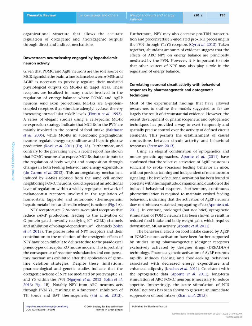
Figure 2
Schematic integration of the different levels of food intake and energy
balance regulation. Food intake and energy balance are coordinately
regulated by homeostatic and non-homeostatic neural mechanisms.
Circulating hormones and vagus stimuli inform the CNS about whole-body
nutritional and energy status. Leptin and insulin are believed to be involved
in the long-term regulation of energy balance, while GI hormones and vagal
afferents represent a short-term regulatory mechanism. These hormones act
in concert to engage specific neuronal circuits in homeostatic and hedonic
centers, establishing dynamic and complex interactions between these
different brain regions to elaborate coordinated endocrine, autonomic,
and behavioral responses to regulate energy balance. Sensory, emotional,
and social cues also influence ingestive behaviors probably through
non-homeostatic and higher brain structures. LHA, lateral hypothalamic
area; VTA, ventral tegmental area; NAc, nucleus accumbens.
JournalofEndocrinology
Thematic Review M SCHNEEBERGER and others Neuronal circuits and energybalance
220 :2 T38
multi-level integration processes that involve multiple
neuronal circuits. The homeostatic system is basically
influenced by long-term (leptin and insulin) and
short-term (GI hormones and vagal inputs) signals that
act in concert to engage specific neuronal circuits in
the hypothalamus and brainstem aimed at fulfilling
whole-body metabolic needs. In addition to this
homeostatic module, the corticolimbic and mesolimbic
centers (which include the ventral tegmental area,
nucleus accumbens, prefrontal cortex, hippocampus,
and amygdala) integrate cognitive, hedonic, and
emotional stimuli in a non-homeostatic process
(Berthoud 2011). Circulating energy balance signals,
such as leptin and ghrelin, also target hedonic networks
to modulate appetite. However, this system may over-
ride homeostatic control and cause energy imbalance
(Berthoud 2011). In fact, striking similarities between
food reward and drug addiction mechanisms have
been reported (DiLeone et al. 2012). Therefore, these
complex interactions between the homeostatic and
non-homeostatic systems culminate in coordinated
http://joe.endocrinology-journals.orgDOI: 10.1530/JOE-13-0398
� 2014 Society for EndocrinologyPrinted in Great Britain
appetite and energy balance regulation through the
modulation of endocrine, autonomic, and behavioral
outputs (Fig. 2). The precise integrative mechanisms
of these different levels of regulation and the gene-
ration of specific physiological outputs are among the
main unsolved enigmas of the central regulation of
energy balance.
Declaration of interest
The authors declare that there is no conflict of interest that could be
perceived as prejudicing the impartiality of the review reported.
Funding
This work was supported by RecerCaixa grant number 2010ACUP_00275;
EFSD/Lilly Fellowship Award; Ministerio de Ciencia e Innovacion (MICINN),
Instituto de Salud Carlos III (ISCIII) grant number PI10/01074; and MICINN
grant number SAF2010-19527 (R G). M S is a recipient of an undergraduate
grant from the University of Barcelona. M C is a recipient of a Miguel Servet
contract (CP09/00233) from MICINN-ISCIII. Some of these grants are
co-financed by the European Regional Development Fund ‘A way to
build Europe’. This work was carried out in part at the Esther Koplowitz
Centre, Barcelona.
Published by Bioscientifica Ltd
Downloaded from Bioscientifica.com at 03/01/2022 01:09:52AMvia free access
JournalofEndocrinology
Thematic Review M SCHNEEBERGER and others Neuronal circuits and energybalance
220 :2 T39
References
Abbott CR, Kennedy AR, Wren AM, Rossi M, Murphy KG, Seal LJ, Todd JF,
Ghatei MA, Small CJ & Bloom SR 2003 Identification of hypothalamic
nuclei involved in the orexigenic effect of melanin-concentrating
hormone. Endocrinology 144 3943–3949. (doi:10.1210/en.2003-0149)
AbbottCR, SmallCJ,KennedyAR,NearyNM,SajediA,GhateiMA&BloomSR
2005 Blockade of the neuropeptide Y Y2 receptor with the specific
antagonist BIIE0246 attenuates the effect of endogenous and exogenous
peptide YY(3–36) on food intake. Brain Research 1043 139–144.
(doi:10.1016/j.brainres.2005.02.065)
Adrian TE, Ferri GL, Bacarese-Hamilton AJ, Fuessl HS, Polak JM & Bloom SR
1985 Human distribution and release of a putative new gut hormone,
peptide YY. Gastroenterology 89 1070–1077.
Air EL, Benoit SC, Blake Smith KA, Clegg DJ & Woods SC 2002 Acute third
ventricular administration of insulin decreases food intake in two
paradigms. Pharmacology, Biochemistry, and Behavior 72 423–429.
(doi:10.1016/S0091-3057(01)00780-8)
Al-Qassab H, Smith MA, Irvine EE, Guillermet-Guibert J, Claret M,
Choudhury AI, Selman C, Piipari K, Clements M, Lingard S et al. 2009
Dominant role of the p110b isoform of PI3K over p110a in energy
homeostasis regulation by POMC and AgRP neurons. Cell Metabolism
10 343–354. (doi:10.1016/j.cmet.2009.09.008)
Amar L, Benoit C, Beaumont G, Vacher CM, Crepin D, Taouis M &
Baroin-Tourancheau A 2012 MicroRNA expression profiling of hypo-
thalamic arcuate and paraventricular nuclei from single rats using
Illumina sequencing technology. Journal of Neuroscience Methods 209
134–143. (doi:10.1016/j.jneumeth.2012.05.033)
Ao Y, Go VL, Toy N, Li T, Wang Y, Song MK, Reeve JR Jr, Liu Y & Yang H
2006 Brainstem thyrotropin-releasing hormone regulates food intake
through vagal-dependent cholinergic stimulation of ghrelin secretion.
Endocrinology 147 6004–6010. (doi:10.1210/en.2006-0820)
Aponte Y, Atasoy D & Sternson SM 2011 AGRP neurons are sufficient to
orchestrate feeding behavior rapidly and without training. Nature
Neuroscience 14 351–355. (doi:10.1038/nn.2739)
Asnicar MA, Smith DP, Yang DD, Heiman ML, Fox N, Chen YF, Hsiung HM
& Koster A 2001 Absence of cocaine- and amphetamine-regulated
transcript results in obesity in mice fed a high caloric diet. Endocrinology
142 4394–4400. (doi:10.1210/en.142.10.4394)
Atasoy D, Betley JN, Su HH & Sternson SM 2012 Deconstruction of a neural
circuit for hunger. Nature 488 172–177. (doi:10.1038/nature11270)
Bagdade JD, Bierman EL & Porte D Jr 1967 The significance of basal insulin
levels in the evaluation of the insulin response to glucose in diabetic
and nondiabetic subjects. Journal of Clinical Investigation 46 1549–1557.
(doi:10.1172/JCI105646)
Balthasar N, Coppari R, McMinn J, Liu SM, Lee CE, Tang V, Kenny CD,
McGovern RA, Chua SC Jr, Elmquist JK et al. 2004 Leptin receptor
signaling in POMC neurons is required for normal body weight
homeostasis. Neuron 42 983–991. (doi:10.1016/j.neuron.2004.06.004)
Balthasar N, Dalgaard LT, Lee CE, Yu J, Funahashi H,Williams T, FerreiraM,
Tang V, McGovern RA, Kenny CD et al. 2005 Divergence of
melanocortin pathways in the control of food intake and energy
expenditure. Cell 123 493–505. (doi:10.1016/j.cell.2005.08.035)
Banno R, Zimmer D, De Jonghe BC, Atienza M, Rak K, Yang W & Bence KK
2010 PTP1B and SHP2 in POMC neurons reciprocally regulate energy
balance in mice. Journal of Clinical Investigation 120 720–734.
(doi:10.1172/JCI39620)
Barrachina MD, Martinez V, Wang L, Wei JY & Tache Y 1997 Synergistic
interaction between leptin and cholecystokinin to reduce short-term
food intake in lean mice. PNAS 94 10455–10460. (doi:10.1073/pnas.
94.19.10455)
Barrera JG, Sandoval DA, D’Alessio DA & Seeley RJ 2011 GLP-1 and energy
balance: an integrated model of short-term and long-term control.
Nature Reviews. Endocrinology 7 507–516. (doi:10.1038/nrendo.2011.77)
http://joe.endocrinology-journals.orgDOI: 10.1530/JOE-13-0398
� 2014 Society for EndocrinologyPrinted in Great Britain
Batterham RL, Cowley MA, Small CJ, Herzog H, Cohen MA, Dakin CL,
Wren AM, Brynes AE, Low MJ, Ghatei MA et al. 2002 Gut hormone
PYY(3–36) physiologically inhibits food intake. Nature 418 650–654.
(doi:10.1038/nature00887)
Batterham RL, Heffron H, Kapoor S, Chivers JE, Chandarana K, Herzog H,
Le Roux CW, Thomas EL, Bell JD & Withers DJ 2006 Critical role for
peptide YY in protein-mediated satiation and body-weight regulation.
Cell Metabolism 4 223–233. (doi:10.1016/j.cmet.2006.08.001)
Baura GD, Foster DM, Porte D Jr, Kahn SE, Bergman RN, Cobelli C &
Schwartz MW 1993 Saturable transport of insulin from plasma into the
central nervous system of dogs in vivo. A mechanism for regulated
insulin delivery to the brain. Journal of Clinical Investigation 92
1824–1830. (doi:10.1172/JCI116773)
Beck B, Jhanwar-Uniyal M, Burlet A, Chapleur-Chateau M, Leibowitz SF &
Burlet C 1990 Rapid and localized alterations of neuropeptide Y in
discrete hypothalamic nuclei with feeding status. Brain Research 528
245–249. (doi:10.1016/0006-8993(90)91664-3)
BegumG, StevensA, Smith EB,ConnorK,Challis JR, Bloomfield F&WhiteA
2012 Epigenetic changes in fetal hypothalamic energy regulating
pathways are associated with maternal undernutrition and twinning.
FASEB Journal 26 1694–1703. (doi:10.1096/fj.11-198762)
Belgardt BF, Husch A, Rother E, Ernst MB, Wunderlich FT, Hampel B,
Klockener T, Alessi D, Kloppenburg P & Bruning JC 2008 PDK1
deficiency in POMC-expressing cells reveals FOXO1-dependent and
-independent pathways in control of energy homeostasis and stress
response. Cell Metabolism 7 291–301. (doi:10.1016/j.cmet.2008.01.006)
Benoit SC, Air EL, Coolen LM, Strauss R, Jackman A, Clegg DJ, Seeley RJ &
Woods SC 2002 The catabolic action of insulin in the brain is mediated
by melanocortins. Journal of Neuroscience 22 9048–9052.
Berthoud HR 2011 Metabolic and hedonic drives in the neural control of
appetite: who is the boss? Current Opinion in Neurobiology 21 888–896.
(doi:10.1016/j.conb.2011.09.004)
BewickGA,Gardiner JV,DhilloWS,KentAS,WhiteNE,WebsterZ,GhateiMA
&Bloom SR 2005 Post-embryonic ablation of AgRPneurons inmice leads
to a lean, hypophagic phenotype. FASEB Journal 19 1680–1682.
(doi:10.1096/fj.04-3434fje)
Bi S, Ladenheim EE, Schwartz GJ & Moran TH 2001 A role for NPY
overexpression in the dorsomedial hypothalamus in hyperphagia and
obesity of OLETF rats. American Journal of Physiology. Regulatory,
Integrative and Comparative Physiology 281 R254–R260.
Bingham NC, Anderson KK, Reuter AL, Stallings NR & Parker KL 2008
Selective loss of leptin receptors in the ventromedial hypothalamic
nucleus results in increased adiposity and a metabolic syndrome.
Endocrinology 149 2138–2148. (doi:10.1210/en.2007-1200)
Blevins JE, Chelikani PK, Haver AC & Reidelberger RD 2008 PYY(3–36)
induces Fos in the arcuate nucleus and in both catecholaminergic and
non-catecholaminergic neurons in the nucleus tractus solitarius of rats.
Peptides 29 112–119. (doi:10.1016/j.peptides.2007.11.003)
BoeyD, Lin S, Enriquez RF, LeeNJ, Slack K, CouzensM, Baldock PA, HerzogH
& Sainsbury A 2008 PYY transgenic mice are protected against diet-
induced and genetic obesity. Neuropeptides 42 19–30. (doi:10.1016/
j.npep.2007.11.003)
Bolborea M & Dale N 2013 Hypothalamic tanycytes: potential roles in the
control of feeding and energy balance. Trends in Neurosciences 36
91–100. (doi:10.1016/j.tins.2012.12.008)
Broadwell RD&BrightmanMW1976Entry of peroxidase intoneurons of the
central and peripheral nervous systems from extracerebral and cerebral
blood. Journal of Comparative Neurology 166 257–283. (doi:10.1002/cne.
901660302)
Broberger C, Johansen J, Johansson C, Schalling M & Hokfelt T 1998 The
neuropeptide Y/agouti gene-related protein (AGRP) brain circuitry in
normal, anorectic, and monosodium glutamate-treated mice. PNAS 95
15043–15048. (doi:10.1073/pnas.95.25.15043)
Butler AA, Kesterson RA, Khong K, Cullen MJ, Pelleymounter MA,
Dekoning J, Baetscher M & Cone RD 2000 A unique metabolic
Published by Bioscientifica Ltd
Downloaded from Bioscientifica.com at 03/01/2022 01:09:52AMvia free access

JournalofEndocrinology
Thematic Review M SCHNEEBERGER and others Neuronal circuits and energybalance
220 :2 T40
syndrome causes obesity in the melanocortin-3 receptor-deficient
mouse. Endocrinology 141 3518–3521. (doi:10.1210/en.141.9.3518)
Cao Y, Nakata M, Okamoto S, Takano E, Yada T, Minokoshi Y, Hirata Y,
Nakajima K, Iskandar K, Hayashi Y et al. 2011 PDK1–Foxo1 in agouti-
related peptide neurons regulates energy homeostasis by modulating
food intake and energy expenditure. PLoS ONE 6 e18324. (doi:10.1371/
journal.pone.0018324)
do Carmo JM, da Silva AA, Rushing JS, Pace BR & Hall JE 2010 Differential
control of metabolic and cardiovascular functions by melanocortin-4
receptors in proopiomelanocortin neurons. American Journal of
Physiology. Regulatory, Integrative and Comparative Physiology 305
R359–R368. (doi:10.1152/ajpregu.00518.2012)
Cason AM, Smith RJ, Tahsili-Fahadan P, Moorman DE, Sartor GC &
Aston-Jones G 2010 Role of orexin/hypocretin in reward-seeking and
addiction: implications for obesity. Physiology & Behavior 100 419–428.
(doi:10.1016/j.physbeh.2010.03.009)
Challis BG, Pinnock SB, Coll AP, Carter RN, Dickson SL & O’Rahilly S 2003
Acute effects of PYY3–36 on food intake and hypothalamic neuro-
peptide expression in the mouse. Biochemical and Biophysical Research
Communications 311 915–919. (doi:10.1016/j.bbrc.2003.10.089)
Chao PT, Yang L, Aja S, Moran TH & Bi S 2011 Knockdown of NPY
expression in the dorsomedial hypothalamus promotes development
of brown adipocytes and prevents diet-induced obesity. Cell Metabolism
13 573–583. (doi:10.1016/j.cmet.2011.02.019)
Chen H, Charlat O, Tartaglia LA, Woolf EA, Weng X, Ellis SJ, Lakey ND,
Culpepper J, Moore KJ, Breitbart RE et al. 1996 Evidence that the
diabetes gene encodes the leptin receptor: identification of a mutation
in the leptin receptor gene in db/db mice. Cell 84 491–495.
(doi:10.1016/S0092-8674(00)81294-5)
Chen HY, Trumbauer ME, Chen AS, Weingarth DT, Adams JR, Frazier EG,
Shen Z, Marsh DJ, Feighner SD, Guan XM et al. 2004 Orexigenic action
of peripheral ghrelin is mediated by neuropeptide Y and agouti-related
protein. Endocrinology 145 2607–2612. (doi:10.1210/en.2003-1596)
Cheung CC, Clifton DK & Steiner RA 1997 Proopiomelanocortin neurons
are direct targets for leptin in the hypothalamus. Endocrinology 138
4489–4492. (doi:10.1210/en.138.10.4489)
Cheung CC, Kurrasch DM, Liang JK & Ingraham HA 2013 Genetic labeling
of steroidogenic factor-1 (SF-1) neurons in mice reveals ventromedial
nucleus of the hypothalamus (VMH) circuitry beginning at neuro-
genesis and development of a separate non-SF-1 neuronal cluster in the
ventrolateral VMH. Journal of Comparative Neurology 521 1268–1288.
(doi:10.1002/cne.23226)
Cheunsuang O & Morris R 2005 Astrocytes in the arcuate nucleus and
median eminence that take up a fluorescent dye from the circulation
express leptin receptors and neuropeptide Y Y1 receptors. Glia 52
228–233. (doi:10.1002/glia.20239)
Choudhury AI, Heffron H, Smith MA, Al-Qassab H, Xu AW, Selman C,
Simmgen M, Clements M, Claret M, Maccoll G et al. 2005 The role of
insulin receptor substrate 2 in hypothalamic and b cell function.
Journal of Clinical Investigation 115 940–950. (doi:10.1172/JCI24445)
Ciriello J, McMurray JC, Babic T & de Oliveira CV 2003 Collateral axonal
projections from hypothalamic hypocretin neurons to cardiovascular
sites in nucleus ambiguus and nucleus tractus solitarius. Brain Research
991 133–141. (doi:10.1016/j.brainres.2003.08.016)
Claret M, Smith MA, Batterham RL, Selman C, Choudhury AI, Fryer LG,
Clements M, Al-Qassab H, Heffron H, Xu AW et al. 2007 AMPK is
essential for energy homeostasis regulation and glucose sensing by
POMC and AgRP neurons. Journal of Clinical Investigation 117
2325–2336. (doi:10.1172/JCI31516)
ClaretM, SmithMA, Knauf C, Al-QassabH,Woods A, Heslegrave A, Piipari K,
Emmanuel JJ, Colom A, Valet P et al. 2011 Deletion of Lkb1 in
pro-opiomelanocortin neurons impairs peripheral glucose homeostasis
in mice.Diabetes 60 735–745. (doi:10.2337/db10-1055)
Clark JT, Kalra PS, CrowleyWR&Kalra SP 1984 Neuropeptide Y and human
pancreatic polypeptide stimulate feeding behavior in rats. Endocrinology
115 427–429. (doi:10.1210/endo-115-1-427)
http://joe.endocrinology-journals.orgDOI: 10.1530/JOE-13-0398
� 2014 Society for EndocrinologyPrinted in Great Britain
ClementK,VaisseC,LahlouN,Cabrol S, PellouxV,CassutoD,GourmelenM,
DinaC,Chambaz J, Lacorte JM et al.1998Amutation in thehuman leptin
receptor gene causes obesity and pituitary dysfunction. Nature 392
398–401. (doi:10.1038/32911)
ConsidineRV,SinhaMK,HeimanML,KriauciunasA,StephensTW,NyceMR,
Ohannesian JP, Marco CC, McKee LJ, Bauer TL et al. 1996 Serum
immunoreactive-leptin concentrations in normal-weight and obese
humans.New England Journal of Medicine 334 292–295. (doi:10.1056/
NEJM199602013340503)
Contreras C, Novelle MG, Leis R, Dieguez C, Skrede S & Lopez M 2013
Effects of neonatal programming on hypothalamic mechanisms
controlling energy balance. Hormone and Metabolic Research 45 935–
944. (doi:10.1055/s-0033-1351281)
Corander MP, Rimmington D, Challis BG, O’Rahilly S & Coll AP 2011 Loss
of agouti-related peptide does not significantly impact the phenotype
of murine POMC deficiency. Endocrinology 152 1819–1828.
(doi:10.1210/en.2010-1450)
Corp ES, Woods SC, Porte D Jr, Dorsa DM, Figlewicz DP & Baskin DG 1986
Localization of 125I-insulin binding sites in the rat hypothalamus by
quantitative autoradiography. Neuroscience Letters 70 17–22.
(doi:10.1016/0304-3940(86)90430-1)
Cowley MA, Smart JL, Rubinstein M, Cerdan MG, Diano S, Horvath TL,
Cone RD & Low MJ 2001 Leptin activates anorexigenic POMC neurons
through a neural network in the arcuate nucleus. Nature 411 480–484.
(doi:10.1038/35078085)
Cowley MA, Smith RG, Diano S, Tschop M, Pronchuk N, Grove KL,
Strasburger CJ, Bidlingmaier M, Esterman M, Heiman ML et al. 2003
The distribution and mechanism of action of ghrelin in the CNS
demonstrates a novel hypothalamic circuit regulating energy homeo-
stasis. Neuron 37 649–661. (doi:10.1016/S0896-6273(03)00063-1)
Crujeiras AB, Campion J, Diaz-Lagares A, Milagro FI, Goyenechea E, Abete I,
Casanueva FF & Martinez JA 2013 Association of weight regain with
specific methylation levels in the NPY and POMC promoters in
leukocytes of obese men: a translational study. Regulatory Peptides 186
1–6. (doi:10.1016/j.regpep.2013.06.012)
Cui Y, Huang L, Elefteriou F, Yang G, Shelton JM, Giles JE, Oz OK,
Pourbahrami T, Lu CY, Richardson JA et al. 2004 Essential role of STAT3
in body weight and glucose homeostasis. Molecular and Cellular Biology
24 258–269. (doi:10.1128/MCB.24.1.258-269.2004)
CyrNE, ToorieAM, Steger JS, SochatMM,Hyner S, PerelloM, Stuart R&Nillni
EA 2013 Mechanisms by which the orexigen NPY regulates anorexigenic
a-MSH and TRH. American Journal of Physiology. Endocrinology and
Metabolism 304 E640–E650. (doi:10.1152/ajpendo.00448.2012)
Dagon Y, Hur E, Zheng B, Wellenstein K, Cantley LC & Kahn BB 2012 p70S6
kinase phosphorylates AMPK on serine 491 to mediate leptin’s effect on
food intake.CellMetabolism16104–112. (doi:10.1016/j.cmet.2012.05.010)
Davis AM, Seney ML, Stallings NR, Zhao L, Parker KL & Tobet SA 2004 Loss
of steroidogenic factor 1 alters cellular topography in the mouse
ventromedial nucleus of the hypothalamus. Journal of Neurobiology 60
424–436. (doi:10.1002/neu.20030)
De Souza CT, Araujo EP, Bordin S, Ashimine R, Zollner RL, Boschero AC, Saad
MJ & Velloso LA 2005 Consumption of a fat-rich diet activates a
proinflammatory response and induces insulin resistance in the hypo-
thalamus. Endocrinology 146 4192–4199. (doi:10.1210/en.2004-1520)
Dietrich MO & Horvath TL 2013 Hypothalamic control of energy balance:
insights into the role of synaptic plasticity. Trends in Neurosciences 36
65–73. (doi:10.1016/j.tins.2012.12.005)
DiLeone RJ, Taylor JR & Picciotto MR 2012 The drive to eat: comparisons
and distinctions between mechanisms of food reward and drug
addiction. Nature Neuroscience 15 1330–1335. (doi:10.1038/nn.3202)
DubeMG,KalraSP&KalraPS1999Foodintakeelicitedbycentraladministration
of orexins/hypocretins: identification of hypothalamic sites of action. Brain
Research 842 473–477. (doi:10.1016/S0006-8993(99)01824-7)
Egawa M, Yoshimatsu H & Bray GA 1991 Neuropeptide Y suppresses
sympathetic activity to interscapular brown adipose tissue in rats.
American Journal of Physiology 260 R328–R334.
Published by Bioscientifica Ltd
Downloaded from Bioscientifica.com at 03/01/2022 01:09:52AMvia free access

JournalofEndocrinology
Thematic Review M SCHNEEBERGER and others Neuronal circuits and energybalance
220 :2 T41
Elias CF, Lee C, Kelly J, Aschkenasi C, Ahima RS, Couceyro PR, Kuhar MJ,
Saper CB & Elmquist JK 1998 Leptin activates hypothalamic CART
neurons projecting to the spinal cord. Neuron 21 1375–1385.
(doi:10.1016/S0896-6273(00)80656-X)
Elias CF, Aschkenasi C, LeeC, Kelly J, Ahima RS, BjorbaekC, Flier JS, SaperCB
& Elmquist JK 1999 Leptin differentially regulates NPY and POMC
neurons projecting to the lateral hypothalamic area.Neuron 23 775–786.
(doi:10.1016/S0896-6273(01)80035-0)
Elmquist JK, Bjorbaek C, Ahima RS, Flier JS & Saper CB 1998 Distributions
of leptin receptor mRNA isoforms in the rat brain. Journal of
Comparative Neurology 395 535–547. (doi:10.1002/(SICI)1096-
9861(19980615)395:4!535::AID-CNE9O3.0.CO;2-2)
Ernst MB, Wunderlich CM, Hess S, Paehler M, Mesaros A, Koralov SB,
Kleinridders A, Husch A, Munzberg H, Hampel B et al. 2009 Enhanced
Stat3 activation in POMC neurons provokes negative feedback
inhibition of leptin and insulin signaling in obesity. Journal of
Neuroscience 29 11582–11593. (doi:10.1523/JNEUROSCI.5712-08.2009)
Fan W, Boston BA, Kesterson RA, Hruby VJ & Cone RD 1997 Role of
melanocortinergic neurons in feeding and the agouti obesity
syndrome. Nature 385 165–168. (doi:10.1038/385165a0)
Fan W, Ellacott KL, Halatchev IG, Takahashi K, Yu P & Cone RD 2004
Cholecystokinin-mediated suppression of feeding involves the brain-
stem melanocortin system. Nature Neuroscience 7 335–336.
(doi:10.1038/nn1214)
Farooqi IS 2008 Monogenic human obesity. Frontiers of Hormone Research
36 1–11. (doi:10.1159/0000115333)
Faulconbridge LF, Cummings DE, Kaplan JM & Grill HJ 2003 Hyperphagic
effects of brainstem ghrelin administration. Diabetes 52 2260–2265.
(doi:10.2337/diabetes.52.9.2260)
Fekete C, Legradi G, Mihaly E, Huang QH, Tatro JB, RandWM, Emerson CH
& Lechan RM 2000 a-Melanocyte-stimulating hormone is contained
in nerve terminals innervating thyrotropin-releasing hormone-
synthesizing neurons in the hypothalamic paraventricular nucleus
and prevents fasting-induced suppression of prothyrotropin-releasing
hormone gene expression. Journal of Neuroscience 20 1550–1558.
Fekete C, Marks DL, Sarkar S, Emerson CH, Rand WM, Cone RD &
Lechan RM 2004 Effect of agouti-related protein in regulation of the
hypothalamic–pituitary–thyroid axis in the melanocortin 4 receptor
knockout mouse. Endocrinology 145 4816–4821. (doi:10.1210/
en.2004-0476)
Florijn WJ, Mulder AH, Versteeg DH & Gispen WH 1993 Adrenocortico-
tropin/a-melanocyte-stimulating hormone (ACTH/MSH)-like peptides
modulate adenylate cyclase activity in rat brain slices: evidence for an
ACTH/MSH receptor-coupled mechanism. Journal of Neurochemistry 60
2204–2211. (doi:10.1111/j.1471-4159.1993.tb03506.x)
Frayling C, Britton R & Dale N 2011 ATP-mediated glucosensing by
hypothalamic tanycytes. Journal of Physiology 589 2275–2286.
(doi:10.1113/jphysiol.2010.202051)
Fuente-Martin E, Garcia-Caceres C, Granado M, de Ceballos ML, Sanchez-
Garrido MA, Sarman B, Liu ZW, Dietrich MO, Tena-Sempere M,
Argente-Arizon P et al. 2012 Leptin regulates glutamate and glucose
transporters in hypothalamic astrocytes. Journal of Clinical Investigation
122 3900–3913. (doi:10.1172/JCI64102)
Garcia-Caceres C, Fuente-Martin E, Burgos-Ramos E, GranadoM, Frago LM,
Barrios V, Horvath T, Argente J & Chowen JA 2011 Differential acute
and chronic effects of leptin on hypothalamic astrocyte morphology
and synaptic protein levels. Endocrinology 152 1809–1818.
(doi:10.1210/en.2010-1252)
Gehlert DR, Chronwall BM, Schafer MP&O’Donohue TL 1987 Localization
of neuropeptide Ymessenger ribonucleic acid in rat andmouse brain by
in situ hybridization. Synapse 1 25–31. (doi:10.1002/syn.890010106)
Gibbs J & Smith GP 1977 Cholecystokinin and satiety in rats and rhesus
monkeys. American Journal of Clinical Nutrition 30 758–761.
Gibbs J, Young RC & Smith GP 1973 Cholecystokinin decreases food intake
in rats. Journal of Comparative and Physiological Psychology 84 488–495.
(doi:10.1037/h0034870)
http://joe.endocrinology-journals.orgDOI: 10.1530/JOE-13-0398
� 2014 Society for EndocrinologyPrinted in Great Britain
Gil K, Bugajski A & Thor P 2011 Electrical vagus nerve stimulation decreases
food consumption and weight gain in rats fed a high-fat diet. Journal of
Physiology and Pharmacology 62 637–646.
Gong L, Yao F, Hockman K, Heng HH, Morton GJ, Takeda K, Akira S,
Low MJ, Rubinstein M & MacKenzie RG 2008 Signal transducer and
activator of transcription-3 is required in hypothalamic agouti-related
protein/neuropeptide Y neurons for normal energy homeostasis.
Endocrinology 149 3346–3354. (doi:10.1210/en.2007-0945)
Graham M, Shutter JR, Sarmiento U, Sarosi I & Stark KL 1997 Over-
expression of Agrt leads to obesity in transgenic mice. Nature Genetics
17 273–274. (doi:10.1038/ng1197-273)
Grayson BE, Levasseur PR, Williams SM, Smith MS, Marks DL & Grove KL
2010 Changes in melanocortin expression and inflammatory pathways
in fetal offspring of nonhuman primates fed a high-fat diet.
Endocrinology 151 1622–1632. (doi:10.1210/en.2009-1019)
GreenmanY,KupermanY,Drori Y,Asa SL,Navon I, ForkoshO,Gil S, SternN
& Chen A 2013 Postnatal ablation of POMC neurons induces an obese
phenotype characterized by decreased food intake and enhanced
anxiety-like behavior. Molecular Endocrinology 27 1091–1102.
(doi:10.1210/me.2012-1344)
Gropp E, Shanabrough M, Borok E, Xu AW, Janoschek R, Buch T, Plum L,
Balthasar N, Hampel B, Waisman A et al. 2005 Agouti-related peptide-
expressing neurons are mandatory for feeding. Nature Neuroscience 8
1289–1291. (doi:10.1038/nn1548)
Guan XM, Yu H, Trumbauer M, Frazier E, Van der Ploeg LH & Chen H 1998
Induction of neuropeptide Y expression in dorsomedial hypothalamus
of diet-induced obese mice. Neuroreport 9 3415–3419. (doi:10.1097/
00001756-199810260-00015)
Guo L, Munzberg H, Stuart RC, Nillni EA & Bjorbaek C 2004 N-acetylation
of hypothalamic a-melanocyte-stimulating hormone and regulation by
leptin. PNAS 101 11797–11802. (doi:10.1073/pnas.0403165101)
Hagan MM, Rushing PA, Pritchard LM, Schwartz MW, Strack AM, Van Der
Ploeg LH, Woods SC & Seeley RJ 2000 Long-term orexigenic effects of
AgRP-(83–132) involve mechanisms other than melanocortin receptor
blockade. American Journal of Physiology. Regulatory, Integrative and
Comparative Physiology 279 R47–R52.
Halaas JL, Gajiwala KS, Maffei M, Cohen SL, Chait BT, Rabinowitz D,
Lallone RL, Burley SK & Friedman JM 1995 Weight-reducing effects of
the plasma protein encoded by the obese gene. Science 269 543–546.
(doi:10.1126/science.7624777)
Harrold JA, Widdowson PS & Williams G 1999 Altered energy balance
causes selective changes in melanocortin-4 (MC4-R), but not melano-
cortin-3 (MC3-R), receptors in specific hypothalamic regions: further
evidence that activation of MC4-R is a physiological inhibitor of
feeding. Diabetes 48 267–271. (doi:10.2337/diabetes.48.2.267)
Haskell-Luevano C & Monck EK 2001 Agouti-related protein functions as
an inverse agonist at a constitutively active brain melanocortin-4
receptor. Regulatory Peptides 99 1–7. (doi:10.1016/S0167-
0115(01)00234-8)
Havrankova J, Roth J & Brownstein M 1978 Insulin receptors are widely
distributed in the central nervous system of the rat. Nature 272
827–829. (doi:10.1038/272827a0)
Hayes MR, Skibicka KP & Grill HJ 2008 Caudal brainstem processing is
sufficient for behavioral, sympathetic, and parasympathetic responses
driven by peripheral and hindbrain glucagon-like-peptide-1 receptor
stimulation. Endocrinology 149 4059–4068. (doi:10.1210/
en.2007-1743)
Herzer S, Silahtaroglu A & Meister B 2012 Locked nucleic acid-based in situ
hybridisation reveals miR-7a as a hypothalamus-enriched microRNA
with a distinct expression pattern. Journal of Neuroendocrinology 24
1492–1504. (doi:10.1111/j.1365-2826.2012.02358.x)
Hill JW, Williams KW, Ye C, Luo J, Balthasar N, Coppari R, Cowley MA,
Cantley LC, Lowell BB & Elmquist JK 2008 Acute effects of leptin
require PI3K signaling in hypothalamic proopiomelanocortin neurons
in mice. Journal of Clinical Investigation 118 1796–1805. (doi:10.1172/
JCI32964)
Published by Bioscientifica Ltd
Downloaded from Bioscientifica.com at 03/01/2022 01:09:52AMvia free access

JournalofEndocrinology
Thematic Review M SCHNEEBERGER and others Neuronal circuits and energybalance
220 :2 T42
Hill JW, Xu Y, Preitner F, Fukuda M, Cho YR, Luo J, Balthasar N, Coppari R,
Cantley LC, Kahn BB et al. 2009 Phosphatidyl inositol 3-kinase
signaling in hypothalamic proopiomelanocortin neurons contributes
to the regulation of glucose homeostasis. Endocrinology 150 4874–4882.
(doi:10.1210/en.2009-0454)
Hill JW,EliasCF, FukudaM,WilliamsKW,BerglundED,HollandWL,ChoYR,
Chuang JC, Xu Y, Choi M et al. 2010 Direct insulin and leptin action
on pro-opiomelanocortin neurons is required for normal glucose
homeostasis and fertility. Cell Metabolism 11 286–297. (doi:10.1016/
j.cmet.2010.03.002)
Horvath TL, Bechmann I, Naftolin F, Kalra SP & Leranth C 1997
Heterogeneity in the neuropeptide Y-containing neurons of the rat
arcuate nucleus: GABAergic and non-GABAergic subpopulations. Brain
Research 756 283–286. (doi:10.1016/S0006-8993(97)00184-4)
Horvath TL, Sarman B, Garcia-Caceres C, Enriori PJ, Sotonyi P,
Shanabrough M, Borok E, Argente J, Chowen JA, Perez-Tilve D et al.
2010 Synaptic input organization of the melanocortin system predicts
diet-induced hypothalamic reactive gliosis and obesity. PNAS 107
14875–14880. (doi:10.1073/pnas.1004282107)
Hsuchou H, He Y, Kastin AJ, Tu H, Markadakis EN, Rogers RC, Fossier PB &
Pan W 2009a Obesity induces functional astrocytic leptin receptors in
hypothalamus. Brain 132 889–902. (doi:10.1093/brain/awp029)
Hsuchou H, Pan W, Barnes MJ & Kastin AJ 2009b Leptin receptor mRNA in
rat brain astrocytes. Peptides 30 2275–2280. (doi:10.1016/j.peptides.
2009.08.023)
Huszar D, Lynch CA, Fairchild-Huntress V, Dunmore JH, Fang Q,
Berkemeier LR, Gu W, Kesterson RA, Boston BA, Cone RD et al. 1997
Targeted disruption of the melanocortin-4 receptor results in obesity in
mice. Cell 88 131–141. (doi:10.1016/S0092-8674(00)81865-6)
Hwa JJ, Ghibaudi L, Gao J & Parker EM 2001 Central melanocortin system
modulates energy intake and expenditure of obese and lean Zucker rats.
American Journal of Physiology. Regulatory, Integrative and Comparative
Physiology 281 R444–R451.
IskandarK,CaoY,HayashiY,NakataM,TakanoE,YadaT,ZhangC,OgawaW,
Oki M, Chua S Jr et al. 2010 PDK-1/FoxO1 pathway in POMC neurons
regulates Pomc expression and food intake. American Journal of Physiology.
Endocrinology and Metabolism 298 E787–E798. (doi:10.1152/ajpendo.
00512.2009)
JayaramB, PanW,Wang Y, HsuchouH,Mace A, Cornelissen-GuillaumeGG,
Mishra PK, Koza RA&KastinAJ 2013Astrocytic leptin-receptor knockout
mice show partial rescue of leptin resistance in diet-induced obesity.
Journal of Applied Physiology 114 734–741. (doi:10.1152/japplphysiol.
01499.2012)
Kalra SP, Dube MG, Sahu A, Phelps CP & Kalra PS 1991 Neuropeptide Y
secretion increases in the paraventricular nucleus in association with
increased appetite for food. PNAS 88 10931–10935. (doi:10.1073/pnas.
88.23.10931)
Kamegai J, Tamura H, Shimizu T, Ishii S, Sugihara H & Wakabayashi I 2001
Chronic central infusion of ghrelin increases hypothalamic neuro-
peptide Y and agouti-related protein mRNA levels and body weight in
rats. Diabetes 50 2438–2443. (doi:10.2337/diabetes.50.11.2438)
Kievit P, Howard JK, Badman MK, Balthasar N, Coppari R, Mori H, Lee CE,
Elmquist JK, Yoshimura A & Flier JS 2006 Enhanced leptin sensitivity
and improved glucose homeostasis in mice lacking suppressor of
cytokine signaling-3 in POMC-expressing cells. Cell Metabolism 4
123–132. (doi:10.1016/j.cmet.2006.06.010)
KimMS, RossiM, Abusnana S, Sunter D,MorganDG, Small CJ, Edwards CM,
Heath MM, Stanley SA, Seal LJ et al. 2000 Hypothalamic localization of
the feeding effect of agouti-related peptide and a-melanocyte-stimulat-
ing hormone. Diabetes 49 177–182. (doi:10.2337/diabetes.49.2.177)
Kim KW, Zhao L, Donato J Jr, Kohno D, Xu Y, Elias CF, Lee C, Parker KL &
Elmquist JK 2011 Steroidogenic factor 1 directs programs regulating
diet-induced thermogenesis and leptin action in the ventral medial
hypothalamic nucleus. PNAS 108 10673–10678. (doi:10.1073/
pnas.1102364108)
http://joe.endocrinology-journals.orgDOI: 10.1530/JOE-13-0398
� 2014 Society for EndocrinologyPrinted in Great Britain
Kirchgessner AL & Sclafani A 1988 PVN–hindbrain pathway involved in the
hypothalamic hyperphagia-obesity syndrome. Physiology & Behavior 42
517–528. (doi:10.1016/0031-9384(88)90153-9)
Kishi T, Aschkenasi CJ, Lee CE, Mountjoy KG, Saper CB & Elmquist JK 2003
Expression of melanocortin 4 receptor mRNA in the central nervous
system of the rat. Journal of Comparative Neurology 457 213–235.
(doi:10.1002/cne.10454)
Kissileff HR, Pi-Sunyer FX, Thornton J & Smith GP 1981 C-terminal
octapeptide of cholecystokinin decreases food intake in man. American
Journal of Clinical Nutrition 34 154–160.
Kitamura T, Feng Y, Kitamura YI, Chua SC Jr, Xu AW, Barsh GS, Rossetti L &
AcciliD 2006 Forkheadprotein FoxO1mediatesAgrp-dependent effects of
leptin on food intake.NatureMedicine12534–540. (doi:10.1038/nm1392)
Koda S, Date Y, Murakami N, Shimbara T, Hanada T, Toshinai K, Niijima A,
Furuya M, Inomata N, Osuye K et al. 2005 The role of the vagal nerve in
peripheral PYY3–36-induced feeding reduction in rats. Endocrinology
146 2369–2375. (doi:10.1210/en.2004-1266)
Kojima M, Hosoda H, Date Y, Nakazato M, Matsuo H & Kangawa K 1999
Ghrelin is a growth-hormone-releasing acylated peptide from stomach.
Nature 402 656–660. (doi:10.1038/45230)
Kong W, Stanley S, Gardiner J, Abbott C, Murphy K, Seth A, Connoley I,
Ghatei M, Stephens D & Bloom S 2003 A role for arcuate cocaine and
amphetamine-regulated transcript in hyperphagia, thermogenesis,
and cold adaptation. FASEB Journal 17 1688–1690. (doi:10.1096/fj.02-
0805fje)
KongD,TongQ,YeC,KodaS,FullerPM,KrashesMJ,VongL,RayRS,OlsonDP
& Lowell BB 2012 GABAergic RIP-Cre neurons in the arcuate nucleus
selectively regulate energy expenditure. Cell 151 645–657. (doi:10.1016/
j.cell.2012.09.020)
Konner AC, Janoschek R, Plum L, Jordan SD, Rother E,MaX, XuC, Enriori P,
Hampel B, BarshGS et al.2007 Insulin action inAgRP-expressingneurons
is required for suppression of hepatic glucose production.Cell Metabolism
5 438–449. (doi:10.1016/j.cmet.2007.05.004)
Kotz CM, Wang CF, Briggs JE, Levine AS & Billington CJ 2000 Effect of NPY
in the hypothalamic paraventricular nucleus on uncoupling proteins 1,
2, and 3 in the rat. American Journal of Physiology. Regulatory, Integrative
and Comparative Physiology 278 R494–R498.
Krashes MJ, Koda S, Ye C, Rogan SC, Adams AC, Cusher DS, Maratos-Flier E,
Roth BL & Lowell BB 2011 Rapid, reversible activation of AgRP neurons
drives feeding behavior in mice. Journal of Clinical Investigation 121
1424–1428. (doi:10.1172/JCI46229)
Kreymann B, Williams G, Ghatei MA & Bloom SR 1987 Glucagon-like
peptide-1 7–36: a physiological incretin in man. Lancet 2 1300–1304.
(doi:10.1016/S0140-6736(87)91194-9)
Kristensen P, Judge ME, Thim L, Ribel U, Christjansen KN, Wulff BS,
Clausen JT, Jensen PB, Madsen OD, Vrang N et al. 1998 Hypothalamic
CART is a new anorectic peptide regulated by leptin. Nature 393 72–76.
(doi:10.1038/29993)
Krolczyk G, Zurowski D, Sobocki J, Slowiaczek MP, Laskiewicz J, Matyja A,
Zaraska K, Zaraska W & Thor PJ 2001 Effects of continuous microchip
(MC) vagal neuromodulation on gastrointestinal function in rats.
Journal of Physiology and Pharmacology 52 705–715.
Krude H, Biebermann H, Luck W, Horn R, Brabant G & Gruters A 1998
Severe early-onset obesity, adrenal insufficiency and red hair pigmen-
tation caused by POMC mutations in humans. Nature Genetics 19
155–157. (doi:10.1038/509)
Langlet F, Levin BE, Luquet S, Mazzone M, Messina A, Dunn-Meynell AA,
Balland E, Lacombe A, Mazur D, Carmeliet P et al. 2013 Tanycytic
VEGF-A boosts blood–hypothalamus barrier plasticity and access of
metabolic signals to the arcuate nucleus in response to fasting. Cell
Metabolism 17 607–617. (doi:10.1016/j.cmet.2013.03.004)
Lee YS, Challis BG, Thompson DA, Yeo GS, Keogh JM, Madonna ME,
Wraight V, Sims M, Vatin V, Meyre D et al. 2006 A POMC variant
implicates b-melanocyte-stimulating hormone in the control of human
energy balance. Cell Metabolism 3 135–140. (doi:10.1016/j.cmet.
2006.01.006)
Published by Bioscientifica Ltd
Downloaded from Bioscientifica.com at 03/01/2022 01:09:52AMvia free access

JournalofEndocrinology
Thematic Review M SCHNEEBERGER and others Neuronal circuits and energybalance
220 :2 T43
Lee M, Kim A, Chua SC Jr, Obici S & Wardlaw SL 2007 Transgenic MSH
overexpression attenuates the metabolic effects of a high-fat diet.
American Journal of Physiology. Endocrinology and Metabolism 293
E121–E131. (doi:10.1152/ajpendo.00555.2006)
Lee J, Martin E, Paulino G, de Lartigue G & Raybould HE 2011 Effect of
ghrelin receptor antagonist on meal patterns in cholecystokinin type 1
receptor null mice. Physiology & Behavior 103 181–187. (doi:10.1016/
j.physbeh.2011.01.018)
Lee DA, Bedont JL, Pak T, Wang H, Song J, Miranda-Angulo A, Takiar V,
Charubhumi V, Balordi F, Takebayashi H et al. 2012 Tanycytes of the
hypothalamic median eminence form a diet-responsive neurogenic
niche. Nature Neuroscience 15 700–702. (doi:10.1038/nn.3079)
Li J, Tang Y & Cai D 2012 IKKb/NF-kB disrupts adult hypothalamic neural
stem cells to mediate a neurodegenerative mechanism of dietary
obesity and pre-diabetes.Nature Cell Biology 14 999–1012. (doi:10.1038/
ncb2562)
Lin HV, Plum L, Ono H, Gutierrez-Juarez R, Shanabrough M, Borok E,
Horvath TL, Rossetti L & Accili D 2010 Divergent regulation of energy
expenditure and hepatic glucose production by insulin receptor in
agouti-related protein and POMC neurons. Diabetes 59 337–346.
(doi:10.2337/db09-1303)
Lopez M, Varela L, Vazquez MJ, Rodriguez-Cuenca S, Gonzalez CR,
Velagapudi VR, Morgan DA, Schoenmakers E, Agassandian K, Lage R
et al. 2010 Hypothalamic AMPK and fatty acid metabolism mediate
thyroid regulation of energy balance. Nature Medicine 16 1001–1008.
(doi:10.1038/nm.2207)
Ludwig DS, Tritos NA, Mastaitis JW, Kulkarni R, Kokkotou E, Elmquist J,
Lowell B, Flier JS & Maratos-Flier E 2001 Melanin-concentrating
hormone overexpression in transgenic mice leads to obesity and
insulin resistance. Journal of Clinical Investigation 107 379–386.
(doi:10.1172/JCI10660)
Luiten PG, ter Horst GJ, Karst H & Steffens AB 1985 The course of
paraventricular hypothalamic efferents to autonomic structures in
medulla and spinal cord. Brain Research 329 374–378. (doi:10.1016/
0006-8993(85)90554-2)
Luquet S, Perez FA, Hnasko TS & Palmiter RD 2005 NPY/AgRP neurons are
essential for feeding in adult mice but can be ablated in neonates.
Science 310 683–685. (doi:10.1126/science.1115524)
Luquet S, Phillips CT & Palmiter RD 2007 NPY/AgRP neurons are not
essential for feeding responses to glucoprivation. Peptides 28 214–225.
(doi:10.1016/j.peptides.2006.08.036)
Lyons WE, Mamounas LA, Ricaurte GA, Coppola V, Reid SW, Bora SH,
Wihler C, Koliatsos VE & Tessarollo L 1999 Brain-derived neurotrophic
factor-deficient mice develop aggressiveness and hyperphagia in
conjunction with brain serotonergic abnormalities. PNAS 96
15239–15244. (doi:10.1073/pnas.96.26.15239)
Marks JL, Porte D Jr, Stahl WL & Baskin DG 1990 Localization of insulin
receptor mRNA in rat brain by in situ hybridization. Endocrinology 127
3234–3236. (doi:10.1210/endo-127-6-3234)
Marsh DJ, Weingarth DT, Novi DE, Chen HY, Trumbauer ME, Chen AS,
Guan XM, Jiang MM, Feng Y, Camacho RE et al. 2002 Melanin-
concentrating hormone 1 receptor-deficientmice are lean, hyperactive,
and hyperphagic and have altered metabolism. PNAS 99 3240–3245.
(doi:10.1073/pnas.052706899)
Martinez de Morentin PB, Whittle AJ, Ferno J, Nogueiras R, Dieguez C,
Vidal-Puig A & Lopez M 2012 Nicotine induces negative energy balance
through hypothalamic AMP-activated protein kinase. Diabetes 61
807–817. (doi:10.2337/db11-1079)
McGowan MK, Andrews KM, Fenner D & Grossman SP 1993 Chronic
intrahypothalamic insulin infusion in the rat: behavioral specificity.
Physiology & Behavior 54 1031–1034. (doi:10.1016/0031-
9384(93)90320-F)
McMahon LR & Wellman PJ 1998 PVN infusion of GLP-1-(7–36) amide
suppresses feeding but does not induce aversion or alter locomotion in
rats. American Journal of Physiology 274 R23–R29.
http://joe.endocrinology-journals.orgDOI: 10.1530/JOE-13-0398
� 2014 Society for EndocrinologyPrinted in Great Britain
Mercer AJ, Hentges ST, Meshul CK & Low MJ 2013 Unraveling the central
proopiomelanocortin neural circuits. Frontiers in Neuroscience 7 19.
(doi:10.3389/fnins.2013.00019)
Merchenthaler I, Lane M & Shughrue P 1999 Distribution of pre–pro-
glucagon and glucagon-like peptide-1 receptor messenger RNAs in the
rat central nervous system. Journal of Comparative Neurology 403
261–280. (doi:10.1002/(SICI)1096-9861(19990111)403:2!261::AID-
CNE8O3.0.CO;2-5)
Mesaros A, Koralov SB, Rother E, Wunderlich FT, Ernst MB, Barsh GS,
Rajewsky K & Bruning JC 2008 Activation of Stat3 signaling in AgRP
neurons promotes locomotor activity. Cell Metabolism 7 236–248.
(doi:10.1016/j.cmet.2008.01.007)
Milanski M, Degasperi G, Coope A, Morari J, Denis R, Cintra DE,
Tsukumo DM, Anhe G, Amaral ME, Takahashi HK et al. 2009
Saturated fatty acids produce an inflammatory response predominantly
through the activation of TLR4 signaling in hypothalamus:
implications for the pathogenesis of obesity. Journal of Neuroscience 29
359–370. (doi:10.1523/JNEUROSCI.2760-08.2009)
Minokoshi Y, Alquier T, FurukawaN, Kim YB, Lee A, Xue B,Mu J, Foufelle F,
Ferre P, Birnbaum MJ et al. 2004 AMP-kinase regulates food intake by
responding to hormonal and nutrient signals in the hypothalamus.
Nature 428 569–574. (doi:10.1038/nature02440)
Mizuno TM & Mobbs CV 1999 Hypothalamic agouti-related protein
messenger ribonucleic acid is inhibited by leptin and stimulated by
fasting. Endocrinology 140 814–817. (doi:10.1210/en.140.2.814)
Mizuno TM, Kleopoulos SP, Bergen HT, Roberts JL, Priest CA & Mobbs CV
1998 Hypothalamic pro-opiomelanocortin mRNA is reduced by fasting
and in ob/ob and db/db mice, but is stimulated by leptin. Diabetes 47
294–297. (doi:10.2337/diab.47.2.294)
Mizuno TM, Kelley KA, Pasinetti GM, Roberts JL & Mobbs CV 2003
Transgenic neuronal expression of proopiomelanocortin attenuates
hyperphagic response to fasting and reverses metabolic impairments in
leptin-deficient obese mice. Diabetes 52 2675–2683. (doi:10.2337/
diabetes.52.11.2675)
Montague CT, Farooqi IS, Whitehead JP, Soos MA, Rau H, Wareham NJ,
Sewter CP, Digby JE, Mohammed SN, Hurst JA et al. 1997 Congenital
leptin deficiency is associated with severe early-onset obesity in
humans. Nature 387 903–908. (doi:10.1038/43185)
Moran TH, Baldessarini AR, Salorio CF, Lowery T & Schwartz GJ 1997 Vagal
afferent and efferent contributions to the inhibition of food intake by
cholecystokinin. American Journal of Physiology 272 R1245–R1251.
Munzberg H, Huo L, Nillni EA, Hollenberg AN & Bjorbaek C 2003 Role of
signal transducer and activator of transcription 3 in regulation of
hypothalamic proopiomelanocortin gene expression by leptin. Endo-
crinology 144 2121–2131. (doi:10.1210/en.2002-221037)
Nakazato M, Murakami N, Date Y, Kojima M, Matsuo H, Kangawa K &
Matsukura S 2001 A role for ghrelin in the central regulation of feeding.
Nature 409 194–198. (doi:10.1038/35051587)
Naleid AM, Grace MK, Cummings DE & Levine AS 2005 Ghrelin induces
feeding in the mesolimbic reward pathway between the ventral
tegmental area and the nucleus accumbens. Peptides 26 2274–2279.
(doi:10.1016/j.peptides.2005.04.025)
Nguyen AD, Mitchell NF, Lin S, Macia L, Yulyaningsih E, Baldock PA,
Enriquez RF, Zhang L, Shi YC, Zolotukhin S et al. 2012 Y1 and Y5
receptors are both required for the regulation of food intake and energy
homeostasis in mice. PLoS ONE 7 e40191. (doi:10.1371/journal.pone.
0040191)
Nijenhuis WA, Oosterom J & Adan RA 2001 AgRP(83–132) acts as an
inverse agonist on the human-melanocortin-4 receptor. Molecular
Endocrinology 15 164–171. (doi:10.1210/me.15.1.164)
Ollmann MM, Wilson BD, Yang YK, Kerns JA, Chen Y, Gantz I & Barsh GS
1997 Antagonism of central melanocortin receptors in vitro and in vivo
by agouti-related protein. Science 278 135–138. (doi:10.1126/science.
278.5335.135)
Olofsson LE, Unger EK, Cheung CC & Xu AW 2013 Modulation of AgRP-
neuronal function by SOCS3 as an initiating event in diet-induced
Published by Bioscientifica Ltd
Downloaded from Bioscientifica.com at 03/01/2022 01:09:52AMvia free access

JournalofEndocrinology
Thematic Review M SCHNEEBERGER and others Neuronal circuits and energybalance
220 :2 T44
hypothalamic leptin resistance. PNAS 110 E697–E706. (doi:10.1073/
pnas.1218284110)
Palkovits M & Eskay RL 1987 Distribution and possible origin of b-
endorphin and ACTH in discrete brainstem nuclei of rats. Neuropeptides
9 123–137. (doi:10.1016/0143-4179(87)90051-5)
Palmiter RD, Erickson JC, Hollopeter G, Baraban SC & Schwartz MW 1998
Life without neuropeptide Y. Recent Progress in Hormone Research 53
163–199.
Parise EM, Lilly N, Kay K, Dossat AM, Seth R, Overton JM & Williams DL
2011 Evidence for the role of hindbrain orexin-1 receptors in the
control ofmeal size. American Journal of Physiology. Regulatory, Integrative
and Comparative Physiology 301 R1692–R1699. (doi:10.1152/ajpregu.
00044.2011)
Parker KL, Rice DA, Lala DS, Ikeda Y, Luo X, Wong M, Bakke M, Zhao L,
Frigeri C, Hanley NA et al. 2002 Steroidogenic factor 1: an essential
mediator of endocrine development. Recent Progress in Hormone Research
57 19–36. (doi:10.1210/rp.57.1.19)
Paternain L, Batlle MA, De la Garza AL, Milagro FI, Martinez JA & Campion J
2012 Transcriptomic and epigenetic changes in the hypothalamus are
involved in an increased susceptibility to a high-fat-sucrose diet in
prenatally stressed female rats. Neuroendocrinology 96 249–260.
(doi:10.1159/000341684)
Peyron C, Tighe DK, van den Pol AN, de Lecea L, Heller HC, Sutcliffe JG &
Kilduff TS 1998 Neurons containing hypocretin (orexin) project to
multiple neuronal systems. Journal of Neuroscience 18 9996–10015.
Pinto S, Roseberry AG, Liu H, Diano S, ShanabroughM, Cai X, Friedman JM
& Horvath TL 2004 Rapid rewiring of arcuate nucleus feeding circuits
by leptin. Science 304 110–115. (doi:10.1126/science.1089459)
Plagemann A, Harder T, Brunn M, Harder A, Roepke K, Wittrock-Staar M,
Ziska T, Schellong K, Rodekamp E, Melchior K et al. 2009 Hypothalamic
proopiomelanocortin promoter methylation becomes altered by early
overfeeding: an epigeneticmodel of obesity and themetabolic syndrome.
Journal of Physiology 587 4963–4976. (doi:10.1113/jphysiol.2009.176156)
Plagemann A, Roepke K, Harder T, Brunn M, Harder A, Wittrock-Staar M,
Ziska T, Schellong K, Rodekamp E, Melchior K et al. 2010 Epigenetic
malprogramming of the insulin receptor promoter due to develop-
mental overfeeding. Journal of Perinatal Medicine 38 393–400.
(doi:10.1515/jpm.2010.051)
Plum L, Lin HV, Dutia R, Tanaka J, Aizawa KS, Matsumoto M, Kim AJ,
Cawley NX, Paik JH, Loh YP et al. 2009 The obesity susceptibility gene
Cpe links FoxO1 signaling in hypothalamic pro-opiomelanocortin
neurons with regulation of food intake. Nature Medicine 15 1195–1201.
(doi:10.1038/nm.2026)
Poggioli R, Vergoni AV & Bertolini A 1986 ACTH-(1–24) and a-MSH
antagonize feeding behavior stimulated by kappa opiate agonists.
Peptides 7 843–848. (doi:10.1016/0196-9781(86)90104-X)
van den Pol AN, Yao Y, Fu LY, Foo K, Huang H, Coppari R, Lowell BB &
Broberger C 2009 Neuromedin B and gastrin-releasing peptide excite
arcuate nucleus neuropeptide Y neurons in a novel transgenic mouse
expressing strong Renilla green fluorescent protein in NPY neurons.
Journal of Neuroscience 29 4622–4639. (doi:10.1523/JNEUROSCI.3249-
08.2009)
Qian S, Chen H, Weingarth D, Trumbauer ME, Novi DE, Guan X, Yu H,
Shen Z, Feng Y, Frazier E et al. 2002 Neither agouti-related protein nor
neuropeptide Y is critically required for the regulation of energy
homeostasis in mice. Molecular and Cellular Biology 22 5027–5035.
(doi:10.1128/MCB.22.14.5027-5035.2002)
Qiu J, Fang Y, Ronnekleiv OK & Kelly MJ 2010 Leptin excites proopio-
melanocortin neurons via activation of TRPC channels. Journal of
Neuroscience 30 1560–1565. (doi:10.1523/JNEUROSCI.4816-09.2010)
Qu D, Ludwig DS, Gammeltoft S, Piper M, Pelleymounter MA, Cullen MJ,
Mathes WF, Przypek R, Kanarek R & Maratos-Flier E 1996 A role for
melanin-concentrating hormone in the central regulation of feeding
behaviour. Nature 380 243–247. (doi:10.1038/380243a0)
Reed AS, Unger EK, Olofsson LE, Piper ML, Myers MG Jr & Xu AW 2010
Functional role of suppressor of cytokine signaling 3 upregulation in
http://joe.endocrinology-journals.orgDOI: 10.1530/JOE-13-0398
� 2014 Society for EndocrinologyPrinted in Great Britain
hypothalamic leptin resistance and long-term energy homeostasis.
Diabetes 59 894–906. (doi:10.2337/db09-1024)
RenH,Orozco IJ, SuY, SuyamaS,Gutierrez-JuarezR,HorvathTL,WardlawSL,
Plum L, Arancio O & Accili D 2012 FoxO1 target Gpr17 activates AgRP
neurons to regulate food intake. Cell 149 1314–1326. (doi:10.1016/
j.cell.2012.04.032)
Richard D & Baraboi D 2004 Circuitries involved in the control of energy
homeostasis and the hypothalamic–pituitary–adrenal axis activity.
Treatments in Endocrinology 3 269–277. (doi:10.2165/00024677-
200403050-00001)
Robertson SA, Leinninger GM & Myers MG Jr 2008 Molecular and
neural mediators of leptin action. Physiology & Behavior 94 637–642.
(doi:10.1016/j.physbeh.2008.04.005)
Roseberry AG, Liu H, Jackson AC, Cai X & Friedman JM 2004 Neuropeptide
Y-mediated inhibition of proopiomelanocortin neurons in the arcuate
nucleus shows enhanced desensitization in ob/ob mice. Neuron 41
711–722. (doi:10.1016/S0896-6273(04)00074-1)
Rossi J, BalthasarN,OlsonD, ScottM,BerglundE, LeeCE,ChoiMJ, LauzonD,
Lowell BB & Elmquist JK 2011 Melanocortin-4 receptors expressed by
cholinergic neurons regulate energy balance and glucose homeostasis.
Cell Metabolism 13 195–204. (doi:10.1016/j.cmet.2011.01.010)
Rother E, Belgardt BF, Tsaousidou E, Hampel B, Waisman A, Myers MG Jr &
Bruning JC 2012 Acute selective ablation of rat insulin promoter-
expressing (RIPHER) neurons defines their orexigenic nature. PNAS 109
18132–18137. (doi:10.1073/pnas.1206147109)
Sakurai T, Amemiya A, Ishii M, Matsuzaki I, Chemelli RM, Tanaka H,
Williams SC, Richardson JA, Kozlowski GP, Wilson S et al. 1998 Orexins
and orexin receptors: a family of hypothalamic neuropeptides and
G protein-coupled receptors that regulate feeding behavior. Cell 92
573–585. (doi:10.1016/S0092-8674(00)80949-6)
Savontaus E, Breen TL, Kim A, Yang LM, Chua SC Jr & Wardlaw SL 2004
Metabolic effects of transgenic melanocyte-stimulating hormone
overexpression in lean and obese mice. Endocrinology 145 3881–3891.
(doi:10.1210/en.2004-0263)
Sawchenko PE & Swanson LW 1982 Immunohistochemical identification
of neurons in the paraventricular nucleus of the hypothalamus that
project to the medulla or to the spinal cord in the rat. Journal of
Comparative Neurology 205 260–272. (doi:10.1002/cne.902050306)
Schneeberger M, Altirriba J, Garcia A, Esteban Y, Castano C, Garcia-
Lavandeira M, Alvarez CV, Gomis R & Claret M 2013 Deletion of
miRNA processing enzyme Dicer in POMC-expressing cells leads to
pituitary dysfunction, neurodegeneration and development of obesity.
Molecular Metabolism 2 74–85. (doi:10.1016/j.molmet.2012.10.001)
Schwartz MW, Sipols AJ, Marks JL, Sanacora G, White JD, Scheurink A,
Kahn SE, Baskin DG, Woods SC, Figlewicz DP et al. 1992 Inhibition of
hypothalamic neuropeptide Y gene expression by insulin. Endocrinology
130 3608–3616. (doi:10.1210/en.130.6.3608)
Schwartz MW, Baskin DG, Bukowski TR, Kuijper JL, Foster D, Lasser G,
Prunkard DE, Porte D Jr, Woods SC, Seeley RJ et al. 1996 Specificity of
leptin action on elevated blood glucose levels and hypothalamic
neuropeptide Y gene expression in ob/ob mice. Diabetes 45 531–535.
(doi:10.2337/diab.45.4.531)
Schwartz MW, Seeley RJ, Woods SC, Weigle DS, Campfield LA, Burn P &
Baskin DG 1997 Leptin increases hypothalamic pro-opiomelanocortin
mRNA expression in the rostral arcuate nucleus. Diabetes 46
2119–2123. (doi:10.2337/diab.46.12.2119)
Schwartz GJ, Salorio CF, Skoglund C & Moran TH 1999 Gut vagal afferent
lesions increase meal size but do not block gastric preload-induced
feeding suppression. American Journal of Physiology 276 R1623–R1629.
Scott V, Kimura N, Stark JA & Luckman SM 2005 Intravenous peptide
YY3–36 and Y2 receptor antagonism in the rat: effects on feeding
behaviour. Journal of Neuroendocrinology 17 452–457. (doi:10.1111/
j.1365-2826.2005.01330.x)
Segal JP, Stallings NR, Lee CE, Zhao L, Socci N, Viale A, Harris TM, SoaresMB,
Childs G, Elmquist JK et al. 2005 Use of laser-capture microdissection
for the identification ofmarker genes for the ventromedial hypothalamic
Published by Bioscientifica Ltd
Downloaded from Bioscientifica.com at 03/01/2022 01:09:52AMvia free access

JournalofEndocrinology
Thematic Review M SCHNEEBERGER and others Neuronal circuits and energybalance
220 :2 T45
nucleus. Journal of Neuroscience 25 4181–4188. (doi:10.1523/JNEUROSCI.
0158-05.2005)
Shi YC, Lau J, Lin Z, Zhang H, Zhai L, Sperk G, Heilbronn R, Mietzsch M,
Weger S, Huang XF et al. 2013 Arcuate NPY controls sympathetic output
andBAT functionvia a relayof tyrosinehydroxylaseneurons in thePVN.
Cell Metabolism 17 236–248. (doi:10.1016/j.cmet.2013.01.006)
Sipols AJ, Baskin DG & Schwartz MW 1995 Effect of intracerebro-
ventricular insulin infusion on diabetic hyperphagia and hypothalamic
neuropeptide gene expression. Diabetes 44 147–151. (doi:10.2337/
diab.44.2.147)
Skibicka KP & Grill HJ 2009a Hindbrain leptin stimulation induces
anorexia and hyperthermia mediated by hindbrain melanocortin
receptors. Endocrinology 150 1705–1711. (doi:10.1210/en.2008-1316)
Skibicka KP & Grill HJ 2009b Hypothalamic and hindbrain melanocortin
receptors contribute to the feeding, thermogenic, and cardiovascular
action of melanocortins. Endocrinology 150 5351–5361. (doi:10.1210/
en.2009-0804)
Small CJ, Liu YL, Stanley SA, Connoley IP, Kennedy A, StockMJ& Bloom SR
2003 Chronic CNS administration of agouti-related protein (Agrp)
reduces energy expenditure. International Journal of Obesity and Related
Metabolic Disorders 27 530–533. (doi:10.1038/sj.ijo.0802253)
Smart JL, Tolle V & Low MJ 2006 Glucocorticoids exacerbate obesity and
insulin resistance in neuron-specific proopiomelanocortin-deficient
mice. Journal of Clinical Investigation 116 495–505. (doi:10.1172/
JCI25243)
Smith MA, Hisadome K, Al-Qassab H, Heffron H, Withers DJ & Ashford ML
2007 Melanocortins and agouti-related protein modulate the
excitability of two arcuate nucleus neuron populations by alteration of
resting potassium conductances. Journal of Physiology 578 425–438.
(doi:10.1113/jphysiol.2006.119479)
Sofroniew MV & Vinters HV 2010 Astrocytes: biology and pathology. Acta
Neuropathologica 119 7–35. (doi:10.1007/s00401-009-0619-8)
Sohn JW, Elmquist JK & Williams KW 2013 Neuronal circuits that regulate
feeding behavior and metabolism. Trends in Neurosciences 36 504–512.
(doi:10.1016/j.tins.2013.05.003)
Stanley BG, Kyrkouli SE, Lampert S & Leibowitz SF 1986 Neuropeptide Y
chronically injected into the hypothalamus: a powerful neurochemical
inducer of hyperphagia and obesity. Peptides 7 1189–1192.
(doi:10.1016/0196-9781(86)90149-X)
Stephens TW, Basinski M, Bristow PK, Bue-Valleskey JM, Burgett SG, Craft L,
Hale J, Hoffmann J, Hsiung HM, Kriauciunas A et al. 1995 The role of
neuropeptideY intheantiobesityactionof theobesegeneproduct.Nature
377 530–532. (doi:10.1038/377530a0)
Sternson SM 2013 Hypothalamic survival circuits: blueprints for purposive
behaviors. Neuron 77 810–824. (doi:10.1016/j.neuron.2013.02.018)
Stevens A, Begum G, Cook A, Connor K, Rumball C, Oliver M, Challis J,
Bloomfield F & White A 2010 Epigenetic changes in the hypothalamic
proopiomelanocortin and glucocorticoid receptor genes in the ovine
fetus after periconceptional undernutrition. Endocrinology 151
3652–3664. (doi:10.1210/en.2010-0094)
Sun Y, Wang P, Zheng H & Smith RG 2004 Ghrelin stimulation of growth
hormone release and appetite is mediated through the growth
hormone secretagogue receptor. PNAS 101 4679–4684. (doi:10.1073/
pnas.0305930101)
Tang-ChristensenM, Larsen PJ, Goke R, Fink-Jensen A, Jessop DS, Moller M
& Sheikh SP 1996 Central administration of GLP-1-(7–36) amide
inhibits food and water intake in rats. American Journal of Physiology 271
R848–R856.
Tapia-Gonzalez S, Garcia-Segura LM, Tena-Sempere M, Frago LM,
Castellano JM,Fuente-MartinE,Garcia-CaceresC,Argente J&ChowenJA
2011 Activation of microglia in specific hypothalamic nuclei and the
cerebellum of adult rats exposed to neonatal overnutrition. Journal of
Neuroendocrinology 23 365–370. (doi:10.1111/j.1365-2826.2011.02113.x)
Tatemoto K & Mutt V 1980 Isolation of two novel candidate hormones
using a chemical method for finding naturally occurring polypeptides.
Nature 285 417–418. (doi:10.1038/285417a0)
http://joe.endocrinology-journals.orgDOI: 10.1530/JOE-13-0398
� 2014 Society for EndocrinologyPrinted in Great Britain
Thaler JP, Choi SJ, Schwartz MW & Wisse BE 2010 Hypothalamic
inflammation and energy homeostasis: resolving the paradox. Frontiers
in Neuroendocrinology 31 79–84. (doi:10.1016/j.yfrne.2009.10.002)
Thaler JP, Yi CX, Schur EA, Guyenet SJ, Hwang BH, Dietrich MO, Zhao X,
Sarruf DA, Izgur V, Maravilla KR et al. 2012 Obesity is associated with
hypothalamic injury in rodents and humans. Journal of Clinical
Investigation 122 153–162. (doi:10.1172/JCI59660)
Thornton JE, Cheung CC, Clifton DK & Steiner RA 1997 Regulation of
hypothalamic proopiomelanocortin mRNA by leptin in ob/ob mice.
Endocrinology 138 5063–5066. (doi:10.1210/en.138.11.5063)
Tolle V & Low MJ 2008 In vivo evidence for inverse agonism of agouti-
related peptide in the central nervous system of proopiomelanocortin-
deficient mice. Diabetes 57 86–94. (doi:10.2337/db07-0733)
Tong Q, Ye CP, Jones JE, Elmquist JK & Lowell BB 2008 Synaptic release of
GABA by AgRP neurons is required for normal regulation of energy
balance. Nature Neuroscience 11 998–1000. (doi:10.1038/nn.2167)
van den Top M, Lee K, Whyment AD, Blanks AM & Spanswick D 2004
Orexigen-sensitive NPY/AgRP pacemaker neurons in the hypothalamic
arcuate nucleus. Nature Neuroscience 7 493–494. (doi:10.1038/nn1226)
Travagli RA, Hermann GE, Browning KN & Rogers RC 2006 Brainstem
circuits regulating gastric function. Annual Review of Physiology 68
279–305. (doi:10.1146/annurev.physiol.68.040504.094635)
Tschop M, Smiley DL & Heiman ML 2000 Ghrelin induces adiposity in
rodents. Nature 407 908–913. (doi:10.1038/35038090)
Turton MD, O’Shea D, Gunn I, Beak SA, Edwards CM, Meeran K, Choi SJ,
Taylor GM, Heath MM, Lambert PD et al. 1996 A role for glucagon-like
peptide-1 in the central regulation of feeding. Nature 379 69–72.
(doi:10.1038/379069a0)
Vong L, Ye C, Yang Z, Choi B, Chua S Jr & Lowell BB 2011 Leptin action
on GABAergic neurons prevents obesity and reduces inhibitory tone
to POMC neurons. Neuron 71 142–154. (doi:10.1016/j.neuron.2011.
05.028)
van de Wall E, Leshan R, Xu AW, Balthasar N, Coppari R, Liu SM, Jo YH,
MacKenzie RG, Allison DB, Dun NJ et al. 2008 Collective and individual
functions of leptin receptor modulated neurons controlling
metabolism and ingestion. Endocrinology 149 1773–1785. (doi:10.1210/
en.2007-1132)
Whittle AJ, Carobbio S, Martins L, Slawik M, Hondares E, Vazquez MJ,
Morgan D, Csikasz RI, Gallego R, Rodriguez-Cuenca S et al. 2012 BMP8B
increases brown adipose tissue thermogenesis through both central and
peripheral actions. Cell 149 871–885. (doi:10.1016/j.cell.2012.02.066)
Willesen MG, Kristensen P & Romer J 1999 Co-localization of growth
hormone secretagogue receptor and NPY mRNA in the arcuate nucleus
of the rat. Neuroendocrinology 70 306–316. (doi:10.1159/000054491)
Williams DL, Kaplan JM & Grill HJ 2000 The role of the dorsal vagal
complex and the vagus nerve in feeding effects of melanocortin-3/4
receptor stimulation. Endocrinology 141 1332–1337. (doi:10.1210/
en.141.4.1332)
Wirth MM, Olszewski PK, Yu C, Levine AS & Giraudo SQ 2001
Paraventricular hypothalamic a-melanocyte-stimulating hormone
and MTII reduce feeding without causing aversive effects. Peptides 22
129–134. (doi:10.1016/S0196-9781(00)00367-3)
Woods SC, Lotter EC, McKay LD & Porte D Jr 1979 Chronic intra-
cerebroventricular infusion of insulin reduces food intake and body
weight of baboons. Nature 282 503–505. (doi:10.1038/282503a0)
Wren AM, Seal LJ, CohenMA, Brynes AE, Frost GS, Murphy KG, Dhillo WS,
Ghatei MA & Bloom SR 2001 Ghrelin enhances appetite and increases
food intake in humans. Journal of Clinical Endocrinology and Metabolism
86 5992. (doi:10.1210/jc.86.12.5992)
Wu Q & Palmiter RD 2011 GABAergic signaling by AgRP neurons prevents
anorexia via a melanocortin-independent mechanism. European
Journal of Pharmacology 660 21–27. (doi:10.1016/j.ejphar.2010.10.110)
Wu Q, Boyle MP & Palmiter RD 2009 Loss of GABAergic signaling by AgRP
neurons to the parabrachial nucleus leads to starvation. Cell 137
1225–1234. (doi:10.1016/j.cell.2009.04.022)
Published by Bioscientifica Ltd
Downloaded from Bioscientifica.com at 03/01/2022 01:09:52AMvia free access

JournalofEndocrinology
Thematic Review M SCHNEEBERGER and others Neuronal circuits and energybalance
220 :2 T46
Wu Q, Clark MS & Palmiter RD 2012 Deciphering a neuronal circuit that
mediates appetite. Nature 483 594–597. (doi:10.1038/nature10899)
Xu B, Goulding EH, Zang K, Cepoi D, Cone RD, Jones KR, Tecott LH &
Reichardt LF 2003 Brain-derived neurotrophic factor regulates energy
balance downstream of melanocortin-4 receptor. Nature Neuroscience 6
736–742. (doi:10.1038/nn1073)
XuAW,KaelinCB,MortonGJ,OgimotoK, StanhopeK,GrahamJ,BaskinDG,
Havel P, Schwartz MW& Barsh GS 2005a Effects of hypothalamic
neurodegeneration on energy balance. PLoS Biology3 e415. (doi:10.1371/
journal.pbio.0030415)
Xu AW, Kaelin CB, Takeda K, Akira S, Schwartz MW& Barsh GS 2005b PI3K
integrates the action of insulin and leptin on hypothalamic neurons.
Journal of Clinical Investigation 115 951–958. (doi:10.1172/JCI24301)
Xu AW, Ste-Marie L, Kaelin CB & Barsh GS 2007 Inactivation of signal
transducer and activator of transcription 3 in proopiomelanocortin
(Pomc) neurons causes decreased pomc expression, mild obesity, and
defects in compensatory refeeding. Endocrinology 148 72–80.
(doi:10.1210/en.2006-1119)
Yang Y, Atasoy D, Su HH & Sternson SM 2011 Hunger states switch a flip–
flop memory circuit via a synaptic AMPK-dependent positive feedback
loop. Cell 146 992–1003. (doi:10.1016/j.cell.2011.07.039)
Yaswen L, Diehl N, BrennanMB&Hochgeschwender U 1999Obesity in the
mouse model of pro-opiomelanocortin deficiency responds to periph-
eral melanocortin. Nature Medicine 5 1066–1070. (doi:10.1038/12506)
Yeo GS, Connie Hung CC, Rochford J, Keogh J, Gray J, Sivaramakrishnan S,
O’Rahilly S & Farooqi IS 2004 A de novomutation affecting human TrkB
associated with severe obesity and developmental delay. Nature
Neuroscience 7 1187–1189. (doi:10.1038/nn1336)
http://joe.endocrinology-journals.orgDOI: 10.1530/JOE-13-0398
� 2014 Society for EndocrinologyPrinted in Great Britain
Yi CX, Al-Massadi O, Donelan E, Lehti M, Weber J, Ress C, Trivedi C,
Muller TD, Woods SC & Hofmann SM 2012 Exercise protects against
high-fat diet-induced hypothalamic inflammation. Physiology &
Behavior 106 485–490. (doi:10.1016/j.physbeh.2012.03.021)
Zeltser LM, Seeley RJ & Tschop MH 2012 Synaptic plasticity in neuronal
circuits regulating energy balance. Nature Neuroscience 15 1336–1342.
(doi:10.1038/nn.3219)
Zhan C, Zhou J, Feng Q, Zhang JE, Lin S, Bao J, Wu P & Luo M 2013 Acute
and long-term suppression of feeding behavior by POMC neurons in
the brainstem and hypothalamus, respectively. Journal of Neuroscience
33 3624–3632. (doi:10.1523/JNEUROSCI.2742-12.2013)
Zhang Y, Proenca R, Maffei M, Barone M, Leopold L & Friedman JM 1994
Positional cloning of the mouse obese gene and its human homologue.
Nature 372 425–432. (doi:10.1038/372425a0)
Zhang R, Dhillon H, Yin H, Yoshimura A, Lowell BB, Maratos-Flier E &
Flier JS 2008 Selective inactivation of Socs3 in SF1 neurons improves
glucose homeostasis without affecting body weight. Endocrinology 149
5654–5661. (doi:10.1210/en.2008-0805)
Zhao K, Ao Y, Harper RM, Go VL & Yang H 2013 Food-intake dysregulation
in type 2 diabetic Goto–Kakizaki rats: hypothesized role of
dysfunctional brainstem thyrotropin-releasing hormone and impaired
vagal output. Neuroscience 247 43–54. (doi:10.1016/j.neuroscience.
2013.05.017)
Zheng H, Patterson LM, Rhodes CJ, Louis GW, Skibicka KP, Grill HJ, Myers
MG Jr & Berthoud HR 2010 A potential role for hypothalamomedullary
POMC projections in leptin-induced suppression of food intake.
American Journal of Physiology. Regulatory, Integrative and Comparative
Physiology 298 R720–R728. (doi:10.1152/ajpregu.00619.2009)
Received in final form 29 October 2013Accepted 7 November 2013
Published by Bioscientifica Ltd
Downloaded from Bioscientifica.com at 03/01/2022 01:09:52AMvia free access