hypertensive disorders in pregnancy - penn · pdf filehypertensive disorders in pregnancy ......
TRANSCRIPT

Hypertensive Disorders in Pregnancy Corrina Oxford, MD*
Educational Objectives:
After completing this seminar, the student should be better able to:
Understand the effects of pregnancy on the maternal cardiovascular system.
Devise a clear and rational plan for antepartum care in women with chronic
hypertension.
Understand the distinction between pregnancy-induced hypertension and
preeclampsia, and between “mild” and “severe” preeclampsia.
Discuss the risks and benefits of the various preventive strategies for women at high-
risk for developing preeclampsia.
Counsel patients with a diagnosis of preeclampsia about their management options.
Recognize and manage appropriately the complications of preeclampsia, including
eclampsia (seizures), uncontrolled hypertension and stroke.
I. Effects of pregnancy on the maternal cardiovascular system
Most physiologic changes of pregnancy are evolutionary protective mechanisms in preparation
for the blood loss associated with delivery.
The cardiac output in pregnancy increases by 30-50% of baseline with an average output of 6
L/min in term gravidas. This is associated with a 20-50% increase in blood volume which is
mainly due to a ~50% increase in plasma volume. The combination of a significant increase in
plasma volume, along with a 33% increase in red cell mass, results in an average blood volume
increase of ~1500 mL above the 5 L baseline average. This volume expansion begins as early as
7 weeks of pregnancy and is even higher in multiple gestations. An expanded blood volume to
this degree is an excellent way to prepare for the average 500 mL blood loss associated with
vaginal delivery (1000 mL in cesarean) without hemodynamic effects. Women can lose a
significant amount of blood before showing signs of hemodynamic compromise. 2
The systemic vascular resistance (SVR) decreases significantly in pregnancy as well. 2 The
formula for blood pressure is BP= CO x SVR. This reflects cardiovascular ability to perfuse-
organs (including fetus). BP decreases ~10% by the 7th week of pregnancy and is lowest at ~18-
24 weeks. Decreased BP due to lower SVR in pregnancy is because of progesterone mediated
vasoldilation. This leads to increased blood volume and a resultant increased C.O. However, the
increased C.O. cannot fully compensate for lower SVR and this explains the lower BP seen
during pregnancy. Blood pressure increases to near baseline levels in the late third trimester.
(figure 1)

Figure 1.
Blood pressure pattern in pregnancy
Measurement Normal value Change in pregnancy
(%)
Heart rate (beats/min)* 71 ± 10 +10-20
Stroke volume (mL)* 73 ± 9 +30
Cardiac output (L/min)* 4.3 ± 0.9 +30-50 (6 L/min)
Blood volume (L) 5 +20-50
Central venous pressure (mm Hg)* 4 ± 3 Not significant
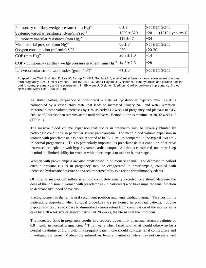
Pulmonary capillary wedge pressure (mm Hg)* 6 ± 2 Not significant
Systemic vascular resistance (dyne/cm/sec)* 1530 ± 520 −20 (1210 dyne/cm/s)
Pulmonary vascular resistance (mm Hg)* 119 ± 47 −34
Mean arterial pressure (mm Hg)* 86 ± 8 Not significant
Oxygen consumption (mL/min) V02 250 +20-30
COP (mm Hg)* 20.8 ± 1.0 −14
COP - pulmonary capillary wedge pressure gradient (mm Hg)* 14.5 ± 2.5 −28
Left ventricular stroke work index (g/min/m2)* 41 ± 8 Not significant
As stated earlier, pregnancy is considered a state of “gestational hypervolemia” as it is
hallmarked by a vasodilatory state that leads to increased urinary Na+ and water retention.
Maternal plasma volume increases by 10% as early as 7 weeks of pregnancy and plateaus to ~45-
50% at ~32 weeks then remains stable until delivery. Hemodilution is maximal at 30-32 weeks. 2
(Table 1)
The massive blood volume expansion that occurs in pregnancy may be severely blunted by
pathologic conditions, in particular severe preeclampsia. The mean blood volume expansion in
women with preeclampsia has been reported to be ~200 mL as compared to the typical 1500 mL
in normal pregnancies. 2 This is particularly important as preeclampsia is a condition of relative
intravascular depletion with hyperdynamic cardiac output. All things considered, one must keep
in mind the limited ability for women with preeclampsia to tolerate hemorrhage.
Women with pre-eclampsia are also predisposed to pulmonary edema. The decrease in colloid
oncotic pressure (COP) in pregnancy may be exaggerated in preeclampsia, coupled with
increased hydrostatic pressure and vascular permeability is a recipe for pulmonary edema.
Of note, as magnesium sulfate is almost completely renally excreted, one should decrease the
dose of the infusion in women with preeclampsia (in particular) who have impaired renal function
to decrease likelihood of toxicity.
Placing women in the left lateral recumbent position augments cardiac output. 3 This position is
particularly important when surgical procedures are performed in pregnant patients. Supine
hypotension occurs secondary to diminished venous return from compression of the inferior vena
cava by a 20 week size or greater uterus. At 20 weeks, the uterus is at the umbilicus.
The increased GFR in pregnancy results in a reduced upper limit of normal serum creatinine of
0.8 mg/dL in normal pregnancies. 3 This means when faced with what would otherwise be a
normal creatinine of 1.0 mg/dL in a pregnant patient, one should consider renal compromise and
investigate the cause. Medications infused via femoral central catheters may not circulate well
Adapted from Clark S, Cotton D, Lee W, Bishop C, Hill T, Southwick J, et al. Central hemodynamic assessment of normal term pregnancy. Am J Obstet Gynecol 1989;161:1439-42; and Elkayam U, Gleicher N. Hemodynamics and cardiac function during normal pregnancy and the puerperium. In: Elkayam U, Gleicher N, editors. Cardiac problems in pregnancy. 3rd ed. New York: Wiley-Liss; 1998. p. 3-19.
with compression from the gravid uterus on the iliac vessels and this may be critical when
resuscitating a patient.
Pregnancy is also a hypercoagulable state designed to attenuate the bleeding with childbirth.
Unfortunately this evolutionary adaptation also increases the risk for venous-thromboembolism in
pregnancy.
Renal plasma flow and glomerular filtration rate (GFR) both increase in pregnancy. The GFR
may increase up to 50% in pregnancy. 3
II. Hypertensive disorders of pregnancy
The hypertensive diseases that are encountered most commonly during pregnancy include:
gestational hypertension (HTN), chronic hypertension, and chronic hypertension with
superimposed pre-eclampsia, mild or severe preeclampsia and HELLP Syndrome.
Development of hypertensive disorders is the most common medical complication during
pregnancy, affecting 10% to 20% of all pregnancies worldwide. Elevated blood pressure (BP) is a
common denominator in a number of clinical entities that complicate pregnancy, including
pregnancy-induced hypertension (PIH), preeclampsia, eclampsia, and hemolysis, elevated liver
enzyme levels, low platelet count (HELLP) syndrome. Preeclampsia complicates 5% to 14% of
pregnancies worldwide and 5% to 8% in the United States. 4
Criteria established by the National High Blood Pressure Working Group, in pregnant women,
gestational hypertension is defined as: SBP ≥ 140 mm Hg or DBP ≥ 90 mm Hg that occurs after
20 weeks of gestation in a woman with previously normal blood pressure. 5 If blood pressure
elevations described above occur before 20 weeks of gestation, this is a patient with chronic
hypertension. If a woman with chronic HTN develops preeclampsia during pregnancy, the
nomenclature to describe this is: chronic hypertension with superimposed preeclampsia. One
does not have to qualify if it is mild or severe when described in patients with chronic HTN.
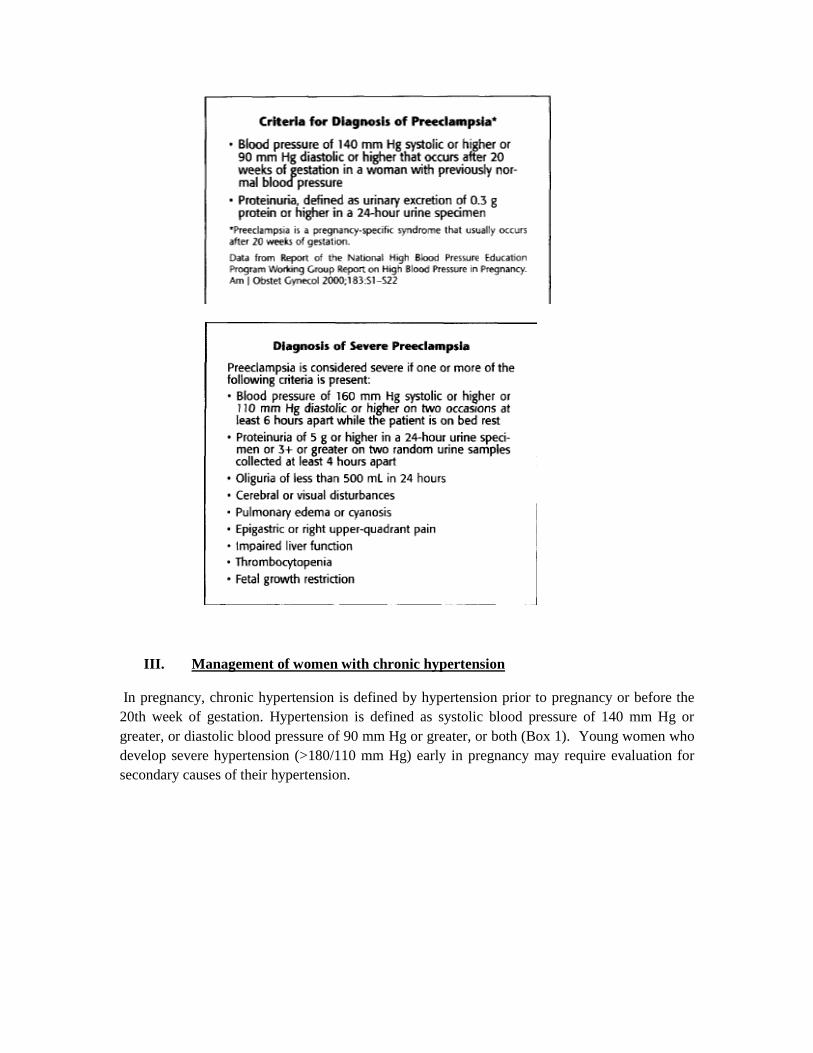
The hallmark of preeclampsia is hypertension and proteinuria. This is in comparison to the
patient with elevated blood pressure without proteinuria who may have gestational hypertension.
Preeclampsia is defined as: SBP >140mm Hg or DBP >90 mm Hg on 2 occasions 6 h apart with
an onset after 20 weeks and urine protein +1 on dipstick on 2 occasions 6 h apart or urine protein
>300 mg/24 h. 5 A screening spot urine protein/creatinine ratio>0.19 is suspicious and depending
on other maternal clinical signs and symptoms will help solidify a diagnosis of preeclampsia.
Severe preeclampsia is defined by severely elevated BPs (SBP >160, DBP >110), proteinuria and
evidence of end organ effects described in the table below. Eclampsia is the development of a
tonic-clonic seizure and this can occur in 1-2% of women with severe preeclampsia. 6
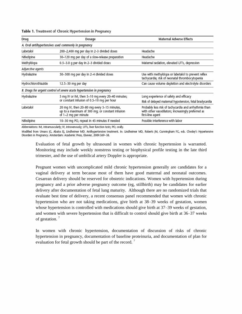
III. Management of women with chronic hypertension
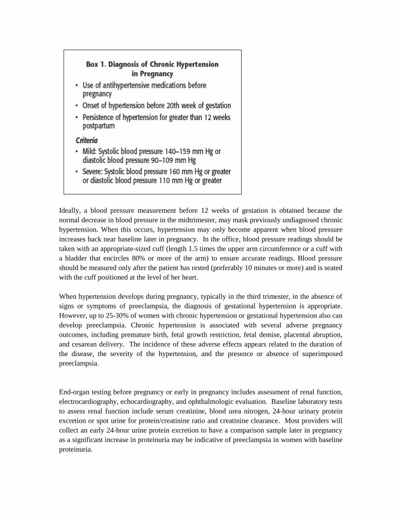
In pregnancy, chronic hypertension is defined by hypertension prior to pregnancy or before the
20th week of gestation. Hypertension is defined as systolic blood pressure of 140 mm Hg or
greater, or diastolic blood pressure of 90 mm Hg or greater, or both (Box 1). Young women who
develop severe hypertension (>180/110 mm Hg) early in pregnancy may require evaluation for
secondary causes of their hypertension.
Ideally, a blood pressure measurement before 12 weeks of gestation is obtained because the
normal decrease in blood pressure in the midtrimester, may mask previously undiagnosed chronic
hypertension. When this occurs, hypertension may only become apparent when blood pressure
increases back near baseline later in pregnancy. In the office, blood pressure readings should be
taken with an appropriate-sized cuff (length 1.5 times the upper arm circumference or a cuff with
a bladder that encircles 80% or more of the arm) to ensure accurate readings. Blood pressure
should be measured only after the patient has rested (preferably 10 minutes or more) and is seated
with the cuff positioned at the level of her heart.
When hypertension develops during pregnancy, typically in the third trimester, in the absence of
signs or symptoms of preeclampsia, the diagnosis of gestational hypertension is appropriate.
However, up to 25-30% of women with chronic hypertension or gestational hypertension also can
develop preeclampsia. Chronic hypertension is associated with several adverse pregnancy
outcomes, including premature birth, fetal growth restriction, fetal demise, placental abruption,
and cesarean delivery. The incidence of these adverse effects appears related to the duration of
the disease, the severity of the hypertension, and the presence or absence of superimposed
preeclampsia.
End-organ testing before pregnancy or early in pregnancy includes assessment of renal function,
electrocardiography, echocardiography, and ophthalmologic evaluation. Baseline laboratory tests
to assess renal function include serum creatinine, blood urea nitrogen, 24-hour urinary protein
excretion or spot urine for protein/creatinine ratio and creatinine clearance. Most providers will
collect an early 24-hour urine protein excretion to have a comparison sample later in pregnancy
as a significant increase in proteinuria may be indicative of preeclampsia in women with baseline
proteinuria.

Women with significant left ventricular hypertrophy and abnormal function secondary to
hypertension are more prone to experience cardiac decompensation and heart failure as pregnancy
progresses because of the increased intravascular volume and cardiac demand. Renal dysfunction
increases the risk of adverse pregnancy outcome.
It is often difficult to distinguish worsening chronic hypertension from superimposed
preeclampsia when the patient presents with elevated blood pressure late in pregnancy. In the
woman with chronic hypertension and renal disease, it may not be possible to distinguish between
the two entities. However, most young, nulliparous women who present with hypertension for the
first time during late pregnancy will have preeclampsia.
Antihypertensive therapy has been shown to reduce the risk of a severe maternal hypertensive
crisis but has not been shown to improve the overall perinatal outcome.
Experts in the United States have recommended that pregnant women with hypertension in the
blood pressure range of 150–160/100–110 mm Hg should be treated with antihypertensive
therapy, and that their blood pressure should be kept lower than 150/100 mm Hg. Guidelines
from Canada and the United Kingdom suggest considering treatment at lower values by main-
taining a blood pressure of 140–159/90–109 mm Hg. In women with evidence of end-organ
damage, such as left ventricular hypertrophy or renal insufficiency, antihypertensive treatment is
recommended to maintain blood pressure in the normal range, thereby reducing the risk of further
end-organ damage. Angiotensin-converting enzyme (ACE) inhibitors and angiotensin receptor
blockers are contraindicated during all trimesters of pregnancy.
The most appropriate management of mild hypertension in women who received antihypertensive
medications at conception is unclear. For women with mild hypertension, antihypertensive
therapy offers long-term maternal benefits, but there have been few if any short-term benefits
noted in pregnancy. 7
Evaluation of fetal growth by ultrasound in women with chronic hypertension is warranted.
Monitoring may include weekly nonstress testing or biophysical profile testing in the late third
trimester, and the use of umbilical artery Doppler is appropriate.
Pregnant women with uncomplicated mild chronic hypertension generally are candidates for a
vaginal delivery at term because most of them have good maternal and neonatal outcomes.
Cesarean delivery should be reserved for obstetric indications. Women with hypertension during
pregnancy and a prior adverse pregnancy outcome (eg, stillbirth) may be candidates for earlier
delivery after documentation of fetal lung maturity. Although there are no randomized trials that
evaluate best time of delivery, a recent consensus panel recommended that women with chronic
hypertension who are not taking medications, give birth at 38–39 weeks of gestation, women
whose hypertension is controlled with medications should give birth at 37–39 weeks of gestation,
and women with severe hypertension that is difficult to control should give birth at 36–37 weeks
of gestation. 7
In women with chronic hypertension, documentation of discussion of risks of chronic
hypertension in pregnancy, documentation of baseline proteinuria, and documentation of plan for
evaluation for fetal growth should be part of the record. 7
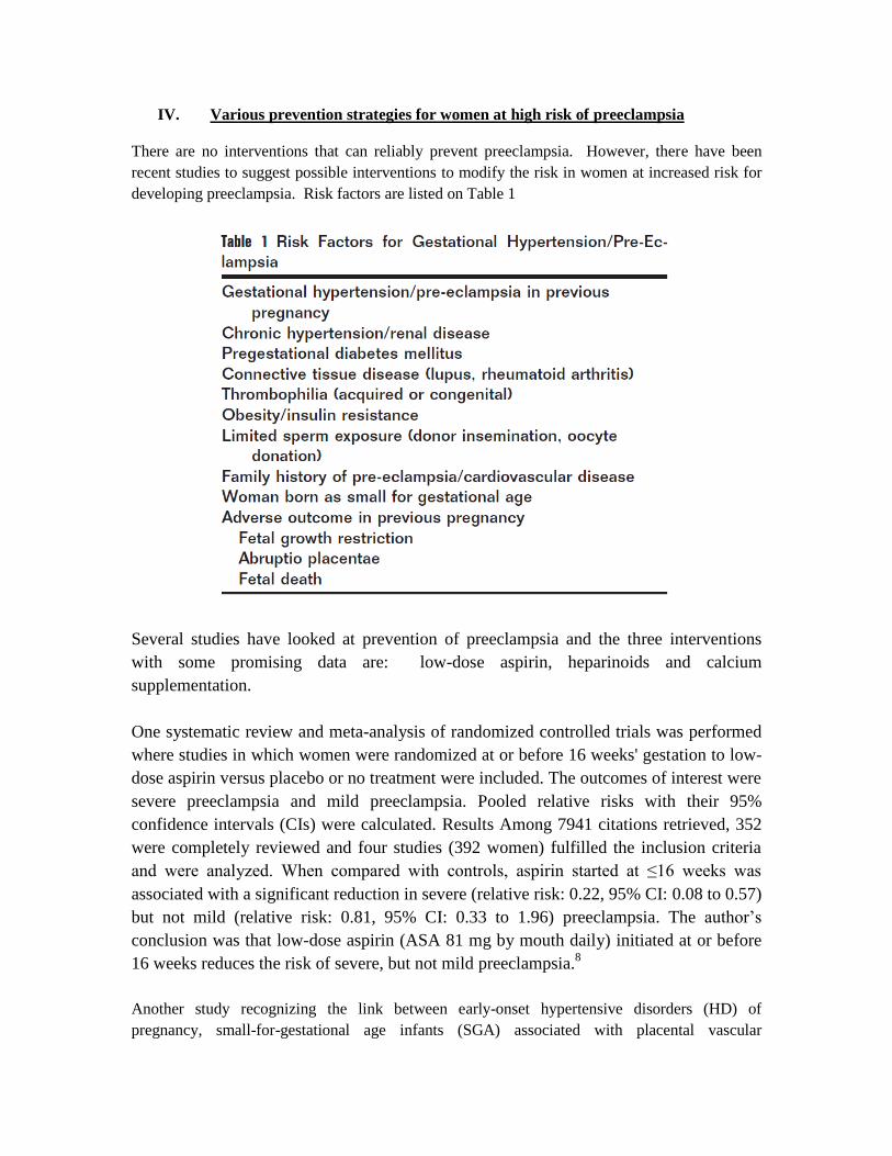
IV. Various prevention strategies for women at high risk of preeclampsia
There are no interventions that can reliably prevent preeclampsia. However, there have been
recent studies to suggest possible interventions to modify the risk in women at increased risk for
developing preeclampsia. Risk factors are listed on Table 1
Several studies have looked at prevention of preeclampsia and the three interventions
with some promising data are: low-dose aspirin, heparinoids and calcium
supplementation.
One systematic review and meta-analysis of randomized controlled trials was performed
where studies in which women were randomized at or before 16 weeks' gestation to low-
dose aspirin versus placebo or no treatment were included. The outcomes of interest were
severe preeclampsia and mild preeclampsia. Pooled relative risks with their 95%
confidence intervals (CIs) were calculated. Results Among 7941 citations retrieved, 352
were completely reviewed and four studies (392 women) fulfilled the inclusion criteria
and were analyzed. When compared with controls, aspirin started at ≤16 weeks was
associated with a significant reduction in severe (relative risk: 0.22, 95% CI: 0.08 to 0.57)
but not mild (relative risk: 0.81, 95% CI: 0.33 to 1.96) preeclampsia. The author’s
conclusion was that low-dose aspirin (ASA 81 mg by mouth daily) initiated at or before
16 weeks reduces the risk of severe, but not mild preeclampsia.8
Another study recognizing the link between early-onset hypertensive disorders (HD) of
pregnancy, small-for-gestational age infants (SGA) associated with placental vascular
thrombosis, and inheritable thrombophilia showed that aspirin reduced the recurrence risk. They
theorized that adding low-molecular-weight heparin (LMWH) to aspirin at < 12 weeks gestation
reduces the recurrence of HD in women with previous early-onset HD (pre-eclampsia, HELLP
syndrome and eclampsia) and/or SGA, in the context of inheritable thrombophilia without
antiphospholipid antibodies. They performed a multicenter randomized control trial (RCT) where
139 women included were < 12 weeks gestation. Inclusion criteria: previous delivery< 34 weeks
gestation with HD and/or SGA; inheritable thrombophilia (protein C deficiency, protein S
deficiency, activated protein C resistance, factor V Leiden heterozygosity and prothrombin gene
G20210A mutation heterozygosity); and no antiphospholipid antibodies detected. Intervention:
either daily LMWH (dalteparin, 5000 IU weight-adjusted dosage) with aspirin 80 mg or aspirin
80 mg alone. Primary outcomes: recurrent HD onset (i) < 34 weeks gestation and (ii) irrespective
of gestational age. Secondary outcomes: recurrent SGA, preterm birth, maternal/neonatal
hospitalization, spontaneous abortion and individual HD. Analysis by intention-to-treat. The
results showed low-molecular-weight heparin with aspirin reduced recurrent HD onset < 34
weeks gestation (risk difference [RD] 8.7%: confidence interval [CI] of RD 1.9–15.5%; P =
0.012; number needed to treat [NNT] 12). Recurrent HD irrespective of gestational age was not
different between the arms. No women withdrew as a result of adverse effects. They concluded
that adding LMWH to aspirin at < 12 weeks gestation reduces recurrent HD onset < 34 weeks
gestation in women with inheritable thrombophilia and prior delivery for HD/SGA <34 weeks.
However, close monitoring of the mother and fetus remains important throughout pregnancy. 9
The Cochrane Group conducted a meta-analysis to assess the effects of calcium
supplementation during pregnancy on hypertensive disorders of pregnancy and related
maternal and child outcomes. The randomized trials compared at least 1 g daily of
calcium during pregnancy with placebo. They included 13 studies of good quality
(involving 15,730 women). The average risk of high blood pressure was reduced with
calcium supplementation rather than placebo (12 trials, 15,470 women: risk ratio (RR)
0.65, 95% confidence interval (CI) 0.53 to 0.81). There was also a reduction in the
average risk of pre-eclampsia associated with calcium supplementation (13 trials, 15,730
women: RR 0.45, 95% CI 0.31 to 0.65). The effect was greatest for high-risk women
(five trials, 587 women: RR 0.22, 95% CI 0.12 to 0.42), and those with low baseline
calcium intake (eight trials, 10,678 women: RR 0.36, 95% CI 0.20 to 0.65).The average
risk of preterm birth was reduced in the calcium group overall (11 trials, 15,275 women:
RR 0.76, 95% CI 0.60 to 0.97) and amongst women at high risk of developing pre-
eclampsia recruited to four small trials (568 women: RR 0.45, 95% CI 0.24 to
0.83).There was no overall effect on the risk of stillbirth or death before discharge from
hospital (11 trials 15,665 babies; RR 0.90, 95% CI 0.74 to 1.09). The composite outcome
maternal death or serious morbidity was reduced (four trials, 9732 women; RR 0.80, 95%
CI 0.65 to 0.97). Most of the women in these trials were low risk and had a low calcium
diet. Maternal deaths were reported in only one trial. One death occurred in the calcium
group and six in the placebo group, a difference which was not statistically significant
(RR 0.17, 95% CI 0.02 to 1.39). Blood pressure in childhood has been assessed in two
studies, only one of which is currently included: childhood systolic blood pressure greater
than 95th percentile was reduced (514 children: RR 0.59, 95% CI 0.39 to 0.91). The
authors concluded that calcium supplementation appears to approximately halve the risk
of pre-eclampsia, reduce the risk of preterm birth and reduce the rare occurrence of the
composite outcome 'death or serious morbidity'. There were no other clear benefits, or
harms.10
V. Management of mild and severe preeclampsia
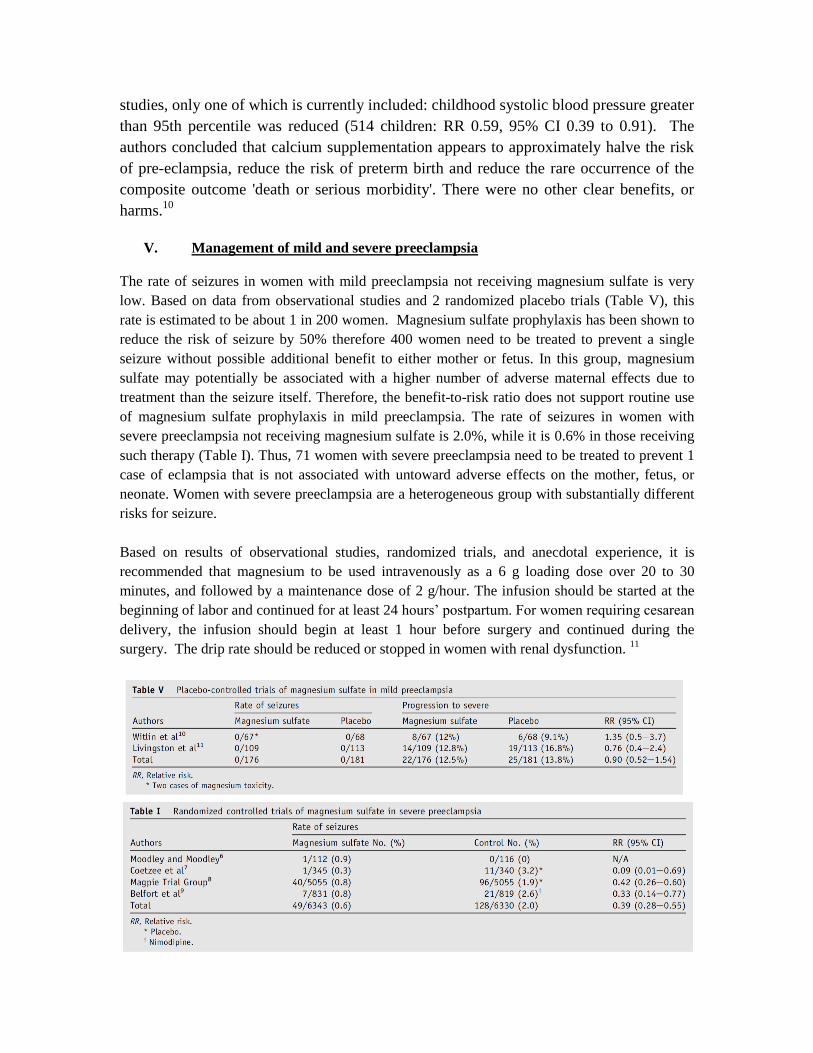
The rate of seizures in women with mild preeclampsia not receiving magnesium sulfate is very
low. Based on data from observational studies and 2 randomized placebo trials (Table V), this
rate is estimated to be about 1 in 200 women. Magnesium sulfate prophylaxis has been shown to
reduce the risk of seizure by 50% therefore 400 women need to be treated to prevent a single
seizure without possible additional benefit to either mother or fetus. In this group, magnesium
sulfate may potentially be associated with a higher number of adverse maternal effects due to
treatment than the seizure itself. Therefore, the benefit-to-risk ratio does not support routine use
of magnesium sulfate prophylaxis in mild preeclampsia. The rate of seizures in women with
severe preeclampsia not receiving magnesium sulfate is 2.0%, while it is 0.6% in those receiving
such therapy (Table I). Thus, 71 women with severe preeclampsia need to be treated to prevent 1
case of eclampsia that is not associated with untoward adverse effects on the mother, fetus, or
neonate. Women with severe preeclampsia are a heterogeneous group with substantially different
risks for seizure.
Based on results of observational studies, randomized trials, and anecdotal experience, it is
recommended that magnesium to be used intravenously as a 6 g loading dose over 20 to 30
minutes, and followed by a maintenance dose of 2 g/hour. The infusion should be started at the
beginning of labor and continued for at least 24 hours’ postpartum. For women requiring cesarean
delivery, the infusion should begin at least 1 hour before surgery and continued during the
surgery. The drip rate should be reduced or stopped in women with renal dysfunction. 11

There is evidence-based data from the HYPITAT trial that suggests delivery is indicated in
pregnancies complicated by mild gestational hypertension or mild pre-eclampsia occurring at
37 or more weeks’ gestation. This is the first multicenter trial designed to compare the risks and
benefits of induction of labor versus expectant monitoring for women with mild gestational
hypertension/pre-eclampsia at >36-0/7 weeks’ gestation. The trial included 756 women with a
singleton pregnancy at 36-0/7 to 41-6/7 weeks who had mild gestational hypertension (n = 496)
or mild pre-eclampsia (n = 246); 377 were allocated to induction and 379 to expectant
monitoring. Women randomized to the induction group had a significant reduction in primary
outcome (Table 3). This reduction was mainly attributable to differences in the rates of
progression to severe hypertension. There were no differences in adverse neonatal outcomes. In
addition, the overall rates of cesarean delivery were not different in both groups; however, in the
induction group, the rate of cesarean delivery was lower in nulliparous women and in those with a
cervical Bishop score <2. This latter finding refutes the belief that induction of labor in these
women increases the rate of cesarean delivery. Therefore, induction of labor and/or delivery at
>37 weeks’ gestation should be offered to all such women provided that gestational age is well
documented and the induction period is not prolonged beyond 48 hours.
The main aim of expectant treatment is to improve perinatal outcome by prolonging gestation and
reducing neonatal morbidities (acute and long-term). There are potential perinatal complications
during expectant treatment; consequently, all reported studies recommended intensive fetal
surveillance for early detection of fetal compromise. The most common indication for delivery in
most studies was deterioration in fetal status. Expectant treatment improves perinatal outcome in
a select group of women with severe preeclampsia at <32 6/7 weeks of gestation. It must be
emphasized that these recommendations are based on only 2 randomized trials (a total of 133
women) and several observational studies on the subject.
The presence of severe preeclampsia at >34 0/7 weeks of gestation mandates immediate
hospitalization in the labor and delivery unit. Intravenous magnesium sulfate therapy should be
started to prevent convulsions and antihypertensive medications to lower severe hypertension
(systolic pressure SBP >160 mm Hg and/or diastolic pressure DBP >110 mm Hg). The goal is to
keep SBP between 140 -155 mm Hg and DBP between 90- 105 mm Hg.
In addition, corticosteroids are administered for fetal lung maturation. During the observation
period, maternal and fetal conditions are assessed, and a decision is made regarding the need for
delivery. After initial clinical and laboratory evaluation, a decision must be made for immediate
delivery vs expectant treatment. Patients with eclampsia, neurologic deficit (blindness, confusion,
motor deficit), pulmonary edema, disseminated intravascular coagulation, suspected abruptio
placentae, or nonreassuring fetal heart rate testing are delivered regardless of the benefit of
corticosteroids after maternal stabilization. Patients with a gestational age of <23 0/7 weeks
should be offered termination of pregnancy because no babies have survived in reported studies
during the expectant treatment of severe preeclampsia at this gestational age. In addition,
expectant treatment in patients with gestational age between 23 0/7 and 23 6/7 results in
extremely high maternal and perinatal morbidity and mortality rates. Therefore, expectant
treatment in these patients should be considered only as an option after extensive counseling.
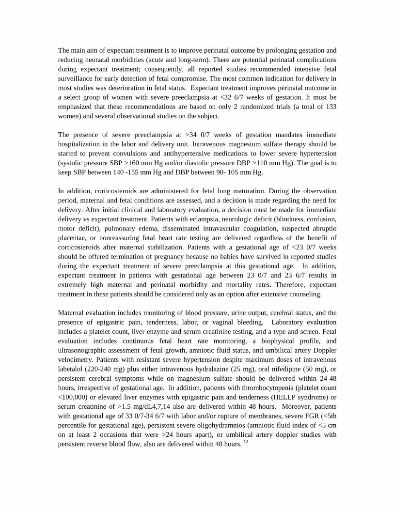
Maternal evaluation includes monitoring of blood pressure, urine output, cerebral status, and the
presence of epigastric pain, tenderness, labor, or vaginal bleeding. Laboratory evaluation
includes a platelet count, liver enzyme and serum creatinine testing, and a type and screen. Fetal
evaluation includes continuous fetal heart rate monitoring, a biophysical profile, and
ultrasonographic assessment of fetal growth, amniotic fluid status, and umbilical artery Doppler
velocimetry. Patients with resistant severe hypertension despite maximum doses of intravenous
labetalol (220-240 mg) plus either intravenous hydralazine (25 mg), oral nifedipine (50 mg), or
persistent cerebral symptoms while on magnesium sulfate should be delivered within 24-48
hours, irrespective of gestational age. In addition, patients with thrombocytopenia (platelet count
<100,000) or elevated liver enzymes with epigastric pain and tenderness (HELLP syndrome) or
serum creatinine of >1.5 mg/dL4,7,14 also are delivered within 48 hours. Moreover, patients
with gestational age of 33 0/7-34 6/7 with labor and/or rupture of membranes, severe FGR (<5th
percentile for gestational age), persistent severe oligohydramnios (amniotic fluid index of <5 cm
on at least 2 occasions that were >24 hours apart), or umbilical artery doppler studies with
persistent reverse blood flow, also are delivered within 48 hours. 12
During observation on the antepartum ward, blood pressure is measured every 4-6 hours. Patients
receive antihypertensive drugs as needed, usually oral nifedipine 10-20 mg every 4-6 hours (40-
120 mg per day) and/or labetalol 200-800 mg every 8-12 hours (600-2400 mg per day), to keep
SBP between 140 and 155 mm Hg and DBP between 90 and 105 mm Hg. Alternatively, a long
acting (XL) version of nifedipine (30 mg every 12 hours) can be used for BP control. During
titration of oral antihypertensive agents, if the patient has a persistent severe hypertensive
episode, blood pressure is assessed every 15 minutes. If the blood pressure remains in the severe
range after 30-60 minutes, the patient should be transferred to the labor and delivery unit for more
intensive monitoring and treatment. The patient should then receive an acute dose of either oral
nifedipine 10mg or labetalol 20 mg intravenously or hydralazine 5-10 mg intravenously, as
needed.
Patients with resistant severe hypertension after maximum doses of IV labetalol should receive
magnesium sulfate and delivered. The patients receive frequent assessment of maternal and fetal
well-being. Maternal assessment includes: frequent evaluation of symptoms (headache, blurred or
double vision, confusion, nausea, vomiting, epigastric or right upper abdominal pain, shortness of
breath, uterine activity, and vaginal bleeding), volume intake and output, and laboratory testing.
Laboratory testing includes complete blood count with platelet count and transaminase, lactate
dehydrogenase, and serum creatinine levels. Fetal assessment includes daily fetal kick counts, at
least daily nonstress test (NST) with uterine activity monitoring with biophysical profile (BPP) if
the NST is nonreactive and twice weekly amniotic fluid assessment. Severe oligohydramnios is
defined as an amniotic fluid index of <5 cm on at least 2 occasions that are at least 24 hours apart.
Severe oligohydramnios is considered an indication for delivery in all patients with a gestational
age of >30 weeks, irrespective of other fetal testing results. In those <30 weeks of gestation,
pregnancy may be continued with reassuring NST and umbilical artery Doppler findings. 12
Umbilical artery Doppler studies are performed weekly, or more often if IUGR is suspected
and/or testing reveals abnormal diastolic flow. Umbilical artery doppler studies with reverse
diastolic blood flow after initial maternal/fetal stabilization is considered an indication for
delivery. Ultrasonographic assessment of fetal growth is performed every 2 weeks. If a patient
experiences headache that does not resolve with oral analgesics within 6 hours and the headache
continues to be severe, they should be transferred to the labor and delivery unit and receive
intravenous magnesium sulfate and antihypertensives as needed. If the headache persists,
preparations should be made for delivery. Patients with new onset epigastric or right upper
abdominal pain, retrosternal pain or pressure, and recurrent heart burn, particularly in association
with nausea and vomiting, are also transferred to the labor and delivery unit for further
assessment. If the symptoms persist and/or the liver enzymes are abnormal, preparations are made
for delivery. In addition, the onset of uterine contractions and/or vaginal bleeding requires
immediate transfer to the labor and delivery unit because it could signify the development of
abruption placentae. At any time during expectant treatment, the development of any of the
findings in the table below is an indication for delivery. 12
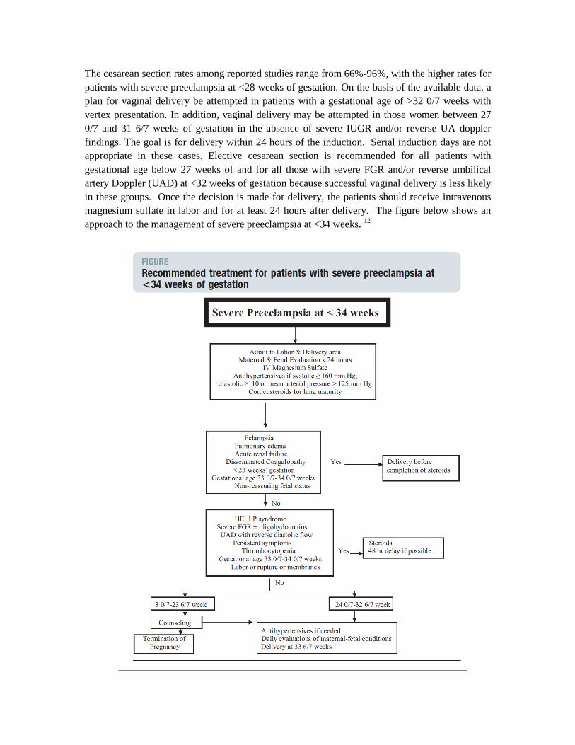
The cesarean section rates among reported studies range from 66%-96%, with the higher rates for
patients with severe preeclampsia at <28 weeks of gestation. On the basis of the available data, a
plan for vaginal delivery be attempted in patients with a gestational age of >32 0/7 weeks with
vertex presentation. In addition, vaginal delivery may be attempted in those women between 27
0/7 and 31 6/7 weeks of gestation in the absence of severe IUGR and/or reverse UA doppler
findings. The goal is for delivery within 24 hours of the induction. Serial induction days are not
appropriate in these cases. Elective cesarean section is recommended for all patients with
gestational age below 27 weeks of and for all those with severe FGR and/or reverse umbilical
artery Doppler (UAD) at <32 weeks of gestation because successful vaginal delivery is less likely
in these groups. Once the decision is made for delivery, the patients should receive intravenous
magnesium sulfate in labor and for at least 24 hours after delivery. The figure below shows an
approach to the management of severe preeclampsia at <34 weeks. 12
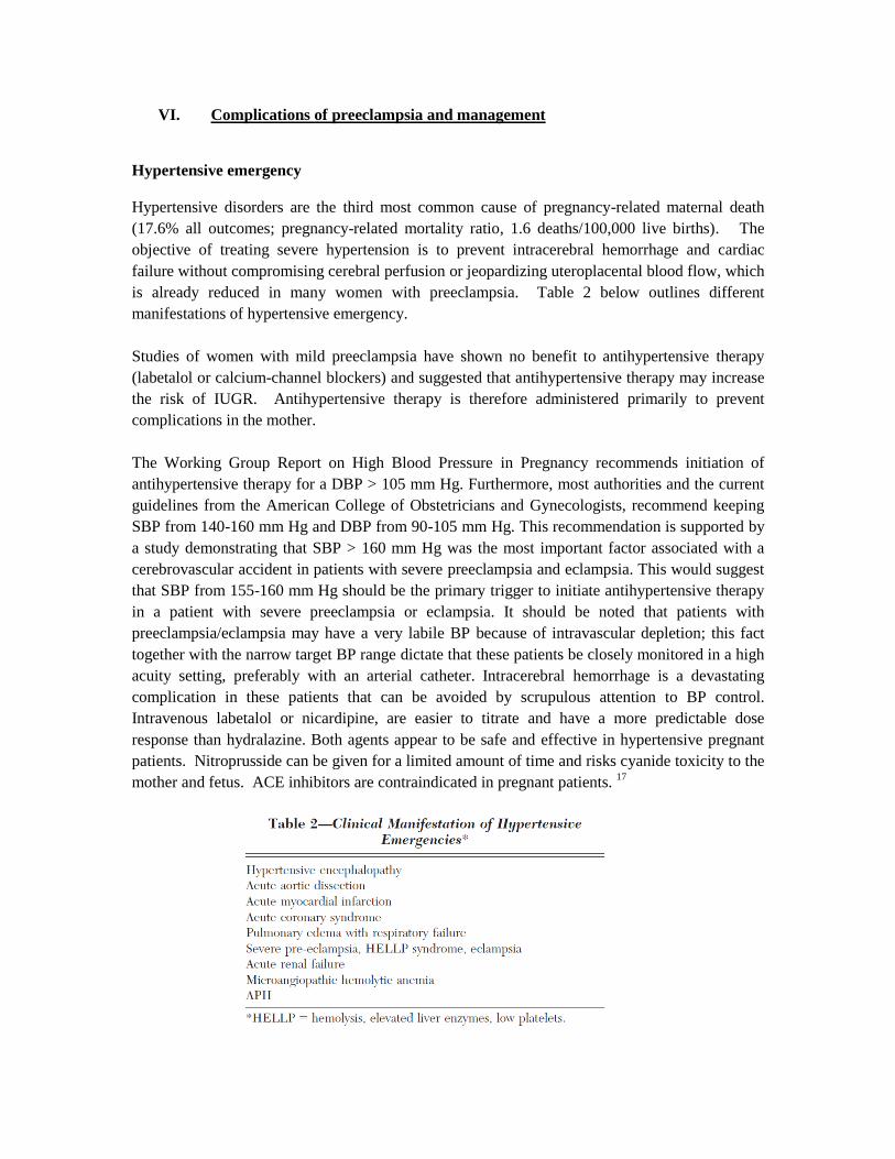
VI. Complications of preeclampsia and management
Hypertensive emergency
Hypertensive disorders are the third most common cause of pregnancy-related maternal death
(17.6% all outcomes; pregnancy-related mortality ratio, 1.6 deaths/100,000 live births). The
objective of treating severe hypertension is to prevent intracerebral hemorrhage and cardiac
failure without compromising cerebral perfusion or jeopardizing uteroplacental blood flow, which
is already reduced in many women with preeclampsia. Table 2 below outlines different
manifestations of hypertensive emergency.
Studies of women with mild preeclampsia have shown no benefit to antihypertensive therapy
(labetalol or calcium-channel blockers) and suggested that antihypertensive therapy may increase
the risk of IUGR. Antihypertensive therapy is therefore administered primarily to prevent
complications in the mother.
The Working Group Report on High Blood Pressure in Pregnancy recommends initiation of
antihypertensive therapy for a DBP > 105 mm Hg. Furthermore, most authorities and the current
guidelines from the American College of Obstetricians and Gynecologists, recommend keeping
SBP from 140-160 mm Hg and DBP from 90-105 mm Hg. This recommendation is supported by
a study demonstrating that SBP > 160 mm Hg was the most important factor associated with a
cerebrovascular accident in patients with severe preeclampsia and eclampsia. This would suggest
that SBP from 155-160 mm Hg should be the primary trigger to initiate antihypertensive therapy
in a patient with severe preeclampsia or eclampsia. It should be noted that patients with
preeclampsia/eclampsia may have a very labile BP because of intravascular depletion; this fact
together with the narrow target BP range dictate that these patients be closely monitored in a high
acuity setting, preferably with an arterial catheter. Intracerebral hemorrhage is a devastating
complication in these patients that can be avoided by scrupulous attention to BP control.
Intravenous labetalol or nicardipine, are easier to titrate and have a more predictable dose
response than hydralazine. Both agents appear to be safe and effective in hypertensive pregnant
patients. Nitroprusside can be given for a limited amount of time and risks cyanide toxicity to the
mother and fetus. ACE inhibitors are contraindicated in pregnant patients. 17
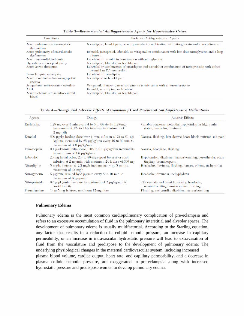
Pulmonary Edema
Pulmonary edema is the most common cardiopulmonary complication of pre-eclampsia and
refers to an excessive accumulation of fluid in the pulmonary interstitial and alveolar spaces. The
development of pulmonary edema is usually multifactorial. According to the Starling equation,
any factor that results in a reduction in colloid osmotic pressure, an increase in capillary
permeability, or an increase in intravascular hydrostatic pressure will lead to extravasation of
fluid from the vasculature and predispose to the development of pulmonary edema. The
underlying physiological changes in the maternal cardiovascular system, including increased
plasma blood volume, cardiac output, heart rate, and capillary permeability, and a decrease in
plasma colloid osmotic pressure, are exaggerated in pre-eclampsia along with increased
hydrostatic pressure and predispose women to develop pulmonary edema.
Medical therapies to treat pulmonary edema should be optimized to expedite treatment results.
Furosemide (Lasix) can be administered intravenously as a single dose of 10-40 mg over 2
minutes to promote diuresis. Bladder catheterization allows for accurate measurement of urine
output. Most patients will respond to initial diuresis therapy. If adequate response is not seen
within 30-60 minutes, the dose should be increased to 40-60 mg administered by slow
intravenous injection to a maximum of 120 mg in 1 hour. Electrolytes should be monitored
closely and repleted as indicated. Morphine sulfate can be administered intravenously as needed
both for pain as well as in an attempt to reduce the adrenergic vasoconstrictor stimuli to the
pulmonary arteriolar and venous beds. As with the management of all parturients with pre-
eclampsia, sodium and water should be modestly restricted, and maternal fluid balance should be
strictly monitored.
Oxygen saturation should be monitored using a pulse oximeter, and oxygen supplementation
using a nonrebreather facemask can be used to treat maternal hypoxemia. Intubation is rarely
required. In addition to these standard measures, it is appropriate to follow the patient’s blood
pressure, electrocardiogram, and fetal heart rate tracing. Afterload reduction using vasodilators
may be necessary, especially in parturients with chronic hypertension and superimposed pre-
eclampsia. 16
ARDS
Pre-eclampsia complicated by HELLP syndrome, pulmonary edema, and/or cardiopulmonary
disease can advance to pregnancy-related ARDS in rare instances. In a series of 83 obstetrical
patients with ARDS from all causes, the antepartum mortality rate was 23% and the postpartum
mortality rate was 50%. 16
Cerebrovascular Accidents
The vast majority of patients with cerebral ischemia present with acutely elevated BP regardless
of the subtype of infarct or preexisting hypertension. The BP elevation decreases spontaneously
over time. The elevated BP is not a manifestation of a hypertensive emergency but rather a
protective physiologic response to maintain cerebral perfusion pressure to the vascular territory
affected by ischemia. Lowering the BP in patients with ischemic strokes may reduce cerebral
blood flow, which because of impaired autoregulation, may result in further ischemic injury. The
common practice of “normalizing” the BP following a cerebrovascular accident is potentially
dangerous.
The American Stroke Association and the European Stroke Initiative guidelines recommend
withholding antihypertensive therapy for acute ischemic stroke unless there is planned
thrombolysis, evidence of concomitant noncerebral acute organ damage, or if the BP is
excessively high, arbitrarily chosen as a SBP > 220 mm Hg or a DBP > 120 mm Hg based on the
upper limit of normal autoregulation. In these patients, the aim is to reduce the mean arterial
pressure by not more than 10 to 15% in the first 24 h. Semplicini and colleagues demonstrated
that a high initial BP was associated with a better neurologic outcome following an acute
ischemic stroke. These authors suggest that hypertension may be protective during an acute
ischemic stroke and that lowering the BP may be potentially harmful. Hypertensive therapy is
required for SBP > 185 mm Hg or DBP >110 mm Hg, with a targeted SBP of 180 mm Hg and a
DBP of 105 mm Hg. The current American Heart Association guidelines recommend the use of
labetalol or nicardipine if the SBP is > 220 mm Hg or the DBP is from 121- 140 mm Hg, and
nitroprusside for a DBP > 140 mm Hg. For the reasons outlined above, we believe nitroprusside
to be a poor choice in patients with intracranial pathology. These parameters are in place for non-
pregnant patients and are applied to the gravid population, however, no well done studies define
appropriate parameters in pregnancy.
In patients with intracerebral hematomas, there is almost always a rise in intracranial pressure
with reflex systemic hypertension. There is no evidence that hypertension provokes further
bleeding in patients with intracranial hemorrhage. However, a precipitous fall in systemic BP will
compromise cerebral perfusion. The controlled lowering of the BP is currently recommended
only when the SBP is >200 mm Hg, the DBP is > 110 mm Hg, or the MAP is > 130 mm Hg.
One study demonstrated that the rapid decline of BP within the first 24 h after presentation of an
intracranial hemorrhage was associated with increased mortality; the rate of decline in BP was
independently associated with increased mortality. Nicardipine has been demonstrated to be an
effective agent for the control of BP in patients with intracerebral hemorrhage. 17
Peripartum Cardiomyopathy
Peripartum cardiomyopathy (PPCM) affects patients late in pregnancy or in the early postpartum
period. It is an infrequent complication of pre-eclampsia, but a history of preeclampsia can be
found in up to 70% of those who develop PPCM. The 4 following criteria are needed to meet the
definition of peripartum cardiomyopathy: (1) development of cardiac failure in the last month of
pregnancy or within 5 months of delivery; (2) absence of an identifiable cause for the cardiac
failure; (3) absence of recognizable heart disease before the last month of pregnancy; (4) left
ventricular systolic dysfunction (for example, left ventricular ejection fraction (EF) below 45%).
The etiology remains unclear, but risks factors include multiple pregnancy, pre-eclampsia,
multiparity, and advanced maternal age. Pre-eclampsia is associated with peripartum
cardiomyopathy as a risk factor; however, peripartum cardiomyopathy is an infrequent
complication of pre-eclampsia. Women with residual cardiac dysfunction postpartum should be
counseled against future pregnancy given associated risk for worsening left ventricular function
and mortality. 16
Myocardial Ischemia and Infarction
Myocardial infarction associated with pregnancy is a rare event, usually related to maternal risk
factors for ischemic heart disease, such as hypertension, diabetes mellitus, and coronary
atherosclerosis. However, a mechanism of coronary spasm has been suggested to be a cause of
myocardial infarction in patients with normal coronary arteries or in those with minimal
nonobstructive coronary artery disease, especially infarction related to pre-eclampsia, after
administration of ergot alkaloids, bromocriptine, oxytocin, and prostaglandin.

Early diagnosis of acute myocardial infarction in pregnancy is often hindered by the normal
changes of pregnancy and low level of suspicion.
Pre-eclampsia and coronary artery disease share common risk factors for endothelial dysfunction
and damage, such as hypertension, diabetes, and obesity. The associated maternal physiological
changes from pre-eclampsia include increased inflammatory markers, dyslipidemia, insulin
resistance, endothelial dysfunction, and oxidative stress and are associated with an increased risk
for cardiovascular disease in later life. A history of pre-eclampsia has been reported to be a risk
factor for several distinct cardiovascular conditions later in life. 16
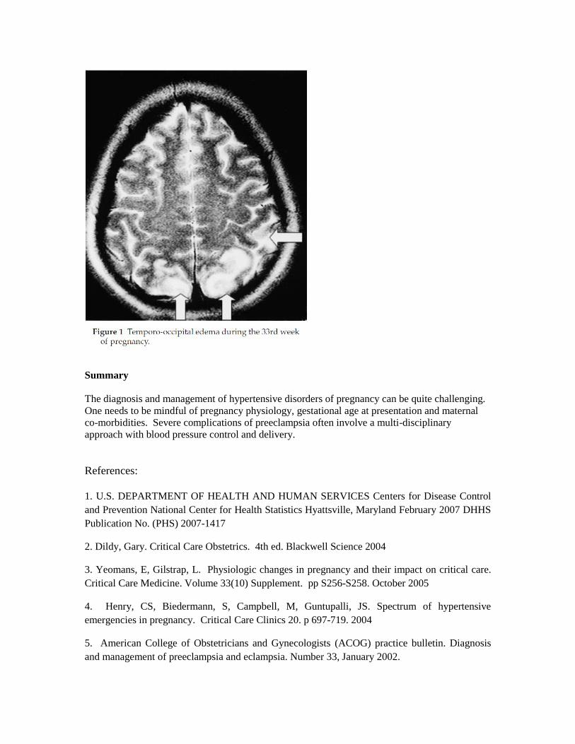
Cortical Blindness and PRESS (aka PLES)
Blindness in pre-eclampsia could be due to ocular pathologies such as retinal detachment or
vascular occlusion, or more frequently cortical origin (Table 1). It can be seen in 1–15% of all
preeclamptic patients and the rates are higher in underdeveloped countries. Visual disturbances
are a well-known clinical entity seen in patients with hypertensive disorders of pregnancy. Exact
central defects of this finding were not known until Hinchey et al. described PLES as a clinical
syndrome in 1996. Although they describe this condition as a puerperal entity, the exact
frequency of cerebral involvement is not known as cranial imaging is not routine in the absence
of obvious neurological symptoms. Therefore, most cases of posterior cerebral edema might go
unnoticed in pre-eclamptic women, even with visual disturbances (figure 1). Acute cortical
blindness is one of the most dramatic presentations of pre-eclampsia and is historically known to
be reversible. However, blindness, especially when secondary to ocular pathologies, may be
irreversible. 15
Summary
The diagnosis and management of hypertensive disorders of pregnancy can be quite challenging.
One needs to be mindful of pregnancy physiology, gestational age at presentation and maternal
co-morbidities. Severe complications of preeclampsia often involve a multi-disciplinary
approach with blood pressure control and delivery.
References:
1. U.S. DEPARTMENT OF HEALTH AND HUMAN SERVICES Centers for Disease Control
and Prevention National Center for Health Statistics Hyattsville, Maryland February 2007 DHHS
Publication No. (PHS) 2007-1417
2. Dildy, Gary. Critical Care Obstetrics. 4th ed. Blackwell Science 2004
3. Yeomans, E, Gilstrap, L. Physiologic changes in pregnancy and their impact on critical care.
Critical Care Medicine. Volume 33(10) Supplement. pp S256-S258. October 2005
4. Henry, CS, Biedermann, S, Campbell, M, Guntupalli, JS. Spectrum of hypertensive
emergencies in pregnancy. Critical Care Clinics 20. p 697-719. 2004
5. American College of Obstetricians and Gynecologists (ACOG) practice bulletin. Diagnosis
and management of preeclampsia and eclampsia. Number 33, January 2002.

6. Eric A P Steegers, Peter von Dadelszen, Johannes J Duvekot, Robert Pijnenborg. Pre-
eclampsia. Lancet 2010; 376: 631–44
7. ACOG practice Bulletin 125: Chronic Hypertension in Pregnancy. Vol 119 (2) part I.
February 2012. p396-402
8. Roberge S, Giguère Y, Villa P, Nicolaides K, Vainio M, Forest JC, von Dadelzen P, Vaiman
D, Tapp S, Bujold E. Early Administration of Low-Dose Aspirin for the Prevention of Severe
and Mild Preeclampsia: A Systematic Review and Meta-Analysis. Am J Perinatol. 2012 Apr 11.
[Epub ahead of print]
9. de Vries JI, van Pampus MG, Hague WM, Bezemer PD, Joosten JH; FRUIT Investigators.
Low-molecular-weight heparin added to aspirin in the prevention of recurrent early-onset pre-
eclampsia in women with inheritable thrombophilia: the FRUIT-RCT. Thromb Haemost. 2012
Jan;10(1):64-72.
10. Hofmeyr GJ, Lawrie TA, Atallah AN, Duley L. Calcium supplementation during pregnancy
for preventing hypertensive disorders and related problems. Cochrane Database Syst Rev. 2010
Aug 4;(8):CD001059.
11. Sibai BM. Management of late preterm and early-term pregnancies complicated by mild
gestational hypertension/pre-eclampsia. Semin Perinatol. 2011 Oct;35(5):292-6.
12. Sibai BM, Barton JR. Expectant management of severe preeclampsia remote from term:
patient selection, treatment, and delivery indications. Am J Obstet Gynecol 2007;196:514.e1-
514.e9.
13. Sibai, BM. Magnesium sulfate prophylaxis in preeclampsia: Lessons learned from recent
trials. American Journal of Obstetrics and Gynecology (2004) 190, 1520e6
14. Bauer, ST, Cleary, KL. Cardiopulmonary Complications of Pre-eclampsia. Semin Perinatol
33:158-165. 2009
15. Lutfu S. Onderoglu, Polat Dursun, Murat Gultekin and Nilufer Y. Celik Posterior
leukoencephalopathy syndrome as a cause of reversible blindness during pregnancy J. Obstet.
Gynaecol. Res. Vol. 33, No. 4: 539–542, August 2007
16. Charles S. Henry, MD, Scott A. Biedermann, MD, Michel F. Campbell, MD, Jayarama S.
Guntupalli, MD Spectrum of hypertensive emergencies in pregnancy. Crit Care Clin 20 (2004)
697– 712
17. Paul E. Marik, MD, FCCP; and Joseph Varon, MD, FCCP Hypertensive Crises Challenges
and Management CHEST 2007; 131:1949–1962