hyperphosphorylationoftauinducedbynaturallysecreted ... · pdf...
TRANSCRIPT

Hyperphosphorylation of Tau Induced by Naturally SecretedAmyloid-� at Nanomolar Concentrations Is Modulated byInsulin-dependent Akt-GSK3� Signaling Pathway*□S
Received for publication, February 8, 2012, and in revised form, August 17, 2012 Published, JBC Papers in Press, August 21, 2012, DOI 10.1074/jbc.M112.348300
Takayoshi Tokutake‡1, Kensaku Kasuga‡1, Ryuji Yajima‡, Yumi Sekine‡, Toshiyuki Tezuka‡, Masatoyo Nishizawa‡,and Takeshi Ikeuchi‡§2
From the ‡Department of Neurology, Brain Research Institute, and the §Center for Transdisciplinary Research, Niigata University,Niigata 951-8585, Japan
Background: Little is known about the underlying mechanisms by which extracellular amyloid-� (A�) induces Tau phos-phorylation in Alzheimer disease (AD).Results: Naturally secreted A� induced hyperphosphorylation of Tau and impaired insulin signal transduction.Conclusion:Adisturbed insulin signaling cascademay be implicated in the pathway of A�-inducedTau hyperphosphorylation.Significance: These findings may explain the molecular link between Alzheimer disease and insulin signaling.
Alzheimer disease (AD) is neuropathologically characterizedby the formation of senile plaques from amyloid-� (A�) andneurofibrillary tangles composed of phosphorylated Tau.Although there is growing evidence for the pathogenic role ofsolubleA� species inAD, themajor question of howA� induceshyperphosphorylation of Tau remains unanswered. To addressthis question, we here developed a novel cell coculture system toassess the effect of extracellular A� at physiologically relevantlevels naturally secreted from donor cells on the phosphoryla-tion of Tau in recipient cells. Using this assay, we demonstratedthat physiologically relevant levels of secreted A� are sufficientto cause hyperphosphorylation of Tau in recipient N2a cellsexpressing human Tau and in primary culture neurons. Thishyperphosphorylation of Tau is inhibited by blocking A� pro-duction in donor cells. The expression of familial AD-linkedPSEN1mutants andAPP�E693mutant that induce the produc-tion of oligomeric A� in donor cells results in a similar hyper-phosphorylation of Tau in recipient cells. The mechanismunderlying the A�-induced Tau hyperphosphorylation is medi-ated by the impaired insulin signal transduction because wedemonstrated that the phosphorylation ofAkt andGSK3�uponinsulin stimulation is less activated under this condition. Treat-ing cells with the insulin-sensitizing drug rosiglitazone, a perox-isome proliferator-activated receptor � agonist, attenuates theA�-dependent hyperphosphorylation of Tau. These findingssuggest that the disturbed insulin signaling cascade may beimplicated in the pathways through which soluble A� inducesTau phosphorylation and further support the notion that cor-
recting insulin signal dysregulation in ADmay offer a potentialtherapeutic approach.
Alzheimer disease (AD)3 is the most prevalent age-relateddementing illness in humans, which is clinically characterizedby memory loss and other intellectual disabilities seriousenough to interfere with daily life. AD is neuropathologicallycharacterized by the loss of neurons and synapses as well as thepresence of senile plaques and neurofibrillary tangles. Senileplaques are extracellular deposits mainly composed of smallpeptides called amyloid-� (A�) that are proteolytically gener-ated from the amyloid precursor protein (APP). Hyperphos-phorylated Tau is the principal component of paired helicalfilaments in intracellular inclusions (i.e. neurofibrillary tan-gles). It is widely recognized that A� deposition occurs prior tothe accumulation of hyperphosphorylated Tau in the AD brain.The monomer A� can assemble into soluble oligomers, proto-fibrils, and fibrils to eventually form insoluble amyloid plaques(1, 2). A growing body of evidence suggests that nonfibrillarsoluble A� species, including oligomers, may facilitate den-dritic spine loss, altered hippocampal synaptic plasticity, andimpaired memory (3–5). Recent studies have suggested thatsoluble A� oligomers prepared from synthetic A� peptidesand isolated from brain extract from AD patients accelerateTau phosphorylation (6–8). Despite these observationsindicating the pathophysiological roles of soluble A� speciesin AD pathogenesis, how A� induces the hyperphosphoryla-tion of Tau in AD brains remains a fundamental and unan-swered question.Recent epidemiological studies have suggested a link
between AD and type 2 diabetes mellitus (T2DM) associated
* This work was supported in part by Grants-in-aid for Scientific Research (C)20590990 and 23591234 from the Ministry of Education and Science/Ja-pan Society for the Promotion of Science (JSPS) (to T. I.), a grant from theSuzuken Memorial Foundation (to T. I.), Grant-in-Aid for Young Scientists21890078 from JSPS (to K. K.), and a grant from the Tsubaki Memorial Foun-dation (to T. T.).
□S This article contains supplemental Table 1 and Figs. 1–5.1 Both authors contributed equally to this work.2 To whom correspondence should be addressed: Dept. of Neurology, Brain
Research Institute, Niigata University, 1-757 Asahimachi, Niigata 951-8585,Japan. Tel.: 81-25-227-0747; Fax: 81-25-227-0682; E-mail: [email protected].
3 The abbreviations used are: AD, Alzheimer disease; A�, amyloid �; APP, amy-loid precursor protein; T2DM, type 2 diabetes mellitus; GSK3�, glycogensynthase kinase 3�; Tricine, N-[2-hydroxy-1,1-bis(hydroxymethyl)ethyl]-glycine; Bicine, N,N-bis(2-hydroxyethyl)glycine; FAD, familial AD; APPswe,APP Swedish mutant; ANOVA, analysis of variance; pAkt and pGSK3�,phosphorylated Akt and GSK3�, respectively.
THE JOURNAL OF BIOLOGICAL CHEMISTRY VOL. 287, NO. 42, pp. 35222–35233, October 12, 2012© 2012 by The American Society for Biochemistry and Molecular Biology, Inc. Published in the U.S.A.
35222 JOURNAL OF BIOLOGICAL CHEMISTRY VOLUME 287 • NUMBER 42 • OCTOBER 12, 2012
by guest on May 23, 2018
http://ww
w.jbc.org/
Dow
nloaded from

with insulin resistance (9, 10). However, little is known aboutthemolecularmechanisms underlying this association. Insulin/insulin receptor signaling normally plays a pivotal role in theregulation of peripheral glucose homeostasis (11). In the brain,insulin receptors are abundantly distributed in synaptic mem-branes of the brain in distinct patterns, with higher concentra-tions in the olfactory bulb, cerebral cortex, hypothalamus, cer-ebellum, and choroid plexus. They play a role in regulatingsynaptic activities that are required for learning and memoryindependent of glucoregulatory functions (12, 13). One pos-sible mechanism relevant to the molecular link betweenT2DM and AD is an alteration of insulin signaling (14). Insu-lin receptors are tyrosine kinases, which autophosphorylateafter their activation by binding to insulin, resulting in rec-ognition by insulin receptor substrate adaptors. Insulinreceptor substrate is then phosphorylated at tyrosines, per-mitting recruitment of Src homology 2 domain-containingproteins, including phosphatidylinositol 3-kinase (PI3K).Activated PI3K induces the activation of Akt, which phos-phorylates various biological substrates, including glycogensynthase kinase 3� (GSK3�). It has been demonstrated thatGSK3� regulates the phosphorylation of Tau (15), the maincomponent of neurofibrillary tangles. Impairment of insulinsignaling may result in a high activity of GSK3� that leads toan enhanced Tau phosphorylation, a critical event in ADpathogenesis.With these as a background, we considered the possibility
that the insulin signal pathway could be involved in the A�-in-duced hyperphosphorylation ofTau. To test this hypothesis, weestablished a novel cell coculture assay for the examination ofthe effect of naturally secreted A� at physiologically relevantlevels on the phosphorylation of Tau.
EXPERIMENTAL PROCEDURES
Constructs—HumanTau cDNAwas isolated by reverse tran-scription PCR using adult human brain mRNA as a template.Several isoforms of human Tau have been cloned; in this study,we chose to use the cDNA encoding the 4R1N isoform ofhuman Tau. The cDNAs encoding the human wild-type (WT)APP and the Swedish variant APP695 in the pCB6 vector,PSEN1 L166P and �T440 in the pAG3 vector, and the humaninsulin receptor in the pcDNA3.1 vector were described previ-ously (16–18). APP �E693 cDNA containing the Osaka muta-tion (19) was generated using PCR-based mutagenesis tech-niques.The resulting cDNAconstructswereverifiedbysequenceanalysis.Antibodies—Full-length APP and the APP C-terminal frag-
ment were detected using a rabbit anti-APP C-terminal frag-ment antibody (Sigma-Aldrich). A�40/42 was detected usingthe monoclonal antibody 82E1 (IBL). The AT8 antibody(Thermo Scientific) that is raised against the phosphorylatedSer-202/Thr-205 epitope and immunoreacts with PHF-Tau inthe AD brain was used to detect phosphorylated Tau. TheAT180 (Thermo Scientific) and PHF6 (Santa Cruz Biotechnol-ogy, Inc.) antibodies that recognize Tau phosphorylated at Thr-231/Ser-235 and Thr-231, respectively, were also used. TheTau-1 antibody (Millipore), which specifically recognizes thedephosphorylated form of an epitope that encompasses Ser-
199 and Ser-202 was used for the detection of dephosphory-lated Tau. Total Tau was detected using the Tau-5 antibody(Abcam). Phosphospecific antibodies against phosphorylatedAkt at serine 473 and phosphorylated GSK3� at serine 9 wereused for the examination of insulin-dependent signaling (CellSignaling Technology). The insulin receptor derivatives weredetected using CT-3 (NeoMarkers), a monoclonal antibodythat recognizes the C-terminal amino acid residues of humaninsulin receptor.Cell Culture and Transfection—Human embryonic kidney
(HEK) 293 cells were maintained in Dulbecco’s modified Eagle’smedium (DMEM) containing 10% fetal bovine serum (FBS).
FIGURE 1. Coculture system in this study. A, schematic illustration of cocul-ture system. Donor HEK293 cells in the upper chamber were transiently trans-fected with various cDNA constructs to produce secreted A�. The recipientcells in the lower chamber are N2a cells stably expressing human Tau (4R1N)or primary neurons prepared from rat embryos. B, detergent lysates of N2a(lanes 1 and 2) and N2a cells stably expressing human Tau (4R1N) (lanes 3 and4) were blotted with the anti-Tau antibodies Tau-1 and Tau-5. Note that exog-enously expressed human Tau was markedly detectable in N2a cells express-ing human Tau. C, the levels of A� in the medium of the donor cells trans-fected with APPswe or WT cDNA were compared with those in cerebrospinalfluid from AD patients by immunoblot analysis (mean age at lumbar punc-ture, 72 years; range, 60 – 86 years).
A�-induced Tau Phosphorylation and Insulin Signal Impairment
OCTOBER 12, 2012 • VOLUME 287 • NUMBER 42 JOURNAL OF BIOLOGICAL CHEMISTRY 35223
by guest on May 23, 2018
http://ww
w.jbc.org/
Dow
nloaded from

Mouse neuroblastoma Neuro2a (N2a) cells were maintained in a1:1 mixture of DMEM and Opti-MEM supplemented with 5%FBS. Primary neuronal cultures were prepared from the neocorti-ces of embryonic day 17 rat embryos. Briefly, cortical fragmentswere dissociated into single cells in a dissociation solution (Sumi-tomo Bakelite) and resuspended in nerve culture medium (Sumi-tomo Bakelite). Neurons were plated onto poly-D-lysine-coated6-well dishes. The cells were transiently transfected with plasmid
DNA using Lipofectamine 2000 (Invitrogen). To generate stablecell lines, N2a cells transfected with 2 �g of cDNA encodinghuman Tau (4R1N) in the pcDNA3.1 vector were selected using1000�g/mlG418and screened for the expressionof the transgenebyWestern blot analysis.Coculture System—The BD Falcon cell culture insert system
containing polyethylene terephthalate membranes with 1-�mpores (BD Biosciences) was utilized in the assay. HEK293 cells
FIGURE 2. Tau phosphorylation in N2a cells cocultured with donor cells secreting A�. A, donor HEK293 cells were mock-transfected or transientlytransfected with APP WT or the APPswe mutant construct. The levels of A� secreted in the medium of the donor cells were analyzed using the anti-A� antibody82E1. Detergent-extracted lysates of N2a cells were evaluated using the indicated antibodies. The level of phosphorylated Tau (p-tau) detected using the AT8and AT180 antibodies significantly increased in the N2a cells cocultured with the donor cells expressing APPswe that generated a high level of A�. The levelsof the dephosphorylated form of Tau detected using the Tau-1 antibody were comparable among the three types of donor cell. �-Actin was visualized fornormalization of loading controls. B, the results of semiquantitative analysis of phosphorylated/total Tau by densitometry are shown as mean � S.E. (error bars)(n � 4). *, p � 0.05; **, p � 0.01 by Tukey’s test versus mock after ANOVA. C, donor HEK293 cells stably expressing WT PSEN1 or the D385A variant were transientlytransfected with cDNA encoding APPswe. Note that hyperphosphorylation of Tau was markedly attenuated when the recipient cells were cocultured with thedonor cells expressing PSEN1 D385A, which inhibits A� production. D, results of semiquantitative analysis of AT8/Tau-5 (total Tau) are shown (n � 3). **, p �0.01 by Student’s t test.
A�-induced Tau Phosphorylation and Insulin Signal Impairment
35224 JOURNAL OF BIOLOGICAL CHEMISTRY VOLUME 287 • NUMBER 42 • OCTOBER 12, 2012
by guest on May 23, 2018
http://ww
w.jbc.org/
Dow
nloaded from

(donor cells) with �90% confluence were transiently trans-fected with the various cDNA constructs. After 24 h, N2a cellsstably expressing human Tau (4R1N) or primary rat neurons ina 6-well cell culture plate were cocultured with the host cellsand incubated in a serum-free medium at 37 °C for 48 h. Afterincubation, the cells were subjected to biochemical analysis.Western Blot Analysis—Cells were solubilized using a lysis
buffer containing radioimmune precipitation assay buffer sup-plemented with 1 mM sodium orthovanadate, 5 mM sodiumfluoride, and a protease inhibitormixture (Sigma-Aldrich). Theprotein concentration of detergent lysates was determinedusing a bicinchoninic acid protein assay kit (Pierce). Cell lysateswere subjected to Tris-glycine SDS-PAGE, and separated pro-teins were transferred to a polyvinylidene difluoridemembrane(Millipore) before incubation with appropriate antibodies. Fordetection of secreted A�, Tris-Tricine and Bicine-urea gel sys-temswere utilized as reported previously (16). Immunoreactivebands were visualized using an enhanced chemiluminescencedetection system (Millipore) and semiquantified using a LASsystem (GE Healthcare).A� Sandwich ELISA—Sandwich ELISA that specifically
detects the C terminus of A�was performed tomeasure A�(1–40) and A�(1–42) levels using the BNT77/BA27 and BNT77/BC05 antibodies (WakoPureChemicals), respectively, in a con-ditioned medium of cells as described previously (20). Tomeasure the levels of oligomeric A� species, we carried out A�
oligomer ELISA that employs the same anti-A� N-terminalmonoclonal antibody, 82E1, as both the capture and reporterantibody (21). Briefly, the conditioned medium was incubatedin microplate wells coated with the 82E1 antibody. Bound A�species were detected using the same antibody labeled withbiotin, followed by development with horseradish peroxidase(HRP)-conjugated streptavidin. The color intensity of the wellsreflects the amount of molecules bound to the 82E1 antibodywith two ormore epitopes.Molarity was calculated as a relativevalue standardized for dimers of A�.Statistical Analyses—Data are shown as means � S.E. The
comparison between two groups was carried out by the two-tailed t test. For comparison between several groups, we usedone-way analysis of variance followed by the Tukey post hocmultiple comparison test. The statistical significance wasdefined as a p value of 0.05 or smaller. Statistical analyses wereperformed using SPSS version 12.0J software (SPSS Japan).
RESULTS
Coculture System Using Dual Chamber Dishes—We at-tempted to establish a new coculture system that enabled theexamination of whether extracellular A� species naturallysecreted from cultured cells have priming effects on Tau phos-phorylation in other populations of cultured cells. Two types ofcultured cell population, donor and recipient cells, were cul-tured separately on a polyethylene terephthalate membrane
FIGURE 3. Hyperphosphorylation of Tau by coculture of N2a cells with cells expressing PSEN1 mutants. A, donor HEK293 cells stably expressing WT PSEN1or FAD-linked L166P and �T440 mutants were transiently transfected with cDNA encoding APP WT. The level of A�42 increased in the medium of the donorcells that stably expressed the L166P and �T440 mutants. Detergent-extracted lysates of N2a cells were evaluated using the indicated antibodies. Note that thehyperphosphorylation of Tau (p-tau) and accumulation of total Tau were observed in N2a cells cocultured with the donor cells stably expressing the PSEN1mutants (L166P and �T440) that produced a high level of A�42. B, results of semiquantitative analysis of AT8/Tau-5 (total Tau) are shown (n � 4). *, p � 0.05 byTukey’s test versus WT PSEN1 after ANOVA. Error bars, S.E.
A�-induced Tau Phosphorylation and Insulin Signal Impairment
OCTOBER 12, 2012 • VOLUME 287 • NUMBER 42 JOURNAL OF BIOLOGICAL CHEMISTRY 35225
by guest on May 23, 2018
http://ww
w.jbc.org/
Dow
nloaded from

with 1-�m pores using a dual chamber. Naturally secreted A�peptides can induce biological activities in recipient cells fol-lowing their transport across the polyethylene terephthalatemembrane (Fig. 1A). N2a cells stably transfected with human4R1N Tau were used as the recipient cells, in which exoge-nously expressed human Tau was easily visualized using theanti-Tau antibodies (Fig. 1B). HEK293 donor cells were tran-siently transfectedwith various cDNAconstructs to secreteA�.When the donor HEK293 cells were transfected with the APPSwedishmutant (APPswe), the level of A� in coculturemediumdetermined by SDS-PAGE was comparable with that found incerebrospinal fluid of AD patients (Fig. 1C). Further quantifica-tion ofA�40 andA�42 by sandwichELISA confirmed that theirlevels in the medium were comparable with those in cerebro-spinal fluid of AD patients (supplemental Table 1). Thus, thiscoculture system allows us to assess the effects of naturallysecreted A� species at physiologically relevant levels on recip-ient cells.Naturally Secreted A�-dependent Hyperphosphorylation of
Tau—We began by asking whether A� peptides naturallysecreted from donor cells can cause any change in Tau phos-phorylation in recipient cells. The recipient N2a cells stablyexpressing human Tau (4R1N) were cocultured with theHEK293 donor cells that were transiently mock-transfected ortransfected with cDNA encoding WT APP or the Swedishmutant. The level of phosphorylated Tau detected using theAT8 and AT180 antibodies in the recipient N2a cells cocul-tured with the APPswe-expressing donor cells was significantlyhigher than that in the recipient cells cocultured with mock-transfecteddonorcells(Fig.2,AandB).Thelevelsofdephosphory-lated Tau in the recipient cells detected using the Tau-1 anti-body were comparable among the three types of donor cell. To
examine the possible effect of overproduction of solubleAPPbydonor cells on phosphorylation of Tau, we transiently trans-fected cDNA encoding soluble APP� into donor cells. Produc-tion of soluble APP� in themedium revealed no apparent alter-ation of the level of phosphorylated Tau in the recipient cells(supplemental Fig. 1, A and B).We then determined whether hyperphosphorylation of Tau
in the recipient cells is dependent on soluble A� generated bythe donor cells. The increase in phosphorylated Tau level in therecipient cells was not observed when these cells were cocul-tured with the donor cells stably expressing the dominant neg-ative PS1 D385A variant, which largely blocked A� production(Fig. 2, C and D). Similarly, treating the donor cells with the�-secretase inhibitor DAPT significantly reduced the level ofphosphorylated Tau in the recipient cells (supplemental Fig. 2,A and B). These results suggest that the hyperphosphorylationof Tau in the recipient cells is induced by A� naturally secretedfrom the donor cells at physiologically relevant levels in anA�-dependent manner.A� Species Generated by Familial AD (FAD)-linkedMutants
Enhance Phosphorylation of Tau—We next examined theeffects of A� species secreted as induced by FAD-linked muta-tions of PSEN1 and APP. We chose to use the donor HEK293cells stably expressing PSEN1 mutations of L166P and �T440,which were further transfected with cDNA encodingWTAPP.The expression of the FAD-linked PSEN1 mutants resulted inoverproduction of A�42 in the medium as reported previously(16, 17) (Fig. 3A). When the N2a cells were cocultured with thedonor cells expressing the PSEN1 mutants, the levels of phos-phorylated and total Tau were markedly higher than thosedetected in cells cocultured with WT PSEN1-expressing cells(Fig. 3, A and B). The levels of dephosphorylated Tau detected
FIGURE 4. Hyperphosphorylation of Tau induced by coculture of N2a cells with cells expressing APP oligomer mutation. A, donor HEK293 cells weretransiently transfected with WT APP or the �E693 mutant, which induced the production of oligomer A�. Detergent-extracted lysates of N2a cells wereanalyzed using the indicated antibodies. The hyperphosphorylation of Tau (p-tau) and accumulation of total Tau were observed in the N2a cells coculturedwith the donor cells transfected with the APP �E693 mutant. B, results of semiquantitative analysis of AT8/Tau-5 (total Tau) are shown (n � 4). *, p � 0.05 byStudent’s t test. Error bars, S.E.
A�-induced Tau Phosphorylation and Insulin Signal Impairment
35226 JOURNAL OF BIOLOGICAL CHEMISTRY VOLUME 287 • NUMBER 42 • OCTOBER 12, 2012
by guest on May 23, 2018
http://ww
w.jbc.org/
Dow
nloaded from
using the Tau-1 antibody were comparable among the threetypes of donor cell.We next examined the effects of the FAD-linked APP �E693
mutant, which has been shown to induce the production of theoligomeric form of A� species (19). Cells expressing thismutant produce an A� variant lacking glutamate 22 (E22�),which was reported to readily induce the formation of oligo-mers but not amyloid fibrils in in vitro and in vivo models (19,22). The donor HEK293 cells were transfected with cDNAencoding APP WT or the �E693 mutant, and the level of A�was examined by sandwich ELISA. Although the levels ofA�(1–40) and A�(1–42) in the medium of cells expressing theAPP �E693 mutant were very low as determined by conven-tional sandwich ELISA, as reported previously (19), oligomer/dimer-specific ELISA revealed that the levels of A� oligomerwere much higher than those in the medium of APP WT-ex-pressing donor cells (supplemental Table 1). The coculture ofthe N2a recipient cells with the donor cells expressing the APP�E693 mutant resulted in a significant increase in the levels ofphosphorylated and total Tau (Fig. 4, A and B).Impeded Insulin Signal Transduction in A�-induced Hyper-
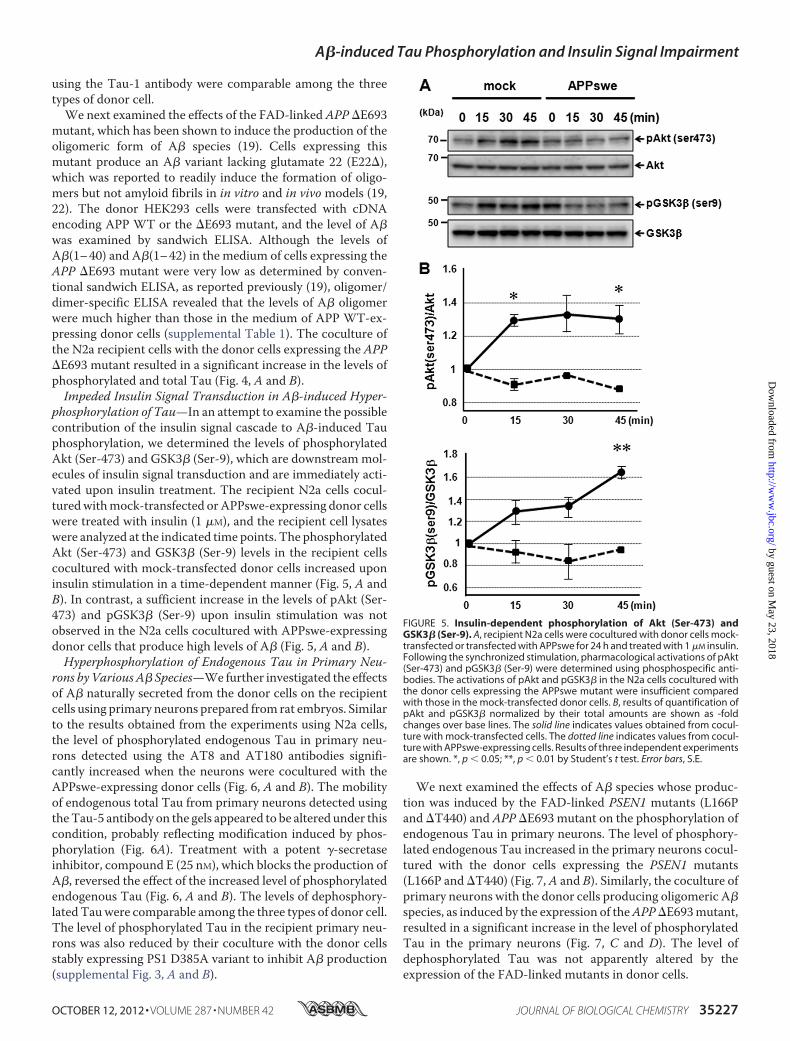
phosphorylation of Tau—In an attempt to examine the possiblecontribution of the insulin signal cascade to A�-induced Tauphosphorylation, we determined the levels of phosphorylatedAkt (Ser-473) and GSK3� (Ser-9), which are downstreammol-ecules of insulin signal transduction and are immediately acti-vated upon insulin treatment. The recipient N2a cells cocul-turedwithmock-transfected orAPPswe-expressing donor cellswere treated with insulin (1 �M), and the recipient cell lysateswere analyzed at the indicated time points. The phosphorylatedAkt (Ser-473) and GSK3� (Ser-9) levels in the recipient cellscocultured with mock-transfected donor cells increased uponinsulin stimulation in a time-dependent manner (Fig. 5, A andB). In contrast, a sufficient increase in the levels of pAkt (Ser-473) and pGSK3� (Ser-9) upon insulin stimulation was notobserved in the N2a cells cocultured with APPswe-expressingdonor cells that produce high levels of A� (Fig. 5, A and B).Hyperphosphorylation of Endogenous Tau in Primary Neu-
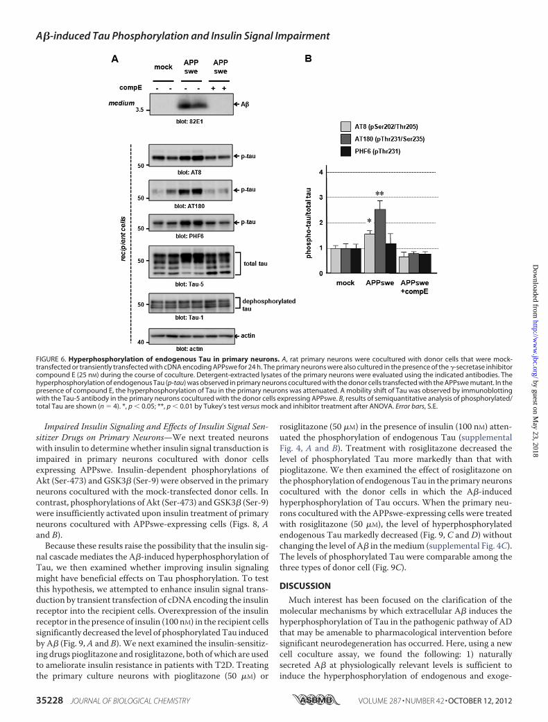
rons byVariousA� Species—We further investigated the effectsof A� naturally secreted from the donor cells on the recipientcells using primary neurons prepared from rat embryos. Similarto the results obtained from the experiments using N2a cells,the level of phosphorylated endogenous Tau in primary neu-rons detected using the AT8 and AT180 antibodies signifi-cantly increased when the neurons were cocultured with theAPPswe-expressing donor cells (Fig. 6, A and B). The mobilityof endogenous total Tau from primary neurons detected usingtheTau-5 antibody on the gels appeared to be altered under thiscondition, probably reflecting modification induced by phos-phorylation (Fig. 6A). Treatment with a potent �-secretaseinhibitor, compound E (25 nM), which blocks the production ofA�, reversed the effect of the increased level of phosphorylatedendogenous Tau (Fig. 6, A and B). The levels of dephosphory-latedTauwere comparable among the three types of donor cell.The level of phosphorylated Tau in the recipient primary neu-rons was also reduced by their coculture with the donor cellsstably expressing PS1 D385A variant to inhibit A� production(supplemental Fig. 3, A and B).
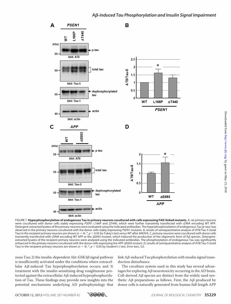
We next examined the effects of A� species whose produc-tion was induced by the FAD-linked PSEN1 mutants (L166Pand �T440) and APP �E693 mutant on the phosphorylation ofendogenous Tau in primary neurons. The level of phosphory-lated endogenous Tau increased in the primary neurons cocul-tured with the donor cells expressing the PSEN1 mutants(L166P and �T440) (Fig. 7,A and B). Similarly, the coculture ofprimary neurons with the donor cells producing oligomeric A�species, as induced by the expression of theAPP�E693mutant,resulted in a significant increase in the level of phosphorylatedTau in the primary neurons (Fig. 7, C and D). The level ofdephosphorylated Tau was not apparently altered by theexpression of the FAD-linked mutants in donor cells.
FIGURE 5. Insulin-dependent phosphorylation of Akt (Ser-473) andGSK3� (Ser-9). A, recipient N2a cells were cocultured with donor cells mock-transfected or transfected with APPswe for 24 h and treated with 1 �M insulin.Following the synchronized stimulation, pharmacological activations of pAkt(Ser-473) and pGSK3� (Ser-9) were determined using phosphospecific anti-bodies. The activations of pAkt and pGSK3� in the N2a cells cocultured withthe donor cells expressing the APPswe mutant were insufficient comparedwith those in the mock-transfected donor cells. B, results of quantification ofpAkt and pGSK3� normalized by their total amounts are shown as -foldchanges over base lines. The solid line indicates values obtained from cocul-ture with mock-transfected cells. The dotted line indicates values from cocul-ture with APPswe-expressing cells. Results of three independent experimentsare shown. *, p � 0.05; **, p � 0.01 by Student’s t test. Error bars, S.E.
A�-induced Tau Phosphorylation and Insulin Signal Impairment
OCTOBER 12, 2012 • VOLUME 287 • NUMBER 42 JOURNAL OF BIOLOGICAL CHEMISTRY 35227
by guest on May 23, 2018
http://ww
w.jbc.org/
Dow
nloaded from
Impaired Insulin Signaling and Effects of Insulin Signal Sen-sitizer Drugs on Primary Neurons—We next treated neuronswith insulin to determinewhether insulin signal transduction isimpaired in primary neurons cocultured with donor cellsexpressing APPswe. Insulin-dependent phosphorylations ofAkt (Ser-473) and GSK3� (Ser-9) were observed in the primaryneurons cocultured with the mock-transfected donor cells. Incontrast, phosphorylations of Akt (Ser-473) andGSK3� (Ser-9)were insufficiently activated upon insulin treatment of primaryneurons cocultured with APPswe-expressing cells (Figs. 8, Aand B).Because these results raise the possibility that the insulin sig-
nal cascade mediates the A�-induced hyperphosphorylation ofTau, we then examined whether improving insulin signalingmight have beneficial effects on Tau phosphorylation. To testthis hypothesis, we attempted to enhance insulin signal trans-duction by transient transfection of cDNA encoding the insulinreceptor into the recipient cells. Overexpression of the insulinreceptor in the presence of insulin (100 nM) in the recipient cellssignificantly decreased the level of phosphorylatedTau inducedby A� (Fig. 9,A and B). We next examined the insulin-sensitiz-ing drugs pioglitazone and rosiglitazone, both ofwhich are usedto ameliorate insulin resistance in patients with T2D. Treatingthe primary culture neurons with pioglitazone (50 �M) or
rosiglitazone (50 �M) in the presence of insulin (100 nM) atten-uated the phosphorylation of endogenous Tau (supplementalFig. 4, A and B). Treatment with rosiglitazone decreased thelevel of phosphorylated Tau more markedly than that withpioglitazone. We then examined the effect of rosiglitazone onthe phosphorylation of endogenousTau in the primary neuronscocultured with the donor cells in which the A�-inducedhyperphosphorylation of Tau occurs. When the primary neu-rons cocultured with the APPswe-expressing cells were treatedwith rosiglitazone (50 �M), the level of hyperphosphorylatedendogenous Tau markedly decreased (Fig. 9, C and D) withoutchanging the level of A� in themedium (supplemental Fig. 4C).The levels of phosphorylated Tau were comparable among thethree types of donor cell (Fig. 9C).
DISCUSSION
Much interest has been focused on the clarification of themolecular mechanisms by which extracellular A� induces thehyperphosphorylation of Tau in the pathogenic pathway of ADthat may be amenable to pharmacological intervention beforesignificant neurodegeneration has occurred. Here, using a newcell coculture assay, we found the following: 1) naturallysecreted A� at physiologically relevant levels is sufficient toinduce the hyperphosphorylation of endogenous and exoge-
FIGURE 6. Hyperphosphorylation of endogenous Tau in primary neurons. A, rat primary neurons were cocultured with donor cells that were mock-transfected or transiently transfected with cDNA encoding APPswe for 24 h. The primary neurons were also cultured in the presence of the �-secretase inhibitorcompound E (25 nM) during the course of coculture. Detergent-extracted lysates of the primary neurons were evaluated using the indicated antibodies. Thehyperphosphorylation of endogenous Tau (p-tau) was observed in primary neurons cocultured with the donor cells transfected with the APPswe mutant. In thepresence of compound E, the hyperphosphorylation of Tau in the primary neurons was attenuated. A mobility shift of Tau was observed by immunoblottingwith the Tau-5 antibody in the primary neurons cocultured with the donor cells expressing APPswe. B, results of semiquantitative analysis of phosphorylated/total Tau are shown (n � 4). *, p � 0.05; **, p � 0.01 by Tukey’s test versus mock and inhibitor treatment after ANOVA. Error bars, S.E.
A�-induced Tau Phosphorylation and Insulin Signal Impairment
35228 JOURNAL OF BIOLOGICAL CHEMISTRY VOLUME 287 • NUMBER 42 • OCTOBER 12, 2012
by guest on May 23, 2018
http://ww
w.jbc.org/
Dow
nloaded from
nous Tau; 2) the insulin-dependent Akt-GSK3� signal pathwayis insufficiently activated under the conditions where extracel-lular A�-induced Tau hyperphosphorylation occurs; and 3)treatment with the insulin-sensitizing drug rosiglitazone pro-tected against the extracellular A�-induced hyperphosphoryla-tion of Tau. These findings may provide new insights into thepotential mechanisms underlying AD pathophysiology that
linkA�-inducedTauphosphorylationwith insulin signal trans-duction disturbance.The coculture system used in this study has several advan-
tages for exploring A� neurotoxicity occurring in the AD brain.Cell-derived A� species are distinct from the widely used syn-thetic A� preparations as follows. First, the A� produced bydonor cells is naturally generated from human full-length APP
FIGURE 7. Hyperphosphorylation of endogenous Tau in primary neurons cocultured with cells expressing FAD-linked mutants. A, rat primary neuronswere cocultured with donor cells stably expressing PSEN1 L166P and �T440, which were further transiently transfected with cDNA encoding WT APP.Detergent-extracted lysates of the primary neurons were evaluated using the indicated antibodies. The hyperphosphorylation of endogenous Tau (p-tau) wasobserved in the primary neurons cocultured with the donor cells stably expressing PSEN1 mutants. B, results of semiquantitative analysis of AT8/Tau-5 (totalTau) in the recipient primary neurons are shown (n � 4). *, p � 0.05 by Tukey’s test versus WT after ANOVA. C, primary neurons were cocultured with donor cellstransiently transfected with cDNA encoding WT APP or the �E693 mutant, which induced the production of the oligomeric form of A� species. Detergent-extracted lysates of the recipient primary neurons were analyzed using the indicated antibodies. The phosphorylation of endogenous Tau was significantlyenhanced in the primary neurons cocultured with the donor cells expressing the APP �E693 mutant. D, results of semiquantitative analysis of AT8/Tau-5 (totalTau) in the recipient primary neurons are shown (n � 4). *, p � 0.05 by Student’s t test. Error bars, S.E.
A�-induced Tau Phosphorylation and Insulin Signal Impairment
OCTOBER 12, 2012 • VOLUME 287 • NUMBER 42 JOURNAL OF BIOLOGICAL CHEMISTRY 35229
by guest on May 23, 2018
http://ww
w.jbc.org/
Dow
nloaded from

by proteolytic processing and is composed of both A�40 andA�42 species similar to those that occur in the brain. In con-trast, syntheticA�peptides have a single defined length, usuallyA�40 or A�42. Furthermore, synthetic A� peptides requireseveral steps of chemical preparation and incubation for thepeptides to form specific conformations, whichmay cause pro-pensity alterations that are distinct from those in the naturallygenerated A� species. Second, naturally secreted A� specieshave biological effects at low nanomolar concentrations similarto those in the humanbrain and cerebrospinal fluid, as shown in
this study, whereas synthetic A� peptides are typically requiredatmicromolar concentrations to obtain similar biological activ-ities (supplemental Fig. 5). The naturally generated A� speciesat physiological concentrations show much higher biologicalactivities than the synthetic A� peptides. In addition, our sys-tem enables the evaluation of the continuous effects of secretedA� on the recipient cells during the course of their coculturewith the donor cells.Recent epidemiological studies, such as the Rotterdam and
Hisayama studies, have provided evidence that the insulinresistance associated with T2DM is a strong risk factor for AD(23, 24). The underlying mechanisms linking the developmentof insulin resistance with AD are not fully understood. Severallines of evidence suggest that insulin regulates the phosphoryl-ation of Tau in neuronal cells (25, 26). Furthermore, insulinreceptor substrate 2 knock-out mice showed an enhanced dep-osition of hyperphosphorylated Tau in hippocampal neurons(27). Recently, Takeda et al. (28) have reported that reducedinsulin levels in brains as well as enhanced neuronal insulinresistance are observed in AD transgenic mice crossed withob/ob diabetic mice, which developed a more severe cognitiveimpairment. Supporting these findings obtained from animalmodels, data from a biochemical study of autopsied brains alsosuggest the presence of insulin resistance in the brains of ADpatients. A recent study has suggested that insulin-PI3K-Aktsignaling is attenuated in the brains of AD patients (29), partic-ularly in those of AD patients with the complication of T2DM(30). Taken together, these results suggest that insulin recep-tor-mediated signaling disturbance may be a mechanistic linkbetween insulin resistance in the brain and AD pathophysiol-ogy. In this regard, our study raises an intriguing issue that theinsulin-mediated signaling pathway can directly result fromextracellular A� toxicity, eventually leading to neuronal dys-function caused by the hyperphosphorylation of Tau.Although our findings may provide new insights into AD
pathogenesis, the mechanism(s) by which soluble A� impairsthe insulin receptor-mediated signaling pathway is not pre-cisely clarified. Previous studies have also shown that thereduced responsiveness of insulin receptors and attenuateddownstream signaling for insulin stimulation are caused by A�oligomers (31–34). It has been shown that the down-regulationof insulin receptors on the cell surface is induced byA�-deriveddiffusible ligands acting as specific pathogenic ligands bindingto particular synapses (34). However, our experiment using acell surface biotinylation assay revealed that the cell surfacelocalization of insulin receptors in primary neurons did notchange under the condition where impaired insulin signaltransduction occurred due to the coculture of the primary neu-rons with the A�-producing donor cells (data not shown). Fur-ther studies will be required to determine the primary effects ofextracellular A� on the function and localization of insulinreceptors. Moreover, Tau has been found to be phosphorylatedat over 30 serine/threonine residues in the brains of ADpatients, and approximately one-half of these are canonicalsites for proline-directed protein kinases, including MAPK,cyclin-dependent kinase, and GSK3� (35–37). Recently, cul-tured neurons differentiated from induced pluripotent stemcells derived fromprimary fibroblasts of FADpatientswithAPP
FIGURE 8. Insulin-dependent phosphorylation of Akt (Ser-473) and GSK3�(Ser-9) in recipient primary neurons. A, rat primary neurons cocultured withdonor cells mock-transfected or transiently transfected with cDNA encodingAPPswe were stimulated with insulin (1 �M) to induce insulin signal cascade acti-vation. Following the synchronized stimulation, the cell lysates of the primaryneurons were collected at the indicated time points. The pharmacological acti-vation of pAkt (Ser-473) and PGSK3� (Ser-9) was examined by immunoblot anal-ysis using phosphospecific antibodies. The activation of pAkt and pGSK3� uponinsulin stimulation was insufficient in the primary neurons cocultured with thedonor cells expressing the APPswe mutant. B, results of quantification of pAktand pGSK3� normalized by their total amount are shown as -fold changes overthe base line. The solid line indicates values obtained from coculture with mock-transfected cells. The dotted line indicates values from coculture with APPswe-expressing cells. Results of three independent experiments are shown. *,p � 0.05 by Student’s t test. Error bars, S.E.
A�-induced Tau Phosphorylation and Insulin Signal Impairment
35230 JOURNAL OF BIOLOGICAL CHEMISTRY VOLUME 287 • NUMBER 42 • OCTOBER 12, 2012
by guest on May 23, 2018
http://ww
w.jbc.org/
Dow
nloaded from

duplication have been reported to show significantly higher lev-els of active GSK3� and phosphorylated Tau (Thr-231) (38).The present study also showed that the inefficient activation ofAkt-GSK3� signaling is relevant to A�-induced Tau phosphor-ylation.However, it is conceivable that different pathways func-tion in concert in Tau phosphorylation in the AD brain.Understanding the effects of A� on the hyperphosphoryla-
tion of Taumediated by the insulin signal pathwaymay providea new potential therapeutic approach. In this study, we showedthat the insulin-sensitizing drug rosiglitazone has a protectiveeffect against A�-induced hyperphosphorylation of Tau in pri-mary neurons. A plausible mechanism underlying this protec-tion is provided by our demonstration that rosiglitazone poten-tiated insulin signal transduction (data not shown), althoughother pleiotropic mechanisms of action exerted by rosiglita-
zone are also possible. The rationale for using rosiglitazone forthe treatment of brain diseases is supported by the findings thatperoxisome proliferator-activated receptor � is expressed inthe brain regions, including the hippocampus (39). Further-more, rosiglitazone can penetrate the blood-brain barrier (40).In the mouse AD model, Pedersen et al. (41) reported thatrosiglitazone attenuated deficits in learning and memory with-out changing A� deposition. Previous human clinical trialsexploring the potential effectiveness of peroxisome prolifera-tor-activated receptor � agonists, including rosiglitazone andpioglitazone, for AD patients showed contradictory resultsregarding the efficacy of these drugs. The results of Watson etal. (42) demonstrated a positive correlation between insulinlevel and cognitive improvement in patients treated withrosiglitazone compared with those treated with placebo. Gold
FIGURE 9. Protective effect due to enhanced insulin signal transduction against endogenous Tau phosphorylation. A, the recipient N2a cells with orwithout transient transfection of cDNA encoding the insulin receptor were cocultured with donor cells expressing APPswe. Detergent-extracted lysates of therecipient cells were analyzed using the indicated antibodies. Expression of insulin receptor in the presence of insulin reduced the level of phosphorylated Tau(p-tau) in the recipient cells. B, results of semiquantitative analysis of AT8/Tau-5 (total Tau) in the recipient Na2 cells are shown (n � 4). *, p � 0.05; **, p � 0.01Tukey’s test versus WT after ANOVA. C, rat primary neurons cocultured with donor cells transiently transfected with cDNA encoding APPswe were treated withvehicle only or with rosiglitazone (50 �M) in the presence of insulin (100 nM). Detergent-extracted lysates of the recipient cells were analyzed using theindicated antibodies. Treatment with rosiglitazone showed a protective effect against the hyperphosphorylation of Tau induced by A� secreted at physiolog-ically relevant levels. D, results of semiquantitative analysis of AT8/Tau-5 (total Tau) in the recipient primary neurons are shown (n � 3). *, p � 0.05 by Tukey’stest versus WT after ANOVA. Error bars, S.E.
A�-induced Tau Phosphorylation and Insulin Signal Impairment
OCTOBER 12, 2012 • VOLUME 287 • NUMBER 42 JOURNAL OF BIOLOGICAL CHEMISTRY 35231
by guest on May 23, 2018
http://ww
w.jbc.org/
Dow
nloaded from
et al. (43) have recently conducted a multicenter randomizeddouble-blind clinical study, which revealed no evidence of effi-cacy of rosiglitazone for improving cognitive function in ADpatients. In contrast, Sato et al. (44) reported the significantimprovements of cognition and regional blood flow in patientswith AD complicated by T2DM treated with pioglitazone com-pared with those treated with placebo controls. A recent clini-cal trial showed that the administration of intranasal insulinimproved cognitive performance in elderly patients with earlyADand amnesticmild cognitive impairment (45). Although theprecise mechanisms underlying these effects of therapies tar-geting brain insulin signaling have not yet been clarified, cor-recting insulin signal dysregulation in the CNSmay be a poten-tial therapeutic approach for some AD patients, particularlywhen AD is a consequence of insulin dysregulation or the ADpatients have the T2DM complication.
Acknowledgments—We thank K. Nishino, A. Isami, M. Shinozaki,and Dr. H. Kaneko for technical assistance, Dr. T. Tomiyama fortechnical advice on oligomer A� analysis, and Dr. O. Onodera forhelpful and critical comments on this work.
REFERENCES1. Glabe, C. G. (2008) Structural classification of toxic amyloid oligomers.
J. Biol. Chem. 283, 29639–296432. Roychaudhuri, R., Yang, M., Hoshi, M. M., and Teplow, D. B. (2009) Am-
yloid �-protein assembly and Alzheimer disease. J. Biol. Chem. 284,4749–4753
3. Gong, Y., Chang, L., Viola, K. L., Lacor, P. N., Lambert, M. P., Finch, C. E.,Krafft, G. A., and Klein, W. L. (2003) Alzheimer’s disease-affected brain.Presence of oligomericA� ligands (ADDLs) suggests amolecular basis forreversible memory loss. Proc. Natl. Acad. Sci. U.S.A. 100, 10417–10422
4. Klyubin, I., Walsh, D. M., Lemere, C. A., Cullen, W. K., Shankar, G. M.,Betts, V., Spooner, E. T., Jiang, L., Anwyl, R., Selkoe, D. J., Rowan, M. J.(2005) Amyloid � protein immunotherapy neutralizes A� oligomers thatdisrupt synaptic plasticity in vivo. Nat. Med. 11, 556–561
5. Shankar, G. M., Bloodgood, B. L., Townsend, M., Walsh, D. M., Selkoe,D. J., and Sabatini, B. L. (2007) Natural oligomers of the Alzheimer amy-loid-� protein induce reversible synapse loss by modulating an NMDA-type glutamate receptor-dependent signaling pathway. J. Neurosci. 27,2866–2875
6. Ma, Q. L., Yang, F., Rosario, E. R., Ubeda, O. J., Beech, W., Gant, D. J.,Chen, P. P., Hudspeth, B., Chen, C., Zhao, Y., Vinters, H. V., Frautschy,S. A., and Cole, G. M. (2009) �-Amyloid oligomers induce phosphoryla-tion of Tau and inactivation of insulin receptor substrate via c-Jun N-ter-minal kinase signaling. Suppression by �-3 fatty acids and curcumin.J. Neurosci. 29, 9078–9089
7. Zempel, H., Thies, E., Mandelkow, E., and Mandelkow, E. M. (2010) A�oligomers cause localized Ca2� elevation, missorting of endogenous Tauinto dendrites, Tau phosphorylation, and destruction ofmicrotubules andspines. J. Neurosci. 30, 11938–11950
8. Jin, M., Shepardson, N., Yang, T., Chen, G., Walsh, D., and Selkoe, D. J.(2011) Soluble amyloid �-protein dimers isolated from Alzheimer cortexdirectly induce Tau hyperphosphorylation and neuritic degeneration.Proc. Natl. Acad. Sci. U.S.A. 108, 5819–5824
9. Biessels, G. J., Staekenborg, S., Brunner, E., Brayne, C., and Scheltens, P.(2006) Risk of dementia in diabetes mellitus. A systematic review. LancetNeurol. 5, 64–74
10. Profenno, L. A., Porsteinsson, A. P., and Faraone, S. V. (2010) Meta-anal-ysis of Alzheimer disease risk with obesity, diabetes, and related disorders.Biol. Psychiat. 67, 505–512
11. Saltiel, A. R., and Kahn, C. R. (2001) Insulin signaling and the regulation ofglucose and lipid metabolism. Nature 414, 799–806
12. Hill, J. M., Lesniak,M. A., Pert, C. B., and Roth, J. (1986) Autoradiographiclocalization of insulin receptors in rat brain. Prominence in olfactory andlimbic areas. Neuroscience 17, 1127–1138
13. Zhao, W., Chen, H., Xu, H., Moore, E., Meiri, N., Quon, M. J., and Alkon,D. L. (1999) Brain insulin receptors and spatial memory. Correlatedchanges in gene expression, tyrosine phosphorylation, and signaling mol-ecules in the hippocampus of water maze-trained rats. J. Biol. Chem. 274,34893–34902
14. Sato, N., Takeda, S., Uchio-Yamada, K., Ueda, H., Fujisawa, T., Rakugi, H.,and Morishita, R. (2011) Role of insulin signaling in the interaction be-tween Alzheimer disease and diabetes mellitus. A missing link to thera-peutic potential. Curr. Aging Sci. 4, 118–127
15. Takashima, A., Noguchi, K.,Michel, G.,Mercken,M., Hoshi,M., Ishiguro,K., and Imahori, K. (1996) Exposure of rat hippocampal neurons to amy-loid � peptide (25–35) induces the inactivation of phosphatidylinositol3-kinase and the activation of Tau protein kinase I/glycogen synthasekinase-3�. Neurosci. Lett. 203, 33–36
16. Ikeuchi, T., Dolios, G., Kim, S. H., Wang, R., and Sisodia, S. S. (2003)Familial Alzheimer disease-linked presenilin 1 variants enhance produc-tion of both A� 1–40 and A� 1–42 peptides that are only partially sensi-tive to a potent aspartyl protease transition state inhibitor of “�-secretase”.J. Biol. Chem. 278, 7010–7018
17. Kaneko, H., Kakita, A., Kasuga, K., Nozaki, H., Ishikawa, A., Miyashita, A.,Kuwano, R., Ito, G., Iwatsubo, T., Takahashi, H., Nishizawa, M., Onodera,O., Sisodia, S. S., and Ikeuchi, T. (2007) Enhanced accumulation of phos-phorylated �-synuclein and elevated �-amyloid 42/40 ratio caused by ex-pression of the presenilin-1 �T440 mutant associated with familial Lewybody disease and variant Alzheimer disease. J. Neurosci. 27, 13092–13097
18. Kasuga, K., Kaneko, H., Nishizawa, M., Onodera, O., and Ikeuchi, T.(2007) Generation of intracellular domain of insulin receptor tyrosinekinase by �-secretase. Biochem. Biophys. Res. Commun. 360, 90–96
19. Tomiyama, T., Nagata, T., Shimada, H., Teraoka, R., Fukushima, A., Kane-mitsu, H., Takuma, H., Kuwano, R., Imagawa, M., Ataka, S., Wada, Y.,Yoshioka, E., Nishizaki, T., Watanabe, Y., and Mori, H. (2008) A newamyloid � variant favoring oligomerization in Alzheimer-type dementia.Ann. Neurol. 63, 377–387
20. Kasuga, K., Ohno, T., Ishihara, T.,Miyashita, A., Kuwano, R., Onodera, O.,Nishizawa, M., and Ikeuchi, T. (2009) Depression and psychiatric symp-toms preceding onset of dementia in a family with early onset Alzheimerdisease with a novel PSEN1 mutation. J. Neurol. 256, 1351–1353
21. Xia, W., Yang, T., Shankar, G., Smith, I. M., Shen, Y., Walsh, D. M., andSelkoe, D. J. (2009) A specific enzyme-linked immunosorbent assay formeasuring�-amyloid protein oligomers in human plasma and brain tissueof patients with Alzheimer disease. Arch. Neurol. 66, 190–199
22. Tomiyama, T., Matsuyama, S., Iso, H., Umeda, T., Takuma, H., Ohnishi,K., Ishibashi, K., Teraoka, R., Sakama, N., Yamashita, T., Nishitsuji, K., Ito,K., Shimada,H., Lambert,M. P., Klein,W. L., andMori, H. (2010)Amousemodel of amyloid � oligomers. Their contribution to synaptic alteration,abnormal Tau phosphorylation, glial activation, and neuronal loss in vivo.J. Neurosci. 30, 4845–4856
23. Ohara, T., Doi, Y., Ninomiya, T., Hirakawa, Y., Hata, J., Iwaki, T., Kanba, S.,and Kiyohara, Y. (2011) Glucose tolerance status and risk of dementia inthe community. The Hisayama study. Neurology 77, 1126–1134
24. Schrijvers, E. M., Witteman, J. C., Sijbrands, E. J., Hofman, A., Koudstaal,P. J., and Breteler, M. M. (2010) Insulin metabolism and the risk of Al-zheimer disease. The Rotterdam study. Neurology 75, 1982–1987
25. Hong, M., and Lee, V. M. (1997) Insulin and insulin-like growth factor-1regulate Tau phosphorylation in cultured human neurons. J. Biol. Chem.272, 19547–19553
26. Lesort, M., and Johnson, G. V. (2000) Insulin-like growth factor-1 andinsulin mediate transient site-selective increases in Tau phosphorylationin primary cortical neurons. Neuroscience 99, 305–316
27. Schubert, M., Brazil, D. P., Burks, D. J., Kushner, J. A., Ye, J., Flint, C. L.,Farhang-Fallah, J., Dikkes, P.,Warot, X.M., Rio, C., Corfas, G., andWhite,M. F. (2003) Insulin receptor substrate-2 deficiency impairs brain growthand promotes Tau phosphorylation. J. Neurosci. 23, 7084–7092
28. Takeda, S., Sato,N., Uchio-Yamada, K., Sawada, K., Kunieda, T., Takeuchi,D., Kurinami, H., Shinohara, M., Rakugi, H., and Morishita, R. (2010)
A�-induced Tau Phosphorylation and Insulin Signal Impairment
35232 JOURNAL OF BIOLOGICAL CHEMISTRY VOLUME 287 • NUMBER 42 • OCTOBER 12, 2012
by guest on May 23, 2018
http://ww
w.jbc.org/
Dow
nloaded from

Diabetes-acceleratedmemory dysfunction via cerebrovascular inflamma-tion and A� deposition in an Alzheimermousemodel with diabetes. Proc.Natl. Acad. Sci. U.S.A. 107, 7036–7041
29. Steen, E., Terry, B. M., Rivera, E. J., Cannon, J. L., Neely, T. R., Tavares, R.,Xu, X. J., Wands, J. R., and de la Monte, S. M. (2005) Impaired insulin andinsulin-like growth factor expression and signaling mechanisms in Al-zheimer disease. Is this type 3 diabetes? J. Alzheimers Dis. 7, 63–80
30. Liu, Y., Liu, F., Grundke-Iqbal, I., Iqbal, K., and Gong, C. X. (2011) Defi-cient brain insulin signaling pathway in Alzheimer disease and diabetes.J. Pathol. 225, 54–62
31. Townsend, M., Mehta, T., and Selkoe, D. J. (2007) Soluble A� inhibitsspecific signal transduction cascades common to the insulin receptorpathway. J. Biol. Chem. 282, 33305–33312
32. Lee, H. K., Kumar, P., Fu, Q., Rosen, K. M., and Querfurth, H. W. (2009)The insulin/Akt signaling pathway is targeted by intracellular �-amyloid.Mol. Biol. Cell 20, 1533–1544
33. Zhao, W. Q., De Felice, F. G., Fernandez, S., Chen, H., Lambert, M. P.,Quon, M. J., Krafft, G. A., and Klein, W. L. (2008) Amyloid � oligomersinduce impairment of neuronal insulin receptors. FASEB J. 22, 246–260
34. De Felice, F. G., Vieira, M. N., Bomfim, T. R., Decker, H., Velasco, P. T.,Lambert, M. P., Viola, K. L., Zhao, W. Q., Ferreira, S. T., and Klein, W. L.(2009) Protection of synapses against Alzheimer-linked toxins. Insulinsignaling prevents the pathogenic binding of A� oligomers. Proc. Natl.Acad. Sci. U.S.A. 106, 1971–1976
35. Morishima-Kawashima, M., Hasegawa, M., Takio, K., Suzuki, M., Yo-shida, H., Titani, K., and Ihara, Y. (1995) Proline-directed and non-pro-line-directed phosphorylation of PHF-Tau. J. Biol. Chem. 270, 823–829
36. Gong, C. X., Liu, F., Grundke-Iqbal, I., and Iqbal, K. (2005) Post-trans-lationalmodificationsofTauprotein inAlzheimerdisease. J.NeuralTransm.112, 813–838
37. Hooper, C., Killick, R., and Lovestone, S. (2008) The GSK3 hypothesis ofAlzheimer disease. J. Neurochem. 104, 1433–1439
38. Israel, M. A., Yuan, S. H., Bardy, C., Reyna, S. M., Mu, Y., Herrera, C.,Hefferan, M. P., Van Gorp, S., Nazor, K. L., Boscolo, F. S., Carson, C. T.,
Laurent, L. C., Marsala, M., Gage, F. H., Remes, A. M., Koo, E. H., andGoldstein, L. S. (2012) Probing sporadic and familial Alzheimer diseaseusing induced pluripotent stem cells. Nature 482, 216–220
39. Inestrosa, N. C., Godoy, J. A., Quintanilla, R. A., Koenig, C. S., and Bronf-man, M. (2005) Peroxisome proliferator-activated receptor � is expressedin hippocampal neurons, and its activation prevents �-amyloid neurode-generation. Role of Wnt signaling. Exp. Cell Res. 304, 91–104
40. Sheu, W. H., Chuang, H. C., Cheng, S. M., Lee, M. R., Chou, C. C., andCheng, F. C. (2011)Microdialysis combined blood sampling technique forthe determination of rosiglitazone and glucose in brain and blood of ger-bils subjected to cerebral ischemia. J. Pharm. Biomed. Anal. 54, 759–764
41. Pedersen,W. A., McMillan, P. J., Kulstad, J. J., Leverenz, J. B., Craft, S., andHaynatzki, G. R. (2006) Rosiglitazone attenuates learning and memorydeficits in Tg2576 Alzheimer mice. Exp. Neurol. 199, 265–273
42. Watson, G. S., Cholerton, B. A., Reger, M. A., Baker, L. D., Plymate, S. R.,Asthana, S., Fishel, M. A., Kulstad, J. J., Green, P. S., Cook, D. G., Kahn,S. E., Keeling, M. L., and Craft, S. (2005) Preserved cognition in patientswith early Alzheimer disease and amnestic mild cognitive impairmentduring treatment with rosiglitazone. A preliminary study. Am. J. Geriatr.Psychiatry 13, 950–958
43. Gold, M., Alderton, C., Zyartau-Hind, M., Egginton, S., Saunders, A. M.,Irizarry, M., Craft, S., Landreth, G., Linnamagi, U., and Sawchak, S. (2010)Rosiglitazone monotherapy in mild-to-moderate Alzheimer disease. Re-sults from a randomized, double-blind, placebo-controlled phase IIIstudy. Dement. Geriatr. Cogn. Disord. 30, 131–146
44. Sato, T., Hanyu, H., Hirao, K., Kanetaka, H., Sakurai, H., and Iwamoto, T.(2011) Efficacy of PPAR-� agonist pioglitazone inmild Alzheimer disease.Neurobiol. Aging 32, 1626–1633
45. Craft, S., Baker, L. D., Montine, T. J., Minoshima, S., Watson, G. S., Clax-ton, A., Arbuckle, M., Callaghan, M., Tsai, E., Plymate, S. R., Green, P. S.,Leverenz, J., Cross, D., andGerton, B. (2012) Intranasal insulin therapy forAlzheimer disease and amnestic mild cognitive impairment. A pilot clin-ical trial. Arch. Neurol. 69, 29–38
A�-induced Tau Phosphorylation and Insulin Signal Impairment
OCTOBER 12, 2012 • VOLUME 287 • NUMBER 42 JOURNAL OF BIOLOGICAL CHEMISTRY 35233
by guest on May 23, 2018
http://ww
w.jbc.org/
Dow
nloaded from
Masatoyo Nishizawa and Takeshi IkeuchiTakayoshi Tokutake, Kensaku Kasuga, Ryuji Yajima, Yumi Sekine, Toshiyuki Tezuka,
Signaling PathwayβNanomolar Concentrations Is Modulated by Insulin-dependent Akt-GSK3
atβHyperphosphorylation of Tau Induced by Naturally Secreted Amyloid-
doi: 10.1074/jbc.M112.348300 originally published online August 21, 20122012, 287:35222-35233.J. Biol. Chem.
10.1074/jbc.M112.348300Access the most updated version of this article at doi:
Alerts:
When a correction for this article is posted•
When this article is cited•
to choose from all of JBC's e-mail alertsClick here
Supplemental material:
http://www.jbc.org/content/suppl/2012/08/21/M112.348300.DC1
http://www.jbc.org/content/287/42/35222.full.html#ref-list-1
This article cites 45 references, 21 of which can be accessed free at
by guest on May 23, 2018
http://ww
w.jbc.org/
Dow
nloaded from