hydrolysis of angiotensin ii receptor blocker prodrug...
TRANSCRIPT

DMD#6163
1
Hydrolysis of Angiotensin II Receptor Blocker Prodrug Olmesartan
Medoxomil by Human Serum Albumin and Identification of Its
Catalytic Active Sites
Shen-Feng Ma, Makoto Anraku, Yasunori Iwao, Keishi Yamasaki, Ulrich
Kragh-Hansen, Noriyuki Yamaotsu, Shuichi Hirono, Toshihiko Ikeda, and Masaki
Otagiri.
Graduate School of Pharmaceutical Sciences, Kumamoto University, Kumamoto
862-0793, Japan (S.-F. M., M. A., Y. I. and M. O.).
Department of Pharmacy, Miyazaki Medical College Hospital, Miyazaki 889-1692,
Japan (K. Y.).
Department of Medical Biochemistry, University of Aarhus, DK-8000 Aarhus C,
Denmark (U. K.-H.).
The School of Pharmaceutical Sciences, Kitasato University, Tokyo 108-8641, Japan (N.
Y. and S. H.).
Drug Metabolism and Pharmacokinetics Research Laboratories, Sankyo Co., Ltd.,
Tokyo 140-8710, Japan (T. I.).
DMD Fast Forward. Published on September 23, 2005 as doi:10.1124/dmd.105.006163
Copyright 2005 by the American Society for Pharmacology and Experimental Therapeutics.
This article has not been copyedited and formatted. The final version may differ from this version.DMD Fast Forward. Published on September 23, 2005 as DOI: 10.1124/dmd.105.006163
at ASPE
T Journals on January 3, 2020
dmd.aspetjournals.org
Dow
nloaded from

DMD#6163
2
a) Running Title: Hydrolysis of CS-866 by HSA and studies of the active sites
b) Corresponding author: Professor Masaki Otagiri, Ph.D.,
Department of Biopharmaceutics, Graduate School of Pharmaceutical
Sciences, Kumamoto University, 5-1 Oe-honmachi, Kumamoto 862-0973,
Japan
Tel: +81-96-371-4150; Fax: +81-96-362-7690
E-mail: [email protected]
c) Number of text pages: 29
Number of figures: 7
Number of tables: 5
Number of references: 34
Number of words in the Abstract: 233
Number of words in the Introduction: 382
Number of words in the Discussion: 1465
d) Abbreviations used: PNPA, p-nitrophenyl acetate; n-butyl-p-AB,
n-butyl-p-aminobenzoate.
This article has not been copyedited and formatted. The final version may differ from this version.DMD Fast Forward. Published on September 23, 2005 as DOI: 10.1124/dmd.105.006163
at ASPE
T Journals on January 3, 2020
dmd.aspetjournals.org
Dow
nloaded from

DMD#6163
3
Abstract
In the present study, we investigated the esterase-like activity of human serum
albumin (HSA) and the mechanism by which it hydrolyzes, and thereby activates,
olmesartan medoxomil (CS-866), a novel angiotensin II receptor antagonist. CS-866 has
previously been shown to be rapidly hydrolyzed in serum in which HSA appeared to
play the most important role in catalyzing the hydrolysis. We found that the hydrolysis
of CS-866 by HSA followed Michaelis-Menten kinetics. Compared with the release of
p-nitrophenol from p-nitrophenyl acetate (PNPA), CS-866 showed lower affinity to
HSA and a lower catalytic rate of hydrolysis. Thermodynamic data indicated that PNPA
has a smaller value of activation entropy (∆S) than CS-866; consequently, PNPA is
more reactive than CS-866. Ibuprofen and warfarin acted as competitive inhibitors of
hydrolysis of CS-866, whereas Dansyl-L-asparagine (DNSA), n-butyl p-aminobenzoate
(n-butyl p-AB) and diazepam did not. These findings suggest that the hydrolytic activity
is associated to parts of site I and site II for ligand binding. All chemically modified
HSA derivatives (Tyr-, Lys-, His- and Trp-modifications) had significantly lower
reactivity than native HSA; Lys-HSA and Trp-HSA had especially low reactivity. All the
mutant HSAs tested (K199A, W214A and Y411A) exhibited a significant decrease in
reactivity, suggesting that Lys-199, Trp-214 and Tyr-411 play important roles in the
hydrolysis. Results obtained using a computer docking model are in agreement with the
experimental results, and strongly support the hypotheses that we derived from the
experiments.
This article has not been copyedited and formatted. The final version may differ from this version.DMD Fast Forward. Published on September 23, 2005 as DOI: 10.1124/dmd.105.006163
at ASPE
T Journals on January 3, 2020
dmd.aspetjournals.org
Dow
nloaded from

DMD#6163
4
Introduction
Ester prodrugs are hydrolyzed to their pharmacologically active metabolites after
absorption. Esterases present in the small intestine, plasma and liver are involved in this
process. In most cases, intestinal esterases serve as the major enzymes in activation of
prodrugs during the first pass through the gut after absorption. However, prodrugs that
are relatively resistant to hydrolysis by intestinal esterases enter the blood circulation,
and are activated by serum (plasma) and liver esterases. The major hydrolyzing
enzymes in serum are cholinesterase, arylesterase, carboxylesterase and albumin. The
relative importance of each serum esterase in prodrug activation varies among animal
species and prodrugs.
Olmesartan medoxomil (CS-866: (5-methyl-2-oxo-1, 3-dioxolen-4-yl) methoxy-4-
(1-hydroxyl-1-methylethyl)-2-propyl-1-{4-[2-(tetrazol-5-yl)-phenyl]phenyl}methylimi-
dazol-5-carboxylate) is a novel nonpeptide angiotensin II receptor antagonist that acts as
an antihypertensive prodrug (Neutel, 2001; Koike et al., 2001; Brousil and Burke, 2003).
After oral administration, CS-866 is rapidly de-esterified, producing an active acid
metabolite, olmesartan (RNH-6270) (Fig. 1) (Neutel, 2001; Koike et al., 2001; Brousil
and Burke, 2003). Hydrolysis of CS-866 in serum has been observed in several species,
and comparison between 5 species has shown that hydrolytic activity is highest in
rabbits, followed by dogs, mice, rats and humans (Ikeda, 2000). Furthermore, it was
found that differences in hydrolytic activity due to serum albumin are large compared to
the combined activity of all serum components. Thus, HSA might make an important
contribution to activation of CS-866 after oral administration.
In the present study, we examined the esterase-like activity of HSA and the
mechanism of its hydrolysis of CS-866. First, the general properties of the hydrolytic
This article has not been copyedited and formatted. The final version may differ from this version.DMD Fast Forward. Published on September 23, 2005 as DOI: 10.1124/dmd.105.006163
at ASPE
T Journals on January 3, 2020
dmd.aspetjournals.org
Dow
nloaded from

DMD#6163
5
reaction of HSA with CS-866 were determined, including the kinetics and
thermodynamics, and compared with those of the hydrolytic reaction between HSA and
p-nitrophenyl acetate (PNPA) (Means and Bender, 1975; Sakurai et al., 2004). Second,
to characterize the effects of exogenous compounds on hydrolysis, we investigated
changes in hydrolytic activity in the presence and absence of various ligands. Then, we
examined the importance of certain types of amino acid residues of HSA for the
hydrolysis of CS-866, using chemical modification techniques. Recombinant HSA
(rHSA) proteins with alterations of specific amino acid residues were prepared using
site-directed mutagenesis techniques, to obtain detailed information about the
contribution of those residues. Finally, computer docking models of CS-866 and HSA
were constructed and were found to be consistent with the experimental results.
This article has not been copyedited and formatted. The final version may differ from this version.DMD Fast Forward. Published on September 23, 2005 as DOI: 10.1124/dmd.105.006163
at ASPE
T Journals on January 3, 2020
dmd.aspetjournals.org
Dow
nloaded from

DMD#6163
6
Materials and Methods Materials
HSA was donated by the Chemo-Sera-Therapeutic Research Institute (Kumamoto,
Japan). HSA was defatted before use (Chen, 1967).
CS-866 and RNH-6270 were donated by Sankyo Co., Ltd. (Tokyo, Japan). PNPA,
succinic anhydride (SA) and n-butyl p-aminobenzoate (n-butyl p-AB) were purchased
form Nakalai Tesque (Kyoto, Japan). Warfarin was obtained from Eisai Co., Tokyo,
Japan; ibuprofen was obtained from Kaken Pharmaceutical Co., Osaka, Japan;
diazepam was obtained from Sumitomo Pharmaceutical Co., Osaka, Japan;
tetranitromethane (TNM) was obtained from Aldrich Chemical Company, Inc. U.S.A.;
and trinitrobenzensulfonic acid (TNBS) was obtained from Wako Pure Chemical
Industries, Ltd., Osaka, Japan. Dansyl-L-asparagine (DNSA), 2-hydroxyl-5-nitrobenzyl
bromide (I-Br) and diethyl pyrocarbonate (DEP) were purchased from the Sigma
Chemical Co. (St Louis, MO, U.S.A.). Restriction enzymes, T4 polynucleotide kinase,
calf intestinal alkaline phosphatase, a DNA ligation kit, TaKaRa EX Taq DNA
polymerase and a site-directed mutagenesis kit (oligonucleotide-directed dual amber
method) were obtained from Takara Shuzo Co., Ltd. (Kyoto, Japan). A DNA sequence
kit was obtained from Perkin-Elmer Applied Biosystems (Tokyo, Japan). The Pichia
Expression kit was purchased from Invitrogen.
All other chemicals were of analytical grade.
Esterase-like Activity Measurement
Procedures for Michaelis-Menten Equation Runs
The reaction was started by adding CS-866 in 100% acetonitrile (5 µl) to
This article has not been copyedited and formatted. The final version may differ from this version.DMD Fast Forward. Published on September 23, 2005 as DOI: 10.1124/dmd.105.006163
at ASPE
T Journals on January 3, 2020
dmd.aspetjournals.org
Dow
nloaded from

DMD#6163
7
preincubated HSA (120 µl, 75 µM), at a final concentration of 10 to 250 µM. Incubation
proceeded for 10 min, and was terminated by adding 500 µl of acetonitrile to the
incubation mixture. We have checked that 4 % acetonitrile has little effect on the
reaction. After centrifugation for 1 min, a 30-µl aliquot of the deproteinized supernatant
was subjected to HPLC, and RNH-6270 was separated from CS-866 on an ODS column
using the following conditions: column, YMC-Pack ODS-AM, AM-302, 150 × 4.6 mm
I.D; column temperature, 40ºC maintained by HITACHI 655A-52 Column Oven; pump,
HITACHI L-6000 Pump; detector, HITACHI FL Detector L-7480 fluorescent monitor;
integrator, HITACHI D-2500 Chromato-Integrator; mobile phase, acetonitrile: water:
acetic acid = 40:60:0.1; wavelength, EX=260 nm, EM=370 nm; flow rate, 1.0 ml/min.
The reaction between CS-866 and HSA took place at 4 ºC. Under that condition,
Michaelis-Menten equation analysis can be applied.
(1)
Here, [S] is the concentration of substrate. That is the case, because previous
studies have revealed a linear relationship between 1/V and 1/S when plotted in a
Lineweaver-Burk plot (Koike et al., 2001):
(2)
Procedures for Kinetic Runs
Hydrolysis of CS-866 (5 µM) by HSA (at least a 5-fold excess concentration over
the substrate) was performed using conditions able to avoid complications due to
=
Vmax[S]
KM+ [S] v
= 1 KM
+ v
1Vmax Vmax[S]
This article has not been copyedited and formatted. The final version may differ from this version.DMD Fast Forward. Published on September 23, 2005 as DOI: 10.1124/dmd.105.006163
at ASPE
T Journals on January 3, 2020
dmd.aspetjournals.org
Dow
nloaded from

DMD#6163
8
multiple reactive sites of albumin. Under such conditions, pseudo-first-order rate
constant analysis can be performed. The pseudo-first-order rate constant for the release
of RNH-6270 (kobs), the dissociation constant of the substrate-HSA complex (KS) and
the catalytic rate constants (kcat) were calculated as reported elsewhere (Sakurai et al.,
2004).
Thermodynamic Analysis
Thermodynamic analysis of the HSA-catalyzed reaction was performed at
temperatures ranging from 20ºC to 40ºC. We calculated the thermodynamic parameters,
the free energy change for the initial reaction between enzyme and substrate (∆GS), the
activation free energy for the rate-determining step (∆G), the free energy difference for
the reaction (∆GT), the activation energy (Ea), the activation enthalpy change (∆H) and
the activation entropy change (∆S), using previously published methods (Sakurai et al.,
2004).
Effects of Ligands
HSA (120 µl, 75 µM) was preincubated, and the enzymatic reaction was started by
adding CS-866 in 100% acetonitrile (5 µl) to the solution, at a final concentration of 100
to 250 µM, in the presence or absence of each ligand (at a final concentration of 0-600
µM) (Ikeda, 2000). Incubation proceeded for 10 min at 37ºC, and the release of
RNH-6270 was measured by HPLC as described above.
Chemical Modification of HSA
Histidine Residues
This article has not been copyedited and formatted. The final version may differ from this version.DMD Fast Forward. Published on September 23, 2005 as DOI: 10.1124/dmd.105.006163
at ASPE
T Journals on January 3, 2020
dmd.aspetjournals.org
Dow
nloaded from

DMD#6163
9
Chemical modification of His residues was performed using diethyl pyrocarbonate
(DEP) (Roosemont, 1978). An average of 2.22 His residues was modified out of the
total of 16 His residues.
Lysine Residues
Chemical modification of Lys residues was performed according to Gounaris and
Perlmann (1967). The modification ratio was calculated as described by Haynes et al.
(1967). An average of 3.80 out of the 59 Lys residues was modified.
Tyrosine Residues
Chemical modification of Tyr residues was performed as outlined by Sokolovsky
et al. (1966). An average of 1.24 out of the 18 Tyr residues was modified.
Tryptophan Residues
Chemical modification of the single Trp residue was performed at room
temperature (Fehske et al., 1978). An average of 0.88 out of the 1 Trp residue was
modified.
Chemical modifications of specific amino acid residues (Tyr, Lys, His and Trp)
were performed with the assumption that the effects on other amino acid residues would
be negligible. The secondary and tertiary protein structures of all the modified HSAs
were examined by circular dichroism (CD) measurements before use, and no significant
difference was observed between the derivatives and native HSA (data not shown).
Synthesis and Purification of rHSA Forms
This article has not been copyedited and formatted. The final version may differ from this version.DMD Fast Forward. Published on September 23, 2005 as DOI: 10.1124/dmd.105.006163
at ASPE
T Journals on January 3, 2020
dmd.aspetjournals.org
Dow
nloaded from
DMD#6163
10
The recombinant DNA techniques used to produce wild-type rHSA and the
single-residue mutants were essentially the same as those described by Watanabe et al.
(2001). A chimaeric plasmid (pJDB-ADH-L10-HSA-A) containing cDNA for the
mature form of HSA and an L10 leader sequence was donated by Tonen Co. (Tokyo,
Japan). The mutagenic primers used (underlined letters indicate mismatches) were as
follows:
5’-CAAACAGAGACTCGCCTGTGCCAGTCTCC-3’ for K199A;
5’-GAGCTTTCAAAGCAGCTGCAGTAGCTCGCCTG-3’ for W214A;
5’-CTATTAGTTCGTGCCACCAAG-3’ for Y411A.
The L10-HSA coding region was amplified by PCR using a forward and a reverse
primer containing a 5’-terminal EcoRI site, and was cloned into the EcoRI-digested
pKF19k vector (Takara Shuzo Co. Kyoto, Japan), and mutagenesis was then performed.
The mutation was confirmed by DNA sequencing of the entire HSA coding region using
the dideoxy chain termination method and a PerkinElmer ABI Prism 310 Genetic
Analyzer. To construct the HSA expression vector pHIL-D2-HSA, an L10-HSA coding
region with or without the desired mutation site was incorporated into the
methanol-inducible pHIL-D2 vector (Invitrogen Co., San Diego, CA, U.S.A.). The
resulting vector was introduced into the yeast species Pichia pastoris (strain GS115) to
express rHSA. Secreted rHSA was isolated from the growth medium by precipitation
with 60% ammonium sulphate at room temperature, and was then purified using a
column of Blue Sepharose CL-6B (Amersham Pharmacia Co. Uppsala, Sweden). The
eluted rHSA was deionized and then defatted using charcoal treatment.
The resulting protein exhibited a single band on an SDS/PAGE gel, and all of the
recombinant proteins migrated to the same position as native HSA (data not shown).
This article has not been copyedited and formatted. The final version may differ from this version.DMD Fast Forward. Published on September 23, 2005 as DOI: 10.1124/dmd.105.006163
at ASPE
T Journals on January 3, 2020
dmd.aspetjournals.org
Dow
nloaded from
DMD#6163
11
Any secondary or tertiary structural differences between native (wild-type) and mutant
rHSAs were analyzed by CD (data not shown). In the far-UV and near-UV regions, all
rHSAs exhibited the same characteristics as native HSA.
Docking of CS-866 to HSA
In order to dock CS-866 to HSA, we used the crystal structure of the
HSA:myristate:S-warfarin complex (PDB ID 1H9Z (Petitpas et al., 2001)). The docking
calculation of CS-866 to HSA was performed using SYBYL FlexX (Rarey et al., 1996).
CS-866 docked at site I and site II. The residues within 5 Å from S-warfarin were
defined as site I, and the residues within 5 Å from myristate-3 and -4 were defined as
site II. During the docking calculation, the structure of HSA was kept rigid. The docking
algorithm generated 275 and 209 different placements of CS-866 in site I and site II,
respectively. All placements were evaluated using the scoring function of FlexX. For
each site, because the top 10 placements exhibited nearly identical binding modes, we
chose the placement with the best value as the candidate binding mode.
Refinement of Docking Models
To refine the docking models, the coordinates of CS-866 and the residues within 10
Å from CS-866 were optimized to reduce the root mean square of the gradients of
potential energy to below 0.05 kcal mol-1Å-1 using SYBYL 6.9.1 (Tripos, Inc., 2003).
The Tripos force field was used for the molecular energy calculation. The AMBER 7
charges (Cornell et al., 1995) were used as the atomic charges for HSA. The
Gasteiger-Hückel charges (Gasteiger and Marsili, 1980, 1981; Marsili and Gasteiger,
1980; Purcel and Singer, 1967) were used as the charges for CS-866. The cut-off
This article has not been copyedited and formatted. The final version may differ from this version.DMD Fast Forward. Published on September 23, 2005 as DOI: 10.1124/dmd.105.006163
at ASPE
T Journals on January 3, 2020
dmd.aspetjournals.org
Dow
nloaded from

DMD#6163
12
distance for the non-bonded interactions was 10 Å. The distance-dependent dielectric
constant of 4r was used. Due to the lack of the 1st and 2nd N-terminal residues and the
lack of the 585th C-terminal residue in the crystal structure of HSA, the 3rd and 584th
residues were protected by an acetyl group and by an N-methyl group, respectively. The
initial positions of the other missing atoms in the crystal structure were generated by
SYBYL.
Statistics
Where possible, statistical analyses were performed using Student’s t test.
This article has not been copyedited and formatted. The final version may differ from this version.DMD Fast Forward. Published on September 23, 2005 as DOI: 10.1124/dmd.105.006163
at ASPE
T Journals on January 3, 2020
dmd.aspetjournals.org
Dow
nloaded from

DMD#6163
13
Results
Hydrolytic Kinetics
First, we were able to confirm that the hydrolysis of CS-866 by HSA followed
Michaelis-Menten kinetics (data not shown). Table 1 shows the KM, Vmax, kcat, and the
specificity constant (kcat/KM) values for the hydrolytic reaction.
To elucidate the reactivity of CS-866, we compared the kinetic parameters for
CS-866 with those determined for the release of p-nitrophenol from PNPA (Table 2)
(Means and Bender, 1975; Sakurai et al., 2004). The KS value was found to be lower for
CS-866, suggesting that PNPA has greater affinity than CS-866 for HSA (Sakurai et al.,
2004; Fersht, 1998). The catalytic rate constant, kcat, was also found to be greater for
PNPA.
Thermodynamics
The relationship between the catalytic rate constants and temperature followed the
Arrhenius equation. Accordingly, a linear relationship was found between ln kcat and 1/T,
where T is the absolute temperature in degrees K (data not shown). The activation
energy of the reaction, Ea, calculated from the Arrhenius plot (Sakurai et al., 2004;
Fersht, 1998), was found to be 37.1 kJ·mol-1.
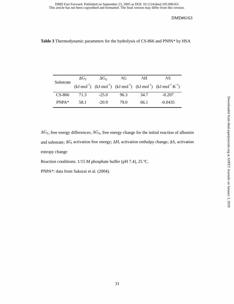
Using the HSA-hydrolysis parameters, we compared energy changes and
thermodynamic parameters between CS-866 and PNPA (Table 3). CS-866 had larger
values of ∆G (96.3 kJ·mol-1
) and ∆S (-0.207 kJ·mol-1
·K-1
) than PNPA.
Effects of Ligands on Hydrolysis
The site I-specific ligands warfarin, DNSA and n-butyl p-AB were used as
This article has not been copyedited and formatted. The final version may differ from this version.DMD Fast Forward. Published on September 23, 2005 as DOI: 10.1124/dmd.105.006163
at ASPE
T Journals on January 3, 2020
dmd.aspetjournals.org
Dow
nloaded from
DMD#6163
14
inhibitors to investigate for any competition with the hydrolytic reaction (Kragh-Hansen
et al., 2002; Yamasaki et al., 1996). Interestingly, warfarin inhibited hydrolysis in a
competitive manner, with a Ki value of 155 µM in a Dixon plot (Fig. 2A). By contrast,
neither n-butyl p-AB nor DNSA inhibited HSA-catalyzed hydrolysis of CS-866 (Fig.
2B and 2C).
The site II-specific ligands ibuprofen and diazepam were used to investigate,
whether there is competition between that ligand-binding site and the catalytic site
(Kragh-Hansen et al., 2002). Diazepam had no inhibitory effect, but competitive
inhibition was observed with ibuprofen, with a Ki value of 235 µM in a Dixon plot (Fig.
3A and 3B).
Effect of Chemical Modification on Hydrolysis
Hydrolytic activities of the 4 specifically modified HSA derivatives (Tyr-, Lys-,
His- and Trp-HSA) were assayed (Fig. 4). Compared with native-type HSA, all
modified HSA derivatives had significantly decreased hydrolytic activity (p< 0.05).
Modification of Lys residues or of the single Trp residue resulted in the most
pronounced reductions in catalytic reactivity.
Examination of Hydrolytic Activity Using Site-directed Mutagenesis
Wild-type rHSA and the HSA single-residue mutants K199A, W214A and Y411A
were used to examine involvement of various amino acid residues in the hydrolysis. The
hydrolytic activity of each rHSA was examined in order to elucidate the contribution of
specific amino acid residues (Fig. 5). Compared with the wild-type rHSA, all mutant
rHSAs showed significant decreases in catalytic activity. K199A exhibited a particularly
This article has not been copyedited and formatted. The final version may differ from this version.DMD Fast Forward. Published on September 23, 2005 as DOI: 10.1124/dmd.105.006163
at ASPE
T Journals on January 3, 2020
dmd.aspetjournals.org
Dow
nloaded from
DMD#6163
15
marked decrease in catalytic activity (P< 0.001), suggesting that Lys-199 plays a
particularly important role in the hydrolysis. These results are in good agreement with
those obtained with the chemically modified HSAs. The W214A and Y411A mutants
showed a significant reduction in catalytic activity (P< 0.05), indicating that the amino
acid residues Trp-214 and Tyr-411 are also involved in the hydrolytic reaction.
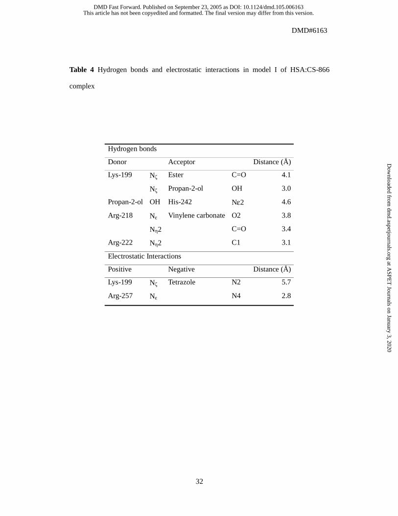
Molecular Interaction of CS-866 and HSA in Docking Models
We obtained 2 docking models: model I for site I, and model II for site II. In model
I, the binding of CS-866 was similar to that of warfarin (Fig. 6). The biphenyl moiety of
CS-866 was bound to the hydrophobic pocket consisting of Leu-219, Leu-238, Val-241,
Leu-260, Ala-261, Ile-264, Ile-290, and Ala-291. The 2-propyl-imidazole moiety and
the propan-2-ol moiety were bound to the other hydrophobic pocket (Phe-211, Trp-214,
Ala-215, Leu-219, and Leu-238). Hydrogen bonds to and electrostatic interactions with
other residues are detailed in Table 4. Oxygen atoms of the vinylene carbonate moiety
formed hydrogen bonds with side chains of Arg-218 and of Arg-222. Negative charges
of the tetrazole moiety interacted electrostatically with side chains of Lys-199 and of
Arg-257. The hydroxyl oxygen atom of the propan-2-ol moiety formed a hydrogen bond
with the side chain of Lys-199, and the hydrogen atom formed a hydrogen bond with
the side chain of His-242. The carbonyl oxygen atom of the ester moiety formed a
hydrogen bond with the side chain of Lys-199. The ester moiety of CS-866 was in the
vicinity of Glu-292. However, the side chain of Glu-292 was distant from the carbonyl
carbon of the ester moiety, because the docking program, FlexX, cannot account for
chemical reactions and remains rigid. We changed the torsion angles of the side chain of
Glu-292 without steric hindrance as Glu-292 became capable of a nucleophilic reaction.
This article has not been copyedited and formatted. The final version may differ from this version.DMD Fast Forward. Published on September 23, 2005 as DOI: 10.1124/dmd.105.006163
at ASPE
T Journals on January 3, 2020
dmd.aspetjournals.org
Dow
nloaded from
DMD#6163
16
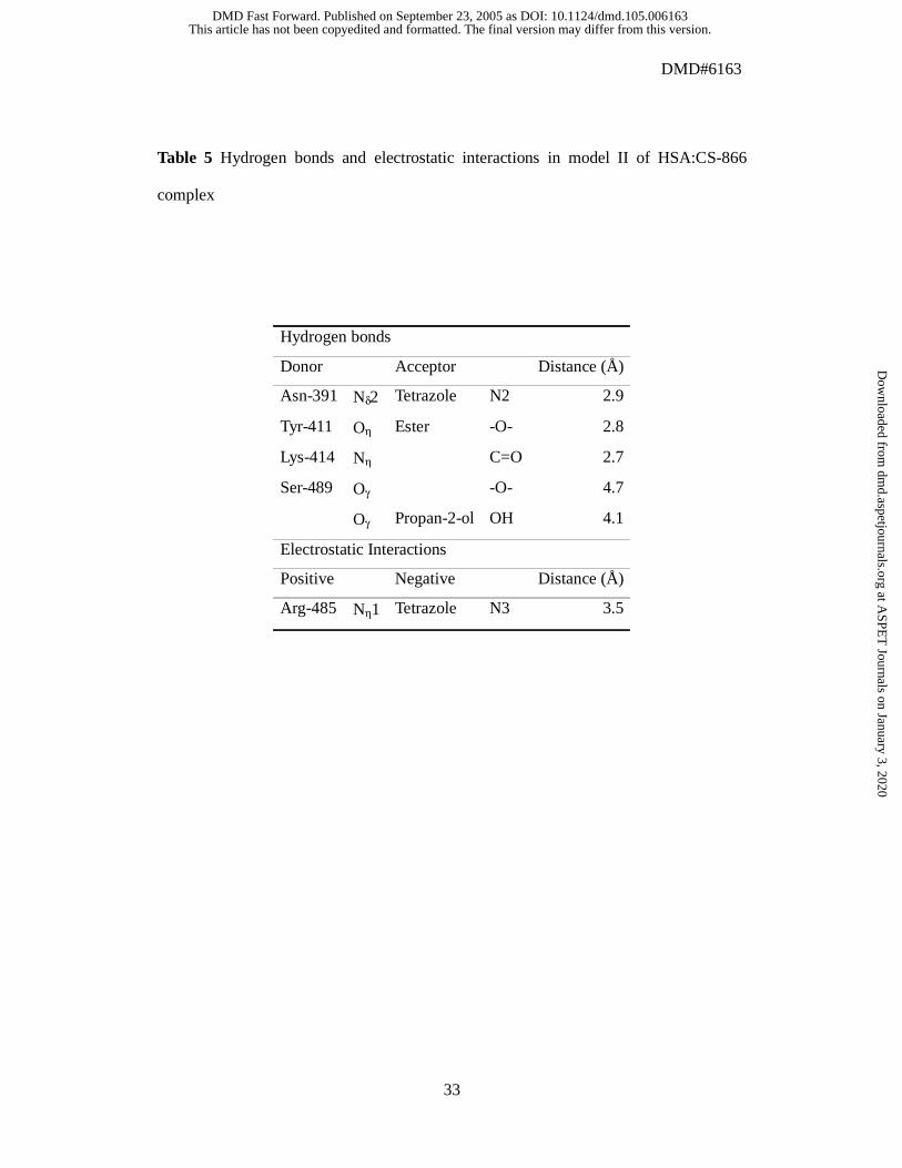
In model II, CS-866 was bound to site II using the pocket for myristate-4 (Fig. 7)
(Curry et al., 1998). The pocket for myristate-3 was not occupied. The biphenyl moiety
and the 2-propyl-imidazole moiety were bound to the hydrophobic pocket consisting of
Leu-387, Pro-486 and Ala-490. The propan-2-ol moiety was surrounded by the
hydrophobic residues (Leu-387, Leu-430 and Leu-453). Table 5 shows hydrogen bonds
and electrostatic interactions in site II. Negative charges of the tetrazole moiety
interacted electrostatically with the side chain of Arg-485, and the tetrazole moiety
formed a hydrogen bond with the side chain of Asn-391. The side chain of Ser-489
formed hydrogen bonds with the hydroxyl oxygen atom of the propan-2-ol moiety and
the carboxyl oxygen atom of the ester moiety. The carbonyl oxygen atom of the ester
moiety formed a hydrogen bond with the side chain of Lys-414. The carboxyl oxygen
atom of the ester moiety formed a hydrogen bond with the side chain of Tyr-411.
This article has not been copyedited and formatted. The final version may differ from this version.DMD Fast Forward. Published on September 23, 2005 as DOI: 10.1124/dmd.105.006163
at ASPE
T Journals on January 3, 2020
dmd.aspetjournals.org
Dow
nloaded from

DMD#6163
17
Discussion
This antihypertensive prodrug, CS-866, is hydrolyzed in the serum. Hydrolysis of
CS-866 in serum has been observed in several species, and comparison between 5
species has shown that hydrolytic activity is highest in rabbits, followed by dogs, mice,
rats and humans (Ikeda et al., 2000). Furthermore, we examined the activity due to
serum albumin. It was found to be highest in humans, followed by rats, mice, rabbits
and dogs (data not shown). This indicates that the mechanisms of hydrolysis of CS-866
in serum differ among those species, and that HSA plays a more important role in
producing RNH-6270 than other serum albumin species.
Thermodynamic Properties
The esterase-like activity of HSA is dependent on the catalytic rate constant, kcat,
and increases with a decrease in the activation free energy change, ∆G. Thus, the
magnitude of ∆G, which is dependent on activation entropy change (∆S), as calculated
from a thermodynamic analysis, can be regarded as an indicator of hydrolytic activity of
HSA (Sakurai et al., 2004; Fersht, 1998). Because PNPA has lower ∆G and ∆S values
than CS-866 (Table 3), PNPA exhibited greater affinity for HSA and a higher catalytic
rate than CS-866 (Table 2). Hydrolysis reactions catalyzed by albumin have previously
been found to have a particularly great entropy difference between the ground state (ES)
and the transition state (ES*) (Sakurai et al., 2004). The active sites of HSA to which
the substrate binds is perfectly oriented to the reactive site of the substrate (the ester
portion) for hydrolysis, and thus has a smaller entropy difference between the transition
state (ES*) and the ground state (ES). This may be the reason why hydrolysis of PNPA
proceeds more readily than hydrolysis of CS-866. That is, compared to CS-866, PNPA
This article has not been copyedited and formatted. The final version may differ from this version.DMD Fast Forward. Published on September 23, 2005 as DOI: 10.1124/dmd.105.006163
at ASPE
T Journals on January 3, 2020
dmd.aspetjournals.org
Dow
nloaded from

DMD#6163
18
has a structure and orientation that are better suited to hydrolysis by HSA.
Relationship Between Ligand Binding Sites and Hydrolytic Active Sites
HSA is the most abundant protein in blood plasma, and serves as a storage protein
and transport protein for many endogenous and exogenous compounds (Kragh-Hansen
et al., 2002; Peters, 1996). The unique capability of HSA to reversibly bind a large
number of compounds is usually explained by the existence of a number of binding
regions (including site I and site II), each of which has a very different specificity
(Kragh-Hansen et al., 2002; Kragh-Hansen, 1991). Furthermore, site I on HSA consists
of 3 subsites: Ia, Ib and Ic (Fehske et al., 1982; Yamasaki et al., 1996). Another
important role of HSA is as a catalyst for the hydrolysis of various compounds, such as
esters, amides and phosphates. It has been suggested that the active site of HSA for
p-nitrophenyl esters is site II, and that Tyr-411 is essential for hydrolysis of
p-nitrophenyl esters (Ozeki et al., 1980; Watanabe et al., 2000). The reactive site and
active residue for nitroaspirin are reportedly site I and Lys-199, respectively (Ikeda and
Kurono, 1986). The relationship between the hydrolytic active sites of HSA for CS-866
and the proteins ligand-binding sites was investigated in the present study.
There are interesting patterns of competition between site-I and site-II ligands for
hydrolysis. Although warfarin, which is regarded as a typical ligand of subsite Ia of
HSA, acts as a competitive inhibitor, this does not necessarily indicate that the HSA
catalytic site for CS-866 is subsite Ia, because ibuprofen, a typical site II ligand, also
exhibited evidence of competitive inhibition (Fig. 2A and 3B). These results suggest
that substrate specificity of the esterase-like region and ligand-binding site of HSA is
inconsistent. In other words, the catalytic site for CS-866 on HSA may recognize
This article has not been copyedited and formatted. The final version may differ from this version.DMD Fast Forward. Published on September 23, 2005 as DOI: 10.1124/dmd.105.006163
at ASPE
T Journals on January 3, 2020
dmd.aspetjournals.org
Dow
nloaded from

DMD#6163
19
CS-866 in a manner different from that of the ligand-binding site.
Roles of Specific Amino Acid Residues
For proteins whose X-ray crystallographic structure is known, the role of each
amino acid residue can be quantitatively determined using the amino acid displacement
(site-directed mutagenesis) technique and information obtained from X-ray analysis.
The present chemical modification experiments indicate that Lys, Trp and Tyr
residues of HSA are important for hydrolysis of CS-866 by HSA, and that His residues
are also involved (Fig. 4). These experiments were performed with mildly modified
HSA, because, for example, only 1.24 of the Tyr residues and 3.8 of the Lys residues
were modified. However, HSA has 59 Lys residues, and the numbers of Trp, Tyr and
His residues are 1, 18 and 16, respectively. Previous findings have demonstrated that
Tyr-411 is most likely the reactive Tyr of HSA (Watanabe et al., 2000). It is also known
that the reactivity of Lys-199 is high (Means and Bender, 1975). Further, it is reported
that this single Trp residue contributes to the esterase-like activity of HSA (Ozeki et al.,
1980; Kurono et al., 1982). In an attempt to identify specific residues of importance for
the hydrolysis of CS-866, we examined the activity of several rHSAs, namely wild-type
HSA and the single-residue mutants K199A, W214A and Y411A.
Because we did not observe great decrease of the hydrolytic activity of HSA for
CS-866, even in the single-residue mutants K199A and Y411A, we conclude that the
catalytic sites of HSA for CS-866 are not solely confined to the Lys-199 and Tyr-411
residues, but rather involves several additional amino acid residues (Fig. 5).
The single Trp residue, Trp-214, is located close to Lys-199, as indicated by X-ray
diffraction analysis, and is an element of a major interdomain cluster of hydrophobic
This article has not been copyedited and formatted. The final version may differ from this version.DMD Fast Forward. Published on September 23, 2005 as DOI: 10.1124/dmd.105.006163
at ASPE
T Journals on January 3, 2020
dmd.aspetjournals.org
Dow
nloaded from

DMD#6163
20
residues (Sugio et al., 1999; He and Carter, 1992). The mutant W214A exhibited a
significant decrease in hydrolytic activity (Fig. 5). In addition, the microenvironment
near Trp-214 was investigated to obtain detailed information about the role of this
residue in the hydrolysis. After incubation with CS-866 for 10 minutes, the relative
fluorescence intensity of HSA decreased by more than half and the λmax was
blue-shifted (data not shown). These results are consistent with a model indicating that
the Trp-214 residue is involved in hydrolytic reaction. These limited data leads us the
idea that a double (or triple) mutation of Lys-199, Trp-214 and Tyr-411 could
completely abolish the hydrolytic activity. Further investigations on this point are under
way at this laboratory.
Structural Mechanism of Hydrolysis Based on Models
The present findings suggest that HSA has 2 catalytic sites for CS-866, for the
following 2 reasons. Mutation at site I or site II diminishes but does not abolish the
hydrolytic activity. The hydrolytic activity is inhibited by both warfarin (site I drug) and
ibuprofen (site II drug).
In model I (Fig. 6), CS-866 occupied the binding site of warfarin; this is consistent
with the results showing that warfarin inhibits the hydrolytic activity of HSA. In site I,
the carbonyl oxygen atom of the ester moiety formed a hydrogen bond with the side
chain of Lys-199, and this hydrogen bond could function as an oxyanion hole. The
importance of Lys-199 indicated by the model is consistent with the decreased
hydrolytic activity of the K199A mutant and the HSA variant produced by chemical
modification of Lys. The catalytic residue may be Glu-292; the distance between the
oxygen atom of the side chain of Glu-292 and the carbonyl carbon of the ester moiety of
This article has not been copyedited and formatted. The final version may differ from this version.DMD Fast Forward. Published on September 23, 2005 as DOI: 10.1124/dmd.105.006163
at ASPE
T Journals on January 3, 2020
dmd.aspetjournals.org
Dow
nloaded from

DMD#6163
21
CS-866 was 4.8 Å. The hydrophobic interaction between CS-866 and Trp-214 indicated
by the model is consistent with the diminished hydrolytic activity of the W214A mutant
and the HSA variant produced by chemical modification of Trp.
Model II (Fig. 7) indicates that the mechanism of hydrolysis of CS-866 is almost
the same as that found in previous studies for p-nitrophenyl esters, with the exception of
the involvement of Arg-410 (Sakurai et al., 2004; Watanabe et al., 2000). Instead of
Arg-410, Lys-414 was used to create an oxyanion hole. The distance between the
hydroxyl oxygen atom of Tyr-411 and the carbonyl carbon of the ester moiety of
CS-866 was 3.3 Å, indicating that it is possible that Tyr-411 plays the role of a cathartic
residue. The importance of Tyr-411 and Lys-414 is consistent with the decreased
hydrolytic activity of the Y411A mutant and of the HSA variants produced by chemical
modification of Tyr or Lys. In our model II, CS-866 was bound to the pocket for
myristate-4 in site II. The binding pockets of the site II ligands ibuprofen and diazepam
are unknown. If ibuprofen binds to the pocket for myristate-4, our model II provides a
mechanism for inhibition of hydrolytic activity of HSA by ibuprofen.
The present findings indicate that hydrolysis of CS-866 by HSA is dependent on
∆S. Another important factor is the orientation between the catalytic active site on HSA
and the ester region of the substrate. There are differences between the catalytic active
sites and the ligand-binding sites of HSA. Furthermore, the residues of Lys-199,
Trp-214 and Tyr-411 play important roles in this catalytic reaction. All of these
experimental findings are consistent with the docking model that we derived from
computer simulation.
This article has not been copyedited and formatted. The final version may differ from this version.DMD Fast Forward. Published on September 23, 2005 as DOI: 10.1124/dmd.105.006163
at ASPE
T Journals on January 3, 2020
dmd.aspetjournals.org
Dow
nloaded from

DMD#6163
22
Acknowledgments
This work was supported in part by Grant-in-Aid for scientific research from the
Ministry of Education, Science and Culture of Japan (11694298 for M.O.) and was also
supported in part by Grant-in-Aid for scientific research, encouragement of young
scientists (B) (13771414 for N.Y.) from Japan Society for the Promotion of Science.
This article has not been copyedited and formatted. The final version may differ from this version.DMD Fast Forward. Published on September 23, 2005 as DOI: 10.1124/dmd.105.006163
at ASPE
T Journals on January 3, 2020
dmd.aspetjournals.org
Dow
nloaded from
DMD#6163
23
References
Brousil JA and Burke JM (2003) Olmesartan medoxomil: an angiotensin II-receptor
blocker. Clin Ther 25: 1041-1055.
Chen RF (1967) Removal of fatty acids from serum albumin by charcoal treatment. J
Biol Chem 242: 173-181.
Curry S, Mandelkow H, Brick P and Franks N (1998) Crystal structure of human serum
albumin complexed with fatty acid reveals an asymmetric distribution of binding sites.
Nat Struct Biol 5:827-835.
Cornell WD, Cieplak P, Bayly CI, Gould IR, Merz KM Jr, Ferguson DM, Spellmeyer
DC, Fox T, Caldwell JW and Kollman PA (1995) A second generation force field for the
simulation of proteins, nucleic acids, and organic molecules. J Am Chem Soc
117:5179-5197.
Fehske KJ, Müller WE and Wollert U (1978) The modification of the lone tryptophan
residue in human serum albumin by 2-hydroxy-5-nitrobenzyl bromide. Characterization
of the modified protein and the binding of L-tryptophan and benzodiazepines to the
tryptophan-modified albumin. Hoppe-Seylers Z Physiol Chem 359: 709-717.
Fehske KJ, Schläfer U, Wollert U and Müller WE (1982) Characterization of an
important drug binding area on human serum albumin including the high-affinity
binding sites of warfarin and azapropazone. Mol Pharmacol 21: 387-393.
Fersht A (1998) Structure and Mechanism in Protein Science, Freeman, New York.
Gasteiger J and Marsili M (1980) Iterative partial equalization of orbital
electronegativity: a rapid access to atomic charges. Tetrahedron 36:3219-3228.
Gasteiger J and Marsili M (1981) Prediction of proton magnetic resonance shifts: the
dependence on hydrogen charges obtained by iterative partial equalization of orbital
This article has not been copyedited and formatted. The final version may differ from this version.DMD Fast Forward. Published on September 23, 2005 as DOI: 10.1124/dmd.105.006163
at ASPE
T Journals on January 3, 2020
dmd.aspetjournals.org
Dow
nloaded from
DMD#6163
24
electronegativity. Organ Magn Reson 15:353-360.
Gounaris AD and Perlmann GE (1967) Succinylation of pepsinogen. J Biol Chem 242:
2739-2745.
Haynes R, Osuga DT and Feeney RE (1967) Modification of amino groups in inhibitors
of proteolytic enzymes. Biochemistry 6: 541-547.
He XM and Carter DC (1992) Atomic structure and chemistry of human serum albumin.
Nature 358: 209-215.
Ikeda K and Kurono Y (1986) Enzymatic activity and drug binding activity of human
serum albumin. Yakugaku Zasshi 106: 841-855.
Ikeda T (2000) Two prodrugs activated by serum esterases including albumin.
Proceedings of the international symposium on Serum Albumin & α1-Acid Glycoprotein
173-180.
Koike H, Sada T and Mizuno M (2001) In vitro and in vivo pharmacology of
olmesartan medoxomil, an angiotensin II type AT1 receptor antagonist. J Hypertens
Suppl 19: 1:S3-14.
Kragh-Hansen U (1991) Octanoate binding to the indole- and benzodiazepine-binding
region of human serum albumin. Biochem J 273: 641-644.
Kragh-Hansen U, Chuang VTG and Otagiri M (2002) Practical aspects of the
ligand-binding and enzymatic properties of human serum albumin. Biol Pharm Bull 25:
695-704.
Kurono Y, Yamada H and Ikeda K (1982) Effects of drug binding on the esterase-like
activity of human serum albumin. V. Reactive site towards substituted aspirins. Chem
Pharm Bull 30: 296-301.
Marsili M and Gasteiger J (1980) Pi-Charge Distributions from Molecular Topology and
This article has not been copyedited and formatted. The final version may differ from this version.DMD Fast Forward. Published on September 23, 2005 as DOI: 10.1124/dmd.105.006163
at ASPE
T Journals on January 3, 2020
dmd.aspetjournals.org
Dow
nloaded from
DMD#6163
25
Pi-Orbital Electronegativity. Croat Chem Acta 53:601-614.
Means GE and Bender ML (1975) Acetylation of human serum albumin by
p-nitrophenyl acetate. Biochemistry 14: 4989-4994.
Neutel JM (2001) Clinical Studies of CS-866, the newest angiotensin II receptor
antagonist. Am J Cardiol 87 (8A): 37C-43C.
Ozeki Y, Kurono Y, Yotsuyanagi T and Ikeda K (1980) Effects of drug binding on the
esterase activity of human serum albumin: inhibition modes and binding sites of anionic
drugs. Chem Pharm Bull 28: 535-540.
Peters T Jr (1996) All about albumin, Biochemistry, Genetics, and Medical Applications,
Academic Press, San Diego.
Petitpas I, Bhattacharya AA, Twine S, East M and Curry S (2001) Crystal structure
analysis warfarin binding to human serum albumin: anatomy of drug site I. J Biol Chem
276:22804-22809.
Purcel WP and Singer JA (1967) A brief review and table of semiempirical parameters
used in the Hüeckel molecular orbital method. J Chem Eng Data 12:235-246.
Rarey M, Kramer B, Lengauer T and Klebe G (1996) A fast flexible docking method
using incremental construction algorithm. J Mol Biol 261:470-489.
Roosemont JL (1978) Reaction of histidine residues in proteins with
diethylpyrocarbonate: differential molar absorptivities and reactivities. Anal Biochem
88: 314-320.
Sakurai Y, Ma SF, Watanabe H, Yamaotsu N, Hirono S, Kurono Y, Kragh-Hansen U and
Otagiri M (2004) Esterase-like activity of serum albumin: characterization of its
structural chemistry using p-nitrophenyl esters as substrates. Pharm Res 21: 285-292.
Sokolovsky M, Riordan JF and Vallee BL (1966) Tetranitromethane. A reagent for the
This article has not been copyedited and formatted. The final version may differ from this version.DMD Fast Forward. Published on September 23, 2005 as DOI: 10.1124/dmd.105.006163
at ASPE
T Journals on January 3, 2020
dmd.aspetjournals.org
Dow
nloaded from

DMD#6163
26
nitration of tyrosyl residues in proteins. Biochemistry 5: 3582-3589.
Sugio S, Kashima A, Mochizuki S, Noda M and Kobayashi K (1999) Crystal structure
of human serum albumin at 2.5 A resolution. Protein Eng 12: 439-446.
Watanabe H, Tanase S, Nakajou K, Maruyama T, Kragh-Hansen U and Otagiri M
(2000) Role of Arg-410 and Tyr-411 in human serum albumin for ligand binding and
esterase-like activity. Biochem J 349: 813-819.
Watanabe H, Yamasaki K, Kragh-Hansen U, Tanase S, Harada K, Suenaga A and
Otagiri M (2001) In vitro and in vivo properties of recombinant human serum albumin
from Pichia pastoris purified by a method of short processing time. Pharm Res 18:
1775-1781.
Yamasaki K, Maruyama T, Kragh-Hansen U and Otagiri M (1996) Characterization of
site I on human serum albumin: concept about the structure of a drug binding site.
Biochim Biophys Acta 1295: 147-157.
This article has not been copyedited and formatted. The final version may differ from this version.DMD Fast Forward. Published on September 23, 2005 as DOI: 10.1124/dmd.105.006163
at ASPE
T Journals on January 3, 2020
dmd.aspetjournals.org
Dow
nloaded from
DMD#6163
27
Figure legends
Fig. 1 Hydrolysis of CS-866 to RNH-6270
Fig. 2 Effect of warfarin (A), n-butyl p-AB (B) and DNSA (C) (0~600 µM) on the
hydrolysis of CS-866 by HSA
Reaction conditions: 75 µM HSA; 100-250 µM CS-866; 1/15 M phosphate buffer (pH
7.4); 37 ºC. Plots represent mean ± S.D. (n=3)
Fig. 3 Effect of diazepam (A) and ibuprofen (B) (0~600 µM) on the hydrolysis of
CS-866 by HSA
Reaction conditions: 75 µM HSA; 100-250 µM CS-866; 1/15 M phosphate buffer (pH
7.4); 37 ºC. Plots represent mean ± S.D. (n=3)
Fig. 4 Hydrolytic activities of chemically modified HSAs as compared with that of
normal HSA
Reaction conditions: 75 µM HSA; 250 µM CS-866; 1/15 M phosphate buffer (pH 7.4);
37 ºC. Each column represents mean ± S.D. (n=3)
# Significantly different from native type HSA (p< 0.05)
Fig. 5 Hydrolytic activities of mutant rHSAs as compared with that of wild type rHSA
Reaction conditions: 75 µM HSA; 250 µM CS-866; 1/15 M phosphate buffer (pH 7.4);
37 ºC. Each column represents mean ± S.D. (n=3)
# p< 0.05 and ## p< 0.001 as compared with wild type rHSA.
This article has not been copyedited and formatted. The final version may differ from this version.DMD Fast Forward. Published on September 23, 2005 as DOI: 10.1124/dmd.105.006163
at ASPE
T Journals on January 3, 2020
dmd.aspetjournals.org
Dow
nloaded from
DMD#6163
28
Fig. 6 Stereo drawings of ligands in site I: A) modeling structure for CS-866. The
torsion angles of the side chain of Glu-292 were changed as Glu-292 becomes capable
of a nucleophilic reaction, B) crystal structure for S-warfarin (PDB ID 1H9Z)
Relaxed stereo viewing.
Fig. 7 Stereo drawings of ligands in site II: A) modeling structure for CS-866, B) crystal
structure for myristate-3 and -4 (PDB ID 1H9Z)
Relaxed stereo viewing.
This article has not been copyedited and formatted. The final version may differ from this version.DMD Fast Forward. Published on September 23, 2005 as DOI: 10.1124/dmd.105.006163
at ASPE
T Journals on January 3, 2020
dmd.aspetjournals.org
Dow
nloaded from

DMD#6163
29
Table 1 Kinetic parameters for the hydrolytic reaction between CS-866 and HSA at pH
7.4 and 37 ºC
KM Vmax kcat kcat/ KM HSA type
(µM) (nmol·min-1
) (min-1
) (µM-1 ·min
-1)
native 48.2 1.02 0.113 0.00232
Reaction conditions: 75 µM HSA; 10-250 µM CS-866; 1/15 M phosphate buffer (pH
7.4); 37 ºC.
This article has not been copyedited and formatted. The final version may differ from this version.DMD Fast Forward. Published on September 23, 2005 as DOI: 10.1124/dmd.105.006163
at ASPE
T Journals on January 3, 2020
dmd.aspetjournals.org
Dow
nloaded from

DMD#6163
30
Table 2 Kinetic parameters for the hydrolysis of CS-866 and PNPA * by HSA at pH 7.4
and 25 ºC
PNPA*: data from Sakurai et al. (2004).
Substrate kcat
(10-3 ·sec-1
)
KS
(µM)
kcat/ KS
(M-1 ·sec
-1)
CS-866 0.845 42.5 19.9
PNPA* 86.8 217 403.4
This article has not been copyedited and formatted. The final version may differ from this version.DMD Fast Forward. Published on September 23, 2005 as DOI: 10.1124/dmd.105.006163
at ASPE
T Journals on January 3, 2020
dmd.aspetjournals.org
Dow
nloaded from
DMD#6163
31
Table 3 Thermodynamic parameters for the hydrolysis of CS-866 and PNPA* by HSA
∆GT ∆GS ∆G ∆H ∆S Substrate
(kJ·mol-1
) (kJ·mol-1
) (kJ·mol-1
) (kJ·mol-1
) (kJ·mol-1
·K-1
)
CS-866 71.3 -25.0 96.3 34.7 -0.207
PNPA* 58.1 -20.9 79.0 66.1 -0.0435
∆GT, free energy differences; ∆GS, free energy change for the initial reaction of albumin
and substrate; ∆G, activation free energy; ∆H, activation enthalpy change; ∆S, activation
entropy change
Reaction conditions: 1/15 M phosphate buffer (pH 7.4), 25 ºC.
PNPA*: data from Sakurai et al. (2004).
This article has not been copyedited and formatted. The final version may differ from this version.DMD Fast Forward. Published on September 23, 2005 as DOI: 10.1124/dmd.105.006163
at ASPE
T Journals on January 3, 2020
dmd.aspetjournals.org
Dow
nloaded from
DMD#6163
32
Table 4 Hydrogen bonds and electrostatic interactions in model I of HSA:CS-866
complex
Hydrogen bonds
Donor Acceptor Distance (Å)
Lys-199 Nζ Ester C=O 4.1
Nζ Propan-2-ol OH 3.0
Propan-2-ol OH His-242 Nε2 4.6
Arg-218 Nε Vinylene carbonate O2 3.8
Nη2 C=O 3.4
Arg-222 Nη2 C1 3.1
Electrostatic Interactions
Positive Negative Distance (Å)
Lys-199 Nζ Tetrazole N2 5.7
Arg-257 Nε N4 2.8
This article has not been copyedited and formatted. The final version may differ from this version.DMD Fast Forward. Published on September 23, 2005 as DOI: 10.1124/dmd.105.006163
at ASPE
T Journals on January 3, 2020
dmd.aspetjournals.org
Dow
nloaded from
DMD#6163
33
Table 5 Hydrogen bonds and electrostatic interactions in model II of HSA:CS-866
complex
Hydrogen bonds
Donor Acceptor Distance (Å)
Asn-391 Nδ2 Tetrazole N2 2.9
Tyr-411 Oη Ester -O- 2.8
Lys-414 Nη C=O 2.7
Ser-489 Oγ -O- 4.7
Oγ Propan-2-ol OH 4.1
Electrostatic Interactions
Positive Negative Distance (Å)
Arg-485 Nη1 Tetrazole N3 3.5
This article has not been copyedited and formatted. The final version may differ from this version.DMD Fast Forward. Published on September 23, 2005 as DOI: 10.1124/dmd.105.006163
at ASPE
T Journals on January 3, 2020
dmd.aspetjournals.org
Dow
nloaded from

Fig. 1
N
N
OH
NN
N NH
CO2
OO
O
CH3CO2
Olmesartan medoxomilCS-866
H2O
N
N COOH
OH
NN
N NH
RNH-6270
C C CH3
O OH3C
Enzymatic hydrolysis
+
Diacetyl
Olmesartan
Ester prodrug Active metabolite
This article has not been copyedited and formatted. The final version may differ from this version.DMD Fast Forward. Published on September 23, 2005 as DOI: 10.1124/dmd.105.006163
at ASPE
T Journals on January 3, 2020
dmd.aspetjournals.org
Dow
nloaded from

Fig. 2
n-butyl p-AB Concentration (µM)
1/V
[(nm
ol/m
in/m
g H
SA)-1
]0
0.4
0.8
1.2
1.6
-400 -200 0 200 400 600
B
Warfarin Concentration (µM)
1/V
[(nm
ol/m
in/m
g H
SA)-1
]
0
1.5
3
4.5
-400 -200 0 200 400 600
A
DNSA Concentration (µM)
1/V
[(nm
ol/m
in/m
g H
SA)-1
]
0
0.3
0.6
0.9
1.2
-400 -200 0 200 400 600
C
100 µM 150 µM 200 µM 250 µM
n-butyl p-AB Concentration (µM)
1/V
[(nm
ol/m
in/m
g H
SA)-1
]0
0.4
0.8
1.2
1.6
-400 -200 0 200 400 600
B
n-butyl p-AB Concentration (µM)
1/V
[(nm
ol/m
in/m
g H
SA)-1
]0
0.4
0.8
1.2
1.6
-400 -200 0 200 400 600
n-butyl p-AB Concentration (µM)
1/V
[(nm
ol/m
in/m
g H
SA)-1
]0
0.4
0.8
1.2
1.6
-400 -200 0 200 400 600
B
Warfarin Concentration (µM)
1/V
[(nm
ol/m
in/m
g H
SA)-1
]
0
1.5
3
4.5
-400 -200 0 200 400 600
A
Warfarin Concentration (µM)
1/V
[(nm
ol/m
in/m
g H
SA)-1
]
0
1.5
3
4.5
-400 -200 0 200 400 600
Warfarin Concentration (µM)
1/V
[(nm
ol/m
in/m
g H
SA)-1
]
0
1.5
3
4.5
-400 -200 0 200 400 600
A
DNSA Concentration (µM)
1/V
[(nm
ol/m
in/m
g H
SA)-1
]
0
0.3
0.6
0.9
1.2
-400 -200 0 200 400 600
C
DNSA Concentration (µM)
1/V
[(nm
ol/m
in/m
g H
SA)-1
]
0
0.3
0.6
0.9
1.2
-400 -200 0 200 400 600
DNSA Concentration (µM)
1/V
[(nm
ol/m
in/m
g H
SA)-1
]
0
0.3
0.6
0.9
1.2
-400 -200 0 200 400 600
C
100 µM 150 µM 200 µM 250 µM
100 µM 100 µM 150 µM 150 µM 200 µM 200 µM 250 µM 250 µM
This article has not been copyedited and formatted. The final version may differ from this version.DMD Fast Forward. Published on September 23, 2005 as DOI: 10.1124/dmd.105.006163
at ASPE
T Journals on January 3, 2020
dmd.aspetjournals.org
Dow
nloaded from

Fig. 3
1/V
[(nm
ol/m
in/m
g H
SA)-1
]
Ibuprofen Concentration (µM)
0
1
2
3
4
-400 -200 0 200 400 600
B
0
0.5
1
1.5
2
2.5
-400 -200 0 200 400 600
Diazepam Concentration (µM)
1/V
[(n
mol
/min
/mg
HSA
)-1]
A
100 µM 150 µM 200 µM 250 µM 1/
V[(
nmol
/min
/mg
HSA
)-1]
Ibuprofen Concentration (µM)
0
1
2
3
4
-400 -200 0 200 400 600
B
1/V
[(nm
ol/m
in/m
g H
SA)-1
]
Ibuprofen Concentration (µM)
0
1
2
3
4
-400 -200 0 200 400 600
B
0
0.5
1
1.5
2
2.5
-400 -200 0 200 400 600
Diazepam Concentration (µM)
1/V
[(n
mol
/min
/mg
HSA
)-1]
A
0
0.5
1
1.5
2
2.5
-400 -200 0 200 400 600
Diazepam Concentration (µM)
1/V
[(n
mol
/min
/mg
HSA
)-1]
A
100 µM 150 µM 200 µM 250 µM
100 µM 100 µM 150 µM 150 µM 200 µM 200 µM 250 µM 250 µM
This article has not been copyedited and formatted. The final version may differ from this version.DMD Fast Forward. Published on September 23, 2005 as DOI: 10.1124/dmd.105.006163
at ASPE
T Journals on January 3, 2020
dmd.aspetjournals.org
Dow
nloaded from

Fig. 4
This article has not been copyedited and formatted. The final version may differ from this version.DMD Fast Forward. Published on September 23, 2005 as DOI: 10.1124/dmd.105.006163
at ASPE
T Journals on January 3, 2020
dmd.aspetjournals.org
Dow
nloaded from
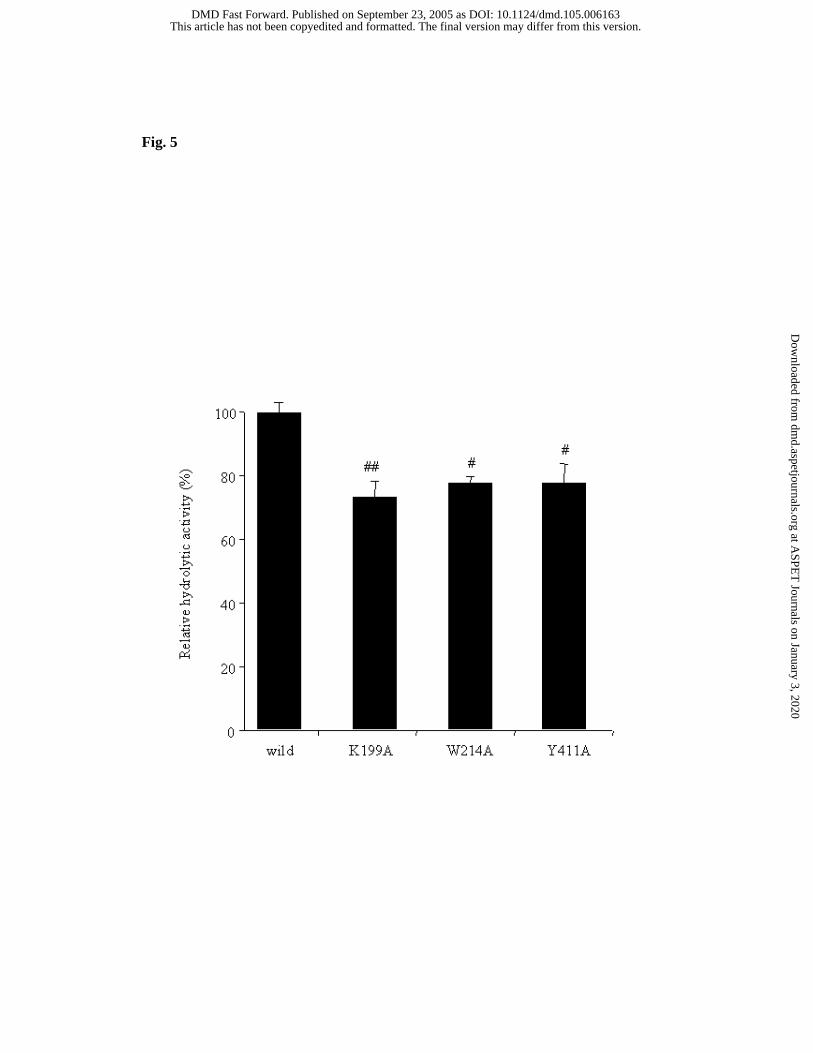
Fig. 5
This article has not been copyedited and formatted. The final version may differ from this version.DMD Fast Forward. Published on September 23, 2005 as DOI: 10.1124/dmd.105.006163
at ASPE
T Journals on January 3, 2020
dmd.aspetjournals.org
Dow
nloaded from

Fig. 6
This article has not been copyedited and formatted. The final version may differ from this version.DMD Fast Forward. Published on September 23, 2005 as DOI: 10.1124/dmd.105.006163
at ASPE
T Journals on January 3, 2020
dmd.aspetjournals.org
Dow
nloaded from
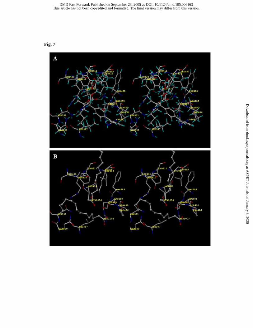
Fig. 7
This article has not been copyedited and formatted. The final version may differ from this version.DMD Fast Forward. Published on September 23, 2005 as DOI: 10.1124/dmd.105.006163
at ASPE
T Journals on January 3, 2020
dmd.aspetjournals.org
Dow
nloaded from