hydrocortisone problems endogenoussorption of hydrocortisone after oral administra-tion have also...
TRANSCRIPT

THE PHYSIOLOGICAL DISPOSITION AND METABOLICFATE OFHYDROCORTISONEIN MAN
BY RALPHE. PETERSON,JAMES B. WYNGAARDEN,SERAFIM L. GUERRA,BERNARDB. BRODIE, ANDJOSEPHJ. BUNIM
(From the National Institute of Arthritis and Metabolic Diseases, and the National HeartInstitute, National Institutes of Health, Bethesda, Md.)
(Submitted for publication July 18, 1955; accepted August 26, 1955)
The present report is concerned primarily withthe physiological disposition and fate of hydro-cortisone in man. Large doses of this steroid havebeen administered intravenously, and the rate ofits disappearance from plasma has been deter-mined in normal subjects and in patients withliver disease and various endocrinopathies. Tracequantities of hydrocortisone-4-C14 have been em-ployed in certain studies, either alone or as alabel for the larger doses. From these studies in-formation has been obtained regarding rates ofmetabolic transformation, rates and routes of ex-cretion of hydrocortisone and its metabolites, andto some extent the identity and amounts of thesemetabolites. Data on the rate and extent of ab-sorption of hydrocortisone after oral administra-tion have also been obtained. Since, in most re-spects, the results obtained upon administeringlarge or trace quantities of hydrocortisone did notdiffer greatly, the findings are to a considerableextent also applicable to problems of endogenoushydrocortisone metabolism.
MATERIALS AND METHODS
Administration of steroid
Fifty to 500 mg. of hydrocortisone were dissolved inethanol and added to 500 ml. of sterile 5 per cent dex-trose in water, producing a final alcohol concentration of2 to 3 per cent. This solution was given intravenouslyover a period of 20 to 30 minutes. Following the infu-sion, blood samples were drawn in heparinized syringesevery 20 to 30 minutes for 2 to 3 hours. Urine sampleswere collected for 24 hours or longer in single or frac-tional specimens, and preserved by freezing.
Procedure for determination of plasma hydrocortisoneThe following procedure is a modification of the re-
cently published method of Silber and Porter (1):Principle-Hydrocortisone is extracted from plasma
into dichloromethane. The dichloromethane extract iswashed with aqueous alkali to remove a considerableamount of "blank" material. The dichloromethane is thenshaken with a sulfuric acid-ethanol reagent, containing
phenylhydrazine. The resulting colored product is meas-ured in the acid phase spectrophotometrically at 410 mny.A correction for material in plasma reacting with sulfuricacid is made by treating an equal aliquot of dichloro-methane extract of plasma with sulfuric acid-ethanolwhich contains no phenylhydrazine.
Reagents.
(a) Dichloromethane-This solvent is purified by pass-ing through a bed of silical gel (average 100 mesh) in a7 by 125 cm. column. Ten to twenty liters can be purifiedin 2 to 3 hours. The effectiveness of purification is de-termined by shaking an aliquot of solvent with the phenyl-hydrazine-sulfuric acid-ethanol reagent (f). No colorshould develop on standing overnight at room tempera-ture. The solvent has remained free of impurities formany months at room temperature.
(b) 01 N Sodium Hydroxide.(c) 70 Per Cent Sulfuric Acid-700 ml. acid C.P.
grade sulfuric acid added to 300 ml. H2O.(d) Ethanol-This is purified by adding 7 gm. silver
nitrate and 15 gm. potassium hydroxide, separately (eachdissolved in 100 ml. ethanol) to 4 liters of absolute ethanol.These are mixed, allowed to stand overnight, and thendistilled, using a Vigreaux column. The first 700 ml. andthe last 100 ml. portions are discarded. The effectivenessof purification is determined by reacting this ethanolwith the phenylhydrazine-sulfuric acid (f). No colorshould develop on standing overnight at room tempera-ture.
(e) Blank Reagent-Two parts (c) with one part (d).(f) Phenylhydrazine-Sulfuric Acid-Ethanol Reagent-
50 mg. of phenylhydrazine hydrochloride (recrystallizedfrom ethanol and water) are dissolved in 50 ml. of theblank reagent (e).
(g) Hydrocortisone Standard-100 mg. of hydrocorti-sone are dissolved in 100 ml. absolute ethanol. A work-ing standard is made by diluting one ml. to a volume of100 ml. with water (10 micrograms per ml. water).
Procedure. (All glassware used must be scrupulouslycleaned with concentrated sulfuric acid.)
1. Extraction-Carefully add 5 ml. plasma to 25 ml.dichloromethane in a 200-ml. Erlenmeyer flask and placeon rotating table (Arthur H. Thomas No. 3623). Ex-tract for 10 to 15 minutes with gentle rotation (this pro-cedure extracts more than 98 per cent of the hydrocorti-sone, free and protein bound, in plasma). Gently transferentire contents to a 25-ml. graduated cylinder.
1779

PETERSON, WYNGAARDEN,GtJERRA, BRODIE, AND BUNIM
2. Washing-After aspirating as much plasma as pos-sible, add 2 ml. 0.1 N sodium hydroxide to dichloro-methane extract, shake for 15 to 20 seconds, and then re-move the alkali layer by aspiration.
3. Aliquots-Transfer two 10-ml. aliquots (for unknownand blank) and place in separate 15-ml. ground glassstoppered conical test tubes.
4. Color Development-For unknowns, add 0.2 ml.phenylhydrazine-sulfuric acid-ethanol reagent (f) to di-chloromethane extract aliquot; for blanks, add 0.2 ml.blank reagent (e). Stopper the tubes, shake vigorouslyfor 15 to 20 seconds, and allow to stand for 30 minutes ormore. The supernatant dichloromethane phase is re-moved by aspiration and the sulfuric acid-ethanol solu-tion allowed to stand at room temperature for 10 or morehours for maximum color development.
5. Spectrophotometry-Transfer the contents of thetubes to microcuvettes (Pyrocell, 1.5 by 10 by 15 mm.)and measure the absorbency of the colored product againsta water blank at 410 myA in Beckman DU spectrophotom-eter.
Note: 5 ml. water run through the entire procedureserves as reagent blank, and 5 ml. water containing 5micrograms of hydrocortisone serves as standard.
Specificity of procedure for determination of hydro-cortisone in plasma after administration of hydrocorti-sone-Specificity of the method was shown by counter-current distribution. Plasma drawn 90 minutes after theend of a 30-minute intravenous infusion of 200 mg. ofhydrocortisone was extracted with dichloromethane, the
extract evaporated to dryness, and the apparent hydro-cortisone subjected to an eight-transfer counter-currentdistribution. The solvents used were water (8 volumes)and chloroform (1 volume). This system gave a par-tition coefficient of about 0.1 for authentic hydrocorti-sone. A trace quantity of hydrocortisone4-C' was addedbefore the counter-current distribution. After distribu-tion, aliquots of both phases were analyzed for carbon"4and by the phenylhydrazine procedure. Figure 1 dem-onstrates the distribution of the phenylhydrazine reactingmaterial and the carbon1' hydrocortisone. These datademonstrated that more than 90 per cent of the ma-terial assayed with phenylhydrazine was distributed ashydrocortisone.
The specificity of the analytical procedure employed wasfurther demonstrated by subjecting dichloromethane ex-tracts of plasma obtained 60 and 120 minutes followingthe end of a 30-minute infusion of 200 mg. of hydrocor-tisone to paper chromatography, using a modification ofthe Bush (2) type system. A small quantity of hydro-cortisone-4-C1' was added to the dichloromethane extractand the specific activity of the phenylhydrazine reactingmaterial was determined. The extract was then chro-matographed at room temperature for 6 hours in abenzene-methanol-water (4:2: 1) system. The hydro-cortisone of the extract, running at the same rate as purehydrocortisone, was located by ultraviolet scanning. Thisarea was cut out and eluted with 3 ml. of 95 per centethanol, and the eluate evaporated to dryness under ni-trogen. The residue was redissolved and the specific ac-
COUNTER-CURRENTDISTRIBUTIONHYDROCORTISONEPLASMA 90 MINUTES AFTER INFUSION
0 I 2 3 4 5 ' 6 T a
TUBE NUMBERFIG. 1. COUNTER-CURRENTDISTRIBUTION ANALYSIS OF PHENYLHYDRAZINERE-
ACTING MATERIAL IN PLASMA DICHLOROMETHANEEXTRACT90 MINUTES FOLLOWINGTERMINATION OF INFUSION OF 200 MG. HYDROCORTISONETO NORMALSUBJECT
1780
4015 H20 ' CHC13_--"
* Phenylhydrazine color reactionA C14- hydrocortisone
O- Represents possiblemetaboliteequiv. 8.7% Totol
-J
crw
H
(fDz
4
Hcn
Wz0C-
LL
s

DISPOSITION AND FATE OF HYDROCORTISONEIN MAN
tivity of the phenylhydrazine reacting material was againdetermined. Table I shows the specific activity of thematerial before and after chromatography. Portions ofthe steroid eluted from the paper were also assayedfluorometrically by a modification of the highly specificmethod described by Sweat (3). Other eluted portionswere treated so as to convert the hydrocortisone to itsacetate which was then chromatographed, eluted, and as-sayed. These procedures confirmed the data obtained bycounter-current distribution studies.
Procedure for determination of hydrocortisone metabo-lites in urine-The method and reagents were the sameas those described for plasma, except that to the urine(10 ml.) 2 ml. 0.5 M phosphate buffer (pH 6.2) and1000 units Sigma bacterial glucuronidase in one ml. ofwater were added. Following incubation for 24 hours at37° C., the mixture was extracted with 8 to 9 volumes ofdichloromethane. After washing the dichloromethaneextract with 1/20 volume 0.1 N NaOH, aliquots (40 ml.)were extracted with 3.0 ml. of the phenylhydrazine-sul-furic acid-ethanol, and the sulfuric acid-ethanol reagentas described for plasma. The resulting colored productwas measured at 410 miu.
Phenylhydrazine reacting materials extracted fromurine either directly or after 8-glucuronidase hydrolysisrepresent many different metabolites of hydrocortisone.
Studies with hydrocortisone-4-C'4Hydrocortisone-4-C14, specific activity 1.47 millicuries
per millimole, was made available through the EndocrineStudy Section of the National Institutes of Health.This compound was shown to be approximately 95 percent pure by counter-current distribution, and isotope di-lution analyses and paper chromatography. It was ad-ministered intravenously in trace quantities in solutionin a small volume of 5 per cent ethanol in water over a
period of 3 to 5 minutes, or mixed with non-isotopic hy-drocortisone given in 500 ml. 3 per cent ethanol in 5 percent dextrose in sterile distilled water. Similar solu-tions were administered orally in certain studies.
After intravenous or oral administration, samples ofblood were taken at 30 to 60-minute intervals for 4 to 8hours, and urine was collected every 2 hours for 12 hours,the following 12 hours, and for 24-hour periods until no
additional radioactivity was demonstrable. Fecal sampleswere collected for 4 days, homogenized with water in a
Waring blendor, and an aliquot lyophilized.Plasma samples were extracted directly with 5 volumes
of dichloromethane. The dichloromethane extract was
washed once with 1/20 volume 0.1 N sodium hydroxide,twice with 1/15 volume water, and then evaporated todryness at 40° C. under nitrogen. The residue was thendissolved in methanol and an aliquot added to theplanchet.
Biliary excretion of radiometabolites of hydrocortisone-Bile collections were made on a 53-year old male pa-tient who gave a history of intermittent colicky pain ofseveral years' duration with signs of recurrent obstructivejaundice for 8 months, and persistent signs of jaundiceand RUQ pain for 3 weeks prior to hospitalization.
TABLE I
Comparison specific activities phenylhydrazine reactingmaterial in plasma extracts following
infusion of hydrocortisone
Specific activities(cpmI/pg.)
Before Afterchroma- chroma-tography tography
Expt. No. 1 (plasma 60 min.after infusion)
1st chromatography 2,370 2,6802nd chromatography 2,7103rd chromatography* 2,500
Expt. No. 2 (plasma 120 min.after infusion)
1st chromatography 635 650t
* Converted to hydrocortisone acetate and rechromato-graphed.
t Six hundred and fifty cpm per microgram with phenyl-hydrazine and fluorometric assay.
Liver function tests were compatible with a diagnosis ofobstructive jaundice. Surgical exploration 1 revealedthe presence of cholecystitis and chololithiasis, and in ad-dition revealed an acute pancreatitis completely obstruc-ting the common bile duct which was dilated to 3 to 4times its normal size. No stones were found on explora-tion of the common bile duct. The sphincter of Oddi wascut. A T-tube was inserted in the common duct, and atits distal end was attached a small "Foley-type" catheter.These two tubes were led to the outside. Bile was per-mitted free access to the T-tube and to the duodenumthrough the common duct; however, the distal end of thecommon duct could be obstructed at will by inflating theFoley catheter bag with 1 to 2 ml. water, thereby drain-ing all the bile through the T-tube. On the morning ofthe study, the bile duct was obstructed and the patient wasgiven 1.5 *c. of hydrocortisone-4-C1' intravenously. Bilesamples were collected every two hours for 12 hours,and for the following 12 hours. Feces and urine werealso collected.
Procedure for carbon"' analyses of samples-All analy-ses for carbon1' were carried out with a Robinson (4)gas-flow counter, using a gas mixture of nine parts argonand one part methane, and connected to a Nuclear Model172 scaler. This apparatus gave a background of 3 to4 cpm and an efficiency for carbon"' of slightly more than53 per cent when counted at infinite thinness. Planchetsof stainless steel with a surface area of 1.6 sq. cm. and adepth of 0.6 cm. were used. They were cleaned by boil-ing with alcoholic potassium hydroxide.
From 0.05 to 0.30 ml. of fresh urine was plated directlyonto tared planchets. The urine was dried on a rotatingplatform with the aid of air and heat from an infrared
1 Weare indebted to Dr. Henry Doubilet of New YorkUniversity College of Medicine, and to Dr. Robert Smith,National Cancer Institute, for the operative proceduresinvolved in carrying out this study.
1781

PETERSON, WYNGAARDEN,GUERRA, BRODIE, AND BUNIM
lamp, and the planchets were then placed in a desiccatoruntil weighed and counted. A correction for self-absorp-tion was made from a self-absorption curve of urine, andall samples were corrected to infinite thinness. Self-ab-sorption curves run on different urines were not foundto show a significant degree of variation. It was notnecessary to correct any samples for self-absorption thatweighed less than 0.1 mg. (infinite thinness). Resultsobtained using a double isotope dilution technique (5)were found to check with results obtained with the self-absorption correction curve data. All counting resultswere corrected for instrument variation by reference toa barium carbonate-C14 standard.
Untreated urine was extracted with 8 volumes of di-chloromethane, an aliquot of the dichloromethane extractevaporated in vacuo, and the residue dissolved in meth-anol and plated. The remaining urine was then hydro-lyzed with P-glucuronidase. This hydrolysate was thenextracted with 8 to 9 volumes of dichloromethane, andan aliquot of the dried dichloromethane residue dissolvedin methanol and plated. Aliquots of this extract and ofthe remaining aqueous residue were then applied toplanchets.
An aliquot of the lyophilized feces was digested usingthe Peters and Gutmann (6) modification of the VanSlyke-Folch (7) procedure for carbon combustion. Thecarbon dioxide was collected in a solution of barium hy-
DISAPPEARANCEOF HYDROCORTISONEFROMPLASMAFOLLOWING INTRAVENOUSADMINISTRATION
-J
a.
0
-j
0
0
cr
a-
z0
I-
0
0
a
I
U)0c10
0
20 40 60 80 100 110 120TIME (MINUTES)
FIG. 2. COMPARATivE RATES OF DISAPPEARANCE OFHYDROCORTISONEFROMPLASMAFOLLOWINGINTRAVENOUSADMINISTRATION TO A NORMALSUBJECT
droxide, and titrated with acid. The barium carbonateprecipitate was filtered dry on a small circle of filterpaper, and this barium carbonate mount placed on aplanchet, counted, and corrected to infinite thickness.
The bile samples were applied directly to planchets;a self-absorption correction factor obtained from a self-absorption curve for bile was employed.
RESULTS
Rate of disappearance of infused steroids fromplasmaRate of disappearance from plasma of hy-
drocortisone administered intravenously-Whenplasma hydrocortisone concentrations determinedat various time intervals following infusion wereplotted on semilogarithmic paper, a straight linewas obtained. In Figure 2 are shown the plasmalevels and the biological half-times obtained in a
single normal subject following the infusion ofquantities of hydrocortisone ranging from 50 to500 mg. The half-times are virtually identical,indicating that the rate of disappearance of hy-drocortisone from plasma is proportional to con-centration over a wide range of plasma levels.This finding permitted a ready comparison of be-havior of infused hydrocortisone in various sub-jects under various conditions. The biologicalhalf-life of hydrocortisone was compared in controlsubjects and in patients with liver disease, rheu-matoid arthritis, hyperthyroidism, hypothyroidism,hypopituitarism, and adrenal cortical insufficiency(Figure 3). In 20 control subjects, representingprimarily normal laboratory personnel, the mean
biological half-time of hydrocortisone disappear-ance was 114 + 6.5 minutes, with a range of 90 to130 minutes. In 12 patients with mild to moder-ately severe cirrhosis, both alcoholic and post-hepatitic types, the disappearance of hydrocorti-sone from plasma was considerably slower, andbiological half-times ranging from 160 to 800 min-utes were obtained. In some cases markedly ab-normal half-times were obtained despite relativelylittle clinical and laboratory evidence of hepaticdysfunction; however, in all but two cases thediagnosis had been confirmed by liver biopsy. Therange of serum bilirubin values in these subjectswas 1.6 to 6.0 mg. per cent.
In 8 patients with rheumatoid arthritis, half-timevalues ranging from 68 to 164 minutes were ob-tained. Essentially normal values were found in
1782

DISPOSITION AND FATE OF IIYDROCORTISONE IN MAN
BIOLOGICAL HALF-TIMES OF INTRAVENOUSLYADMINISTERED HYDROCORTISONE
(800)o
0
0
0
) _0
0
0*- -~~.- *-. --*
s@w- 7 TT iS-So;T;t :: e iL>s-et Ef *00 <* 0..:: * *: :o :v7 .i 0- -f'o: :.: 7:;_*f f*< sI-:;-iE- --;t0:--.0-.-;I ta -: -v -*_ --t * ; *--w -r;-,i-':E_
NORMALS CIRRHOSIS RHEUMATOID HYPO- THYRO- MYXEDEMA
ARTHRITIS ADRENALISM TOXICOSIS
FIG. 3. BIOLOGICAL HALF-TIMES OF INTRAVENOUSLY ADMINISTEREDHYDROCORTISONEIN NORMALSUBJECTS AND IN PATIENTS WITH VARIOUSDISEASES
*
0
3 patients with hypoadrenalism. In 3 subjectswith hyperthyroidism (BMR in + 25 to + 35range) somewhat increased rates of hydrocorti-sone disappearances were found in 2, whereas a
normal rate was found in one. In this subjecthepatomegaly was present at the time of the study,and later resolved during antithyroid therapy.In 3 subjects with hypothyroidism (BMR in -40to - 25 range) delayed rates of hydrocortisonedisappearances were observed in 2 and a normalrate in one.
No studies were carried out on patients withhyperadrenalcorticism; however, the rate of disap-pearance of hydrocortisone from the plasma of a
patient with rheumatoid arthritis who had beenmaintained on 100 mg. of hydrocortisone for 6weeks, and of another who had been taking 75 mg.
a day for more than a year, was within the normalrange.
Comparative rates of disappearance from plasmaof steroids structurally related to hydrocortisone-Figure 4 demonstrates the rate of disappearance
from the plasma in a normal subject of six differ-ent steroids 2 structurally related to hydrocorti-sone, including one with physiologically similaractions (cortisone). Figure 5 demonstrates thedisappearance from the plasma of the same steroidsin a patient with cirrhosis of the liver. With theexception of hydrocortisone, whose disappearancewas greatly delayed in the subject with liver dis-
2 The cortisone, lla-hydrocortisone, and tetrahydro-cortisone were assayed by the phenylhydrazine procedureas described in the Methods section. The corticosterone,lla-corticosterone, and 20#-ol-hydrocortisone were as-
sayed by a modification of the fluorometric method ofSweat (3). This method involves exactly the same
extraction procedure as described for the phenylhydra-zine method. However, the steroid was extracted fromthe washed dichloromethane extract with one ml. of a
reagent containing 7 parts by volume concentrated sul-furic acid and 3 parts by volume redistilled ethanol. Thefluorescence of this solution was then determined by us-
ing a Farrand Fluorimeter with a primary filter madeup of a 470 mu interference and Corning No. 5113 filter,and a secondary filter containing a 540 my interferenceand Corning No. 3486 filter.
1783
500 _-
400 _-
300 -
(Aw
-
-J
I-,< 100
J
-j
0
co

1784 PETERSON, WYNGAARDEN,
DISAPPEARANCEOF STEROIDS FROMPLASMA INNORMALSUBJECT FOLLOWING INTRAVENOUSADMINISTRATION
100.l
50 -
E ~~\ ^ t
Z HYDROCORTISONE \ \ \\c* \
I) - CORTISONEZ CORTICOSTERONE , * \\
Z --- I - HYDROCORTISONEE208-OL HYDROCORTISONE * \.
L1 - Ila -CORTICOSTERONE
TETRAHYDROCORTISONE .. \ \
w
0t 100 50 100 150TIME (MINUTES)
FIG. 4. DISAPPEARANCE OF INTRAVENOUSLY ADMINIS-TERED STEROIDS FROM PLASMA IN NORMALSUBJECT
ease, the rate of disappearance of each steroidtested was not strikingly different in the normaland in the cirrhotic subject. Furthermore, allof these steroids disappeared at rates faster thanhydrocortisone in both subjects.3
Disappearance of radioactive hydrocortisonefrom pkasma-Following intravenous infusion oftracer quantities of hydrocortisone-4-C14, the con-
centration in plasma of isotopic substances extrac-table with dichloromethane decreased logarith-mically with time (Figure 6). Half-times of 60to 90 minutes (10 subjects) were obtained. Notall the extracted material represented unalteredhydrocortisone, however, and in 3 subjects theresults of chromatographic analyses of plasma ex-
SThe method described for phenylhydrazine reactingsteroids in plasma following their infusion, has beenfound not to be specific for cortisone (8). Approxi-mately 50 per cent of the material reacting with phenyl-hydrazine 60 minutes after termination of the infusionis not cortisone. The specificities of the methods usedfor the other steroids have not been determined.
, GUERRA, BRODIE, AND BUNIM
tracts obtained from 40 minutes to 4 hours afterinfusion indicated that from 40 to 60 per cent ofthe radioactivity of this fraction was attributableto the presence of other products. In a givensubject the fraction of unconjugated steroid thatwas hydrocortisone was constant over this timespan, however. The identity of the other labeledproducts was not established, although a reason-able guess as to their nature may be made fromthe analyses of the freely extractable group oflabeled urinary metabolites described in this paper.In addition to the unconjugated metabolites inplasma, there is present a fraction of labeled me-tabolites that is rendered dichloromethane solublefollowing ,-glucuronidase hydrolysis. Such aclass of conjugated plasma corticoid constituentshas previously been demonstrated, using chromato-graphic (9) and colorimetric (10) methods ofanalysis of plasma extracts. The data of Bongio-vanni, Eberlein, Grumbach, VanWyk, and Clay-
DISAPPEARANCE OF INTRAVENOUSLY ADMINISTEREDSTEROIDS FROM PLASMA IN PATIENT WITHCIRRHOSIS OF LIVER
a
-i
IL
z
0
z
hia
cc
(0
at
FIG. 5. DISAPPEARANCEOF INTRAVENOUSLYADMINIS-TERED STEROIDS FROM PLASMA IN PATIENT WITHCIRRHOSIS
N.,\ \.\1

DISPOSITION AND FATE OF HYDROCORTISONEIN MAN
FREE AND GLUCURONIDE-CONJUGATEDSTEROIDS IN PLASMA
200 l l l
00
-J
e .1LBA A A
w ~~~~~AZa..
104
O 1 02__~ ~~~TM INHOR
SUBJTSUBJECT FREE CON.
00
ton (tLB A t
0
0 2 3 4 5 6 7 8
TIME IN HOURS
FIG. 6. DIrEcT DicHLOROMETHANte ExTRACTABLE
("FRE") AND GLUCURONIDECONJUGATEDRADIOACTIVE
STEROIDS IN PLASMA FOLLOWING INFUSION OF TRAcER
QUANTITIS OF HYDRocoRTISONE-4C1' To NoRMAL
SUBJECTS
ton ( 10) suggest that quantitatively the plasma
glucuronide fraction of conjugated adrenal hor-
mone metabolites is approximately equal to theconcentration of unconjugated phenylhydrazinereactive corticoids which normally consist pre-
dominantly of hydrocortisone (11). In the pres-
ent study, the rate of appearance of labeled me-
tabolites of hydrocortisone in conjugated form inplasma has also been followed (Figure 6). Asfree steroid disappears from plasma, conjugatedderivatives hydrolyzable with 8-glucuronidase ap-
pear, and reach a maximum at about 2 hours, atwhich time the free and conjugated fractions are ofequal magnitude. Thereafter, the conjugatedfraction greatly exceeds the free fraction of la-beled steroids. Following extraction of both freeand conjugated labeled products, the residualplasma was analyzed for radioactivity, and none
was detected. However, self-absorption factorswere large so that small quantities of labeled sub-stances may have escaped detection.
Distribution of hydrocortisone in body fluids
Plasma protein binding of hydrocortisone-Fresh human plasma obtained from four normaldonors, and isosmotic phosphate buffer of pH 7.4(0.1 M buffer in M/15 sodium chloride) were
equilibrated across a cellophane membrane for 18
hours at 370 C. with gentle continuous motion.To one or both phases, graded amounts of hy-drocortisone, labeled with a tracer quantity ofradioactive hormone, were added. At the end ofthe equilibration period, aliquots of both phaseswere extracted with dichloromethane, and the ex-tract reduced in volume in vacuo. The concen-trated extracts were then analyzed for hydrocor-tisone by phenylhydrazine and radioactivity as-say procedures. Over the concentration range of2 to 1,000 micrograms per 100 ml., 75 per cent ofthe hydrocortisone present in plasma was boundto the non-diffusible elements of plasma.
Uptake of hydrocortisone by erythrocytes-Three in vitro and one in vivo experiments wereperformed to evaluate the extent of diffusion ofhydrocortisone into the erythrocyte. Fresh he-parinized whole blood drawn from one subjectwas incubated with a tracer quantity of labeledhydrocortisone at 250 C. and 370 C. for one andtwo hours, with gentle agitation. After the incuba-tion period, aliquots of whole blood, of separatedplasma, and of packed cells (hemolyzed in water)were extracted with dichloromethane, and radio-activity measurements made on the extracts. The
CUMULATIVE URINARY EXCRETION OF STEROIDS FOLLOWINGINFUSION OF 200 MG. OF HYDROCORTISONE
40.0
NORMAL
35.0o
/CIRRHOSIS
30.0-0w
25.0Z
w 20.0-
AFTER fi-GLUCURONIDASEw
-I S.OHYDROLYSIS
5.0/ ---AFTER DIRECT EXTRACTION
10.0 - k._,--A CIRRHOSIS-J
zI 5.0 /_ *-@ NORMAL
0 4 8 12 16 20 24 28
TIME (HOURS)
FIG. 7. COMPARATIVERATES OF URINARY ExCRETION(CUMULATIVE) OF PHENYLHYDRAZINEREACTING STER-OIDS FOLLOWING INFUSION OF 200 MG. HYDROCORTISONETO NORMALSUBJECTAND PATIENT WITH CIRRHOSIS (ALLANALYSES MADE ON DICHLOROMETHANEExTRAcTs)
1785

PETERSON, WYNGAARDEN,GLTERRA, BRODIE, AND BUNIM
counting values obtained by analysis of the hemo-lyzed erythrocytes were compared with valuescalculated for erythrocytes from whole blood andplasma radioactivity concentration data and he-matocrit measurements. A similar study wasmade on whole blood drawn from a normal sub-ject 10 minutes following the intravenous injec-tion of 2.5 ,uc. of labeled hydrocortisone over a3-minute period. One fraction of blood was keptat 370 C. for one hour, and another at 250 C. forone hour prior to extraction with dichloromethane.The results of all experiments were similar andindicated that erythrocytes took up hydrocortisone.Solution of hydrocortisone in red cell water ap-peared to account for only about one-half of thetotal. Intracellular binding was calculated to ac-count for 49 to 66 per cent of the hydrocortisonepresent.
Metabolism of the hydrocortisone by wholeblood in vitro was considered unlikely since vary-ing the time or temperature of incubation did notalter the results. Furthermore, hydrocortisonewas not metabolized to a measurable extent in
plasma in vitro at 250 C. for 72 hours, as judgedby chromatography and radioautography of thedichloromethane extract in which the added ster-oid was recovered quantitatively.
From the above data, it can be calculated that75 to 80 per cent of the hydrocortisone in a givenvolume of whole blood (hematocrit, 40 per cent)was in the plasma, and 20 to 25 per cent in the redcells, as calculated by the difference betweenwhole blood and plasma counting values.
Excretion of hydrocortisone and metabolites
Urinary excretion of hydrocortisone and itsmetabolites-Following intravenous administra-tion of hydrocortisone, 20 to 30 per cent of the ad-ministered dose was recovered in the dichloro-methane extract of glucuronidase treated urine ofthe first 24 hours as phenylhydrazine reacting ma-terial (Figure 7). An additional 1 to 2 per centwas recovered during the following 24 hours.Approximately one-half of the recovered materialappeared in the urine in the first 4 hours. Direct
COUNTER- CURRENTDISTRIBUTION PHENYLHYDRAZINEREACTING
STEROIDS IN URINE AFTER INFUSION HYDROCORTISONE
9.0 I l l l llOml H20: :lOml [CH2 Cl2:ccI4]
8.0
7.0-I
6.0-F
z 4.0
u3.0-
zw I.0
0a: /..
0 8 16 24 32 40 48 56 64 72 80 88 96
TUBE NUMBER
FIG. 8. COUNTER-CURRENTDISTRIBUTION ANALYSIS OF PHENYLHYDRAZINEREACTINGMATERIAL IN GLUCURONIDASEHYDROLYZEDURINE DICHLOROMETHANEEXTRACTAFTER INFUSION OF 200 MG. HYDROCORTISONE(FIRST 24-HouR URINE)- Experimental curve; - - - Calculated curve of tetrahydrocortisone (K =
0.94); -* - Calculated curve of tetrahydrohydrocortisone (K = 3.50).
1786

DISPOSITION AND FATE OF HYDROCORTISONEIN MAN
COMPARATIVE RATES CUMULATIVE URINARYEXCRETION OF MICROGRAMvs. MILLIGRAMQUANTITIES OF HYDROCORTISONE- 4-C14
90
>50
1-
o 40
cc-
30w
cc.
C)
z
CtJLa.
8 16 24 32 40 48
TIME (HOURS)
FIG. 9. COMPARATIVERATES OF URINARY EXCRETION(CUMULATIVE) OF RADIOACTIVITY FOLLOWINGADMINIS-TRATION OF MICROGRAMVS. MILLIGRAM QUANTITIES OF
HYDROCORTISONE4-C'
extraction of urine with dichloromethane recov-
ered phenylhydrazine substances equivalent toonly 2 to 3 per cent of the administered dose.(Most of the extractable steroid was excreted dur-ing the first 4 to 6 hours.) Most of the conju-
gated metabolites were relatively polar compoundsas indicated by a counter-current distribution ofa dichloromethane extract of a 24-hour urine ina 99-tube transfer analysis (Figure 8). Two pro-
nounced areas appeared among the polar frac-tions; however, paper chromatographic fractiona-tion of the material comprising these two areas
showed at least two phenylhydrazine reactingcompounds in the area on the left and three in thearea on the right (more polar). The two com-
ponents of the area on the left migrated on paper
as hydrocortisone and tetrahydrocortisone. Oneof the compounds in the more polar area on theright migrated as the tetrahydro derivative of hy-drocortisone. One of the compounds migratedslightly faster and one slightly slower than thetetrahydro derivative of hydrocortisone. All of
these substances (hydrocortisone, tetrahydrocor-tisone, tetrahydrohydrocortisone) gave the appro-
priate color reactions with phenylhydrazine andblue tetrazolium, and the anticipated absorptioncurves in sulfuric acid. In addition, when run as
mixed chromatograms, both before and after con-
version to the acetates, they behaved in the same
manner as the authentic compounds.Following the infusion of hydrocortisone into
a patient with cirrhosis of the liver, the metabo-lites appeared in urine more slowly than in thenormal subject (Figure 7). Also, the total ex-
cretion of freely extracted steroid was greater thanthat found in the normal subject.
Urinary excretion of radiometabolites of hydro-cortisone-Figure 9 demonstrates the cumulativeurinary excretion of labeled hydrocortisone andmetabolites following intravenous administrationof a tracer quantity of hydrocortisone-4-Cl".Eighty per cent of the injected dose was excretedwithin the first 24 hours. A small additional quan-
tity appeared during the second and third 24-hourperiods, and by the fourth day no labeled urinaryproducts could be demonstrated. In a total offive such studies, an average of more than 90 per
cent of the radioactivity was accounted for throughurinary excretion (Table II). The half-time ofthe initial excretion rate of these products aver-
aged 4 hours. During the first several hours fol-lowing the infusion of labeled hydrocortisone,there was excreted a urinary fraction which was
extractable with dichloromethane without priorhydrolysis. This fraction may constitute 15 to 20per cent of radiometabolites present in the first2-hour fraction but after about 8 hours virtually
TABLE II
Urinary excretion of carbon'4 labeled steroid following admin-istration of hydrocortisone-4-C'4 (200 to 800 ug.)
%Total excretion
Excreted Glucu-24 hrs. 72 hrs. "Free" ronide ti hrs.*
L. B. i.v. 79 92 4.0 60 4.0J. B. i.v. 81 89 4.0 4.2L. B. i.V.t 80 90 4.0 50 4.0S. H. p.o. 82 91 3.0 55 3.5H. E. P-o-t 70 76 8.0 45 4.8R. U. i.v. 77 89 3.3 60R. U. i.v. 79 94W. R. i.v. 74 86 3.0 50 3.8
* Determined as half-time of the initial rapid excretoryrate.
t Plus 200 mg. carrier hydrocortisone.
1787

PETERSON, WYNGAARDEN,GUERRA, BRODIE, AND BUNIM
all products were present in conjugated form.Analysis of a 4-day pool of feces collected follow-ing infusion of the steroid revealed an additional2 to 4 per cent radioactive metabolites of hydro-cortisone.
Following intravenous administration of labeledhydrocortisone plus an additional 200 mg. of non-
isotopic hydrocortisone, a similar curve of cumu-
lative excretion of hydrocortisone radiometaboliteswas obtained (Figure 9).
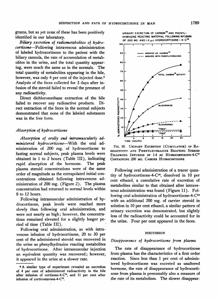
Nature of the hydrocortisone metabolites in theurine-Determination of the nature of the urinarymetabolites excreted following intravenous adminis-tration of 200 mg. of hydrocortisone labeled withhydrocortisone-4-CI4 yielded the following results(Figure 10): Direct dichloromethane extraction ofthe fresh untreated urine recovered 4 per cent ofthe infused radioactivity, and 3 per cent of the ad-ministered steroid as measured with phenylhydra-zine. However, following glucuronidase hydroly-sis and dichloromethane extraction of the urine,50 per cent of the administered radioactivity 4 was
recovered, but only 17 per cent as measured byphenylhydrazine analysis. An additional 15 per
.cent of the administered radioactivity could be ex-
tracted with dichloromethane following hydroly-sis with boiling 15 per cent hydrochloric acid for
4An additional 2 to 3 per cent more radioactivity can
be obtained by reextracting the urine residue with an ad-ditional 8 to 9 volumes of dichloromethane. Furthertreatment of the urine residue with glucuronidase andincubating for an additional 48 hours does not result inthe release of an additional amount of radioactivity.Also, treatment of the urine residue with mammalianP-glucuronidase (Viobin 1000 units per ml. urine, or
Ketodase 500 units per ml. urine) at pH 4.8, 37° C. for5 additional days, did not release any more radioactivemetabolites extractable into dichloromethane.
15 minutes. Direct extraction of the urine with2 or 10 volumes of n-butanol saturated with wa-
ter (pH 2.0) removed 80 per cent of the infusedradioactivity. Treatment of the residual urinewith either glucuronidase or boiling 15 per cent
hydrochloric acid, and re-extracting with dichloro-methane failed to release any additional radio-activity.
Some information regarding the identity ofmetabolites comprising the free and conjugatedfractions has been obtained. By paper chromatog-raphy, the direct dichloromethane extract was
shown to contain several labeled products, con-
sisting of hydrocortisone (equivalent to less thanone-fourth the total of labeled metabolites of thisfraction, or less than 1 per cent of the administereddose), 20f8-hydroxyhydrocortisone, 6,8-hydroxy-hydrocortisone, tetrahydrohydrocortisone, andother unidentified products.5
By similar techniques, the metabolites releasedby ,8-glucuronidase hydrolysis and reacting withphenylhydrazine to form a yellow color were
shown to consist predominantly of tetrahydrocor-tisone and tetrahydrohydrocortisone. The cor-
responding dihydro-derivatives were detected onlyin small amounts. Evidence for the presence ofseveral additional more polar metabolites was alsoobtained by radioautography of these chromato-
5These results have been based on a comparison of therates of migration with the authentic pure compounds, byuse of mixed chromatograms, and by conversion of un-
known steroids to the acetates and comparing their chro-matographic behavior with that of the authentic com-
pounds. These substances also gave the appropriate re-
actions with phenylhydrazine and with blue tetrazolium,the appropriate sulfuric acid fluorescence results, and thecharacteristic spectral curves in sulfuric acid.
TABLE III
Plasma levels phenylhydrazine reacting steroid after oral and intramuscular administration of 200 mg. hydrocortisone *
Hours following administration
0 0.5 1 2 3 .4 8 12 24
T. R. p.o. 17 115 220 148 107 91 44 22 15i.m. 19 42 57 55 47 37 24
V. L. p.o. 17 96 160 92 55 18I.m. 16 33 37 30 27 21 15
T. L. p.o. 25 100 150 190 120 70 25 23i.m. 25 26 40 56 56 46 35 28
R. H. p.o. 13 100 260 255 190C. S. p.o. 16 70 100 140 200 130W. R. p.o. 15 180 260 180 86 18
* Values are expressed as micrograms per 100 ml, plasma,
1788
DISPOSITION AND FATE OF HYDROCORTISONEIN MAN
grams, but as yet none of these has been positivelyidentified in our laboratory.
Biliary excretion of radiometabolites of hydro-cortisone-Following intravenous administrationof labeled hydrocortisone to the patient with thebiliary cannula, the rate of accumulation of metab-olites in the urine, and the total quantity appear-ing, were much the same as in the normals. Thetotal quantity of metabolites appearing in the bile,however, was only 4 per cent of the injected dose.6Analysis of the feces collected for 3 days after in-fusion of the steroid failed to reveal the presence ofany radioactivity.
Direct dichloromethane extraction of the bilefailed to recover any radioactive products. Di-rect extraction of the feces in the normal subjectsdemonstrated that none of the labeled substanceswas in the free form.
Absorption of hydrocortisone
Absorption of orally and intramuscularly ad-ministered hydrocortisone-With the oral ad-ministration of 200 mg. of hydrocortisone tofasting normal subjects, peak plasma levels wereobtained in 1 to 2 hours (Table III), indicatingrapid absorption of the hormone. The peakplasma steroid concentrations were of the sameorder of magnitude as the extrapolated initial con-centrations obtained following intravenous ad-ministration of 200 mg. (Figure 2). The plasmaconcentration had returned to normal levels within8 to 12 hours.
Following intramuscular administration of hy-drocortisone, peak levels were reached moreslowly than following oral administration, andwere not nearly as high; however, the concentra-tions remained elevated for a slightly longer pe-riod of time (Table III).
Following oral administration, as with intra-venous infusion of hydrocortisone, 20 to 30 percent of the administered steroid was recovered inthe urine as phenylhydrazine reacting metabolitesof hydrocortisone. After intramuscular injectionan equivalent quantity was recovered; however,it appeared in the urine at a slower rate.
sA similar type of experiment revealed an excretionof 4 per cent of administered radioactivity in the bileafter infusion of cortisone-4-C14, and 11 per cent afterinfusion of corticosterone-4-C14.
URINARY EXCRETION OF CARBON14AND PHENYL-HYDRAZINE REACTING MATERIAL FOLLOWINGINFUSIONOF 200 MG. AND 1.4 Fc HYDROCORTISONE-4-C14
100 II I
90
Il- 40
4
1-
o 30
4t
20w 20
I.-:
U,Z 10ia4t
TIME (HOURS)
FIG. 10. URINARY EXCRETION (CUMULATIVE) OF RA-DIOACTIVITY AND PHENYLHYDRAZINEREACTING STERoIDFOLLOWING INFUSION OF 1.4 iLc HYDROCORTISONE4-C'CONTAINING 200 MG. CARRIER HYDROCORTISONE
Following oral administration of a tracer quan-tity of hydrocortisone-4-C14, dissolved in 10 percent ethanol, a cumulative rate of excretion ofmetabolites similar to that obtained after intrave-nous administration was found (Figure 11). Fol-lowing oral administration of hydrocortisone-4-C"with an additional 200 mg. of carrier steroid insolution in 10 per cent ethanol, a similar pattern ofurinary excretion was demonstrated, but slightlyless of the radioactivity could be accounted for inthe urine. Four per cent appeared in the feces.
DISCUSSION
Disappearance of hydrocortisone from plasma
The rate of disappearance of hydrocortisonefrom plasma has the characteristics of a first orderreaction. Since less than 1 per cent of adminis-tered hydrocortisone was excreted as unalteredhormone, the rate of disappearance of hydrocorti-sone from plasma is presumably also a measure ofthe rate of its metabolism. The slower disappear-
1789

PETERSON, WYNGAARDEN,GUERRA, BRODIE, AND BUNIM
COMPARATIVERATES CUMULATIVE URINARY EXCRETIONOF VARIOUS DOSAGESORALLY AND INTRAVENOUSLY
ADMINISTERED C14 HYDROCORTISONE
80
I-
ct T0a
a: 600wcr
50
z
E 40
Z 30w
wa-. 20
24 32 40 48
TIME (HOURS)
FIG. 11. COMPARATIVERATES OF URINARY EXCRETION(CUMULATIVE) OF RADIOMETABOLITESFOLLOWINGINTRA-VENOUSOR ORAL ADMINISTRATION OF HYDROCORTISONE-4-Ct4
ance of infused hydrocortisone in patients withliver disease, also noted by Brown, Willardson,Samuels, and Tyler (12), is of particular interest.Since this defect is demonstrable in patients withrelatively minor liver dysfunction, a test of a sub-ject's ability to handle infused hydrocortisone may
serve as a sensitive indicator of hepatic function.In most cirrhotic patients studied, hydrocorti-
sone was the only steroid for which a striking de-fect in rate of metabolism was demonstrable.Tomkins and Isselbacher (13, 14) have describeda specific triphosphopyridine nucleotide-linkedenzyme present in rat liver catalyzing the reduc-tion of the A4 bond of hydrocortisone, but with-out effect on the M4 bond of other steroids. Theproduct of hydrocortisone reduction is dihydro-hydrocortisone, which is then further reduced bya second less specific enzyme (3-hydroxysteroid.dehydrogenase [14] ) to tetrahydrohydrocorti-sone. The first reaction is rate controlling, so
that the intermediate product does not normallyaccumulate. Moreover, the dihydro-derivatives
do not appear in urine of man to any significantextent (15). These findings suggest that therate-controlling step in the removal of hydrocor-tisone from plasma may be the reduction of the Aring of the hormone in the liver, and that it is theenzyme catalyzing this transformation that is de-ficient in hepatic diseases of the types studied. Afinding consistent with this thesis is the observa-tion that tetrahydrocortisone disappears fromplasma at a normal rate in patients with liverdisease (12) (Figure 5).
The ameliorating effects of jaundice (16) uponactive rheumatoid arthritis may well be related toa defect in biological transformation of endogenoushydrocortisone resulting from liver dysfunction.The rate of disappearance of hydrocortisone wasclearly delayed in only one of eight rheumatoidarthritics studied.
In two of three patients with thyrotoxicosis, therate of disappearance of infused hydrocortisonewas increased. In two of three with myxedema,hydrocortisone disappearance was delayed. Sincesimilar disturbances were noted after infusion ofcortisone, these alterations presumably resultfrom general disturbances of rates of metabolicprocesses associated with alterations of thyroidfunction.
In adrenal insufficiency, both primary and sec-ondary, the rates of disappearance of hydrocorti-sone were normal. Also, in patients who had pre-viously received large doses of hydrocortisone forlong periods, the disappearance of infused hydro-cortisone was normal. Hellman, Bradlow, Ades-man, Fukushima, Kulp, and Gallagher (17) havefound the rate of metabolism of hydrocortisone-4-C14 normal in adrenal insufficiency as judged byrates and extents of urinary excretion of the la-beled metabolites. Apparently hepatic processeswhich metabolized hydrocortisone proceed withoutconsideration of the peripheral requirements forhormone, being governed by the concentration ofhydrocortisone in the circulating plasma, and bythe level of enzyme activity in the liver.
Distribution of hydrocortisone in body fluids
The value of the apparent volume of distribu-tion of hydrocortisone depends upon the basis se-
lected for its calculation. Thus, if one utilizes theconcentration of hydrocortisone in plasma at zero
0.740 MGC14 Hydrocortisone [2.8Lc]200 MG. C rHydrocort,Isone [1.4 j c] J
* 0.249 MG. C 14Hydrocortisone [1.7 H±c3oroiA 200 MG.C'4Hydrocortisone [1.6Lc] orol
1790

DISPOSITION AND FATE OF HYDROCORTISONEIN MAN
time as obtained from an extrapolation of thesemilogarithmic plot of plasma hydrocortisoneconcentrations (Figure 2), considered togetherwith the quantity of hydrocortisone injected, anapparent volume of distribution of 50 to 90 literscan be calculated.7 (Average = 70 liters 8 in 20normal and 12 cirrhotic subjects given 50 to 500mg. of hydrocortisone intravenously.)
In the strictest sense these calculations meanthat at the moment of complete mixing it wouldtake a volume of 70 liters to contain all the in-jected hydrocortisone in the concentration at whichit exists in plasma. Since the figures obtained arein all cases close to total body weight, it followsthat hydrocortisone is concentrated in some por-tions of the body at much higher levels than arepresent in plasma. Tissue localization has beendemonstrated to occur in the red cell and in thenon-diffusible elements of plasma, and probablyoccurs in other tissues as well.
When the hydrocortisone space is calculatedfrom the value for the miscible pool as determinedby conventional treatment of serial specific ac-tivity values of circulating hydrocortisone (18),and the plasma level of phenylhydrazine reactingsteroid (largely hydrocortisone [11]), hydrocor-tisone spaces of 8 to 16 liters (12 subjects) areobtained. The method for determination of themiscible pool depends upon knowledge of the ini-tial concentration of isotopic hydrocortisone inplasma. The addition of new unlabeled hormonewill dilute the pool and change its isotope con-centration; the removal of hormone from thepool will not. Therefore, only that hydrocorti-sone capable of mixing with circulating hydro-cortisone will be determined by this method.
The discrepancy between values obtained bythese two methods is at least in part attributableto the fact that in the estimation of a volume ofdistribution, equilibration of solute is assumed
7 Similar calculations made on the basis of the ex-trapolated value for the concentration of dichloromethaneextractable radioactive substances present in plasma atzero time, after infusion of tracer quantities of hydro-cortisone-4-C'4 can have no significance, since it has beenshown that a large fraction of these substances is nothydrocortisone.
8This figure of 70 liters is based on the concentrationsof free plus plasma protein bound steroid. By employingthe free steroid concentration for the calculation, an ap-parent volume of 280 liters is obtained.
to have occurred prior to any irreversible trans-formation or excretion of the test substance. Thisassumption cannot be valid in the case of hydro-cortisone which is known to be metabolized rap-idly. The hydrocortisone which is enitering suchirreversible fates undoubtedly makes a larger con-tribution to the volume of distribution when meas-ured from hydrocortisone disappearance data thanwhen measured from the isotope dilution data.The value arrived at from isotope dilution data,8 to 16 liters, is perhaps a fairly accurate indica-tion of the volume of water required to contain themiscible hydrocortisone at the concenitration atwhich it occurs in plasma.
Excretory pathways of wietabolites of hydrocorti-sone
The major route of excretion of labeled metabo-lites of hydrocortisone was via the urine, in which86 to 94 per cent of the administered carbon14 ap-peared in 72 hours. These figures are in goodagreement with those of Hellman and his associ-ates (17) and Plager, Tyler, Hecht, and Samuels(19). Less than one per cent of the administeredhormone was excreted as free hydrocortisone.These data suggest that no accumulation of hy-drocortisone within the body would result fromrenal failure such as might be anticipated fromhepatic failure.
Only a minor fraction, about 3 per cent, of theadministered isotope was recovered in feces. Thisvalue is somew-hat lower than those reported byHellman (17) and by Plager and their coworkers(19). The quantity of labeled products appear-ing in feces was only slightly less than that ex-creted in bile, suggesting that the bile is the ma-jor and perhaps the sole source of fecal products.Furthermore, it appears unlikely that appreciableintestinal reabsorption of biliary metabolites ofhydrocortisone occurs. These data are in strikingcontrast with those obtained in rats and guineapigs in which 60 to 80 per cent of the administeredhydrocortisone-4-C14 appeared in biliary metabo-lites, much of it destined subsequently to partici-pate in an active entero-hepatic circulation in bothspecies (20). These findings are also differentfrom those obtained with progesterone-4-C14 inman, where 60 per cent of the radiometabolitesappeared in bile (21).
1791

PETERSON, WYNGAARDEN,GUERRA, BRODIE, AND BUNIM
Hellman and his coworkers (17) have reportedthat less than 0.5 per cent of administered hydro-cortisone appeared in the expired air as carbon14dioxide. Similar results have been obtained in theguinea pig and rat (20). These findings indicatethat the biotransformation of hydrocortisone is notassociated with appreciable degradation of the ringstructure of the steroid.
Excretion of inetabolites in urine
Previous studies have shown that about 20 to 30per cent of an administered dose of hydrocortisoneis excreted as metabolites that are extractable withbutanol (22), or become extractable with chloro-form or similar solvents following p-glucuronidasehydrolysis (23, 24), and react wN-ith phenylhydra-zine. Small additional quantities are detectableas 17-ketosteroids, but the sum of all classes ofurinary metabolites recognizable by these variousmethods is less than 40 per cent of the adminis-tered dose.
In the present study, recoveries of metabolitesin urine, as determined by the phenylhydrazinemethod after glucuronide hydrolysis agreed wellwith these earlier reports. However, both in thefreely extractable and the glucuronidase hydro-lyzed fractions, the percentage of the administeredhormone detected by radioactivity analysis wasconsiderably larger than that determined by colori-metric assay. The presence of 20,B-hydroxyhydro-cortisone as a labeled metabolite of hy-drocortisone-C14 offers a likely explanation for the discrepancybetween results of carbon14 and phenylhvdrazineanalyses of the freely extractable fraction sincethis substance does not react with the phenylhy-drazine reagent.
A similar explanation probably applies to thedisparity between carbon14 and phenylhydrazineanalyses of the p-glucuronidase-hydrolyzable frac-tion of conjugated metabolites. Metabolites equiv-alent to 50 to 60 per cent of administered hormonewere released by enzymatic hydrolysis as deter-mined by radioactivity analysis whereas this frac-tion was 20 to 30 per cent of infused steroid by thephenylhydrazine reaction. Recently, four new
urinary metabolites of adrenal cortical hormonehave been identified (25). These are derivativesof tetrahydrohydrocortisone and tetrahydrocorti-sone possessing a 20-ol group in either a or 8 con-
figuration, apparently present as glucuronides,presumably conjugated at the 3-ol group of eachmetabolite. These metabolites accounted forabout 30 per cent of the neutral extractable steroidproducts found after the administration of labeledhydrocortisone. These 20-ol derivatives wouldnot react with phenylhydrazine and their presencein urine may well explain the major portion ofthe difference (equivalent to about 30 per cent ofthe administered hormone) encountered betweenthe results of the two analytical methods em-ployed in this study.
An additional fraction of 10 to 20 per cent ofthe infused isotope could be extracted with n-bu-tanol saturated with water, or following hydroly-sis with boiling hydrochloric acid followed by di-chloromethane extraction. Whether this frac-tion represents glucuronides resisting hydrolysiswith bacterial enzyme. or sulfate or other conju-gation products, remains to be determined, as doesthe nature of the metabolites in the residue com-pletely resisting extraction.
Expressed in terms of the total radioactivity inthe urine excreted during the first 24 hours fol-lowing infusion of hydrocortisone-4-C14, 4 percent is extracted directly with dichloromethane.An additional 60 to 70 per cent is released follow-ing glucuronidase hydrolIysis and dichloromlethaneextraction, and an additional 15 per cent follow-ing either n-butanol extraction or dichloromethaneextraction after treatment with boiling 15 percent hydrochloric acid. This accounts for a totalof about 85 per cent of the radioactivity presentin the urine and leaves approximately 15 per centunextractable.
Oral absorption
The presence of comparable peak plasma levelsafter oral and intravenous administration of hy-drocortisone, and the similarities of recoveries andrates of excretion of metabolites, suggest that oralabsorption of the hormone is both rapid and comii-plete. The fact that the radiometabolites ap-peared in the urine did not of course exclude thepossibility that some of the orally administeredsteroid was metabolically transformed in the gas-trointestinal tract. However, studies carried out
on the stability of hydrocortisone-4-C14 in fluidsfrom various parts of the gastrointestinal tract
1 792

DISPOSITION AND FATE OF HYDROCORTISONEIN MAN
(26) have shown that in gastric juice (pH 1.0)and bile (pH 7.0) no transformation takes placefor at least 36 hours (370 C.). In feces, how-ever, after 4 hours all hydrocortisone had beenmetabolized.
SUMMARY
The physiological disposition and metabolicfate of hydrocortisone have been studied in manfollowing intravenous infusion of hormone indoses up to 500 mg. In normal subjects the half-life of infused hydrocortisone in plasma averaged1.9 hours. Following the infusion of tracer quan-tities of hydrocortisone-4-C14, labeled conjugatedmetabolites appeared in plasma, and at 2 hourstheir concentration was equal to that of labeledunconjugated substances. Normal subjects ex-creted over 90 per cent of the administered hor-mone as urinary metabolites in 72 hours, as judgedfrom excretion of carbon14 following infusion oftracer or labeled larger quantities of hormone.Feces of three normal subjects contained 2 to 4per cent of the administered label, and in one sub-
100- C4 HYDROCORTISONE1. V.
ject with a biliary catheter, 4 per cent of the in-fused label appeared in the bile. About 4 per centof the administered hormone was excreted inurine as unconjugated substances; unchanged hy-drocortisone represented less than 1 per cent ofthe administered hormone. The routes of hydro-cortisone disposition in man are schematically il-lustrated in Figure 12. About 60 per cent of theadministered hydrocortisone was excreted as me-tabolites hydrolyzable with bacterial f8-glucuroni-dase, although only about half of this fraction was(letectable by the phenylhydrazine reaction.
In patients with liver disease disappearance ofinfused hydrocortisone was slower than normal.There was an associated increase in the fraction ofinfused hormone excreted in unconjugated form,and also a slower appearance of conjugated metab-olites in urine. The defect in metabolism of hydro-cortisone in patients with liver disease was dis-cussed in terms of a low hepatic activity of thespecific enzyme catalyzing the reduction of the A4bond of hydrocortisone.
The rates of disappearance of hydrocortisone
<I
___- I:~39 .
KIDNEY
3
FfREE-4*CONJUGATE-87
ROUTES OF HYDROCORTISONEDISPOSITION IN MANF-IG. 12. ROUTESOF DISPOSITION OF HYDROCORTISONEIN MANAFTER INTRAVENOUS
ADMINISTRATION OF "100 UNITS" HYDROCORTISONE-4-C"Urinary excretion of unchanged hydrocortisone; --- Urinary excretion of
hydrocortisone metabolized by the liver but not conjugated; Urinary andbiliary excretion of conjugated metabolites of hydrocortisone.
LIVER
4
1793

PETERSON, WYNGAARDEN,GUERRA, BRODIE, AND BUNIM
from plasma were increased in two of three pa-tients with moderately severe thyrotoxicosis. andslowed in two of three with imyxedema. Disap-pearance rates were normal in three subjects withhypoadrenalcorticisimi.
The absorption of tracer quantities of hydro-cortisone from the gastrointestinal tract wvas shownto be rapid and comiiplete. No destruction of hy-drocortisone was detected after incubation of la-beled hormone with gastric juice, bile, or serum,but extensive destruiction occturred (luring inctuba-tion with feces.
ACKNOWLEDGMENT
We wish to express our gratitude to Charles Pierce,George Senigstack, and Margaret Bollier for valuabletechnical assistance, and to Upjohn, Schering, and Merckpharmaceutical companies for generous supplies ofsteroids.
REFERENCES
1. Silber, R. H., and Porter, C. C., The determinationof 17,21-dihydroxy-20-ketosteroids in urine andplasma. J. Biol. Chem., 1954, 210, 923.
2. Bush, I. E., Methods of paper chromatography ofsteroids applicable to the study of steroids in mam-malian blood and tissues. Biochem. J., 1952, 50,370.
3. Sweat, M. L., Sulfuric acid-induced fluorescence ofcorticosteroids. Anal. Chem., 1954, 26, 773.
4. Robinson, C. V., \Vindowless, flow type, proportionalcounter for counting C". Science, 1950, 112, 198.
5. Heard, R. D. H., Thompson, L., and Yates, C., Adifferential metlhod for counting C"4 in biologicaltissues and fluids. Rev. canad. de biol., 1952, 11,66.
6. Peters, J. H., and Gutmanni, H. R., Micrometlhod formeasurement of carbon-14-labeled material. Anal.Chem., 1953, 25, 987.
7. Van Slyke, D. D., Plazin, J., and Weisiger, J. R.,Reagents for the Van Slyke-Folch wet carboncombustion. J. Biol. Chem., 1951, 191, 299.
8. Peterson, R. E., The physiological disposition andmetabolism of cortisone in man. (In preparation.)
9. Savard, K., Kolff, W. J., and Corcoran, A. C., Cor-ticosteroids of peripheral blood. Endocrinology,1952, 50, 366.
10. Bongiovanni, A. M., Eberlein, W. R., Grumbach,M. M., VanWyk, J. J., and Clayton, G., Conju-gates of adrenal corticoids in human plasma. Proc.Soc. Exper. Biol. & Med., 1954, 87, 282.
11. Peterson, R. E., A method for the determinationof hydrocortisone in plasma, and an evaluation ofits specificity. (In preparation.)
12. Brown, H., \Villardson, D. G., Samuels, L. T., anidTyler, F. H.., 17-hlydroxycorticosteroid metabo-lism- in liver disease. J. Clin. Invest., 1954, 33,1524.
13. Tomkins, G., and Isselbacher, K. J., Enzymatic re-ductioni of cortisone. J. Am. Chemii. Soc., 1954,76, 3100.
14. Tomkins, G., A mammalian 3-hydroxysteroid de-hydrogenase. J. Biol. Chenm., In press.
15. Schneider, J. J., Further isolation of adrenocorticalcompounds from male urine. J. Biol. Chem.. 1952,194, 337.
16. Hench, P. S., Effect of jaundice on chronic infectious(atrophic) arthritis and on primary fibrositis.Arch. IJt. Mled., 1938, 61, 451.
17. Hellman, L., Bradlow, H. L., Adesmanl, J., Fnku-shima, D. K., Kulp, J. L., and Gallagher, T. F.,The fate of hydrocortisone-4-C" in man. J. Clin.Invest., 1954, 33, 1106.
18. Peterson, R. E., and \V-yngaardeni, J. B., The misciblepool and turnover rate of endogenious hydrocorti-sone in man. (In preparation.)
19. Plager, J. E., Tyler, F. H., Hechit, H. H., anid Sam-uels, L. T., 'Metabolism and excretion of intrave-n1ously administered 4-C'4-17-hydrox-corticosterone.J. Clin. Endocrinol. & Metab., 1954, 14, 780.
20. \Vyngaardeni, J. B., Peterson, R. E., and Wolff, A. R.,Physiologic disposition of radiometabolites of hy-drocortisone-4-C" in the rat and guinea pig. J.Biol. Chem., 1955, 212, 963.
21. Wiest, WV. G., Fujimoto, G. I., and Sandberg, A. A.,Metabolism of progesterone-4-C" in man. Federa-tion Proc., 1955, 14, 304.
22. Reddy, WV'. J., Jenkinis, D., and Thorn, G. WV., Esti-mationi of 17-hydroxycorticoids in urine. Metabo-lismii, 1952, 1, 511.
23. Sandberg, A. A., Nelson, D. H., Glenn, F. 'M., Tyler,F. H., and Samuels, L. T., 17-hydroxycorticoster-oids and 17-ketosteroids in urine of human sub-jects: Clinical application of a method employing/-glucuronidase hydrolysis. J. Clin. Endocriniol. &Metab., 1953, 13, 1445.
24. Vestergaard, P., Investigationis oni the estimationi of17-hydroxycorticoids in urine using the Porter/Silber reactioin. Acta endocrinol., 1953, 13, 241.
25. Fukushima, D. K., Leeds, N. S., Bradlow, H. L.,Kritchevsky, T. H., Stokem, M. B., and Gallagher,T. F., The characterization of four new metabo-lites of adrenocortical hormones. J. Biol. Chem.,1955, 212, 449.
26. Petersoin, R. E., Unpublished data.
1794