hyaluronan mixed esters of butyric and … molecola.pdf · hyaluronan mixed esters of butyric and...
TRANSCRIPT
1
HYALURONAN MIXED ESTERS OF BUTYRIC AND RETINOIC ACIDAFFORDING MYOCARDIAL SURVIVAL AND REPAIR
WITHOUT STEM CELL TRANSPLANTATION*Vincenzo Lionetti,1,3,8 Silvia Cantoni,2,8 Claudia Cavallini,2 Francesca Bianchi,2 Sabrina Valente,7
Irene Frascari,2 Elena Olivi,2 Giovanni D. Aquaro,3 Francesca Bonavita,2 Ignazio Scarlata,2Margherita Maioli,4 Valentina Vaccari,2 Riccardo Tassinari,2 Antonietta Bartoli,5 Fabio A.
Recchia,1,6 Gianandrea Pasquinelli,7 and Carlo Ventura2
From Sector of Medicine, Scuola Superiore S. Anna, Pisa, Italy1, Laboratory of Molecular Biology andStem Cell Engineering, Cardiovascular Department-National Institute of Biostructures and Biosystems, S.Orsola - Malpighi Hospital, University of Bologna, Bologna, Italy, and Bioscience Institute, Republic ofSan Marino2, Institute of Clinical Physiology, CNR Fondazione G. Monasterio, Pisa, Italy3, Departmentof Biomedical Sciences, University of Sassari, Sassari, Italy4, Department of Physics, University of Pisa,
Pisa, Italy5, Department of Physiology, New York Medical College, Valhalla, NY, USA6, and Departmentof Hematology, Oncology and Clinical Pathology, University of Bologna, Bologna, Italy7
Vincenzo Lionetti and Silvia Cantoni contributed equally to this work8
Running head: Endogenous Cell Therapy with a Synthetic MoleculeAddress correspondence to: Carlo Ventura, MD, PhD, Laboratory of Molecular Biology and Stem CellEngineering, Cardiovascular Department-National Institute of Biostructures and Biosystems, S. Orsola -Malpighi Hospital, University of Bologna, Via Massarenti 9, I-40138 Bologna, Italy. Fax/Tel: +39-051340339. E-mail: [email protected] or [email protected]
Possible cardiac repair by adult stemcell transplantation is currently hampered bypoor cell viability and delivery efficiency,uncertain differentiating fate in vivo, the needsfor ex vivo cell expansion, and consequent delayin transplantation after the onset of heartattack. By the aid of Magnetic ResonanceImaging, Positron Emission TomographyImaging, and immunohistochemistry, we showthat injection of a hyaluronan mixed ester ofbutyric and retinoic acid (HBR) into infarctedrat hearts afforded substantial cardiovascularrepair and recover of myocardial performance.HBR restored cardiac 18F-FDG uptake,increased capillary density and led to therecruitment of endogenous Stro-1-positive stemcells. TUNEL assay demonstrated that HBR-treated hearts exhibited a decrease in thenumber of apoptotic cardiomyocytes. Inisolated rat cardiomyocytes and Stro-1 stemcells HBR enhanced the transcription of VEGF,HGF, KDR, Akt, and Pim-1. HBR alsoincreased the secretion of VEGF and HGF,suggesting that the mixed ester may haverecruited both myocardial and Stro-1 cells intoa pro-angiogenic paracrine circuitry of cardiacrepair. An increase in capillarogenesis wasinduced in vitro with medium obtained fromHBR-exposed cells. In the infarcted heart
tissue, HBR injection increased histone H4acetylation, as compared with non-injectedsamples. Acetyl-H4 immunoreactivity was alsohigher in rat cardiomyocytes and Stro-1 cellsexposed to HBR, compared to untreated cells.
In conclusion, efficient amelioration ofcardiac function can be afforded by HBRwithout the need of stem cell transplantation orvector-mediated gene delivery. The currentfindings may pave new perspectives inregenerative medicine.
Cardiomyocyte loss during myocardialinfarction (MI) is associated with dysfunction ofunderperfused myocardium, eventuallyprogressing toward heart failure.
Analysis of the rescuing potentialassociated with transplantation of humanmesenchymal stem cells (hMSCs) in animalmodels of MI has recently led to the conclusionthat paracrine actions exerted by adult stem cellsthrough the release of soluble factors might beimportant mechanisms of tissue repair andfunctional improvement (1,2). To this end, wehave recently shown that hMSCs isolated fromfetal membranes of human term placenta(FMhMSCs) secreted large amounts ofangiogenic, mitogenic, antiapoptotic andantifibrotic factors, as compared with hMSCs
http://www.jbc.org/cgi/doi/10.1074/jbc.M109.087254The latest version is at JBC Papers in Press. Published on January 22, 2010 as Manuscript M109.087254
Copyright 2010 by The American Society for Biochemistry and Molecular Biology, Inc.
by guest, on January 23, 2010w
ww
.jbc.orgD
ownloaded from

2
isolated from the human bone marrow,significantly contributing to improvedcardiovascular function in infarcted rat hearts (3).Noteworthy, ex vivo preconditioning of FMhMSCswith a mixed ester of hyaluronan with butyric andretinoic acid (HBR) acted transcriptionally toincrease both the commitment to cardiovascularlineages and the secretion of trophic mediators,remarkably enhancing stem cell-mediatedimprovement in vivo (3).
Here, we directly injected HBR into themyocardium of infarcted rat hearts, and provideevidence that the mixed ester afforded substantialrecover of myocardial performance without theneed of stem cell transplantation. The HBR actionwas also associated with an increase in the numberof Stro-1-positive cells within the injectedmyocardium. These responses likely involved theactivation of a gene program of paracrinepatterning for myocardial protection andangiogenesis, and the enhanced survival of locallyrecruited stem cells.
Experimental Procedures
Synthesis of HBR- The procedure for the synthesisand characterization of HBR, and the relatedchemical structure are reported in detail elsewhere(4). The primary hydroxyl group in position 6 ofthe N-acetyl-D-glucosamine residues in thepolysaccharide backbone is the most reactivetowards esterification. Briefly, we prepared adouble salt of tetrabutylammonium with twofunctional groups of hyaluronan, specifically itscarboxyl and 6-hydroxyl, in order to achieve goodsolubility in polar aprotic organic solvents and toincrease the nucleophilicity of the oxygen atom atC-6. Retinoylation with retinoyl chloride, which isthe rate-limiting step, was carried out beforebutyrylation by means of butyric anhydride and 4-(dimethylamino)pyridine as a hypernucleophilicacylation catalyst (4). The degree of substitution(DS) was considered as the number of theesterified OH groups for each repeating unit ofhyaluronic acid (GlcNAc-GlcUA dimer). Theweight-average molecular weight (Mw) of HBR,referred to as the Mw of sodium hyaluronate, wasdetermined by high-performance size-exclusionchromatography (HP-SEC) (4).Myocardial Infarction (MI)- Studies wereperformed on male Winstar rats (n = 30, 250 - 300
g in size). Animals were sedated (xylazine 14mg/kg, i.p.), anesthetised (Zoletil100, 40 mg/kg,i.p), ventilated with a mixture of air and oxygen(1:1) and MI was induced as previously described(3). Briefly, a thoracotomy was performed in theleft fourth intercostal space and a permanentsurgical ligation was placed around the leftanterior descending coronary artery near its originwith a 6-0 silk suture, during electrocardiographic(ECG) monitoring for ST changes andarrhythmias. Chest was closed in layers andpneumothorax was reduced. Experimentalprotocols were approved by the Animal CareCommittee of the Italian Ministry of Health, inaccordance with the Italian law (DL-116, Jan. 27,1992).
Healthy animals were randomly dividedinto three experimental groups: 1) MI treated with100 µl of phosphate buffer solution (PBS) asvehicle, (Control group, MI + PBS, n = 10), 2) MItreated with 100 µl of HBR solution (0.2 mg ofHBR / 100 g of rat weight) (Treated group, MI +HBR, n = 10), 3) Sham-operated rats (SHAM, n =10), in which left anterior descending coronaryartery was not occluded. The sterile solutions wereinjected into the viable myocardium bordering theinfarct zone and the infarcted site 45 minutes afterthe coronary ligation by a syringe with a needle of24 Gauge. The infarct zone was identified by thepale colour of the myocardium. Small-animalPositron Emission Tomography (mPET) and 1.5 TMagnetic Resonance Imaging (MRI) wereperformed four weeks after coronary ligation.Functional Assessment- Regional left ventricular(LV) myocardial glucose uptake was measured bymPET to assess oxidative metabolism, whereasregional contractility and infarct size werequantified by conventional MRI. For both types ofimaging, we used three cross-sectional planes, i.e.,basal, middle, apical, and six circumferentialregions, i.e., anterior, anterior-lateral, inferolateral,inferior, inferoseptal, anterior-septal. LV regionswere selected as previously described (5). Briefly,the infarcted area comprised segments with morethan 25% of its area occupied by scar tissue andthe border zone comprised segments containingless than 25% of scar tissue area and wasimmediately contiguous (either circumferentiallyor longitudinally) to the infarcted area. The remotesegments that did not contain scar tissue were
by guest, on January 23, 2010w
ww
.jbc.orgD
ownloaded from

3
those located outside the border zone. To assess therelationship between in vivo measurements ofmyocardial contractility and metabolism, mPETwas performed within 24 - 36 hours after MRI.MRI measurements- MRI protocol was performedwith a 1.5 T clinical whole body scanner (SignaCVI, GE Medical Systems) by using a phasedarray send-receive coil for a human knee (knee PAcoil), as previously described (6). An ECG-triggered SSFP (FIESTA) pulse sequence wasacquired to assess LV function (parameters: 200mm of field of view, 3 mm slice thickness, no gap,5 NEX, 2 views per segment, TE/TR 1.6/3.2, flipangle 45°, matrix 192 x 192, and reconstructionmatrix 256 x 256). In each rodent, a total of 4 LVshort-axis (to completely cover ventricular mainaxis in end-diastole) and 2 LV long-axis views(vertical and horizontal long axis) were acquired.For each view, 10 cine-frames were acquired. Fordetection of non-viable myocardium, whichappears hyper-intense, and for assessment of LVinfarct size, delayed enhancement images wereobtained 3 minutes after bolus injection ofGadobutrol (Gadovist®; 0.02 mmol/100 grams ofbody weight) via tail vein; images were acquiredin the same short-axis and long-axis slices as usedfor cine MRI. An ECG-triggered fast GradientEcho Inversion Recovery pulse sequence wasutilized with the following parameters: TR 4.2milliseconds, TE minimum, flip angle 20°, matrix192 x 192, NEX 5, FOV 20-20 mm, slicethickness 3 mm, no inter-slice gap, 1 R-R interval.The inversion time was fixed to 100 millisecondsand eventually optimized to null signal from thenormal myocardium when appropriate.Image processing- To assess the infarct size, theextent of delayed-enhanced areas was measuredusing a semi-automatic software, previouslyvalidated by us (7). For this purpose, we used allshort-axis images and two additional long-axisimages for the analysis of the cardiac apex. In eachimage, the boundaries of contrast-enhanced areaswere traced and manually corrected when needed.Contrast-enhanced regions, namely the infarctedregions, were expressed in grams, as well as inpercent of the entire left ventricle. Left ventricleend-diastolic and end-systolic volumes, LV mass,and ejection fraction were measured from the cine-images using a previously validated software(Mass®, MEDIS).
The infarcted region was detected in cine-images by comparison with the respective delayedenhancement image. Then, regional end-diastolicand end-systolic wall thickness was measured inthe core of infarcted area, in the border regionsand in the remote myocardium. Regionalcontractility was assessed by employing the end-systolic wall thickening in three short-axissegments (basal, middle, and apical) forcorrelations with matched PET slices. Absoluteregional wall thickening was calculated in thesame regions by the difference between end-systolic and end-diastolic wall thickness. Relativeregional wall thickening was calculated with theformula: end-systolic/end-diastolic wall thicknessx 100, and expressed as percentage.PET measurements- Each sedated rat was placedon a scanner bed in prone position and received anintravenous injection of [F-18]fluorodeoxyglucose(18F-FDG, 45 MBq) via the tail vein in a 0.15 mlvolume. The residual dose in the syringe wasmeasured to verify the effective injected dose. Theheart was centered on the field of view. PET wasperformed with a Small Animal PET tomograph(GE eXplore Vista DR; GE Healthcare) (8). Thedynamic list mode acquisition was immediatelystarted for a total time of 30 minutes. Once scanwas finished, animals were placed in a recoverybox with warm temperature until completerecovery. All images were reconstructed withiterative reconstruction on OSEM 2D (Siemens),and visualized frame by frame with a dedicatedsoftware in three planes (axial, sagittal, andcoronal), as previously described (9). To performviability analysis in each experimental condition,dynamic scans were reformatted by adding allframes of the last 10 minutes of acquisition inorder to obtain a static acquisition image. Semi-quantitative analysis of regional LV 18F-FDGuptake was performed with a specific softwareadapted for animals (ECTB) (9,10), using restingscores of 0 - 4: 0 = normal, 1 = equivocal, 2 =moderately reduced, 3 = severely reduced, 4 =absent.Tissue Immunohistochemistry- Hearts werearrested in diastole and 3-mm-thick transverseslices were cut through the short axis of bothventricles at midseptal level; macroscopic infarctsize was assessed by nitro blue tetrazoliumstaining; samples were fixed in 10% bufferedformalin, and embedded in paraffin; four µm-thick
by guest, on January 23, 2010w
ww
.jbc.orgD
ownloaded from

4
sections were used for histological,immunohistochemical and immunofluorescenceanalysis. For conventional histopathologicalanalysis, sections were stained with hematoxylin& eosin (H&E). Picro-Mallory trichrome stainingwas used for determining the degree of fibrosis.Images were digitalized through a video camera(JVC 3CCD video camera, KY-F55B) connectedwith a Leitz Diaplan light microscope; originalimages were taken at 10x and analyzed using theImage-Pro Plus® 6 software (Media Cybernetics,Inc.).
Four µm-thick dewaxed sections were usedfor immunohistochemical studies; capillarydensity was assessed by a polyclonal antibodyagainst von Willebrand Factor (vWF); stem cellrecruitment (mesenchymal cells or mononuclearcells) into the infarcted myocardium was assessedusing antibodies directed against Stro-1 and c-kitantigens, respectively; cycling cells wereidentified with an antibody against nucleartranscription factor Ki-67; the accumulation andthe spatial distribution of acetyl-histones wereinvestigated with an antibody directed againstacetyl-histone H4; perivascular cells werecharacterized using antibodies against NG2 andPDGF-Rβ; expression of Vascular EndothelialGrowth Factor (VEGF) was detected with anantibody recognizing all VEGF isoforms.Specimens were deparaffinated with xylene,rehydrated through decreasing concentrations ofethanol, rinsed in distilled water and subjected toan antigen retrieval treatment. Antigens wereunmasked with citrate buffer pH 6.0, at 120°C, 1atm for 21 minutes. After cooling and washing,endogenous peroxidase activity was neutralizedusing a 3% H2O2 solution in methanol absolute for10 minutes at room temperature (RT) in the dark;sect ions were then processed forimmunohistochemistry with a non-biotin-amplified method (NovoLinkTM PolymerDetection System, Novocastra Laboratories Ltd.).After washing with TBS 1X, the slides wereincubated with NovocastraTM Protein Block for 5minutes in a wet chamber to reduce the non-specific binding of primary antibody and polymerreagent, and rinsed twice with TBS 1X. Tissuesections were subsequently stained usingmonoclonal antibodies (mAbs) against Stro-1(1:100, R&D System, Inc.), Ki-67 (1:230, cloneMM1, Novocastra), NG2 (1:100, R&D System,
Inc.), PDGF-Rβ (1:100, R&D System, Inc.) andpolyclonal antibodies (pAbs) against acetyl-histone H4 (1:150, Lys8, Upstate Biotechnology),c-kit (1:200, DakoCytomation), VEGF (1:50,JH121 clone, Abcam), vWF (1:2000,DakoCytomation) in 1% Bovine Serum Albuminin PBS overnight at 4°C. After washing, slideswere incubated for 30 minutes at RT withNovocastraTM Post Primary Block to enhancepenetration of the next polymer reagent, rinsed inTBS 1X and incubated with NovoLinkTM Polymerfor 30 minutes at RT. After washing, the sectionswere exposed to the substrate/chromogen 3,3’-diaminobenzidine (DAB) prepared fromNovocastraTM DAB Chromogen and NovoLinkTM
DAB Substrate Buffer, rinsed in distilled waterand counterstained with Gill’s hematoxylin. Then,samples were dehydrated, coverslipped andviewed in a light microscopy using the Image-ProPlus® program. Negative control was obtained byomitting the primary Abs.
Assessment of apoptotic programmed celldeath by TUNEL (Terminal deoxynucleotidyltransferase-mediated dUTP Nick End-Labeling)assay is described in the Supplemental Data.Immunofluorescence- To investigate the co-expression of different antigens in the same cell, adouble immunofluorescence procedure was used.We aimed to demonstrate whether Stro-1-positivecells co-expressed c-kit, vWF, CD45, and α -sarcomeric actin, respectively. Four µm-thickdewaxed sections were rehydrated with decreasingconcentrations of ethanol, and rinsed in distilledwater. Antigen retrieval was performed asdescribed above. Then, sections were incubatedfor 30 minutes at RT in wet chamber withblocking solution containing sheep serum (1:10) in1% Bovine Serum Albumin to reduce non-specificstaining. Tissue sections were labeled with theStro-1 mAb (1:100, R&D System) at 37°C for 45minutes in a wet chamber. After rinses, slides wereincubated with a Cy3-conjugated sheep anti-mouse secondary antibody (1:1000, Sigma-Aldrich), in 1% BSA in PBS for 45 minutes at37°C; sections were then treated with goat serum(1:10) or 2% rabbit serum in 1% BSA in PBS for30 minutes at RT, and stained with anti-CD45(1:100, Santa Cruz Biotechnology, Inc.) or anti α-sarcomeric actin mAbs (1:500, Sigma-Aldrich) oranti c-kit (1:200, DakoCytomation) or anti-vWF
by guest, on January 23, 2010w
ww
.jbc.orgD
ownloaded from
5
pAbs ( 1 : 2 0 0 0 , DakoCytomat ion) ; allimmunostainings were performed for 45 minutesat 37°C in the dark. The slides were then incubatedin polyclonal goat anti-rabbit FITC-conjugated(fluoresceine isothiocyanate, 1:500, Sigma-Aldrich) or rabbit anti-mouse FITC-conjugated( f luoresce ine i so th iocyana te , 1 :250 ,DakoCytomation) antibodies for 45 minutes at37°C in the dark. Finally, after several rinses, thesamples were coverslipped with ProLong AntifadeReagent with DAPI (Molecular Probes). Fornegative control, sections were processed omittingthe primary antibody.Gene Expression- Total RNA was extracted usingTRIzol Reagent (Invitrogen) and 1 µg wasreverse-transcribed into cDNA in a 21 µl reactionvolume with SuperScriptTM III ReverseTranscriptase. To assess gene expression, 2 µl ofcDNA were used for Real Time PCR performedwith a Lightcycler system (Roche Diagnostics)and with the SYBR Green I FastStart kit(Lightcycler® FastStart DNA MasterPLUS SYBRGreen I) following the manufacturer’s instructions.Primer sequence is reported in the SupplementalData.
Data were normalized using GAPDH asan index of cDNA content after reversetranscription. Amplification included initialdenaturation at 95°C for 10 minutes, 50 cycles ofdenaturation at 95°C for 10 seconds, annealing at59 - 63°C for 6 - 10 seconds, and extension at72°C for 10 seconds, performed at a temperaturetransition rate of 20°C / second. Fluorescence wasmeasured at the end of each extension step.Specificity of the product was determined by amelting curve analysis, conducted after completionof the cycling process with the aid of atemperature ramp (from 55 - 95°C at 0.1°C /second) and continuous fluorescence monitoring.Samples were run in duplicate, and the averagethreshold cycle (Ct) value was used forcalculations. Relative quantification of mRNAexpression was calculated with the comparative Ctmethod using the “Delta-delta method” forcomparing relative expression results betweentreatments in Real Time PCR (11).Nuclear Run-off Transcription Assay- Isolation ofnuclei and assessment of nuclear purity wereperformed as detailed elsewhere (3). Only freshlyisolated nuclei were used in each experiment.
Nuclear run-off experiments were carried out aspreviously described (3). Nuclear RNA wasisolated by using guanidine thiocyanate and acidphenol extraction, followed by purification onRNAMATRIX™. Equal counts of 32P-labeledRNA (about 5·106 cpm) were then subjected to asolution hybridization RNase protection assay andwere hybridized for 12 hours at 55°C in thepresence of unlabeled antisense VEGF and Pim-1mRNA. To generate these cRNA probes, cDNAfragments of rat VEGF (597 bp), Pim-1 (609 bp),or GAPDH (574 bp) genes were inserted into apCRII-TOPO vector. Transcription of plasmidslinearized with BamHI generated antisense strandsof Pim-1, and GAPDH mRNA, whereastranscription of plasmids linearized with XbaIproduced an antisense strand of VEGF mRNA.Samples were then incubated with a combinationof RNase A and T1 and exposed to proteinase K.The protected fragments were recovered afterpheno l ch lo roform ex t rac t ion andelectrophoretically separated in a polyacrylamidenon-denaturing gel. Autoradiographic exposurewas for 48 hours.In vitro Vasculogenesis Assay- Analysis ofcapillary-like tube formation was performed usingECM gel (Sigma). Fifty µl of gel matrix solution,diluted 1:2 with DMEM, was applied to each wellon a 96-well plate and incubated for 1 hour at37°C. Human umbilical vein endothelial cells(HUVECs) (Lonza) and rat aortic endothelial cells(RAOECs) (Cell Application) were cultured inendothelial growth medium (EGM-2 from Lonza,and RAOEC growth medium from CellApplication, respectively) until confluence. Theywere then trypsinized and seeded on the pre-prepared polymerized gel: for each well, 1x10⁴
cells were suspended in 50 µl of conditionedmedium obtained from rat neonatalcardiomyocytes (RCm) (Cell Application) or ratStro-1-positive cells (ScienCell) cultured for 24hours in the absence or presence of HBR (2mg/ml) and incubated at 37°C. Capillary-likestructures were observed after 2 hours and atregular intervals during the following 24 hours,and photographed using an inverted opticalmicroscope equipped with a digital sight camera(Nikon).ELISA- VEGF and Hepatocyte Growth Factor(HGF) were determined in supernatants harvested
by guest, on January 23, 2010w
ww
.jbc.orgD
ownloaded from

6
from RCm and Stro-1-positive cells cultured inpresence or absence of HBR (2 mg/ml), atdifferent time (12, 24 hours, and 3, 6 days) byusing commercially available kits (Rat VEGFELISA Kit, R&D Systems and Rat HGF ELISAKit, B-Bridge International), according to the usermanual. All samples were assayed at least induplicate.Western Blotting- Total heart lysate and totallysate of RCm and Stro-1-positive cells weresubjected to SDS-PAGE. Acetyl-histone H4 andGAPDH were detected by incubation with apolyclonal rabbit Lys8-acetyl-histone H4-specificantibody (1:1000 dilution, Upstate Biotechnology)and a monoclonal rabbit GAPDH-specificantibody (1:1000 dilution, Cell Signaling)respectively, followed by incubation withhorseradish peroxidase–conjugated antibody torabbit IgG (Cell Signaling). Antigen-antibodycomplexes were visualized by using ECL Westernblotting detection reagents (GE Healthcare)according to the manufacturer’s instructions.Statistical Analysis- Statistical analysis wasperformed using GraphPad Prism ver. 4(http://www.graphpad.com). Data were evaluatedby using a two-tailed, unpaired Student’s t-test andANOVA as appropriate, with Bonferroni post hoctest, assuming a P value less than 0.05 as the limitof significance.
RESULTS
The glycoconjugate HBR is an esterbetween the hydroxyl groups of hyaluronan (HA)and the carboxyl groups of both butyric acid (BU)and retinoic acid (RA). All the synthesized HBRexhibited a DS with BU (DSBU) ranging between0.05 and 1.5, while the DS with RA (DSRA) wasbetween 0.002 and 0.1. The DSBU/DSRA ratio wasat least 6. In the HBR used in the present study,DSBU and DSRA were 1.44 and 0.032, respectively.The weight-average molecular weight (Mw) rangedbetween 10,000 and 30,000 Daltons.
HBR Induced No Adverse Effects inHealthy Rats. No adverse reactions were observedduring and after intra-myocardial injection of HBRin healthy rats. Within four weeks, the animalsexhibited no cardiovascular complications, such asarrhythmias, pulmonary edema, ascites, orthrombosis. Animal behavior was normal.
Histological analysis did not show interstitialedema and inflammatory infiltrates.
HBR Injection into the InfarctedMyocardium Enhanced Myocardial Performance.MRI analysis showed a marked recovery ofcardiac performance in infarcted rats treated withHBR, compared to untreated animals(Supplemental Movies 1 and 2). The ejectionfraction and cardiac output remarkably recoveredin infarcted rats receiving HBR, with significantreduction in LV end-diastolic volume four weeksafter MI (Fig. 1A). No major recovery of LVglobal function was observed one or two weeksfollowing the injection of HBR (data not shown).LV end-systolic wall thickening, an index ofregional contractile function, and LV end-diastolicthickness, an index of regional mass, werepreserved in LV border zone of infarcted HBR-treated hearts (Fig. 1B). Conversely, no increase incontractility and mass was observed in the remotezone (Fig. 1B). Cardiac MRI was performed todetect scar tissue, which appears hyper-intense,and for infarct size assessment in LV injected withPBS or HBR (Fig. 2A). Noteworthy, we found asignificant reduction in the extension of thedelayed enhancement of LV infarct zone in HBR-injected hearts, compared with the untreated group(Fig. 2A and 2B).
HBR Primed Recovery of Cardiac 18F-FDG Uptake. Figure 3 displays a polar map ofregional distribution, and short- and long-axisimages of 18F-FDG uptake in each experimentalcondition. The ischemic injury in the anterior wallsignificantly recovered in infarcted heartsreceiving HBR (Fig. 3). Glucose metabolismassessed by 18F-FDG uptake, an index ofmyocardial viability, was also preserved in theborder and remote regions of infarcted, HBR-treated hearts (Fig. 3).
HBR Increased Capillary Density andInduced Recruitment of Stro-1-Positive Cells.Gross pathologic examination of ischemicmyocardium after nitro blue tetrazolium stainingrevealed that HBR injection substantiallydecreased the percentage of LV occupied byfibrosis. Picro-Mallory trichrome staining andquantitative analyses showed that the infarct areawas significantly smaller in animals receivingHBR than in the untreated group (Fig. 4A, upperimages). Interestingly, in many of the HBR-treatedsamples the blue stained areas, reflecting infarct
by guest, on January 23, 2010w
ww
.jbc.orgD
ownloaded from

7
scarring, were mainly confined to a limited area inthe subendocardial zone which also exhibitedregions of viable, red stained, tissue. A TUNELassay kit demonstrated that HBR-treated heartsexhibited a decreased number of apoptoticcardiomyocytes when compared to untreatedhearts (Fig. 4A, lower images).
Immunohistochemical analysis of vWFexpression revealed that the density of capillaryvessels was significantly increased at the infarctborder zone of animals injected with HBR, ascompared with infarcted tissue receiving saline asa placebo (Fig. 4B). Unlike control samples, in theHBR-treated tissue sections the regeneration ofvWF-positive vascular structures was associatedwith the presence of loose connective tissue inwhich scattered round and spindle elements wereseen embedded; in some samples this “vascularfront” extended from the subepicardial ventricularmyocardium and spread into adjacent clusters of“viable” cardiomyocytes. Immunohistochemistryrevealed an increased number of perivascular Stro-1-positive cells nearby newly formed capillaries inHBR-treated hearts after 4 weeks, significantlyexceeding the few Stro-1-positive cells detected inthe non-injected group (Fig. 4C). Interestingly,most of the Stro-1-positive cells lacked stainingwith vWF (data not shown). On the contrary, someStro-1-positive cells coexpressed cardiac-specificα-sarcomeric actin (data not shown). On thewhole, differently from the Stro-1 expressingelements, the number of c-kit-positive cells did notdiffer significantly among treated and untreatedanimals. This discrepancy may suggest that, at 4weeks, Stro-1 resident perivascular mesenchymalcells, but not c-kit bone marrow-derivedmononuclear cells, were selectively embedded inthe site of tissue repair following HBR injection.
The presence of Ki-67 was used todetermine whether there were cycling cells in themyocardium at the time of sacrifice; after 24hours, a significant increase in Ki-67 expressionwas found in the examined sections from HBR-injected animals, as compared with the untreatedgroup (Fig. 4D). In the HBR-treated hearts, the Ki-67 nuclear antigen was markedly expressed byperivascular spindle- and round-shaped stromalcells located between cardiomyocytes. These cellsexpressed NG2, and PDGF-Rβ (Fig. 4E), a set ofmarkers that have been previously shown to
represent a phenotype indicator ofpericyte/perivascular identity (12). The same cellslacked expression of hematopoietic cell markers(data not shown). Other Ki-67-positive cytotypesincluded endothelial cells and polymorphonuclearleukocytes. VEGF cytoplasmic expression alsosignificantly increased in the cardiomyocytesfollowing HBR injection (Fig. 4F).
HBR Enhanced the Gene Expression andSecretion of Angiogenic, Antiapoptotic andAntifibrotic Factors in both Rat VentricularCardiomyocytes and Stro-1-Positive Stem Cells.The infarcted myocardium has been shown torelease a number of factors, including thegranulocyte colony stimulating factor, and thegranulocyte-macrophage-colony stimulatingfactor, involved in bone marrow cell mobilizationand homing (13,14). No significant difference inplasma levels of each factor was observedthroughout a 24-hour period between ratssubjected to MI in the absence or presence of HBR(data not shown).
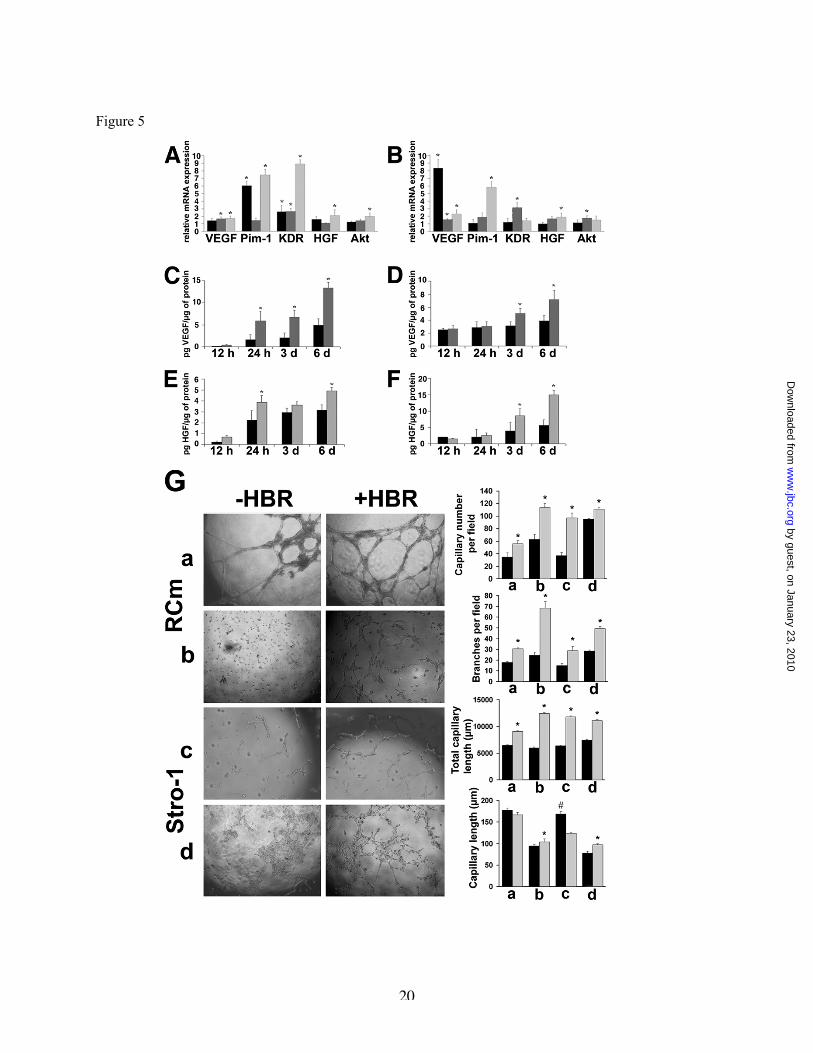
Real-Time PCR analysis revealed that invitro exposure to HBR of both RCm and rat Stro-1-positive stem cells significantly enhanced thegene expression of VEGF, KDR (encoding amajor VEGF receptor), HGF, Akt, and Pim-1 (Fig.5A and 5B). In RCm, HBR-mediated stimulationof Pim-1 gene expression occurred in a time-dependent, biphasic fashion. Increased Pim-1mRNA was detected within 24 hours after HBRstimulation, then declining toward basal valuesafter 3 days. A second elevation in Pim-1 geneexpression was observed after 6 days of RCmexposure to the mixed ester (Fig.5A).
The HBR treatment enhanced thesecretion of VEGF and HGF in the culturemedium from both RCm and Stro-1 cells. Time-course analyses revealed that the stimulatoryaction progressively increased during the first 24hours, persisting throughout a 6-day period (Fig.5C-F). HBR did not appreciably affect thesecretion of VEGF or HGF from HUVECs orRAOECs, nor it induced these cells to formcapillary-like structures in a semisolid medium invitro (data not shown). However, a remarkableincrease in capillarogenesis was observed wheneither HUVECs or RAOECs were cultured withmedium obtained from cardiomyocytes or Stro-1-positive cells exposed to the mixed ester (Fig. 5G).
by guest, on January 23, 2010w
ww
.jbc.orgD
ownloaded from

8
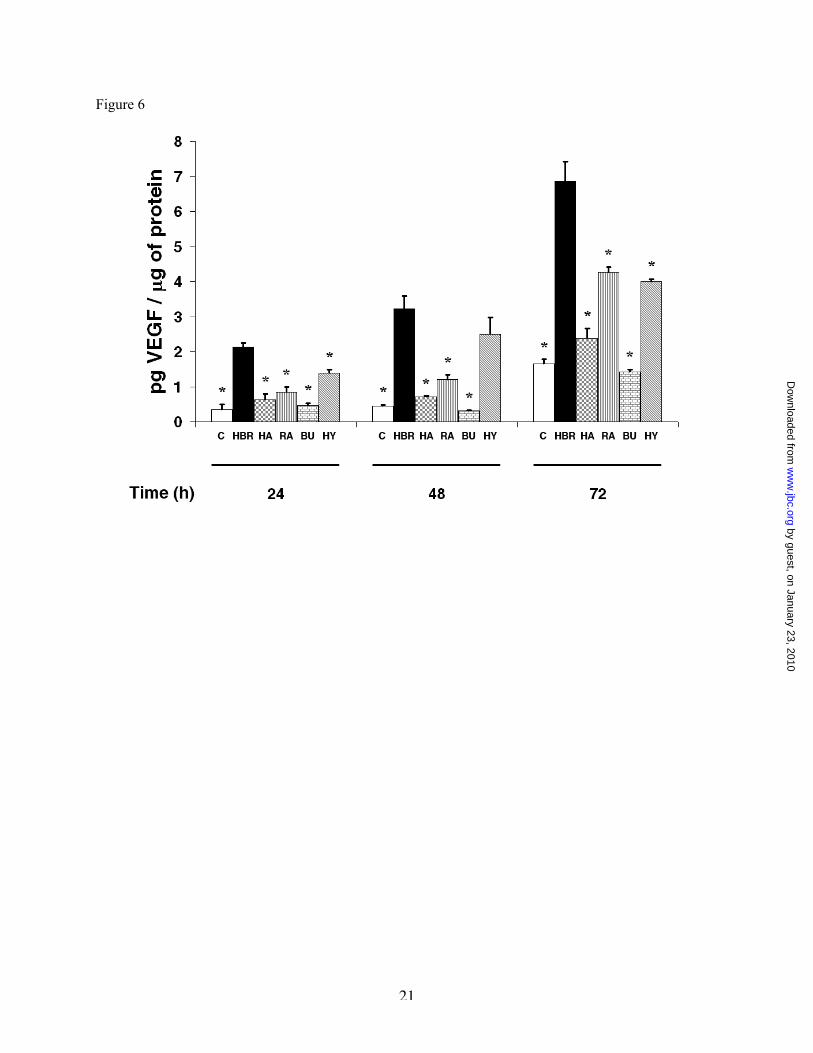
The amount of VEGF released by Stro-1 cells wasslightly increased by BU but not HA alone, beingsignificantly augmented by 10-8 M all-trans RA,and further enhanced following exposure tohydrolized HBR, resulting in the release of allmoieties grafted within the mixed ester (Fig. 6).Nevertheless, under these experimental conditions,the yield of growth factor secretion wasconsiderably lower than that detected in HBR-exposed cells (Fig. 6). Cumulatively, thesefindings suggest the activation of prominentparacrine effects by HBR on both cell populations,enhancing the expression of cytokines and geneswith a crucial role in cell survival andangiogenesis.
HBR acted at transcriptional level. Tofurther dissect the cellular response to HBR,nuclear run-off experiments were designed toassess whether HBR may have affected the rate ofgene transcription and whether, in the affirmative,it may have acted as a unit or after hydrolysis ofhyaluronan grafted moieties. Figure 7 shows thatnuclei isolated from HBR-treated Stro-1-positivecells exhibited a consistent increase in thetranscription rate of VEGF and Pim-1 genes, ascompared with nuclei isolated from untreatedcells. In separate experiments, nuclei were isolatedfrom untreated cells and subsequently incubatedwith HBR, or exposed to HA, BU, or RAadministered alone or in combination. Whilenuclear exposure to HBR or HA failed to trigger atranscriptional response, the incubation with BUor RA enhanced gene transcription (Fig. 7). Thetranscription rate was further enhanced whennuclei were exposed to a combination of BU andRA (Fig. 7).
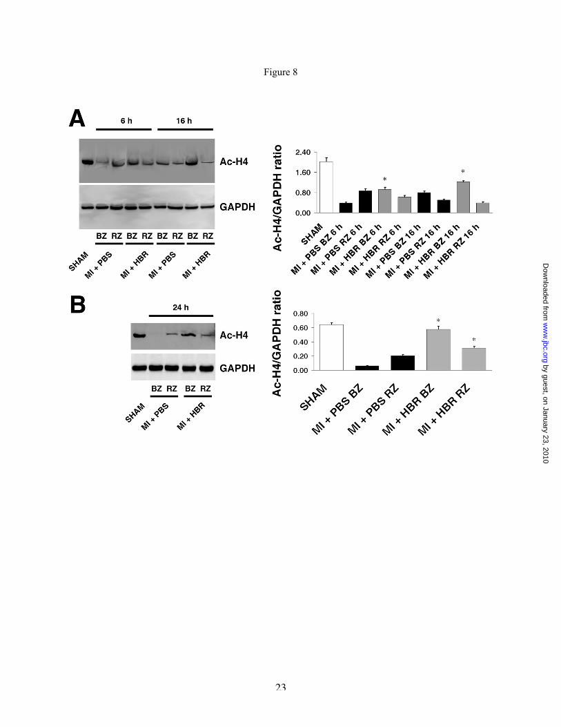
HBR Increased Histone Acetylation.Western blot analysis performed in tissue extractsrevealed that, consonant with previousobservations (15,16), histone H4 acetylationdecreased in the infarcted hearts. Six hours afterHBR injection, acetyl-histone H4 signal rose in theborder zone, progressively increasing up to 16hours, as compared with the untreated group (Fig.8A). After 24 hours, histone H4 acetylationincreased both in border and remote zone ofinfarcted HBR-injected hearts, as compared withthe non-injected tissue (Fig. 8B). Following HBRtreatment, a more pronounced signal was detectedin the border zone, compared to the remote area.A k i n t o W e s t e r n b l o t s t u d i e s ,
immunohistochemical analyses showed an overallincrease in the signal of acetyl-histone H4 inhearts of HBR-treated animals, as compared withuntreated hearts (Fig. 9A). The number ofcardiomyocyte nuclei stained with acetyl-histoneH4 antibody was significantly increased by HBRtreatment in the border zone (Fig. 9A). In vitroexperiments provided evidence that acetyl-H4immunoreactivity was also higher in HBR-exposed R C m and Stro-1-positive cells, ascompared with unexposed cells (Fig. 9B).
DISCUSSION
The present study shows for the first timethat acute myocardial injection of HBR, ahyaluronan ester previously shown to enhancehMSC-mediated cardiac repair in vivo (3), rescuedinfarcted rat hearts by increasing vascularization,promoting cardiomyocyte survival, and restoringnormal tissue function. The HBR action involvedan early increase in the number of pericytes, areserve of progenitor cells that may be integral tothe origin of MSCs and other related adult stemcells (12), suggesting a role of perivascular cells incapillary density enhancement and tissue repairelicited by the mixed ester.
It is now evident that an extremely limitedpercentage of stem cells will engraft and survivewithin the recipient myocardium followingintracoronary infusion or transendocardialinjection (17). Moreover, the use of injectablescaffolds to augment stem cell engraftment is notdevoid of harmful decrease in myocardialperfusion. Hence, the development of deliverablemolecules promoting paracrine mechanisms ofcardiac repair, and survival of endogenouslyrecruited stem cells, without immediate need ofstem cell transplantation, would have remarkablebiomedical implications. Here, we show that HBRmay fulfill both requirements. Four weeks afterHBR injection a significant increase inperivascular Stro-1-positive cells could bedetected within the infarcted hearts. This effectwas associated with a reverse myocardialremodeling (decrease of fibrosis and dilatation)and consistent increase in myocardialperformance. A possible explanation may resultfrom in vivo studies demonstrating that HBR-treated hearts had a significant decrease of
by guest, on January 23, 2010w
ww
.jbc.orgD
ownloaded from

9
apoptotic cardiomyocytes, as revealed by TUNELassay, as well as from in vitro experimentsshowing enhanced gene expression and secretionof VEGF and HGF in both RCm and Stro-1-positive cells exposed to HBR. Growth factorsecretion may have primed a pro-angiogeniccontext (18), reinforced by the stimulatory effectof HBR on the expression of KDR gene, encodinga VEGF receptor involved in autocrine/paracrineamplification of VEGF secretion and angiogenicsignaling (19,20). Moreover, HGF gene transferinto the myocardium improved myocardialfunction and geometry (21), owing to antifibroticeffects through inhibition of transforming growthfactor-β expression. HBR-mediated recruitment ofboth myocardial and Stro-1 cells into a pro-angiogenic paracrine circuitry of cardiac repair isfurther supported by the finding that: (i) anincrease in capillarogenesis was induced in vitro inboth HUVECs or RAOECs with a mediumobtained from HBR-exposed cells; (ii) nosecretion of VEGF or HGF, nor in vitroangiogenesis, were found following a directHUVEC or RAOEC exposure to HBR; and (iii) invivo, a large number of capillary vessels in HBR-injected hearts were “decorated” by Stro-1-positive cells, lacking the expression of vWF, anendothelial marker. Although some Stro-1-positivecells in HBR-injected hearts expressed cardiac-specific α-sarcomeric actin, these few elementsappeared to lack mature sarcomeric organizationand their role in rescuing infarcted myocardiumremains questionable.
The observation that exposure of RCmand Stro-1 cells to HBR increased VEGF, HGF, aswell as Akt and Pim-1 gene expression, may havefurther relevant implications. These genes aredeeply involved in cardioprotective signaling,including antiapoptotic and antifibrotic responses(21-25). Enhanced expression of both Akt andPim-1 promotes cardiomyocyte survival andgrowth in transgenic mice subjected to acutemyocardial infarction (26,27). Here, we show thatin RCm Pim-1 gene expression was augmented ina biphasic manner by the addition of HBR.Biphasic kinetics in Pim-1 gene expression havebeen previously demonstrated in other cell typesexposed to agents regulating cell growth anddifferentiation (28,29), and appeared to involve acomplex interplay between cell signalingnetworks, cell cycle progression, gene
transcription and/or mRNA stability (29). Cardiaccontrol of Pim-1 gene expression is still poorlyunderstood and future work is required to dissectthe molecular mechanisms underlying Pim-1regulation by HBR in myocardial cells.
HBR may also have increased the survivaland rescuing potential of Stro-1-positive cellsrecruited to the injured myocardium. In thisregard, both VEGF and HGF have been shown topromote MSC survival and therapeutic potential( 3 0 ) , enhancing stem cell-mediatedcardioprotection in infarcted hearts (31,32). Up-regulation of both Akt and Pim-1 gene expressionin HBR-treated Stro-1 cells is also worthy ofconsideration, due to the prominent role of theseserine/threonine kinases in stem cell survival anddifferentiation (30,33). MSCs overexpressing Aktprevent remodeling and restore performance ofinfarcted hearts (1,34). Moreover, Pim-1 has beenshown to be required for endothelial and muralcell differentiation in vitro (35).
It is relevant that a “program” of genesinvolved in cardiac protection and stem cellsurvival can be chemically induced by a syntheticmolecule, without the need of viral vector-mediated gene transfer technologies. Themolecular dissection of mechanisms underlyingHBR-mediated responses in vitro and in vivoremains to be fully elucidated. The finding thatVEGF secretion from these cells was significantlyhigher in the presence of HBR, as compared to theresponse to HA, BU, or RA alone, or to a mixtureof the three moieties released by hydrolized HBRsuggests that a maximal response may be achievedwhen both BU and RA are concomitantlyinternalized by the mixed ester. Probably, theexposure to HBR may have afforded an optimalintracellular BU/RA ratio and/or timely action. Tothis end, nuclear run-off experiments indicate thatthe action of HBR was mediated at thetranscriptional level, and that while thetranscription rate of VEGF and Pim-1 wasunaffected following nuclear exposure to HBR, itwas conversely enhanced by BU and RA withsuperimposable time courses and additive effects.These results also prompt the hypothesis that, atleast at nuclear level, HBR may have actedfollowing the hydrolysis of its grafted moieties.
HBR-induced transcription may berelated, at least in part, to the observed increase inhystone acetylation, likely attributable to a
by guest, on January 23, 2010w
ww
.jbc.orgD
ownloaded from

10
decrease in HDAC activity by the butyric moietyof the mixed ester. It is evident that chromatinremodeling by HDAC inhibitors can largely affecttranscription factor accessibility to target cis-acting regulatory sites (36). These so-calledepigenetic changes have profound effects on geneexpression, both in physiological and pathologicalprocesses (36). An inference of the retinoid moietyof HBR with the observed changes in geneexpression is supported by the finding that retinoicacid plays a crucial role in mammalian vasculardevelopment (37) and enhances angiogenesis bytriggering VEGF, KDR, and HGF genetranscription and signaling (38). Moreover,RXR/RAR heterodimer action is enhanced byhistone deacetylase inhibitors, promoting majordevelopmental pathways in pluripotent cells (39).Studies are in progress to further address thespectrum of growth factor and transcriptionalprofiles recruited by HBR.
The finding that a syntheticglycoconjugate displays a therapeutic effect in theacute phase of MI may have several biomedicalimplications. In fact, the clinical use of humanadult stem cells will be hampered in a near futureby a number of interrelated challenges, including:
(i) high-throughput bioprocess development andimproved downstream processing problems; (ii)significant modification, improvement and re-testing of current strategies of stem cell culturingand cardiovascular commitment complying withall standards of Good Manufacturing Practice(GMP); (iii) analytical methodologies for controlof GMP bioprocessing and differentiationefficiencies. Therefore, the timing for cell cultureand expansion within a GMP setting will involve asubstantial delay (several weeks) in autologousstem cell transplantation with respect to the acutephase of a heart attack. Meanwhile, the cardiacrepair afforded by a myocardial injection of HBRmay serve as first aid to rescue a damaged heart.This intervention may be followed by delayedtransplantation of autologous stem cells,eventually preconditioned ex vivo with the samemolecule, to enhance a long-term potential forcardiovascular cell therapy.
REFERENCES
1. Mangi, A.A., Noiseux, N., Kong, D., He, H., Rezvani, M., Ingwall, J.S., and Dzau, V.J. (2003) NatMed 9(9), 1195-1201
2. Gnecchi, M., He, H., Noiseux, N., Liang, O.D., Zhang, L., Morello, F., Mu, H., Melo, L.G., Pratt,R.E., Ingwall, J.S., and Dzau, V.J. (2006) FASEB J 20(6), 661-669
3. Ventura, C., Cantoni, S., Bianchi, F., Lionetti, V., Cavallini, C., Scarlata, I., Foroni, L., Maioli, M.,Bonsi, L., Alviano, F., Fossati, V., Bagnara, G.P., Pasquinelli, G., Recchia, F.A., and Perbellini, A.(2007) J Biol Chem 282(19), 14243-14252
4. Ventura, C., Maioli, M., Asara, Y., Santoni, D., Scarlata, I., Cantoni, S., and Perbellini, A. (2004) JBiol Chem 279 (22), 23574-23579
5. Fernandes, V.R., Wu, K.C., Rosen, B.D., Schmidt, A., Lardo, A.C., Osman, N., Halperin, H.R.,Tomaselli, G., Berger, R., Bluemke, D.A., Marbán, E., and Lima, J.A. (2007) Radiology 245(3), 712-719
6. Higuchi, T., Nekolla, S.G., Jankaukas, A., Weber, A.W., Huisman, M.C., Reder, S., Ziegler, S.I.,Schwaiger, M., and Bengel, F.M. (2007) J Nucl Med 48(2), 288-294
7. Positano, V., Pingitore, A., Giorgetti, A., Favilli, B., Santarelli, M.F., Landini, L., Marzullo, P., andLombardi, M. (2005) J Cardiovasc Magn Reson 7(2), 487-494
8. Spinelli, A.E., D'Ambrosio, D., Pettinato, C., Trespidi, S., Nanni, C., Ambrosini, V., Baldazzi, G.,Bergamini, C., and Marengo, M. (2006) Nucl Instrum Methods in Physics Research A 571(1-2), 215-218
9. Bonacchi, M., Nistri, S., Nanni, C., Gelsomino, S., Pini, A., Cinci, L., Maiani, M., Zecchi-Orlandini,S., Lorusso, R., Fanti, S., Silvertown, J., and Bani, D. (2008 Sep 15) [Epub ahead of print] J Cell MolMed
by guest, on January 23, 2010w
ww
.jbc.orgD
ownloaded from

11
10. Shi, H., Zhang, X., Chen, S., Zhu, W., and Liu, W. (2006) J Nucl Med 47, (Supplement 1) 271P11. Pfaffl, M.W. (2001) A new mathematical model for relative quantification in Real Time PCR.
Nucleic Acids Res 29(9), e4512. Crisan, M., Yap, S., Casteilla, L., Chen, C.W., Corselli, M., Park, T.S., Andriolo, G., Sun, B., Zheng,
B., Zhang, L., Norotte, C., Teng, P.N., Traas, J., Schugar, R., Deasy, B.M., Badylak, S., Buhring,H.J., Giacobino, J.P., Lazzari, L., Huard, J., and Péault, B. (2008) Cell Stem Cell 3(3), 301-313
13. Son, B.R., Marquez-Curtis, L.A., Kucia, M., Wysoczynski, M., Turner, A.R., Ratajczak, J.,Ratajczak, M.Z., and Janowska-Wieczorek, A. (2006) Stem Cells 24(5), 1254-1264
14. Napoli, C., Maione, C., Schiano, C., Fiorito, C., and Ignarro, L.J. (2007) Trends Mol Med 13(7), 278-286
15. Lee, T.M., Lin, M.S., and Chang, N.C. (2007) Am J Physiol Heart Circ Physiol 293(2), H968-H97716. Granger, A., Abdullah, I., Huebner, F., Stout, A., Wang, T., Huebner, T., Epstein, J.A., and Gruber,
P.J. (2008) FASEB J 22(10), 3549-356017. Bonaros, N., Rauf, R., Schachner, T., Laufer, G., and Kocher, A. (2008) Transplantation 86(9), 1151-
116018. Tang, Y.L., Zhao, Q., Zhang, Y.C., Cheng, L., Liu, M., Shi, J., Yang, Y.Z., Pan, C., Ge, J., and
Phillips, M.I. (2004) Regul Pept 117(1), 3-1019. Millauer, B., Wizigmann-Voos, S., Schnurch, H., Martinez, R., Moller, N.P.H., Risau, W., and
Ullrich, A. (1993) Cell 72(6), 835-84620. Ancelin, M., Chollet-Martin, S., Hervé, M.A., Legrand, C., El Benna, J., and Perrot-Applanat M.
(2004) Lab Invest 84(4), 502-51221. Li, Y., Takemura, G., Kosai, K., Yuge, K., Nagano, S., Esaki, M., Goto, K., Takahashi, T.,
Hayakawa, K., Koda, M., Kawase, Y., Maruyama, R., Okada, H., Minatoguchi, S., Mizuguchi, H.,Fujiwara, T., and Fujiwara, H. (2003) Circulation 107(19), 2499-2506
22. Kaga, S., Zhan, L., Altaf, E., and Maulik, N. (2006) J Mol Cell Cardiol 40(1), 138-14723. Guzman, M.J., Crisostomo, P.R., Wang, M., Markel, T.A., Wang, Y., and Meldrum, D.R. (2008) J
Surg Res 150(2), 286-29224. Thirunavukkarasu, M., Addya, S., Juhasz, B., Pant, R., Zhan, L., Surrey, S., Maulik, G., Menon, V.P.,
and Maulik, N. (2008) J Cell Mol Med 12(4), 1284-130225. Nakamura, T., Mizuno, S., Matsumoto, K., Sawa, Y., Matsuda, H., and Nakamura, T. (2000) J Clin
Invest 106(12), 1511-151926. Matsui, T., Li, L., Wu, J.C., Cook, S.A., Nagoshi, T., Picard, M.H., Liao, R., and Rosenzweig, A.
(2002) J Biol Chem 277(25), 22896-2290127. Muraski, J.A., Rota, M., Misao, Y., Fransioli, J., Cottage, C., Gude, N., Esposito, G., Delucchi, F.,
Arcarese, M., Alvarez, R., Siddiqi, S., Emmanuel, G.N., Wu, W., Fischer, K., Martindale, J.J.,Glembotski, C.C., Leri, A., Kajstura, J., Magnuson, N., Berns, A., Beretta, R.M., Houser, S.R.,Schaefer, E.M., Anversa, P., and Sussman, M.A. (2007) Nat Med 13(12), 1467-1475
28. Buckley, A.R., Leff, M.A., Buckley, D.J., Magnuson, N.S., de Jong, G., and Gout, P.W. (1996) CellGrowth Differ 7(12), 1713-1721
29. Buckley, A.R., and Buckley, D.J. (2000) Ann N Y Acad Sci 917, 522-53330. Rosová, I., Dao, M., Capoccia, B., Link, D., and Nolta, J.A. (2008) Stem Cells 26(8), 2173-218231. Forte, G., Minieri, M., Cossa, P., Antenucci, D., Sala, M., Gnocchi, V., Fiaccavento, R., Carotenuto,
F., De Vito, P., Baldini, P.M., Prat, M., and Di Nardo, P. (2006) Stem Cells 24(1), 23-3332. Markel, T.A., Wang, Y., Herrmann, J.L., Crisostomo, P.R., Wang, M., Novotny, N.M., Herring,
C.M., Tan, J., Lahm, T., and Meldrum, D.R. (2008) Am J Physiol Heart Circ Physiol 295(6), H2308-H2314
33. Choi, S.C., Kim, S.J., Choi, J.H., Park, C.Y., Shim, W.J., and Lim, D.S. (2008) Stem Cells Dev 17(4),725-736
34. Hammerman, P.S., Fox, C.J., Birnbaum, M.J., and Thompson, C.B. (2005) Blood 105(11), 4477-448335. Noiseux, N., Gnecchi, M., Lopez-Ilasaca, M., Zhang, L., Solomon, S.D., Deb, A., Dzau, V.J., and
Pratt, R.E. (2006) Mol Ther 14(6), 840-850
by guest, on January 23, 2010w
ww
.jbc.orgD
ownloaded from

12
36. Santini, V., Gozzini, A., and Ferrari, G. (2007) Curr Drug Metab 8(4), 383-39337. Lai, L., Bohnsack, B.L., Niederreither, K., and Hirschi, K.K. (2003) Development 130(26), 6465-
647438. Saito, A., Sugawara, A., Uruno, A., Kudo, M., Kagechika, H., Sato, Y., Owada, Y., Kondo, H., Sato,
M., Kurabayashi, M., Imaizumi, M., Tsuchiya, S., and Ito, S. (2007) Endocrinology 148(3), 1412-1423
39. Dilworth, F.J., Fromental-Ramain, C., Yamamoto, K., and Chambon, P. (2000) Mol Cell 6(5), 1049-1058
by guest, on January 23, 2010w
ww
.jbc.orgD
ownloaded from

13
FOOTNOTES
*Our thanks are due to Dr. Cristina Nanni (Department of Nuclear Medicine, S. Orsola – MalpighiHospital, Bologna, Italy) for her technical support in PET studies. This work was supported by RegioneEmilia Romagna, Programma di Ricerca Regione – Università 2007/2009, Area 1b “Medicinarigenerativa”, Italy; Fondazione Luisa Fanti Melloni, Bologna, Italy; Sintofarm S.p.A. (Guastalla, ReggioEmilia), Italy; Tavola Valdese, Rome, Italy, and “Compagnia di San Paolo”, Torino, Italy.
Key Words: Hyaluronan, Glycoconjugates, Angiogenesis, Reperfusion, Survival
The abbreviations used are: HBR, hyaluronan mixed ester of butyric and retinoic acid; VEGF, VascularEndothelial Growth Factor; HGF, Hepatocyte Growth Factor; MI, myocardial infarction; hMSCs, humanmesenchymal stem cells; MRI, Magnetic Resonance Imaging; mPET, Small-animal Positron EmissionTomography; 18F-FDG, [F-18]fluorodeoxyglucose; RCm, rat neonatal cardiomyocytes; RAOECs, rataortic endothelial cells; HUVEC, human umbilical vein endothelial cells.
FIGURE LEGENDS
Fig. 1. MRI-derived measures of left ventricular function. Global (A) and regional (B) LVfunction. Values are means ± SEM; n = 11 animals for each experimental condition. MI, myocardialinfarction; LVEF, LV ejection fraction; CO, cardiac output; LVEDV, LV end-diastolic volume; LVESV,LV end-systolic volume; ED, end-diastolic; ES, end-systolic. The HBR-treated group received 100 µl ofHBR solution (0.2 mg of HBR / 100 g of rat weight). * P < 0.05 vs. SHAM; † P < 0.05 vs. MI + PBS(one-way ANOVA with subsequent Bonferroni test).
Fig. 2. MRI delayed contrast enhancement of myocardial scar. Representative LV short- andlong-axis gadolinium-delayed contrast-enhanced MRI images for each experimental condition (A).Regional LV delayed enhancement in infarcted LV treated with 100 µl of HBR solution (0.2 mg of HBR /100 g of rat weight) or PBS (B). Values are means ± SEM; n = 11 animals for each experimentalcondition. SA, short axis; LVDE, LV delayed contrast-enhancement. * P < 0.05 vs. SHAM; † P < 0.05vs. MI + PBS (one-way ANOVA with subsequent Bonferroni test).
Fig. 3. Myocardial glucose uptake measured by 18F-deoxyglucose (18F-FDG). (A) RepresentativeLV polar map, short- and long-axis mPET images for each experimental condition; (B) mean score valuesof myocardial 18F-FDG of infarcted LV treated with 100 µl of HBR solution (0.2 mg of HBR / 100 g ofrat weight) or PBS. n = 11 animals for each experimental condition. SA, short-axis; LA, long-axis.
Fig. 4. HBR increased the number of capillary vessels, Stro-1-positive cells and perivascularelements. (A-C) Four weeks following myocardial infarction. Transversally cut left ventricularmyocardium from HBR-treated (100 µl of HBR solution) hearts showed a reduced scar, compared withPBS-treated animals. (A, upper images, the arrows demarcate the infarcted area). Picro-Mallory stains inblue the area of scar and in red the myocardium parenchyma. In the border zone of HBR-treated hearts,scar reduction was associated with fewer apoptotic cardiomyocytes (A, lower images; scale bars = 20µm), and increased number of capillary vessels (B). vWF expression highlights endothelial cells (arrows)lining the capillary inner wall (B); scale bars: upper images = 300 µm, lower images = 50 µm. (C) InHBR-treated samples, Stro-1 positive cells (arrows) increased in number and were closely associated withthe outer capillary wall, while c-kit-positive cells did not vary significantly, compared to untreatedanimals. Scale bars = 20 µm. (D-F) 24 hours following myocardial infarction. (D) The number of Ki-67-positive cells significantly increased in the HBR-treated animals (arrows); scale bars = 50 µm. (E) Cellsexpressing NG2, and PDGF-Rβ (arrows); scale bars = 30 µm (upper images), and 100 µm (lower
by guest, on January 23, 2010w
ww
.jbc.orgD
ownloaded from

14
images). (F) VEGF expression (arrows). Scale bars = 100 µm (left and mid images), and 50 µm (rightimage). * Significantly different from PBS-treated hearts, P < 0.05. (Statistical test: two-tailed, unpairedStudent’s t-test). IHC: immunohistochemistry. Full size images of each individual panel are presented inFig. 1 in the Supplemental Data.
Fig. 5. HBR affects gene expression and secretion of trophic mediators. (A,B) VEGF, KDR,HGF, Akt, and Pim-1 gene expression was assessed by Real Time PCR. RCm (A) or Stro-1 cells (B) werecultured for 24 hours (black bar), 3 days (dark gray bar), and 6 days (light gray bar) in the absence orpresence of HBR (2 mg/ml). The abundance of each mRNA in untreated cells was defined as 1, and theamounts of VEGF, KDR, HGF, Akt, and Pim-1 mRNA from HBR-treated cells were plotted relative tothat value (mean ± SEM; n = 6). (C-F) Time-course analysis of VEGF and HGF respectively released byRCm (C and E) or Stro-1 cells (D and F) cultured in the absence (black bar) or presence (gray bar) ofHBR (mean ± SEM; n = 6). * Significantly different from untreated cells (controls), P < 0.05 (GraphPadPrism ver. 4 (http://www.graphpad.com), two-tailed, unpaired Student’s t-test). (G) In vitrocapillarogenesis was assessed in HUVECs (a,c) and RAOECs (b,d) exposed to a conditioned mediumobtained from RCm or Stro-1 cells cultured for 24 hours in the absence (black bar) or presence (gray bar)of HBR. Morphological characteristics of capillary-like networks were evaluated by using NIS-ElementsD Nikon software (ver.3.06). Data are the mean ± SEM; n = 3. * Significantly different from –HBR; #significantly different from +HBR; P < 0.05.
Fig. 6. Comparative analysis of the effect of HBR, HA, BU, and RA on the secretion of VEGFfrom rat Stro-1 positive cells. Cells were incubated for the indicated times in the absence or presence of2.0 mg/ml HBR, 1.5 mg/ml HA, 2.5 mM BU, 10-8M RA alone, or exposed to a hydrolyzed (HY) HBRsolution (2.0 mg/ml), obtained from a 2-hour basic HBR hydrolysis followed by pH neutralization. Thisprocedure has been shown to afford a complete release of each single HBR grafted moiety (Ventura et al.unpublished observations). Data are the mean ± SEM of four separate experiments. * Significantlydifferent from HBR alone (GraphPad Prism ver. 4 (http://www.graphpad.com), two-tailed, unpairedStudent’s t-test).
Fig. 7. Analysis of VEGF and Pim-1 gene transcription in isolated nuclei. (A,B) Nuclei wereisolated from rat Stro-1 positive cells cultured for 24 hours (VEGF gene transcription) or 6 days (Pim-1gene transcription) in the absence or presence of 2.0 mg/ml HBR, respectively. From lanes C through H,nuclei were isolated from untreated cells and then directly incubated for 12 hours without any drug (C) orin the presence of 2.0 mg/ml HBR (D), 1.5 mg/ml hyaluronic acid (HA) (E), 2.5 mM butyric acid (BU)(F), 10-8M retinoic acid (RA) (G), or with a combination of BU and RA (H). Autoradiographic exposurewas for 2 days on Kodak X-Omat film with an intensifying screen. The right side of each panel reportsthe position of radiolabeled DNA markers showing that the single protected fragments migrated with amolecular size comparable to VEGF (597 bases), Pim-1 (609 bases), or GAPDH (574 bases) mRNA. Dueto the similar size of VEGF-, Pim-1-, and GAPDH-protected fragments,
32P-labeled nuclear RNA was
hybridized separately with cRNA probes and the corresponding hybrids were run onto different gels.Autoradiograms are representative of six separate experiments.
Fig. 8. Time-course analysis of the effect of HBR on myocardial histone acetylation. Western blotanalysis of histone H4 acetylation was performed in total tissue extracts from border (BZ) and remotezone (RZ) of infarcted hearts (MI) 6 or 16 hours (A), or 24 hours (B) after injection in the absence (PBS)or presence of 100 µl of HBR solution (0.2 mg of HBR / 100 g of rat weight). GAPDH was used as aloading control. Acetylation level was estimated by densitometric quantification (n = 3). * Significantlydifferent from the same control time point within the MI BZ (A), or from MI BZ or MI RZ (B). P < 0.05(GraphPad Prism ver. 4 (http://www.graphpad.com), two-tailed, unpaired Student’s t-test). All Westernblots were performed at least three times with similar results.
by guest, on January 23, 2010w
ww
.jbc.orgD
ownloaded from

15
Fig. 9. HBR increased histone acetylation in infarcted myocardium, isolated cardiomyocytes andStro-1 cells. (A) Immunohistochemistry of acetyl-histone H4. Acetylation level was estimated in infarctedhearts by intensity quantification and counting of stained cardiomyocyte nuclei 24 hours after injection inthe absence (PBS) or presence of HBR solution (0.2 mg of HBR / 100 g of rat weight). Scale bar = 30µm. * Significantly different from PBS-treated, P < 0.05. (B) Western blot analysis of total cell extractsfrom RCm and Stro-1-positive cells cultured in the absence (CTR) or presence of HBR (2 mg/ml) (n = 3).* Significantly different from the same control time point, P < 0.05 (GraphPad Prism ver. 4(http://www.graphpad.com), two-tailed, unpaired Student’s t-test). All Western blots were performed atleast three times with similar results.
by guest, on January 23, 2010w
ww
.jbc.orgD
ownloaded from