human t-lymphocyte response in vitro to synthetic peptides
TRANSCRIPT

Proc. Natl. Acad. Sci. USAVol. 82, pp. 3425-3429, May 1985Immunology
Human T-lymphocyte response in vitro to synthetic peptides ofherpes simplex virus glycoprotein D
(human lymphocyte activation)
ELAINE C. DEFREITAS, BERNHARD DIETZSCHOLD, AND HILARY KOPROWSKIThe Wistar Institute of Anatomy and Biology, 36th Street at Spruce, Philadelphia, PA 19104
Contributed by Hilary Koprowski, January 22, 1985
ABSTRACT Immunization of mice with a synthetic pep-tide that corresponds to a murine antibody-defined immuno-dominant domain of herpes simplex virus (HSV) glycoproteinD (gD) induced neutralizing antibodies against HSV types 1and 2 and protected animals against a lethal challenge withHSV type 2 (Dietzschold, B., Eisenberg, R., Ponce de Leon,M., Golub, E., Hudecz, F., Varicchio, A. & Cohen, G. (1984)J. Virol. 52, 431-435). We report here that human peripheralblood T cells from HSV-seropositive and -seronegative adultdonors are activated by this synthetic peptide in vitro. Interleu-kin-2-dependent T-cell lines established from these cultures re-spond specifically to peptides containing residues 1-23 of HSVgD and to a panel of overlapping peptides within this domain.The T-cell proliferative response was maximal when the ma-jority of interleukin-2-propagated T cells were of the helperphenotype and the peptides were at least 16 amino acids long.Peptides of 8 or 12 amino acids from the carboxyl terminuswere nonstimulatory. Peptide-activated T-cell lines from sero-negative donors less than 11 years old could be established invitro, but most cells were of the suppressor/cytotoxic pheno-type and demonstrated no antigen-specificity when tested withthe panel of synthetic peptides.
The immune response to herpes simplex virus (HSV) hasbeen shown to involve a variety of effector mechanismswhich include production of virus-neutralizing antibody, cy-totoxic T lymphocytes, natural killer (NK) cells, antibody-dependent cellular cytotoxicity, and T-cell-derived lympho-kines (43). The pivotal role of helper T-cells in the inductionof these responses has been well documented (44, 45).The nature of the HSV-specified antigens that act as im-
munostimulants has not been completely identified, but sev-eral lines of evidence implicate the major viral glycoproteins(1). One of these, glycoprotein D (gD), has been shown toinduce type-common viral-neutralizing antibody (2, 3), andpassive administration of monoclonal antibody (mAb) spe-cific for gD protects mice from a lethal challenge with HSV(4). Moreover, gD appears to be recognized on HSV-infect-ed cells by cytolytic effector cells (5).An antibody-defined immunodominant determinant of
HSV type 1 (HSV-1) gD was localized and a peptide corre-sponding to residues 8-23 of the mature glycoprotein wassynthesized. This peptide was recognized by an anti-gDmAb known to neutralize HSV-1 and -2 (6). Moreover, im-munization of mice with this synthetic molecule induces vi-ral-neutralizing antibodies and confers protection against alethal challenge with HSV-2 (7).
MATERIALS AND METHODS
Synthesis of HSV gD Peptides. Peptides were manuallysynthesized as described (6) by using the Merrifield solid-
phase method (8). The completed peptides were simulta-neously deprotected and cleaved from the resin with anhy-drous HF containing thioanisole as an organic cation scaven-ger. The crude peptides were purified by gel filtration onBio-Gel P-2 or P-4 (Bio-Rad) columns. Homogeneity of thepeptides was demonstrated by thin-layer chromatography,amino acid analysis, and amino acid sequencing (9). Forthin-layer chromatography on Silica Gel 60-precoated alumi-num sheets (Merck), two solvent systems were used: 1-bu-tanol/acetic acid/water, 4:1:1 (vol/vol), or ethyl acetate/pyridine/acetic acid/water, 60:20:6:11 (vol/vol); peptideswere detected with ninhydrin or chlorine/toluidine.Primary in Vitro Proliferation Assay. Whole blood was ob-
tained by venipuncture from normal adult volunteers. Pe-ripheral blood mononuclear cells (PBMC) were purified bydensity centrifugation on Lymphoprep (Nyegaard, Oslo),washed in Ca2 - and Mg2'-free Hanks' balanced salts solu-tion, and seeded in microtiter plates (Costar, Cambridge,MA) at 3 x 10⁵ per well in RPMI 1640 medium (M. A. Bio-products, Walkersville, MD) plus 10% human AB serum(heat-inactivated), 2 mM glutamine, penicillin, and strepto-mycin (complete medium). Synthetic peptide 1-23, phytohe-magglutinin (PHA; Burroughs Wellcome, Kent, England) at1 ,ug/ml, or tetanus toxoid (Connaught Laboratories, Wil-lowdale, ON) at 10 ,ug/ml were added to wells in triplicate.All cultures were incubated for 5 days at 5% C02; 1 ,uCi (1 Ci= 37 GBq) of [3H]thymidine (ICN) per well was present forthe final 18 hr. Cells were harvested with a Skatron cell har-vester (Eflab Oy, Helsinki, Finland) onto glass fiber discsfor determination of radioactivity by liquid scintillationcounting.
Induction of Synthetic Peptide-Activated T-Cell Lines.PBMC were isolated as described and suspended at 2 x 10⁶per ml in complete medium with synthetic peptide 1-23 at100 ,ug/ml; 2 ml were seeded in each well of a 24-well clusterplate (Costar) and incubated at 37°C in 5% CO2. Cultureswere fed twice weekly with complete medium, containinglectin-free, partially purified human interleukin 2 (IL-2) (Cel-lular Bioproducts, Buffalo, NY) at 10 units/ml, after largeblastoid cells appeared.- For all donors tested in this study,this occurred between days 10 and 15. When viable blastsreached a concentration of 106 per ml, the nonadherent cellswere removed from the plate, washed, resuspended at 105per ml in complete medium with 5% lectin-free IL-2, and 50ml was seeded in a T75 flask (Costar). Cells from all donorsgrew logarithmically for the next 10-14 days. At this time, Tcells were restimulated with synthetic peptide 1-23, autolo-gous y-irradiated PBMC (accessory cells), and lectin-freeIL-2. All lines could be propagated indefinitely (>9 mo) withthis protocol. At various times, cells were tested for specific-ity in a proliferation assay and for phenotype by flow cytom-etry.
Abbreviations: HSV, herpes simplex virus; gD, glycoprotein D;PBMC, peripheral blood mononuclear cell(s); IL-2, interleukin 2;mAb, monoclonal antibody; PHA, phytohemagglutinin; NK, naturalkiller.
3425
The publication costs of this article were defrayed in part by page chargepayment. This article must therefore be hereby marked "advertisement"in accordance with 18 U.S.C. §1734 solely to indicate this fact.
Dow
nloa
ded
by g
uest
on
Nov
embe
r 24
, 202
1
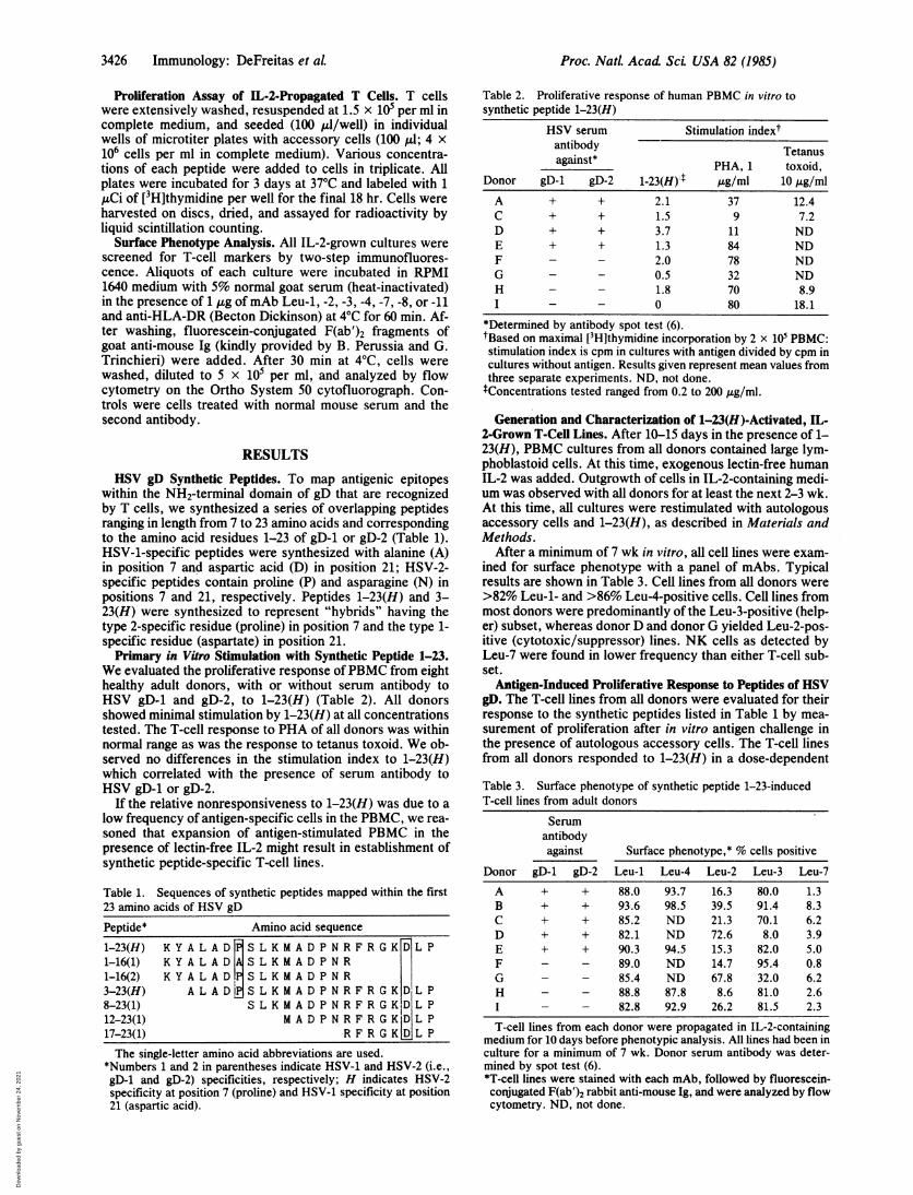
3426 Immunology: DeFreitas etaLP
Proliferation Assay of IL-2-Propagated T Cells. T cellswere extensively washed, resuspended at 1.5 x 10⁵ per ml incomplete medium, and seeded (100 il/well) in individualwells of microtiter plates with accessory cells (100 gl; 4 x10⁶ cells per ml in complete medium). Various concentra-tions of each peptide were added to cells in triplicate. Allplates were incubated for 3 days at 370C and labeled with 1,4Ci of [3H]thymidine per well for the final 18 hr. Cells wereharvested on discs, dried, and assayed for radioactivity byliquid scintillation counting.
Surface Phenotype Analysis. All IL-2-grown cultures werescreened for T-cell markers by two-step immunofluores-cence. Aliquots of each culture were incubated in RPMI1640 medium with 5% normal goat serum (heat-inactivated)in the presence of 1 ,ug ofmAb Leu-1, -2, -3, -4, -7, -8, or -11and anti-HLA-DR (Becton Dickinson) at 40C for 60 min. Af-ter washing, fluorescein-conjugated F(ab')2 fragments ofgoat anti-mouse Ig (kindly provided by B. Perussia and G.Trinchieri) were added. After 30 min at 40C, cells werewashed, diluted to 5 x 10⁵ per ml, and analyzed by flowcytometry on the Ortho System 50 cytofluorograph. Con-trols were cells treated with normal mouse serum and thesecond antibody.
RESULTSHSV gD Synthetic Peptides. To map antigenic epitopes
within the NH2-terminal domain of gD that are recognizedby T cells, we synthesized a series of overlapping peptidesranging in length from 7 to 23 amino acids and correspondingto the amino acid residues 1-23 of gD-1 or gD-2 (Table 1).HSV-1-specific peptides were synthesized with alanine (A)in position 7 and aspartic acid (D) in position 21; HSV-2-specific peptides contain proline (P) and asparagine (N) inpositions 7 and 21, respectively. Peptides 1-23(H) and 3-23(H) were synthesized to represent "hybrids" having thetype 2-specific residue (proline) in position 7 and the type 1-specific residue (aspartate) in position 21.Primary in Vitro Stimulation with Synthetic Peptide 1-23.
We evaluated the proliferative response ofPBMC from eighthealthy adult donors, with or without serum antibody toHSV gD-1 and gD-2, to 1-23(H) (Table 2). All donorsshowed minimal stimulation by 1-23(H) at all concentrationstested. The T-cell response to PHA of all donors was withinnormal range as was the response to tetanus toxoid. We ob-served no differences in the stimulation index to 1-23(H)which correlated with the presence of serum antibody toHSV gD-1 or gD-2.
If the relative nonresponsiveness to 1-23(H) was due to alow frequency of antigen-specific cells in the PBMC, we rea-soned that expansion of antigen-stimulated PBMC in thepresence of lectin-free IL-2 might result in establishment ofsynthetic peptide-specific T-cell lines.
Table 1. Sequences of synthetic peptides mapped within the first23 amino acids of HSV gD
Peptide* Amino acid sequence
1-23(H) KYALAD SLKMADPNRFRGKDLP1-16(1) KYALAD ASLKMADPNR1-16(2) KYALAD,PSLKMADPNR3-23(H) ALAD1HSLKMADPNRFRGKDLP8-23(1) SLKMADPNRFRGKD LP12-23(1) MADPNRFRGK ID L P17-23(1) R F R G K L P
The single-letter amino acid abbreviations are used.
Table 2. Proliferative response of human PBMC in vitro tosynthetic peptide 1-23(H)
HSV serum Stimulation indextantibody Tetanusagainst* PHA, 1 toxoid,
Donor gD-1 gD-2 1-23(H) t gg/ml 10 ig/mlA + + 2.1 37 12.4C + + 1.5 9 7.2D + + 3.7 11 NDE + + 1.3 84 NDF - - 2.0 78 NDG - - 0.5 32 NDH - - 1.8 70 8.9I - - 0 80 18.1
*Determined by antibody spot test (6).tBased on maximal [3H]thymidine incorporation by 2 x 101 PBMC:stimulation index is cpm in cultures with antigen divided by cpm incultures without antigen. Results given represent mean values fromthree separate experiments. ND, not done.:Concentrations tested ranged from 0.2 to 200 tug/ml.Generation and Characterization of 1-23(H)-Activated, IL-
2-Grown T-Cell Lines. After 10-15 days in the presence of 1-23(H), PBMC cultures from all donors contained large lym-phoblastoid cells. At this time, exogenous lectin-free humanIL-2 was added. Outgrowth of cells in IL-2-containing medi-um was observed with all donors for at least the next 2-3 wk.At this time, all cultures were restimulated with autologousaccessory cells and 1-23(H), as described in Materials andMethods.
After a minimum of 7 wk in vitro, all cell lines were exam-ined for surface phenotype with a panel of mAbs. Typicalresults are shown in Table 3. Cell lines from all donors were>82% Leu-1- and >86% Leu-4-positive cells. Cell lines frommost donors were predominantly of the Leu-3-positive (help-er) subset, whereas donor D and donor G yielded Leu-2-pos-itive (cytotoxic/suppressor) lines. NK cells as detected byLeu-7 were found in lower frequency than either T-cell sub-set.
Antigen-Induced Proliferative Response to Peptides of HSVgD. The T-cell lines from all donors were evaluated for theirresponse to the synthetic peptides listed in Table 1 by mea-surement of proliferation after in vitro antigen challenge inthe presence of autologous accessory cells. The T-cell linesfrom all donors responded to 1-23(H) in a dose-dependent
Table 3. Surface phenotype of synthetic peptide 1-23-inducedT-cell lines from adult donors
Serumantibodyagainst Surface phenotype,* % cells positive
Donor gD-1 gD-2 Leu-1 Leu-4 Leu-2 Leu-3 Leu-7
A + + 88.0 93.7 16.3 80.0 1.3B + + 93.6 98.5 39.5 91.4 8.3C + + 85.2 ND 21.3 70.1 6.2D + + 82.1 ND 72.6 8.0 3.9E + + 90.3 94.5 15.3 82.0 5.0F - - 89.0 ND 14.7 95.4 0.8G - - 85.4 ND 67.8 32.0 6.2H - - 88.8 87.8 8.6 81.0 2.6I - - 82.8 92.9 26.2 81.5 2.3
T-cell lines from each donor were propagated in IL-2-containingmedium for 10 days before phenotypic analysis. All lines had been inculture for a minimum of 7 wk. Donor serum antibody was deter-mined by spot test (6).*T-cell lines were stained with each mAb, followed by fluorescein-conjugated F(ab')2 rabbit anti-mouse Ig, and were analyzed by flowcytometry. ND, not done.
*Numbers 1 and 2 in parentheses indicate HSV-1 and HSV-2 (i.e.,gD-i and gD-2) specificities, respectively; H indicates HSV-2specificity at position 7 (proline) and HSV-1 specificity at position21 (aspartic acid).
Proc. NatL Acad ScL USA 82 (1985)
Dow
nloa
ded
by g
uest
on
Nov
embe
r 24
, 202
1

Immunology: DeFreitas et al. Proc. NatL. Acad Sci USA 82 (1985) 3427
B105..
PHCA C
. 1 23 8
&3
23
I 04
1l 23
i*. -.1 16,PROI: -~ 1 7 23
03! t- - - _t| 3 23131b* 1 16 ALA
102L- _o 2 20 200 0
Anti(2 20 200
igen concentration ag ml2 20 200
FIG. 1. Proliferative response of synthetic peptide 1-23-stimulated, IL-2-propagated T-cell lines from donor A (A), donor E (B), and donor D(C). T cells (104) were added to microtiter wells containing 2 x 10⁵ autologous accessory cells in the presence of various concentrations of eachsynthetic peptide (see Table 1) or in the presence of PHA (1 Ag/ml), as described in Materials and Methods. Peptides 1-16(ALA) and 1-16(PRO) contain alanine and proline, respectively, at position 7. Neither T cells alone nor irradiated accessory cells alone responded to anysynthetic peptide at any concentration (<210 ± 19 cpm). Tetanus toxoid (10 pg/ml)-stimulated parallel cultures incorporated <542 ± 28 cpm.Results are representative of three separate experiments. Broken horizontal lines show level of [3H]thymidine incorporation in the absence ofboth antigen and PHA.
fashion irrespective of the presence of serum HSV gD anti-body in these donors. The recognition of synthetic peptidefragments varied among donors in that certain synthetic pep-tides were markedly stimulatory for one donor but not foranother, as illustrated in Fig. 1. Fig. 1A (donor A) shows thatT cells primed to 1-23(H) can recognize fragments 3-23 and8-23 but not 17-23 or 13-23. Sixteen-residue fragments fromthe NH2 terminus with proline or alanine at position 7 areequally stimulatory. In contrast, donor E's (Fig. 1B) T-cellline recognizes only 1-23(H), 3-23(H), and 8-23(1). The Tcells derived from donor D (Fig. 1C) show a marginal re-sponse to 1-23(H) at 200 ,ug/ml but no response to any othersynthetic peptide. Interestingly, this line is predominantly ofLeu-2-positive (cytotoxic/suppressor) phenotype and wasobtained from a donor with recurrent herpes facial labialis.The PHA stimulation index of this line was normal, indicat-ing that the T cells are viable and mitogen-responsive.
Fig. 2 illustrates the T-cell response of two seronegativedonors. Fig. 2A (donor G) indicates that the most immuno-genic peptides are those which have the three NH2-terminalamino acids possessed by the priming antigen, 1-23(H).
10o5|IPHA
E0
C lo.-.9 10-,i
0a
0
.0
-5
.E 103
I
1 o2
A
a 1-23-o4 1 16iPRO!
.1 16 ALAI
l0t I
104
..._
.......A:.---3-23
-*- - --* - - 13-23- -. 172 13 1
200 102 A _
200ign concentration,°gm0Antigen concentration, pug/mi
Those synthetic peptides without these determinants do notstimulate this line. Fig. 2B (donor F) indicates that syntheticpeptide 1-16(2) (proline at position 7) is markedly stimula-tory, whereas 1-16(1) (alanine at position 7) is not. Thus, thisT-cell line appears capable of distinguishing a single aminoacid substitution in this synthetic peptide fragment.The patterns of reactivity for each donor were reproduc-
ible: repetitive blood samples obtained over a period of sev-eral months yielded peptide-specific T-cell lines which rec-ognized the same determinants. These data suggest that thehuman T-cell response to these peptides may be controlledby immune response genes, as has been shown for randomcopolymers of amino acids (10).
Since T-cell lines from all seronegative donors respondedto 1-23(H) and at least one of its analogs, it was possible thateither 1-23(H) was inducing a primary antigen-induced T-cell response in vitro or, alternatively, the absence of HSVgD serum antibody belies the fact that these individuals hadencountered HSV previously. To address this question, weevaluated the ability of 1-23(H) to stimulate PBMC fromseronegative children less than 11 years old with no clinical
B
11 23
* 1 1691, ... iPRO:
* 3 23
-- - --- --
2 20
* 116[ALA!
* 8 23
17 23
-.13 23
200
FIG. 2. Proliferative response ofsynthetic peptide 1-23-stimulated,IL-2-grown T-cell lines from donor G(A) and donor F (B). Experimentswere performed as described in thelegend to Fig. 1 and in Materials andMethods. Results are representativemean of three separate experiments.
1o5I
E
IC0 104
C)0
C:
E 103-If
1 031
A
4 t 230.
- * s l 1E.PRO-
41 *823
I :.~-- 41 a 116 ALA
If ., 1 7 23
X~~~~~~~~~~~~~*13 23i ,.
102
0 2 20
lo:),
PHAPHA
1 02L 0
Dow
nloa
ded
by g
uest
on
Nov
embe
r 24
, 202
1

3428 Immunology: DeFreitas et al.
Table 4. Phenotype of synthetic peptide 1-23-induced T-cell lines from seronegative children
Surface phenotype, % cells positive
Donor Age, yr Leu-1 Leu-2 Leu-3 Leu-4 Leu-7 Leu-8 Leu-11 HLA-DR
X 6 92.7 89.2 13.3 97.7 6.4 30.0 12.8 90.0Y 4 93.4 90.1 15.1 97.7 6.4 24.3 3.5 71.8Z 10 96.6 81.3 17.4 98.4 6.6 27.8 11.0 68.0
See legend to Table 3.
evidence of previous infection with HSV. Using the identicalprotocol as with the adults, IL-2-propagated lines were es-tablished, phenotyped, and tested for antigen-induced prolif-eration.The phenotype of the IL-2-propagated lines from these
children are shown in Table 4. More than 92% of cells prolif-erating in response to IL-2 were Leu-1-positive. Leu-4,which detects peripheral T cells and NK cells, represented>97% of the population. More than 80% of T cells were ofthe Leu-2 (cytotoxic/suppressor) phenotype, whereas -15%were helper T cells (Leu-3-positive). NK cells, as detectedby Leu-7 (11) and Leu-11 (12) represented 3.5-12.8%. Mostof the T cells were DR-positive, as has been reported foractivated T cells (13). Leu-8-positive cells, which representregulatory T cells in both the Leu-2 and Leu-3 populations,were also detected (14).When tested for antigen recognition, individual lines from
all three children showed no response to any synthetic pep-tide at any concentration tested (up to 200 ,g/ml) (Table 5).Mitogenic stimulation with PHA was within normal ranges
for their ages. PBMC from all children responded to tetanustoxoid in a 4-day proliferation assay with stimulation indicesfrom 28 to 46, indicating T-cell immunocompetence to anantigen against which they had been parenterally immunized(data not shown).
DISCUSSION
The antibody-defined antigenic determinants have beenidentified for several purified or synthetic protein antigens(review in ref. 15). More recently, the immunodominant re-gions of proteins recognized by T cells have been examined.Lamb et al. (16) identified a 25 amino acid region of the influ-enza virus hemagglutinin which was recognized by human Tcells. Hackett et al. (17) isolated a murine T-cell clone withspecificity for a nine amino acid fragment from the samemolecule.
Peptides of similar size have also been shown to be T-cellantigens. T cells from insulin B chain-primed guinea pigs rec-
ognize an octapeptide from that molecule (18), whereas anonapeptide of myelin basic protein is seen by T cells fromguinea pigs with experimental allergic encephalomyelitis(19). Similarly, murine T cells primed to myoglobin werestimulated in vitro by 13-residue synthetic fragments of themolecule (20).HSV gD is a major structural antigen of the virus, which
has been implicated in the induction of humoral and cell-me-diated responses to the live virus and to virally infected and
transformed cells (5, 21, 22). Recently, the immunodominantdomains of HSV gD have been mapped by using a panel ofgD-specific mAbs (23). Group VII mAbs identified a sequen-
tial or continuous determinant on gD and were capable ofneutralizing live HSV-1 and -2 (23). Synthesis of overlappingpeptides within this domain was undertaken in efforts to-ward the production of a type-common synthetic vaccine. A16 amino acid synthetic peptide was identified which, whencoupled to protein carriers, induced HSV type-common neu-
tralizing antibody in mice and protected them against a lethalchallenge of HSV-2 (6, 7). We report here that human T cellscan be activated by and specifically recognize this syntheticpeptide.Although synthetic or purified sequenced peptides were
stimulatory in vitro in the animal models previously cited, allanimals used were previously primed in vivo with nativeantigen in adjuvants. Similarly, the human donors used byLamb and co-workers to induce influenza virus hemaggluti-nin-specific T cells were specifically selected for their previ-ous exposure to influenza as determined by serum antibodyat high-titer (16, 24). In contrast to these reports, we foundthat HSV gD peptide-specific T cells were generated fromindividuals who were not immunized in vivo to HSV as de-termined by their lack of serum antibody to HSV gD-1 or gD-2. Moreover, the T-cell lines generated from HSV-naiveadult donors were qualitatively and quantitatively indistin-guishable from those obtained from HSV-primed donors.These findings may be the result of primary in vitro immu-
nization with synthetic peptides. However, reports of in vi-tro immunization have been rare, presumably due to poor
success. The exceptions have centered on in vitro antibodyresponses which rely on the addition of polyclonal activators(25-27) or antigens that have mitogenic activity (28). Fur-thermore, native synthetic peptides have been found to bepoor immunogens even in vivo, so that the induction of spe-cific antibody in animals against these molecules has reliedon the use of high molecular weight carriers coupled to them(7, 29, 30). Our inability to generate specific T cells fromchildren by using the same in vitro immunization protocol aswith adults further argues that we are not inducing a primaryimmunization in vitro.An alternate hypothesis is that the 16 amino acid se-
quences in these synthetic peptides mimic a sequence foundin an HSV-unrelated antigen to which the adult donors, butnot the children, have been exposed in vivo. Similar conclu-sions were reached by Hsu et al., who found a heterogenousT-cell proliferative response to the synthetic copolymerpoly(LTyr, LGlu)-poly(DLAla)-poly(LLys) in adult but not
Table 5. Maximal proliferative response of T-cell lines from seronegative children
[3H]Thymidine incorporation, cpm
Donor PHA, 1,ug/ml 1-23 3-23 8-23 13-23 17-23 1-16 (Pro7) 1-16 (Ala7)X 3,763 470 1519 746 812 556 650 769Y 7,767 310 841 871 503 653 374 691Z 40,588 243 910 713 706 960 475 818
T-cell lines induced from PBMC of seronegative children with synthetic peptides were grown inIL-2-containing medium for a minimum of 7 wk and then were tested for proliferation with syntheticpeptides at 0.2-200 /.g/ml. [3H]Thymidine incorporation was measured after 48 and 96 hr in culture(mean of two separate experiments).
Proc. Nad Acad ScL USA 82 (1985)
Dow
nloa
ded
by g
uest
on
Nov
embe
r 24
, 202
1

Proc. NatL Acad ScL USA 82 (1985) 3429
neonatal PBMC (10). Although the gD-analogous syntheticpeptides were not mitogenic for peripheral-blood T cellsfrom HSV-seronegative or -seropositive donors, continuousexposure to IL-2 and periodic stimulation with antigen ex-panded the 1-23 synthetic peptide-specific clones to the de-gree that both types of lines recognized synthetic peptidescorresponding to sequences in this domain.An implication of these results is that gD synthetic peptide
1-23 is a promising candidate for an HSV-directed vaccine inhumans. The inability of the unmodified peptide to induceHSV-neutralizing antibody in mice does not diminish its vac-cine potential, since there appears to be little correlation be-tween the presence of HSV-neutralizing antibody and pro-tection in humans. For example, killed whole-HSV vaccinesthat elicit neutralizing antibody in animals have not inducedimmune responses in HSV-naive humans. Virus-neutralizingantibodies, found in abundance in HSV carriers, do not af-fect the clinical course of recurrences (31).More recently, the role of T cells in the generation and
persistence of cell-mediated immunity to HSV has been ex-amined. Both protection against and recovery from HSV in-fections were shown to depend on the presence of I-region-restricted helper T cells (32, 33). The mechanisms underlyingthese results may involve the production of y-interferon (34)and the differentiation of HSV-specific cytotoxic T cells(35), both of which are mediated by T helper cells. Thus, ourdemonstration that HSV gD synthetic peptides activatethese cells in vitro may reflect the potential of synthetic pep-tide 1-23 as a T-cell-directed immunoadjuvant in adults.The T-cell lines derived from children's PBMC stimulated
with synthetic peptide and supplemented with IL-2 werefound to be qualitatively different from the adult lines.Whereas most adult lines responded in a dose-dependentfashion to 1-23(H) and other analogs, no specific prolifera-tive response was detected with the children's lines. Despitethe lack of antigen-induced proliferation by the children'slines, 1-23(H) was most likely responsible for their induc-tion, since the ability of T cells to grow in IL-2 is dependenton prior activation with antigen (36) and lack of restimulationwith antigen resulted in total death of these lines after 10-12days with IL-2 alone (data not shown).The phenotypes of the major population (Leu-1- and -2-
positive; DR-positive) suggest that these cells may be sup-pressor effectors (37). Indeed, a recent report shows thatsuppressor T cells are induced in neonatal mononuclear cellcultures at antigen concentrations that induce helper cells inadult cultures (38). The suppressor cells' inability to prolifer-ate in response to antigen is analogous to murine suppressoreffector T-cell lines against human gamma globulin, shownby Levich et al. (39) to be inducible by but nonresponsive tohuman gamma globulin in a proliferation assay.The presence of low but significant numbers of phenotyp-
ic NK cells in adult lines and even higher proportions ofthese cells in activated lines from seronegative children sug-gests that 1-23(H) may induce NK activation, as shown withmature native HSV gD (40). Alternatively, NK cells in thesecultures may have been activated by y-interferon, which isknown to be produced by activated T cells in vitro (41) and tostimulate human NK cells (42).
In conclusion, peptides of HSV gD can activate antigen-specific T cells in vitro from adults whether or not these do-nors have serum antibody to HSV. T-cell lines from seroneg-ative children were also induced by this molecule but did notproliferate in response to any fragment tested. The functionof these cells and their relevance to protection against HSVawait further determination.We are indebted to Drs. Gary Cohen and Roslyn Eisenberg (Uni-
versity of Pennsylvania Dental School) for performing the antigen
spot test for HSV gD-1 and gD-2 on the serum samples. We thankDr. V. Maino (Becton Dickinson) for mAbs Leu-7, -8, and -11.Technical assistance was provided by Ms. K. Haines and Ms. A.Varrichio. Mr. J. Faust kindly performed the flow cytometry. Thiswork was supported by National Institutes of Health Grant A119987-01.
1. Spear, P. G. (1976) J. Virol. 17, 991-1008.2. Cohen, G., Katze, M., Hydrean-Stern, C. & Eisenberg, R. (1978) J.
Virol. 27, 172-181.3. Eisenberg, E., Ponce de Leon, M., Pereira, L., Long, D. & Cohen, G.
(1982) J. Virol. 41, 1099-1104.4. Balachandran, N., Bacchetti, S. & Rawls, W. (1982) Infect. Immun. 37,
1132-1137.5. Norrild, B., Shore, S. & Nahmias, A. (1979) J. Virol. 32, 741-748.6. Cohen, G., Dietzschold, B., Ponce de Leon, M., Long, D., Golub, E.,
Varrichio, A. & Eisenberg, R. (1984) J. Virol. 49, 102-108.7. Dietzschold, B., Eisenberg, R., Ponce de Leon, M., Golub, E., Hudecz,
F., Varrichio, A. & Cohen, G. (1984) J. Virol. 52, 431-435.8. Merrifield, R. B. (1963) J. Am. Chem. Soc. 85, 2149-2155.9. Hunkapillar, M. W. & Hood, L. E. (1978) Biochemistry 17, 2124-
2130.10. Hsu, S., Chan, M. & Bias, W. (1981) Proc. Natl. Acad. Sci. USA 78,
440 444.11. Abo, T. & Balch, C. (1981) J. Immunol. 127, 1024-1029.12. Lanier, L., Le, A., Phillips, J., Warner, N. & Babcock, G. (1983) J.
Immunol. 131, 1789-17%.13. Engleman, E., Warnke, R., Fox, R. & Levy, R. (1981) Proc. Natl. Acad.
Sci. USA 78, 1791-17%.14. Gatenby, P., Kansas, G., Chen, Y., Evans, R. & Engleman, E. (1982) J.
Immunol. 129, 1997-2000.15. Atassi, M. (1980) Mol. Cell. Biochem. 32, 21-44.16. Lamb, J., Eckels, D., Lake, P., Woody, J. & Green, N. (1982) Nature
(London) 300, 66-68.17. Hacrett, C., Dietzschold, B., Gerhard, W., Ghrist, B., Knorr, R., Gilles-
sen, D. & Melchers, F. (1983) J. Exp. Med. 158, 294-302.18. Thomas, D., Hsieh, K., Schauster, J. & Wilner, G. (1981) J. Exp. Med.
153, 583-591.19. Arnon, R. (1981) Immunol. Rev. 55, 5-30.20. Yashioka, M., Bixier, G. & Atassi, M. (1983) Mol. Immunol. 20, 1133-
1137.21. Cohen, G., Ponce de Leon, M. & Nichols, C. (1972) J. Virol. 10, 1021-
1030.22. Reed, C., Cohen, G. & Rapp, F. (1975) J. Virol. 15, 668-670.23. Eisenberg, R., Long, D., Pereira, L., Hampar, B., Zweig, M. & Cohen,
G. (1982) J. Virol. 41, 478-488.24. Lamb, J., Eckels, D., Phelan, M., Lake, P. & Woody, J. (1982) J. Immu-
nol. 128, 1428-1432.25. Fauci, A. & Pratt, K. (1976) J. Exp. Med. 144, 674-684.26. Luzzati, A., Hengartner, H. & Schreier, M. (1977) Nature (London) 269,
419.27. Hoffmann, M. (1980) Proc. Nati. Acad. Sci. USA 77, 1139-1143.28. Delfaissy, J., Galanaud, P., Dormont, J. & Wallon, C. (1977) J. Immu-
nol. 118, 630-635.29. Bittle, J., Houghten, R., Alexander, M., Shinnick, T., Sutcliffe, J., Ler-
ner, R., Rowlands, D. & Brown, F. (1982) Nature (London) 298, 30-31.
30. Green, N., Alexander, M., Olson, A., Alexander, S., Shinnick, T., Sut-cliffe, J. & Lerner, R. (1982) Cell 28, 477-487.
31. Hilleman, M. (1976) Cancer Res. 36, 857-858.32. Nagafuchi, S., Hayashidi, I., Higa, K., Wada, T. & Mori, R. (1982) Mi-
crobiol. Immunol. 26, 359-362.33. Nash, A., Field, H. & Quartey-Papafio, R. (1980) J. Gen. Virol. 48, 351-
357.34. Palacios, R. & Martinez-Maza, 0. (1983) in Interleukins, Lymphokines
and Cytokines, eds. Oppenheim, J., Cohen, S. & Landy, M. (Academic,New York), pp. 375-381.
35. Schmid, D. & Rouse, B. (1983) J. Immunol. 131, 479-484.36. Schreier, M., Iscove, N., Tees, R., Aarden, L. & von Boehmer, H.
(1980) Immunol. Rev. 51, 315.37. Ballieux, R. & Heijnen, C. (1983) Immunol. Rev. 74, 5-28.38. Van Tol, M., Zijlstra, J., Heijnen, C., Kuis, W., Zegers, B. & Ballieux,
R. (1983) Eur. J. Immunol. 13, 390-397.39. Levich, J., Weigle, W. & Parks, E. (1984) J. Cell. Biochem., Suppl. A, 8,
215.40. Bishop, G., Glorioso, J. & Schwartz, S. (1983) J. Exp. Med. 157, 1544-
1561.41. Epstein, L., Kreth, H. & Herzenberg, L. (1974) Cell. Immunol. 12, 407-
421.42. Santoli, D., Trinchieri, G. & Koprowski, H. (1978) J. Immunol. 121,
532-538.43. Nahmias, A., Shore, S., Kohl, S., Starr, S. & Ashman, R. (1976) Cancer
Res. 36, 836-844.44. Pilarski, L. (1979) Eur. J. Immunol. 9, 454-459.45. Schmid, D., Larsen, H. & Rouse, B. (1981) Immunology 44, 755-763.
Immunology: DeFreitas et aL
Dow
nloa
ded
by g
uest
on
Nov
embe
r 24
, 202
1