human sperm nuclear dna fragmentation assays and their values … · 2014-05-30 · human sperm...
TRANSCRIPT
Human Sperm Nuclear DNA Fragmentation Assays and Their Values in Assisted Conception
Mahmood Morshedi, Ph.D., HCLD(ABB)
Outline Of The Talk
• Structure of DNA
• Changes in DNA to facilitate the nuclear matrix attachment and inclusion within the nucleus– Somatic cells and Gametes (sperm)
– “Each of us has enough DNA to reach from here to the sun and back, more than 300 times. How is all of that DNA packaged so tightly into chromosomes and squeezed into a tiny nucleus?” Annunziato, A. (2008)
• Sperm nuclear DNA damage/fragmentation
• Tests to assess sperm nuclear damage
• Influence of sperm nuclear damage on reproduction

Outline Of The Talk
• Our experience with sperm nuclear damage
• Association of DNA damage and basic semen parameters
• Two brief case presentations
• Some thoughts on the utility of sperm nuclear DNA fragmentation assays in reproduction

DNA Structure and Points of Damage
Google Images

Sperm Chromatin Dispersion
DNA loops and SCD
Lysines in histones
S. Ward, 1991

• What are the protamines?

Protamines, Rich in Arginine and Cysteine
- NH2
- NH2
=NH
- NH
27-65 AA (many Arginine & Cysteine AAs)
-SH
Ref??

Protamine Alignment in DNA Double Helix(minor groves)
Arginine
Cysteine
ArginineArginine
Ref??

Protamine-DNA Complex
in 3-D
What other property does the
binding of protamines give
the DNA molecule? Disulfide
bond formation and further
stabilization of the “now”
compact molecules.
Ref??

The consequence of protamine placement: Tightly Packed Very Stable DNA (Charge stabilization, S-S)
Ref??

What binds to the nuclear DNA of the sperm that makes it
compact as is in mature sperm? Something that has lots of
positive charges (-N+H
2) and lots of –SH groups to form –S-S-
Spermiogenesis
When the histones are replaced by protamines in germ
cells/sperm?
Textbook of
Andrology,
Springer,
2000
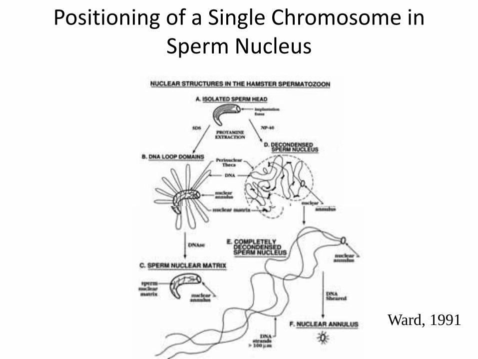
Positioning of a Single Chromosome in Sperm Nucleus
Ward, 1991
• With the compact structure described, how and where the sperm chromatin/DNA can be damaged?
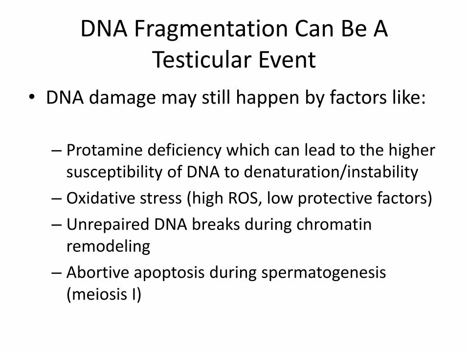
DNA Fragmentation Can Be A Testicular Event
• DNA damage may still happen by factors like:
– Protamine deficiency which can lead to the higher susceptibility of DNA to denaturation/instability
– Oxidative stress (high ROS, low protective factors)
– Unrepaired DNA breaks during chromatin remodeling
– Abortive apoptosis during spermatogenesis (meiosis I)

DNA Fragmentation Can Also Be A Post-Testicular Event
• As a post-testicular event often occurs in epididymis due to:
– Unfavorable epididymal environment
– Endogenous endonucleases, excess estrogens?
– Caspases
– Exogenous gonadotoxic agents
– The reactive oxygen species (ROS)
• The main sources of ROS in semen include leukocytes, abnormal/immature sperm, increased, scrotal temperature, varicocele, advanced male age , smoking, estrogens …

• Different theories and presentations of DNA damage are shown in the next several slides

A two-step hypothesis of DNA damage in the male germ line
(Aitken et al, 2009)
(Aitken et al, 2009)

Google Image
DNA
denaturation
DNA
Fragmentation
(Double or
single stranded
DNA pieces)

What assays are used to assess sperm nuclear DNA status?
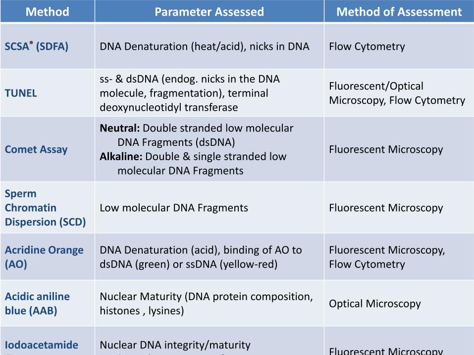
Method Parameter Assessed Method of Assessment
SCSA® (SDFA) DNA Denaturation (heat/acid), nicks in DNA Flow Cytometry
TUNELss- & dsDNA (endog. nicks in the DNA molecule, fragmentation), terminal deoxynucleotidyl transferase
Fluorescent/Optical Microscopy, Flow Cytometry
Comet Assay
Neutral: Double stranded low molecular DNA Fragments (dsDNA)
Alkaline: Double & single stranded low molecular DNA Fragments
Fluorescent Microscopy
Sperm Chromatin Dispersion (SCD)
Low molecular DNA Fragments Fluorescent Microscopy
Acridine Orange (AO)
DNA Denaturation (acid), binding of AO to dsDNA (green) or ssDNA (yellow-red)
Fluorescent Microscopy, Flow Cytometry
Acidic aniline blue (AAB)
Nuclear Maturity (DNA protein composition, histones , lysines)
Optical Microscopy
Iodoacetamidefluorescein
Nuclear DNA integrity/maturity Binds to the SH groups of cysteines in
Fluorescent Microscopy

Method Parameter Assessed Method of Assessment
Toluidine Blue Stain DNA Fragmentation Optical Microscopy
Chromomycin A3
Nuclear Maturity (DNA protein composition)
Fluorescent Microscopy
DNA Breakage Detection via FISH (Fernandez for SCD)
Single Stranded DNA Fragmentation (ssDNA)
Fluorescent Microscopy
In SituNick TranslationSingle Stranded DNA Fragmentation (ssDNA)
Fluorescent Microscopy, Flow Cytometry
8-OHdG Determination8-Hydroxydeoxyguanosine 8-OHdG HPLC
DNA Diffusion AssayDNA Fragmentation using YOYO-1 stain
Fluorescent Microscopy
Gene-specific DNA Damage
-globin, IGF-2, telomericsequences
PCR
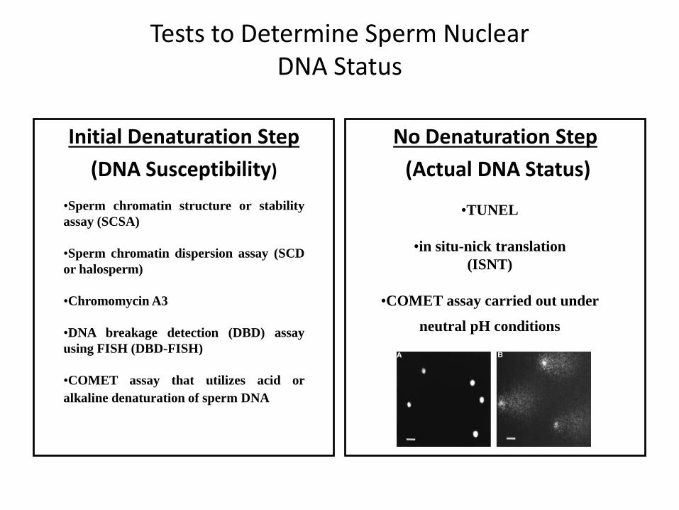
Tests to Determine Sperm Nuclear DNA Status
Initial Denaturation Step
(DNA Susceptibility)
No Denaturation Step
(Actual DNA Status)
•Sperm chromatin structure or stability
assay (SCSA)
•Sperm chromatin dispersion assay (SCD
or halosperm)
•Chromomycin A3
•DNA breakage detection (DBD) assay
using FISH (DBD-FISH)
•COMET assay that utilizes acid or
alkaline denaturation of sperm DNA
•TUNEL
•in situ-nick translation
(ISNT)
•COMET assay carried out under
neutral pH conditions

Principles Behind A Few Commonly Performed Sperm Nuclear DNA Assays• TUNEL
• SCSA
• COMET
• Sperm Chromatin Dispersion Assay (SCD, Halosperm)
• Some of these assays assess DNA status as exists (physiological pH) within the sperm and some determine the DNA status and stability by treating the DNA with acids, bases or heat

Principles Behind the TUNEL Assay
• Name: Terminal deoxynucleotidyl transferase dUTP nick end labeling (TUNEL)
• If sperm have damaged (fragmented) nuclear DNA, there will be many pieces of DNA with 3’ and 5’ ends within their nuclei
• If you have a compound (a nucleotide) that can bind to one of the DNA fragment ends (to the 3’-hydoxyl end) and this compound or nucleotide is attached to a fluorescent dye (FITC-conjugated), then we can read the percentage of sperm with damaged DNA. These cells exhibit strong green fluorescence (TUNEL positive)

Figure 5.
Schematic illustration of DNA strand-break labeling by TdT-mediated Br-dUTP attachment to 3′OH ends and polymerization, followed by immunocytochemical (FITC) detection of BrdU using an antibody to d-UTP (Br-dU Ab FITC).
Cell Prolif. 2005 August; 38(4): 223–243.dUTP= deoxyuridinetriphosphate; TdT= terminal
deoxyribonucleotidyl transferase
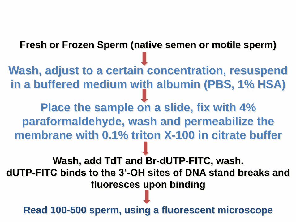
Wash, add TdT and Br-dUTP-FITC, wash.
dUTP-FITC binds to the 3’-OH sites of DNA stand breaks and
fluoresces upon binding
Read 100-500 sperm, using a fluorescent microscope
Fresh or Frozen Sperm (native semen or motile sperm)
Place the sample on a slide, fix with 4%
paraformaldehyde, wash and permeabilize the
membrane with 0.1% triton X-100 in citrate buffer
Wash, adjust to a certain concentration, resuspend
in a buffered medium with albumin (PBS, 1% HSA)
TUNEL Protocol

Questions
• What will be a negative control for the TUNEL assay?
– No dUTP in the mixture
• How about the positive control for the assay?
– Add DNAse to break DNA strands

Calculation: (# of green/total sperm) x 100
TUNEL +
Sperm
TUNEL negative
Sperm
Phase Contrast
Phase Contrast
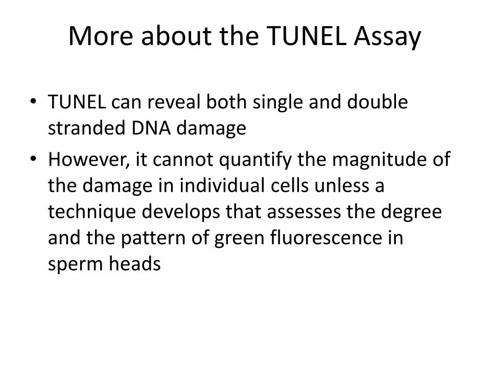
More about the TUNEL Assay
• TUNEL can reveal both single and double stranded DNA damage
• However, it cannot quantify the magnitude of the damage in individual cells unless a technique develops that assesses the degree and the pattern of green fluorescence in sperm heads
Calculation: (# of green/total sperm) x 100
TUNEL +
Sperm
TUNEL negative
Sperm
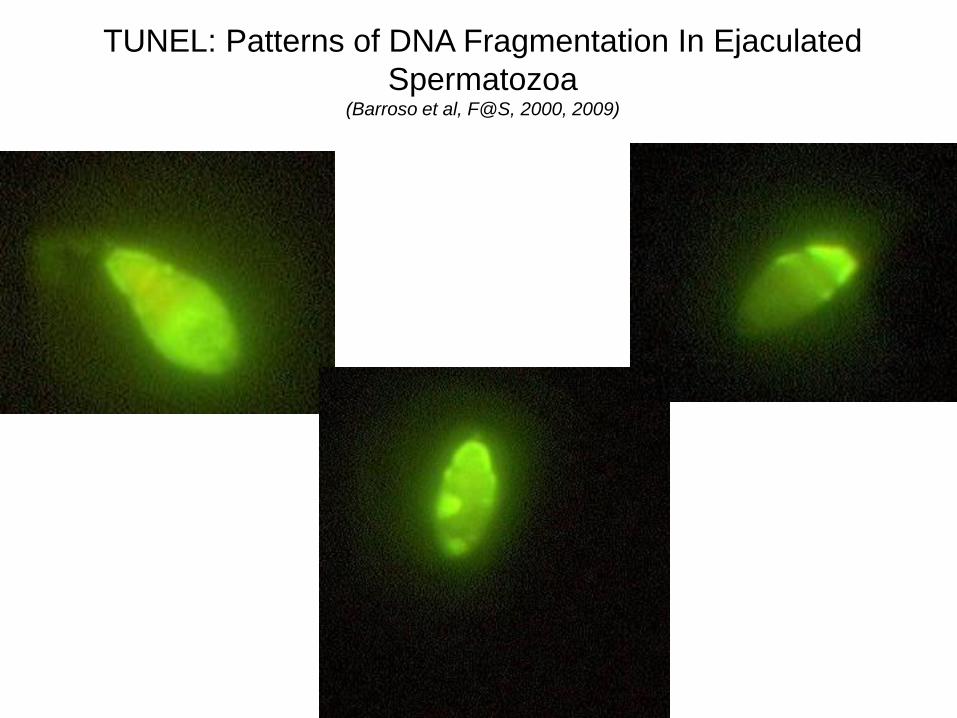
TUNEL: Patterns of DNA Fragmentation In Ejaculated
Spermatozoa(Barroso et al, F@S, 2000, 2009)
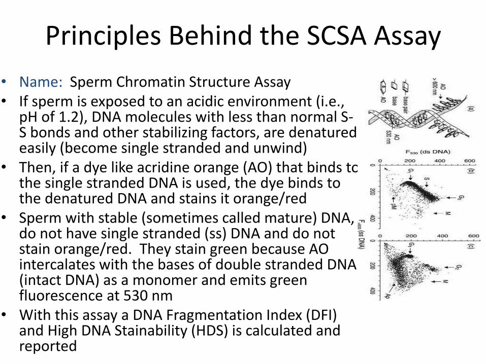
Principles Behind the SCSA Assay
• Name: Sperm Chromatin Structure Assay• If sperm is exposed to an acidic environment (i.e.,
pH of 1.2), DNA molecules with less than normal S-S bonds and other stabilizing factors, are denatured easily (become single stranded and unwind)
• Then, if a dye like acridine orange (AO) that binds to the single stranded DNA is used, the dye binds to the denatured DNA and stains it orange/red
• Sperm with stable (sometimes called mature) DNA, do not have single stranded (ss) DNA and do not stain orange/red. They stain green because AO intercalates with the bases of double stranded DNA (intact DNA) as a monomer and emits green fluorescence at 530 nm
• With this assay a DNA Fragmentation Index (DFI) and High DNA Stainability (HDS) is calculated and reported

Flow Cytometry Determination of Green or Red Sperm
Photomultiplier
Tube
Collecting Lens
Excitation Source
(Laser Beam)
Stained Cells
Slide, courtesy
of Evenson

Fragmented /Denatured DNA (red fluorescence)
Na
tive
DN
A S
tain
abili
ty
(gre
en flu
ore
sce
nce)
SCSA® - Acridine
Orange Stained DNA
100
0100
Slide, courtesy
of Evenson

SCSA® Parameters
DFI = DNA Fragmentation Index
DFI = red fluorescence/ red + green
HDS = =
sperm with defective DNA
High DNA Stainability
More pertinent for natural conception and IUIs rather
than IVF or ICSI

Clinical Results of the SCSA®
Pregnancy OutcomesPregnant:
DFI of <10%
Measurement DFI (%) HDS (%)
1 6.8 5.0
2 8.3 5.4
mean 7.5 5.2
sd 1.1 0.2
Measurement DFI (%) HDS (%)
1 64.9 6.4
2 64.9 7.2
mean 64.9 6.8
sd 0.0 0.4
Non-Pregnant
NOTE: Published results have not been consistent. In general, DFI
of >30% and HDS of >15% are indicative of subfertility SCSA’s
intra-individual coefficient of variation has been calculated as
30+21.5% (range: 0–130%)
Evanson, et al

The Principles Behind the Comet Assay
• The principle behind comet assay is that the negatively-charged broken DNA molecules are free to migrate in an electric field towards the anode (+ pole), with the shorter fragments moving faster. The pattern of migration produces a profile resembling the shape of a comet. Two main principles determine comet formation patterns: the size of DNA fragments and the number of fragments.
+

The Comet Assay: Methodology• Performed at neutral and basic pH• At neutral pH, it reveals only double stranded
DNA breaks• At basic pH, it detects both double and single
stranded DNA breaks
• The assay also is capable of measuring the magnitude of DNA damage
• The assay has not been standardized so techniques are different from lab to lab and the results cannot be compared due to this lack of standardization.
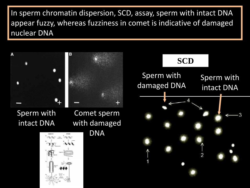
The Comet Assay versus Sperm Chromatin Dispersion Assay
Sperm with intact DNA
Comet sperm with damaged
DNA
In sperm chromatin dispersion, SCD, assay, sperm with intact DNA appear fuzzy, whereas fuzziness in comet is indicative of damaged nuclear DNA
Sperm with intact DNA
Sperm with damaged DNA
SCD
++
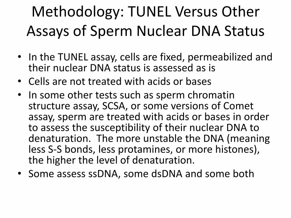
Methodology: TUNEL Versus Other Assays of Sperm Nuclear DNA Status
• In the TUNEL assay, cells are fixed, permeabilized and their nuclear DNA status is assessed as is
• Cells are not treated with acids or bases• In some other tests such as sperm chromatin
structure assay, SCSA, or some versions of Comet assay, sperm are treated with acids or bases in order to assess the susceptibility of their nuclear DNA to denaturation. The more unstable the DNA (meaning less S-S bonds, less protamines, or more histones), the higher the level of denaturation.
• Some assess ssDNA, some dsDNA and some both

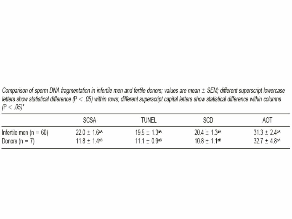
Are there any correlations among the most frequently performed assays of sperm nuclear DNA?

Chohan et al Compared:
• TUNEL with
• SCSA® (SDFA) and
• SCD (sperm chromatin dispersion)
• AO
• 60 men attending the andrology lab and 7 fertile men
• Semen samples were washed only
Chohan K. et al, J Androl, 27(1):2006
Chohan et al J Androl 2006


Ribas-Maynou et al, 2013
Samples were frozen, then tested. All assays except neutral
Comet assay were able to differentiate between fertile
donors and infertile patients
Comet (alkaline): The best, with the threshold value of ~
45% for infertility followed by
TUNEL: Threshold value for infertility ~ 20%
SCD: Threshold value for infertility ~ 23%
SCSA: Threshold value for infertility ~ 19%
Comet (alkaline): No predictive power (Threshold: 34%)

Ribas-Maynou et al, 2013

• What is reported in the literature about the influence of sperm nuclear damage on the outcome of ART?
Use of Sperm with Nuclear DamageMay Result In:
• Poor embryonic development
• Decreased implantation
• Lower pregnancy rates
• Fetal mutations
• Recurrent pregnancy losses
• Increased risk of cancer in offspring

Sperm DNA Damage and Outcome of ART
Study Assay IVF, cases ICSI, cases FR EQ PR
Host, 2000 TUNEL 50 61 Dec., IVF No Report No Report
Tomlinson, 01 TUNEL 140 -- No change No change Decreased
Benchaib, 03 TUNEL 50 54 Dec., ICSI -- Dec., ICSI
Henkel, 04 TUNEL 249 -- No change No Report Decreased
Seli, 04 TUNEL 49 -- No Report Decreased No change
Haung, 05 TUNEL 217 86 Decreased No change No change
Tomsu, 02 COMET 40 -- No change Decreased Decreased
Morris, 02 COMET 20 40 No change Decreased No Report
Larson, 03 SCSA 55 34 No change No change Decreased
Virro, 04 (blast) SCSA 249 -- No change Decreased Decreased
Payne, 05 SCSA 46 54 Decreased No Report Increased
Zini, 05 SCSA -- 60 Decreased Decreased Decreased
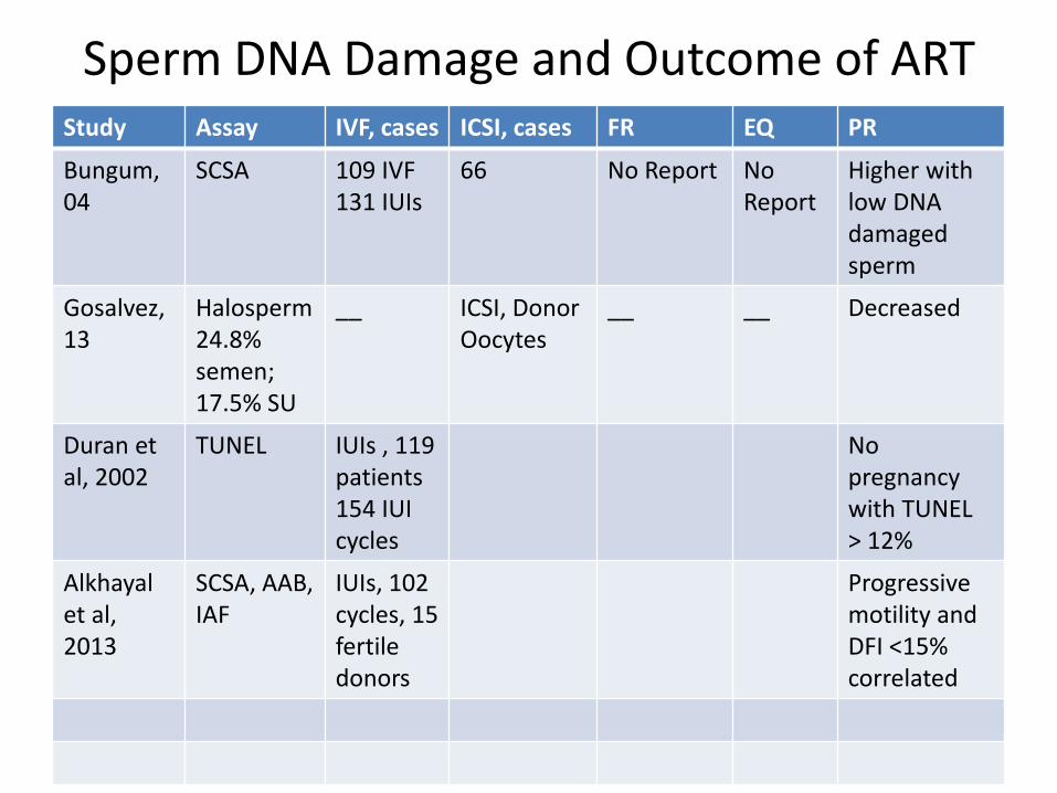
Sperm DNA Damage and Outcome of ARTStudy Assay IVF, cases ICSI, cases FR EQ PR
Bungum, 04
SCSA 109 IVF131 IUIs
66 No Report No Report
Higher with low DNA damaged sperm
Gosalvez, 13
Halosperm24.8% semen; 17.5% SU
__ ICSI, Donor Oocytes
__ __ Decreased
Duran et al, 2002
TUNEL IUIs , 119 patients 154 IUI cycles
No pregnancy with TUNEL > 12%
Alkhayal et al, 2013
SCSA, AAB,IAF
IUIs, 102cycles, 15 fertile donors
Progressive motility and DFI <15% correlated

Some Studies assessed DNA status in neat semen some in the motile fraction
Ranges of DNA-fragmentation (reviewed in Sergerie et al., 2005) have been very wide.
Several studies, working with SCSA, TUNEL and Comet assays, attempted to establish threshold values with which success or failure of natural conception and assisted reproduction treatment could be predicted (see next slide)
The values recommended in different studies showed a high degree of variability with a clear relationship with the type of assay used and the type of assisted reproduction treatment considered
Problems with DNA Fragmentation Studies
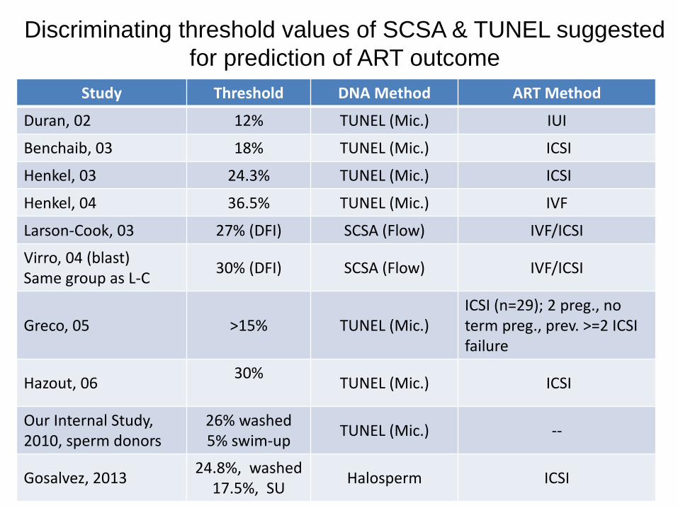
Discriminating threshold values of SCSA & TUNEL suggested
for prediction of ART outcome
Study Threshold DNA Method ART Method
Duran, 02 12% TUNEL (Mic.) IUI
Benchaib, 03 18% TUNEL (Mic.) ICSI
Henkel, 03 24.3% TUNEL (Mic.) ICSI
Henkel, 04 36.5% TUNEL (Mic.) IVF
Larson-Cook, 03 27% (DFI) SCSA (Flow) IVF/ICSI
Virro, 04 (blast)Same group as L-C
30% (DFI) SCSA (Flow) IVF/ICSI
Greco, 05 >15% TUNEL (Mic.)ICSI (n=29); 2 preg., no term preg., prev. >=2 ICSI failure
Hazout, 0630%
TUNEL (Mic.) ICSI
Our Internal Study,2010, sperm donors
26% washed5% swim-up
TUNEL (Mic.) --
Gosalvez, 201324.8%, washed
17.5%, SUHalosperm ICSI

In our donor population, The mean for TUNEL positive sperm in semen was 26% (27+ 13) and for the donor motile sperm was 5%
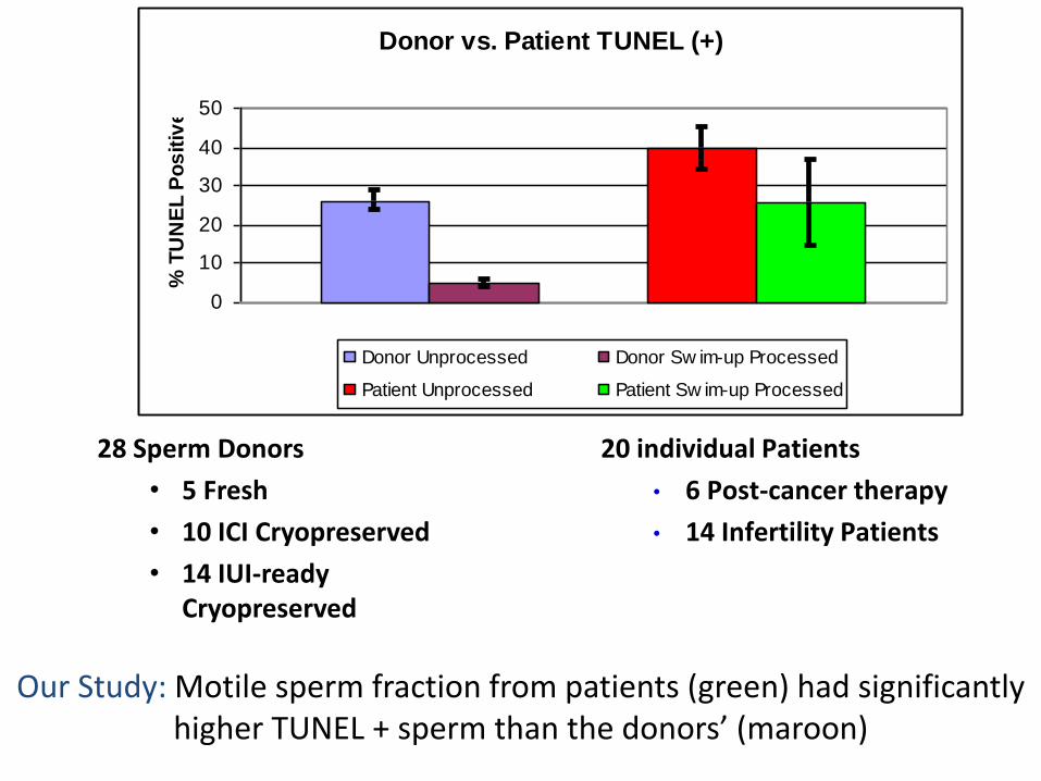
Donor vs. Patient TUNEL (+)
0
10
20
30
40
50
% T
UN
EL
Po
sit
ive
Donor Unprocessed Donor Sw im-up Processed
Patient Unprocessed Patient Sw im-up Processed
28 Sperm Donors
• 5 Fresh
• 10 ICI Cryopreserved
• 14 IUI-ready Cryopreserved
Our Study: Motile sperm fraction from patients (green) had significantly higher TUNEL + sperm than the donors’ (maroon)
20 individual Patients
• 6 Post-cancer therapy
• 14 Infertility Patients

DNA Fragmentation Over Time
0
20
40
60
80
100
T=0 T=4 T=24 T=70
Time (Hours After Swim-Up at RT)
%TUNEL (+) % Motility At Stop
Our small study: DNA fragmentation does not increase significantly upon storage of swim up sample for up to 70 hours at room
temperature (n=3)
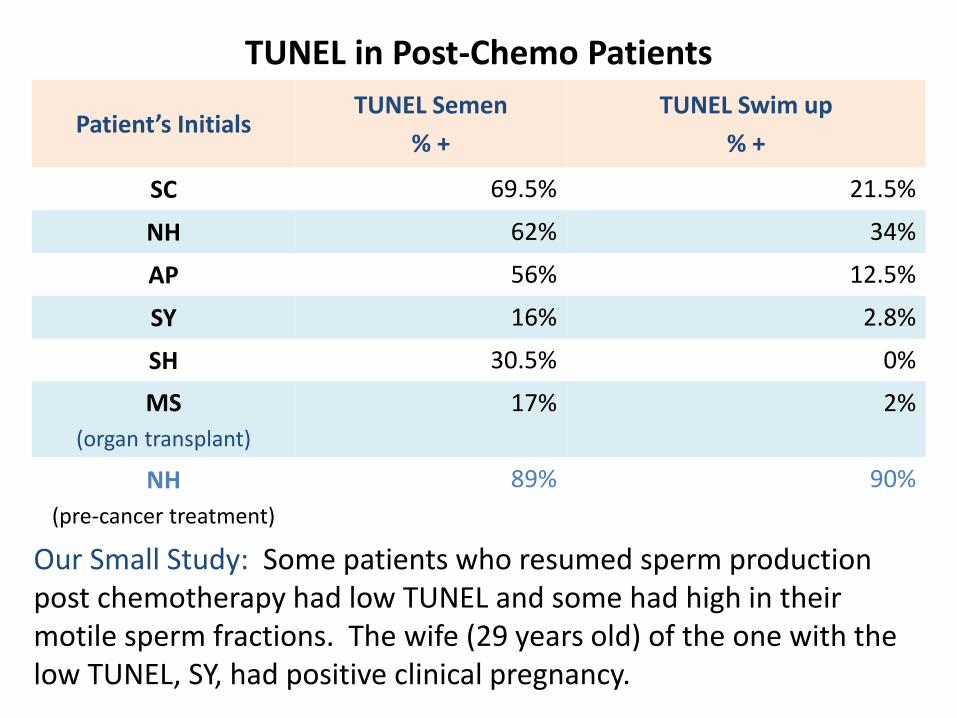
TUNEL in Post-Chemo Patients
Patient’s InitialsTUNEL Semen
% +
TUNEL Swim up
% +
SC 69.5% 21.5%
NH 62% 34%
AP 56% 12.5%
SY 16% 2.8%
SH 30.5% 0%
MS
(organ transplant)
17% 2%
NH
(pre-cancer treatment)
89% 90%
Our Small Study: Some patients who resumed sperm production post chemotherapy had low TUNEL and some had high in their motile sperm fractions. The wife (29 years old) of the one with the low TUNEL, SY, had positive clinical pregnancy.

Does DNA Fragmentation Relate to
Major Semen Parameters?
• Published Studies
• Our Studies
– Sperm morphology and DNA fragmentation (TUNEL)
– Sperm morphology, DNA fragmentation and embryo quality

• Next several slides show that the morphology of sperm selected for ICSI may not necessarily gurantee that the sperm has intact nuclear DNA

N

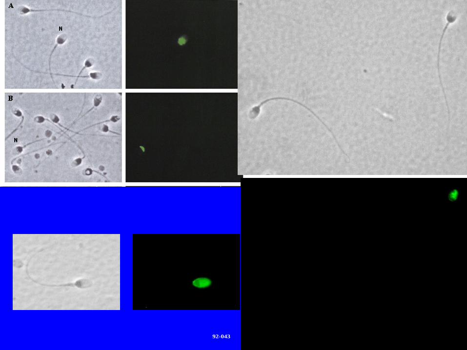
92-043

What is the impact of the proportion of morphologically normal sperm with DNA fragmentation on embryo quality and ICSI outcome?
92-043

If the % of normal-SFD was 17.6 %, the likelihood of pregnancy was 3.5 times higher
Parameter
Area
Under
the
Curve
95%
Confidence
Intervals
P
Cut-off
Point
Sensitivity Specificity
Positive
Likelihood
Ratio
Negative
Likelihood
Ratio
Positive
Predictive
Value
Negative
Predictive
Value
Normal-SFD
0.70 0.53-0.84 0.02 ≤17.6% 61.5 82.6 3.5 0.5 66.7 79.2

DNA Fragmentation (TUNEL) In Fertile, Subfertile and Infertile Men
Normal sperm with DNA fragmentation: 0% (0/4 cases). 30% (1/5
cases), 43% (10/10 cases)

OUR TUNEL REPORTS
• Information about basic semen parameters
• DNA fragmentation in neat semen
• DNA fragmentation in the motile sperm fraction
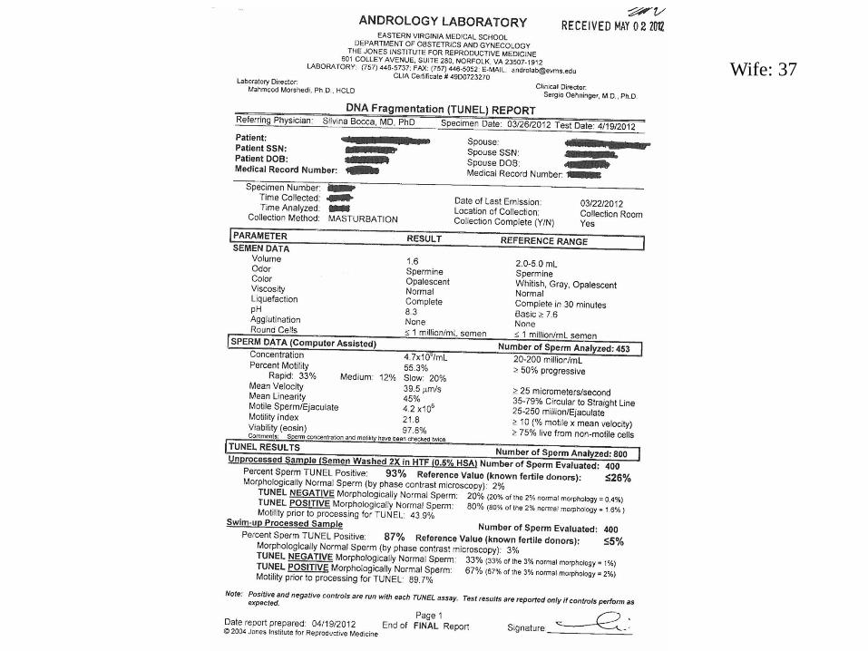
Wife: 37

Two Case Studies
• Case #1

Wife: 37

Wife: 37
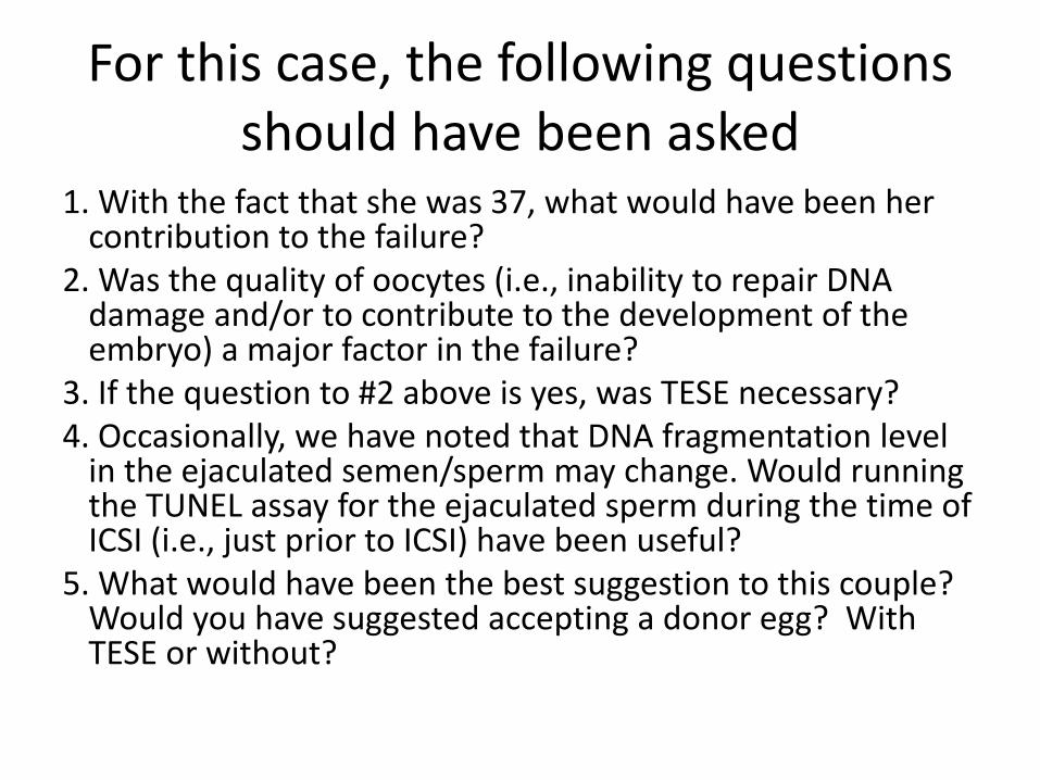
For this case, the following questions should have been asked
1. With the fact that she was 37, what would have been her contribution to the failure?
2. Was the quality of oocytes (i.e., inability to repair DNA damage and/or to contribute to the development of the embryo) a major factor in the failure?
3. If the question to #2 above is yes, was TESE necessary? 4. Occasionally, we have noted that DNA fragmentation level
in the ejaculated semen/sperm may change. Would running the TUNEL assay for the ejaculated sperm during the time of ICSI (i.e., just prior to ICSI) have been useful?
5. What would have been the best suggestion to this couple? Would you have suggested accepting a donor egg? With TESE or without?

Case #2
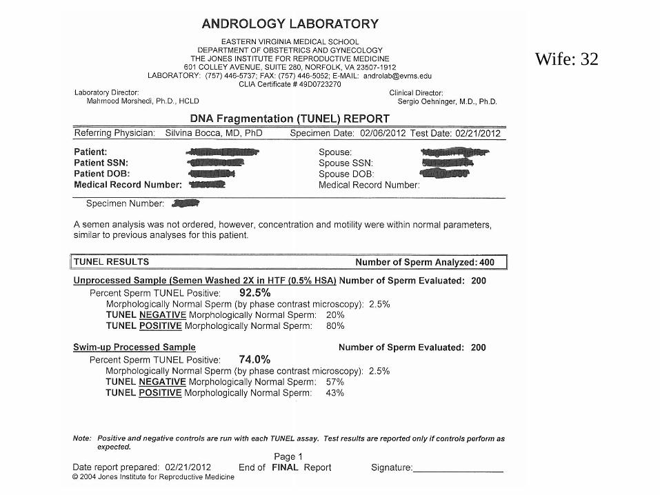
Wife: 32

Wife: 32

The Value Of Sperm Nuclear DNA Assessment In Assisted Conception: Some Thoughts
• Different methodologies has hampered our judgment about the value of the tests
• Tests give only the percentage of cells with damaged DNA. They do not reveal the extent of the damage in the sample being used.
• Setting thresholds is misleading as may vary in various sites, patients, ART methods. They can be used as a guide.

• We often do not ask if the DNA fragmentation is the sole or partial cause of the problem
• Focusing on the sperm DNA status, have negated the contribution of the oocyte to the success or to the failure
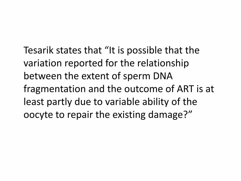
Tesarik states that “It is possible that the variation reported for the relationship between the extent of sperm DNA fragmentation and the outcome of ART is at least partly due to variable ability of the oocyte to repair the existing damage?”

Tesarik, 03-04, DNA Damage (TUNEL, microscopy) in 4 Groups
• Those achieving term pregnancy in their first ICSI attempt
• Those not achieving term pregnancy in their first ICSI attempt
• Those achieving term pregnancy in their third ICSI attempt after two previous failures
• Those not achieving term pregnancy in their third ICSI attempt after two previous failures
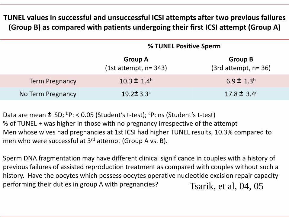
TUNEL values in successful and unsuccessful ICSI attempts after two previous failures (Group B) as compared with patients undergoing their first ICSI attempt (Group A)
% TUNEL Positive Sperm
Group A(1st attempt, n= 343)
Group B (3rd attempt, n= 36)
Term Pregnancy 10.3 ± 1.4b 6.9 ± 1.3b
No Term Pregnancy 19.2± 3.3c 17.8 ± 3.4c
Data are mean ± SD; bP: < 0.05 (Student’s t-test); cP: ns (Student’s t-test)% of TUNEL + was higher in those with no pregnancy irrespective of the attemptMen whose wives had pregnancies at 1st ICSI had higher TUNEL results, 10.3% compared to men who were successful at 3rd attempt (Group A vs. B).
Sperm DNA fragmentation may have different clinical significance in couples with a history of previous failures of assisted reproduction treatment as compared with couples without such a history. Have the oocytes which possess oocytes operative nucleotide excision repair capacity performing their duties in group A with pregnancies? Tsarik, et al, 04, 05

TUNEL values in successful and unsuccessful ICSI attempts performed with the patients’ own oocytes (Group A) as compared with attempts performed with
donated oocytes (Group B)
% TUNEL Positive Sperm
Group A: Pt’s oocytes n= 268
Group B: Donor oocytesn= 281
Woman’s ageb 34.7± 3.2c 21.8 ± 1.1c
Term Pregnancy 6.4 ± 1.4d 11.8 ± 2.9d
No Term Pregnancy 13.0 ± 2.8c 13.7 ± 3.2e
Data are mean ± SD; bAge of the female from which oocytes were retrievedcP < 0.001 (Student’s t-test); dP < 0.001 (Student’s t-test); eP: ns (Student’s t-test)TUNEL results were higher in patients who did not achieve pregnancy, irrespective of the age of the women from which oocytes were retrieved. Oocytes from young donors enables the establishment of a term pregnancy with higher percentages of DNA-fragmented spermatozoa as compared with attempts performed with oocytes coming from the significantly older patient
wives population.
Tesarik et al, 04, 05

Treatments to Reduce the Impact of Sperm DNA Damage
• ICSI using surgically retrieved testicular sperm instead of ejaculated ones (Tesarik, 04; Greco, 05)
• ICSI with ejaculated sperm after 2 months of oral antioxidant treatment (Greco, 05)
• ICSI with sperm selected with the use of a high-magnification optical system (high-magnification ICSI) (Hazout, 06)
• Oral antioxidant therapy??
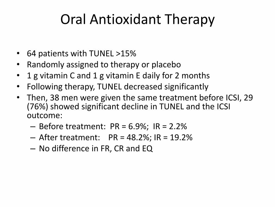
Oral Antioxidant Therapy
• 64 patients with TUNEL >15%• Randomly assigned to therapy or placebo• 1 g vitamin C and 1 g vitamin E daily for 2 months• Following therapy, TUNEL decreased significantly• Then, 38 men were given the same treatment before ICSI, 29
(76%) showed significant decline in TUNEL and the ICSI outcome:– Before treatment: PR = 6.9%; IR = 2.2%– After treatment: PR = 48.2%; IR = 19.2%– No difference in FR, CR and EQ

• Sort of using the same sperm evaluated for DNA damage, it is not possible to know the extent of DNA damage in an individual sperm fertilizing the oocyte
• It is debatable whether any one of these tests is more preferable than the others to optimize clinical decision-making
• Finally, The ASRM, after meta-analysis of eligible studies, concluded that there is no proven role for routine DNA fragmentation testing in the evaluation of infertility

• Regardless of what has been published, the importance of sperm unclear DNA status assays cannot be disputed.
• We must learn how to utilize the results obtained particularly in conjunction with the female associated factors

END!