human myelin proteome and comparative analysis with mouse … · cgi doi 10.1073 pnas.0905936106...
TRANSCRIPT

Human myelin proteome and comparative analysiswith mouse myelinAkihiro Ishiia, Ranjan Duttab, Greg M. Warka, Sun-Il Hwangc, David K. Hanc, Bruce D. Trappb, Steven E. Pfeiffera,and Rashmi Bansala,1
Departments of aNeuroscience and cCell Biology and Center for Vascular Biology, University of Connecticut Medical School, Farmington, CT, 06030;and bDepartment of Neurosciences, Lerner Research Institute, Cleveland Clinic, Cleveland, OH 44195
Communicated by Gordon H. Sato, Manzanar Project Foundation, Wenham, MA, July 13, 2009 (received for review April 15, 2008)
Myelin, formed by oligodendrocytes (OLs) in the CNS, is critical foraxonal functions, and its damage leads to debilitating neurologicaldisorders such as multiple sclerosis. Understanding the molecularmechanisms of myelination and the pathogenesis of human myelindisease has been limited partly by the relative lack of identificationand functional characterization of the repertoire of human myelinproteins. Here, we present a large-scale analysis of the myelinproteome, using the shotgun approach of 1-dimensional PAGE andliquid chromatography/tandem MS. Three hundred eight proteinswere commonly identified from human and mouse myelin frac-tions. Comparative microarray analysis of human white and graymatter showed that transcripts of several of these were elevatedin OL-rich white matter compared with gray matter, providingconfidence in their detection in myelin. Comparison with otherdatabases showed that 111 of the identified proteins/transcriptsalso were expressed in OLs, rather than in astrocytes or neurons.Comparison with 4 previous myelin proteomes further confirmedmore than 50% of the identified proteins and revealed the pres-ence of 163 additional proteins. A select group of identifiedproteins also were verified by immunoblotting. We classified theidentified proteins into biological subgroups and discussed theirrelevance in myelin biogenesis and maintenance. Taken together,the study provides insights into the complexity of this metaboli-cally active membrane and creates a valuable resource for futurein-depth study of specific proteins in myelin with relevance tohuman demyelinating diseases.
microarray � oligodendrocyte � proteomics � mass spectrometry
Myelin is a biologically active multilamellar membrane thatis formed by oligodendrocytes (OLs) in the CNS and
ensheathes axons. Communication between myelin and axons iscritical for axonal functions, including rapid nerve conduction,survival, and cytoskeletal organization (1). The loss or damageof the myelin sheath results in serious neurological diseases suchas multiple sclerosis. In addition, defects in myelin are correlatedwith schizophrenia (2) and age-dependent decline in brainfunction (3).
To prevent or ameliorate diseases of myelin, it is essential tounderstand the mechanism of its biogenesis and maintenance,which requires the identification and functional characterizationof all myelin proteins—that is, the myelin proteome. Taylor et al.(4) reported the first rodent myelin proteome, and severalstudies then expanded it (5–7). Each study identified someadditional proteins and others that were identified in previousproteomic studies, suggesting that no single identificationmethod could successfully identify all myelin proteins. Never-theless, these studies highlighted the complexity of myelin inrodents, which commonly are used as models for human disease.However, to apply this knowledge to diseases of human myelin,it is essential to establish a human myelin proteome and compareit with that in rodents.
Here, we present a large-scale undertaking that combinesproteomics of human and mouse myelin and microarray analysisof human white and gray matter. It brings together information
from multiple sources, including the extensive transcriptomedatabase of Cahoy et al. (8), to provide the field with acomprehensive picture of the numerous proteins not only ofhuman myelin but also of those whose transcripts are enrichedin human CNS white matter over gray matter and in purifiedrodent OLs over astrocytes and neurons, providing increasedconfidence in their identification in myelin. Performing humanand mouse myelin proteomic analyses in parallel has allowed usto present at least 308 target proteins that are definitively presentin myelin fractions of both species; thus, an investigation of themin mice clearly will be relevant for human studies. Further, acomparison of all the proteins that have been identified in 4previous proteomes (4–7) has provided the identity of 163additional proteins, many of which previously were unrecog-nized as being components of myelin. Based on our compre-hensive analysis, we have short-listed 64 potentially interestingproteins for future in-depth analysis and, as an example, dem-onstrated by immunoblot analysis that 8 of these proteins areindeed reproducibly present in multiple human myelin samples.These analyses provide a valuable foundation and database forfuture studies to understand the functional implications of thepresence of these proteins in normal human myelin and in thepathology of human myelin diseases.
Results and DiscussionCharacterization of Human Myelin Samples for Proteomic Analysis.Myelin was purified from whole mouse brain and from multiplesmaller sections of human brain from 4 subjects [supportinginformation (SI) Fig. S1]. Because these brain sections varied intheir yield of myelin as a result of differences in their whitematter content, we first asked if myelin that was purified fromthese sections using sucrose density gradient fractionation (seeMaterials and Methods) was similar with respect to its content ofmyelin proteins. Therefore, levels of major myelin proteins [2�,3�-cyclic-nucleotide 3�-phosphodiesterase (CNP), myelin-associated glycoprotein (MAG), myelin basic protein (MBP),myelin OL glycoprotein (MOG) and OL-specific protein (OSP),myelin proteolipid protein (PLP)] were examined in 11 differentsamples from 4 human subjects through immunoblotting of the‘‘main band fraction’’ of myelin (Fig. 1A). Each myelin sampleshowed similar intensities of signal, suggesting similar contentsof myelin proteins in these samples.
It has been noted in rodents that myelin purification by sucrosedensity gradient centrifugation enriches compact myelin pro-teins in the lowest-density main band fraction, whereas axolem-mal proteins are enriched in the high-density ‘‘pellet fraction’’(9). To examine if this holds true for human myelin and to
Author contributions: S.E.P. and R.B. designed research; A.I., R.D., S.-I.H., and G.M.W.performed research; D.K.H. and B.D.T. contributed new reagents/analytic tools; A.I., R.D.,G.M.W., and R.B. analyzed data; and A.I., G.M.W., and R.B. wrote the paper.
The authors declare no conflict of interest.
1To whom correspondence should be addressed. E-mail: [email protected]
This article contains supporting information online at www.pnas.org/cgi/content/full/0905936106/DCSupplemental.
www.pnas.org�cgi�doi�10.1073�pnas.0905936106 PNAS � August 25, 2009 � vol. 106 � no. 34 � 14605–14610
NEU
ROSC
IEN
CE

evaluate the relative contamination by neuronal and astrocyticproteins of the main band fractions, we analyzed 3 major myelinproteins (MOG, MBP, and PLP), 2 neuronal proteins [contactin-associated protein-2 (Caspr-2) and postsynaptic density-95(PSD95)], and 1 astrocytic protein (GFAP) by immunoblotting(Fig. 1B). Our results indicate that all samples of human myelinfractionated in a similar manner as rodent myelin, with enrich-ment of myelin proteins in the main band fraction and relativelyminimal contamination of this fraction by neuronal and astro-cytic proteins.
Therefore, we conclude that sections of human brain do notneed to be precisely matched for white matter content, patientage, or sex for representative proteomic analysis of humanmyelin and that the main band fractions from any of the myelinsamples are suitable for representative MS analysis of humanmyelin proteins.
Protein Identification by Sequential 1-Dimensional PAGE and LiquidChromatography/Tandem MS. Myelin samples from human sub-jects 3 and 4 (Fig. S1) and P35 mouse brains were separated by1-dimensional gel electrophoresis, followed by peptide extrac-tion and analysis by liquid chromatography and tandem MS(LC-MS/MS). This method avoids some of the limitations thatare inherent to the commonly used 2D-PAGE separationmethod. For example, in 2D-PAGE, a protein must be prevalentenough to be excised. Using the 1-dimensional method, in-geltryptic digestion of slices from the whole gel avoids the need forindividual proteins to be recognized. One-dimensional PAGEalso avoids the bias of 2D-PAGE that causes exclusion of specificprotein populations (e.g., high-molecular-weight and basic pro-teins), which often remain unidentified; for example, MAG andMOG, which were not detected in previous studies (4, 6), weredetected by the present technique.
Duplicate analysis was performed on all samples becauseearlier studies indicated that 1 MS analysis is not sufficient forexhaustive protein coverage (10). Consistently, in the presentstudy, we detected additional proteins upon reanalysis of thesame sample (average of 26% more proteins for human subjects3 and 4 and 21% for mouse). However, despite this increase,certain known myelin proteins, such as connexin-32, remainedunidentified.
Combining identified proteins from the duplicate analysis ofboth human subjects, we detected a total of 678 proteins inhuman myelin fractions (Fig. 2A). Similarly, 515 proteins wereidentified from mouse myelin fractions. Venn diagram compar-isons showed that 308 proteins overlapped between human andmouse myelin. These proteins, belonging to various functionalgroups (Fig. 2B), are listed in Fig. 3 and will be the main focusof the present study. The complete list of all of the identifiedproteins is provided in Table S1.
Although some of the proteins that fell outside the commonlyidentified protein category may represent true biological differ-ences between the species, for the most part it seems unlikely tobe the case because of the technical limitations of MS. Forexample, proteins with slightly different amino acid sequences(e.g., human and mouse homologues) may have peptides withdifferent physicochemical characteristics, resulting in the detec-tion of these proteins in one species but not in the other. Inaddition, incomplete sampling of complex mixtures may pre-clude the identification of all proteins in a given sample (asdescribed earlier). Consistent with this, immunoblot analysis of
Fig. 2. Myelin proteins identified by 1-dimensional GelC-MS/MS. (A) Venndiagram shows the numbers of proteins identified by 1-dimensional GeLC-MS/MS shotgun approach in myelin fractions purified from human and mousebrains. A total of 678 proteins were identified from human and 515 frommouse. To allow for comparison, the mouse identifications were converted tothe corresponding human UniProtKB database entry. Although 475 proteinsconverted successfully, 40 remained unconverted. The 308 proteins that over-lapped are listed in Fig. 3, and the total proteins that were identified in thestudy are listed in supplementary Table S1. (B) Classification of 308 commonlyidentified proteins was done according to their function using SwissProt or viaa literature search.
Fig. 1. Characterization of human myelin samples for proteomic analysis. (A) Myelin was purified from 11 sections of normal human brain, obtained from differentsubjects (Fig. S1). Yield (%) is based on the amount of myelin protein (main band fraction) obtained from the total protein present in the starting brain homogenate.Equal amounts of proteins (5 �g) from main band myelin fractions were analyzed by immunoblotting for 6 specific major myelin proteins (CNP, MAG, MOG, MBP, OSP,and PLP). Note that each myelin sample showed similar intensities of signal, suggesting similar contents of myelin proteins in these samples. (B) Homogenate (Ho),prepared from 6 samples of normal human brain that were obtained from different regions of 4 subjects and from a whole mouse brain, was fractionated by sucrosegradient centrifugation, and 4 fractions floating at increasing densities of sucrose were collected (MB, main band; Di, dispersion; HB, heavy band; Pe pellet). For eachsample, an equal amount of total protein (5 �g) was analyzed by immunoblotting for 3 major myelin proteins (MOG, MBP, PLP), 2 neuronal proteins (Caspr-2, PSD-95),and 1 astrocytic protein (GFAP). Blots are from different experiments and are intended only to show the relative distribution of each protein in the 4 fractions. Notethatall samplesofhumanmyelin fractionated inasimilarmanneras rodentmyelin,withenrichmentofmyelinproteins in themainbandfractionandrelativelyminimalcontamination of this fraction by neuronal and astrocytic proteins. *Samples used for MS analysis.
14606 � www.pnas.org�cgi�doi�10.1073�pnas.0905936106 Ishii et al.
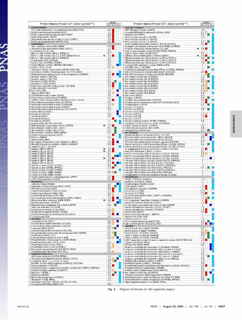
Fig. 3. (Figure continues on the opposite page.)
Ishii et al. PNAS � August 25, 2009 � vol. 106 � no. 34 � 14607
NEU
ROSC
IEN
CE

6 proteins, as an example, showed that proteins that were notidentified by MS in one species could be identified by immuno-blot analysis in that species (Fig. S3). MS identifications also mayvary from protein to protein, even within the same family. Forexample, among the tetraspanin family, CD9 and plasmolipinwere identified only in human, even though they also wereexpressed in mouse myelin, as shown by immunoblotting for CD9(Fig. S3) and by a literature search for plasmolipin (11). CD81,to the contrary, was identified in both species, whereas MAL/MVP17 was not detected in either human or mouse samplesbecause its amino acid sequence precludes it from MS detection(see SI Methods). These data emphasize the limitations of MSanalysis and show that non-identification of a protein by MS doesnot prove conclusively that it is absent from the sample. Thus, thecurrent overlap that is seen between human and rodent pro-teomes is most likely an underestimation.
Comparison of the commonly identified proteins in this studywith 4 other myelin proteomes (4–7) has validated more than 50%of these proteins. Considering the technical differences among the5 approaches, the significant overlap provides considerable confi-dence in their identification. This comparison also has revealed thepresence of 163 proteins that have not previously been described inany other proteome, thus providing insights into the complexcomposition of myelin (Fig. 3 and Fig. S2).
Validation of MS Identification. The rigorously purified main bandmyelin fraction that was used in this study was highly enriched
in myelin proteins, and negligible amounts of neuronal andastrocytic proteins were detected on the immunoblots (Fig. 1B).However, it is difficult, if not impossible, to obtain totally ‘‘puremyelin,’’ presumably because of high-affinity interactions be-tween myelin and axolemmal membranes (9). Given the highsensitivity of MS detection, it is likely that some axonal orastrocytic proteins exist in our (and previous) myelin proteomes.To address this issue and increase confidence in the MS iden-tification of proteins in purified myelin fractions, we argued thatif a protein or mRNA that encodes a protein is expressed by OLs,the cellular source of myelin, it should be a true myelin com-ponent. We therefore used different criteria to help ascertain thecellular origins of proteins that were identified in our proteome.
First, we compared our database with other studies, primarilythe transcriptome database of OLs, neurons, and astrocytes thatwere isolated from rodent brains (8, 12–18). Among the 308proteins, there were at least 111 proteins in our myelin proteomewhose mRNA or protein was expressed by OLs (Fig. 3, columnO, red and tan boxes). Interestingly, 64 of these also were highlyenriched in OLs, not in neurons, and rarely in astrocytes[supplemental tables 4–6 and 17–19 of Cahoy et al (8); see SIMethods]. This group of proteins, which are short-listed in Fig.S2, is likely to play a preferential role in OL/myelin biology.Some mRNAs, which were expressed in 2 or more cell types,possibly have common functions in these cell types. For example,mRNA that encodes RAC1 is expressed by OLs and astrocytes.
Fig. 3. Proteins of human myelin and microarray analysis of human white and gray matter: Functional categorization of identified proteins and comparisonwith other myelin proteomes and transcriptomes of OL-, astrocyte-, or neuron-enriched genes. The 308 proteins commonly identified by MS in human and mousemyelin fractions are categorized into several functional subgroups. Relative intensities of mRNA signals for these proteins was determined by microarray analysesof human white (W) and gray matter (G). Many of these transcripts are expressed at higher levels in OL-rich white over gray matter, shown as fold enrichment(i.e., W/G ratio). In addition, to help ascertain the cellular origins of the proteins identified in our proteome, the relative enrichment of their transcripts in purifiedOLs (red squares in column O), neurons (column N), or astrocytes (column A) was determined through analysis of the transcriptome database of Cahoy et al. (8)and other studies (12–18) of OLs (tan squares in column O). Comparison of the identified proteins in this study with other myelin proteomes (4–7) has confirmedmore than 50% of the identified proteins and revealed the presence of 163 additional proteins in myelin that have not previously been reported in any otherproteome (column U). *Human protein ID in the UniProt Knowledgebase; see Table S1 for corresponding mouse IDs. **Gene symbols as described in NationalCenter for Biotechnology Information database are given only when different from protein ID. mRNA, microarray analysis; X, not present in array chip ortranscripts below reliable detection threshold; IB, proteins validated by immunoblotting in Fig. 4.
14608 � www.pnas.org�cgi�doi�10.1073�pnas.0905936106 Ishii et al.

RAC1 signaling is important for myelin sheath formation (19)and growth of astrocytic processes (20).
Second, we quantified mRNAs that encoded the 308 proteinsin human CNS white and gray matter by microarray analysis.Transcripts of several proteins were highly expressed in theOL-rich white matter compared with gray matter, suggesting anassociation of these transcripts with myelin-producing OLs invivo. Conversely, some other proteins, such as neurofilamentpeptides, which were found at low levels in human whitecompared with gray matter, were obviously axonal proteins thatco-purified with myelin fractions. White-to-gray matter (W/G)ratios are shown in Fig. 3 to facilitate such comparisons for allthe proteins. In addition, when the W/G ratios of the short-listedproteins were examined (Fig. S2), we found that approximatelyhalf the transcripts that were enriched in OLs also were elevated1.5 to 7.5 fold in human white over gray matter. Combining the2 criteria has further narrowed the selection of candidates thatare true myelin proteins, many of which have not been reportedpreviously (Fig. S2).
Third, we searched for the tissue localization of mRNAs of selectproteins in mouse brain from the Allen Brain Atlas (http://www.brain-map.org). The in situ hybridization results showed thatexpression of several transcripts that encoded these proteins wasdetected in the white matter tracts of adult mouse brain.
Last, to verify the MS and microarray identification, as anexample, we performed immunoblot analysis (Fig. 4) on 8proteins that were selected among 64 proteins from the short list(Fig. S2) and belonged to different functional subgroups whosemRNAs were expressed at high levels in human white matterover gray matter and whose mRNA was enriched in purifiedrodent OLs but not neurons or astrocytes. We reliably detectedthe presence of all 8 proteins [band 4.1 G, sirtuin-2, septin-4,septin-7, tubulin polymerization-promoting protein, chlorideintracellular channel protein 4 (CLIC4), dihydropteridine re-ductase (DHPR), and N-myc downstream-regulated gene 1(NDRG1)] on immunoblots of 11 different human myelinpreparations from 4 human subjects. In addition, we also verified6 other identified proteins (Rac-1, CD81, contactin-1, reticu-lon-4, RalA, and creatine kinase B) by immunoblotting.
Together, this analysis provides increased confidence in our
MS detection of numerous proteins that are present in purifiedhuman myelin.
Functional Implications of Proteins Identified in Myelin Proteome. Asexpected, several classical myelin proteins, such as CNP, MAG,MBP, MOG, OSP, and PLP, were found in both human andmouse myelin, and are known to constitute more than 90% oftotal myelin proteins in the rodent. It is becoming clear fromnumerous proteomic analyses (4–7) that there are in fact myriadadditional proteins that are present in rodent myelin. The highestnumber that has been identified in a single study is approxi-mately 160 (7), but the number has been increased to 515 in thepresent analysis. These quantitatively ‘‘minor’’ proteins of myelinmay not necessarily be ‘‘functionally minor,’’ as many of themplay important roles in OL and myelin biology. For example,ermin, an OL protein involved in cytoskeletal rearrangementduring the late wrapping and/or compaction of myelin (17), andNecl4, involved in myelination in the peripheral nervous system(21, 22), were identified in this study but not in previous studies,illustrating the usefulness of this sensitive proteomic approachfor identifying functionally relevant minor myelin proteins.
Among the 308 proteins that were identified by this approach,we detected a variety of proteins that were involved in signaling,cytoskeletal organization, cell adhesion, protein trafficking andvesicular transport, ion transport, endoplasmic reticulum andmitochondrial function, and energy metabolism (Figs. 2B and 3and Fig. S4). All these functions are critical for myelin biogenesisand maintenance.
The presence of a large number of cytoskeletal proteins in themyelin proteome was not unexpected, because the cytoskeletonis a necessary component for formation and maintenance of themyelin sheath (23), including targeting of the major myelinproteins and mRNA to the myelin membrane (24, 25). Inaddition to septin-4 and septin-7, which were verified by immu-noblot analysis, several other members of the septin family(septins 2, 5, 6, 8, 9, and 11) were also detected in the myelinproteome. Microtubule proteins that were identified included�4-tubulin, selectively expressed by OLs (16); microtubule-associated protein (MAP) 1B, expressed by OLs in vitro (12);and MAP1A, MAP2, and Tau. Interestingly, Band 4.1G andtubulin polymerization-promoting protein, reported in OLs (26),have been identified and verified by immunoblot to be presentin myelin.
Biogenesis and maintenance of myelin require vesicular trans-port for the sorting and trafficking of proteins and lipids by OLsto the myelin membrane. Several transport proteins from relatedfamilies were identified in our study from both human androdent. For example, the small GTPase family of proteins,including those that are expressed in OLs (Rab3a, 5a, 5c, 6, andRalA) (13, 27); syntaxins, involved in vesicle fusion, includingsyntaxin-4, which is up-regulated during OL differentiation (15);and clathrin coat-related proteins, involved in vesicle budding,including clathrin heavy chain, AP1b1, APa1, and AP2a2, wereidentified. The multifunctional protein CLIC4, which exists inboth soluble and membrane forms and is known to be localizedto several membrane systems, including the trans-Golgi network,secretory vesicles, and plasma membrane (28), was detected forthe first time in myelin membrane by both MS and immuno-blotting (Figs. 3 and 4). NDRG1, a cytoplasmic protein that isexpressed by OLs (29) and validated by immunoblotting inhuman myelin (Fig. 4), has been identified as a Rab4a effectorrecruited to recycling/sorting endosomes in cell lines (30).
Several mitochondrial proteins have been consistently iden-tified in ‘‘purified’’ myelin fractions by proteomic analysis (4–7).The present study also found a large number of mitochondrialproteins in myelin of both human and mouse, including TCAcycle enzymes (e.g., aconitate hydratase) and respiratory chainenzymes (e.g., cytochrome c). Although the possibility remains
Fig. 4. Immunoblot analysis of select proteins from different subgroupsidentified by MS to validate their presence in human myelins. Eight proteins,belonging to different functional subgroups, were selected as an example tovalidate their identification by MS in human myelin fractions. The criteria forselection was that (i) their mRNA was enriched in OLs (column O) but not inneurons (column N) or astrocytes (column A), and (ii) their mRNA expressionwas at least 1.7 fold higher in human white matter than gray matter. All 8proteins were detected reproducibly by immunoblotting in each of the 11human myelin samples from 4 subjects, providing increased confidence intheir MS detection. Three of them have not been identified in previous rodentmyelin proteomes (column U). TPPP/P25, tubulin polymerization-promotingprotein; CLIC4, chloride intracellular channel protein 4; DHPR, dihydropteri-dine reductase; NDRG1, N-myc downstream-regulated gene 1; Vesic. Trans,vesicular transport; Metb, metabolism.
Ishii et al. PNAS � August 25, 2009 � vol. 106 � no. 34 � 14609
NEU
ROSC
IEN
CE

that they represent a contamination of myelin (5, 7), mitochon-dria are known to localize in OL processes (31) and are presentin peripheral nervous system (32) and CNS myelin (33). It islikely that myelin internode mitochondria provide the ATP thatis needed for the expansion of myelin membranes during my-elination. In addition, mitochondrial dysfunction occurs in var-ious demyelinating neurological diseases, suggesting the impor-tance to human diseases.
In this large-scale proteomic analysis of human and mousemyelin, with the use of multifocal approaches, we have presentedevidence that promotes confidence in our MS identification.This study has vastly expanded the repertoire of myelin proteinsand revealed a more complete picture of the composition ofmyelin proteins that have a variety of possible functions that arerelevant for myelin biogenesis and maintenance. This databaseis expected to serve as a roadmap for future in-depth study ofboth human and mouse myelin biology and the understanding ofhuman diseases that pertain to myelin pathology.
Materials and MethodsMyelin Purification. Postmortem brains from 4 human subjects with no neu-rological disorders were dissected as described previously (34) and summa-rized in Fig. S1. Eleven sections with varying white matter content were cutfrom these brains. Myelin was purified from these samples and from mousebrain (postnatal day 35; C57BL/6) by sucrose density gradient centrifugation asdescribed previously (9). Four fractions were collected, floating at differentsucrose densities: main band (12.0%–22.5%), ‘‘dispersion’’ (22.5%–25.4%),‘‘heavy band’’ (25.5%–26.9%), and ‘‘pellet.’’ Total protein concentrationswere determined by BCA protein assay (Bio-Rad).
MS Analysis. MS was performed as described previously (35). Briefly, mainband myelin fractions (150 �g protein) from human subjects 3 and 4 andmouse were separated by 1-dimensional gel electrophoresis, followed bycolloidal Coomassie staining. Gel lanes were sliced, trypsinized, and extracted.Peptides were analyzed by LC-MS/MS using an LTQ ion-trap mass spectrome-ter. Proteins that were identified by the presence of at least 2 distinct peptidesin the same sample were accepted as high-confidence identification. Dupli-cate analyses were performed for each sample. Further details of MS and dataanalysis are provided in SI Methods.
Immunoblotting. Total proteins from purified human and mouse myelin wereseparated by SDS/PAGE, transferred onto polyvinylidene difluoride mem-branes, incubated with primary antibody (1 h at room temperature) andhorseradish peroxidase-conjugated secondary antibodies (1 h at room tem-perature), and diluted in blocking solution (5% nonfat dry milk in Tris-buffered saline solution with 0.2% Tween 20). Blots were visualized usingenhanced chemiluminescence (GE Healthcare). Details of antibodies are givenin SI Methods.
Microarray Analysis. Eight samples each of white matter or gray matter tissuefrom the same tissue block were collected from 3 postmortem human brains(Fig. S1). RNA was extracted, and microarray and data analysis were per-formed as described previously (34) (see SI Methods).
ACKNOWLEDGMENTS. We dedicate this article to the memory of Dr. S.E.Pfeiffer, who was a source of inspiration and guidance for this work and willcontinue to be remembered in the field of myelin biology, especially for hispioneering contributions in the quest for new myelin proteins through pro-teomics approaches. We thank Dr. B.A. Eipper for insightful suggestions andreview of the manuscript. This work was supported by National Institutes ofHealth (NIH) Grant R01 NS41078 (to S.E.P. and R.B.) and in part by NIH grantsR01 HL 67569 (to D.K.H.) and P01 NS038667 (to B.D.T.).
1. Trapp BD, Kidd GJ (2004) Structure of the myelinated axon. Myelin Biology andDisorders, ed Lazzarini RA (Elsevier, New York), pp 3–27.
2. Davis KL, et al. (2003) White matter changes in schizophrenia: evidence for myelin-related dysfunction. Arch Gen Psychiatry 60:443–456.
3. Peters A (2002) The effects of normal aging on myelin and nerve fibers: a review.J Neurocytol 31:581–593.
4. Taylor CM, et al. (2004) Proteomic mapping provides powerful insights into functionalmyelin biology. Proc Natl Acad Sci USA 101:4643–4648.
5. Roth AD, Ivanova A, Colman DR (2005) New observations on the compact myelinproteome. Neuron Glia Biol 2:15–21.
6. Vanrobaeys F, Van Coster R, Dhondt G, Devreese B, Van Beeumen J (2005) Profiling ofmyelin proteins by 2D-gel electrophoresis and multidimensional liquid chromatogra-phy coupled to MALDI TOF-TOF mass spectrometry. J Proteome Res 4:2283–2293.
7. Werner HB, et al. (2007) Proteolipid protein is required for transport of sirtuin 2 intoCNS myelin. J Neurosci 27:7717–7730.
8. Cahoy JD, et al. (2008) A transcriptome database for astrocytes, neurons, and oligo-dendrocytes: a new resource for understanding brain development and function.J Neurosci 28:264–278.
9. Menon K, et al. (2003) The myelin-axolemmal complex: biochemical dissection and therole of galactosphingolipids. J Neurochem 87:995–1009.
10. Wu L, et al. (2007) Global survey of human T leukemic cells by integrating proteomicsand transcriptomics profiling. Mol Cell Proteomics 6:1343–1353.
11. Cochary EF, Bizzozero OA, Sapirstein VS, Nolan CE, Fischer I (1990) Presence of theplasma membrane proteolipid (plasmolipin) in myelin. J Neurochem 55:602–610.
12. Fischer I, Konola J, Cochary E (1990) Microtubule associated protein (MAP1B) ispresent in cultured oligodendrocytes and co-localizes with tubulin. J Neurosci Res27:112–124.
13. Huber LA, Madison DL, Simons K, Pfeiffer SE (1994) Myelin membrane biogenesis byoligodendrocytes. Developmental regulation of low molecular weight GTP-bindingproteins. FEBS Lett 347:273–278.
14. Fink D, Knapp PE, Mata M (1996) Differential expression of Na,K-ATPase isoforms inoligodendrocytes and astrocytes. Dev Neurosci 18:319–326.
15. Madison DL, Krueger WH, Cheng D, Trapp BD, Pfeiffer SE (1999) SNARE complexproteins, including the cognate pair VAMP-2 and syntaxin-4, are expressed in culturedoligodendrocytes. J Neurochem 72:988–998.
16. Terada N, et al. (2005) Beta IV tubulin is selectively expressed by oligodendrocytes inthe central nervous system. Glia 50:212–222.
17. Brockschnieder D, Sabanay H, Riethmacher D, Peles E (2006) Ermin, a myelinating oligo-dendrocyte-specific protein that regulates cell morphology. J Neurosci 26:757–762.
18. Dumont D, et al. (2007) Characterization of mature rat oligodendrocytes: a proteomicapproach. J Neurochem 102:562–576.
19. Thurnherr T, et al. (2006) Cdc42 and Rac1 signaling are both required for and actsynergistically in the correct formation of myelin sheaths in the CNS. J Neurosci26:10110–10119.
20. Kalman D, Gomperts SN, Hardy S, Kitamura M, Bishop JM (1999) Ras family GTPasescontrol growth of astrocyte processes. Mol Biol Cell 10:1665–1683.
21. Maurel P, et al. (2007) Nectin-like proteins mediate axon Schwann cell interactionsalong the internode and are essential for myelination. J Cell Biol 178:861–874.
22. Spiegel I, et al. (2007) A central role for Necl4 (SynCAM4) in Schwann cell-axoninteraction and myelination. Nat Neurosci 10:861–869.
23. Trapp BD, Pfeiffer SE, Anitei M, Kidd GJ (2004) Cell biology of myelin assembly. MyelinBiology and Disorders, ed Lazzarini RA (Elsevier, New York), pp 29–55.
24. Benjamins JA, Nedelkoska L (1994) Maintenance of membrane sheets by culturedoligodendrocytes requires continuous microtubule turnover and Golgi transport. Neu-rochem Res 19:631–639.
25. Ainger K, et al. (1993) Transport and localization of exogenous myelin basic proteinmRNA microinjected into oligodendrocytes. J Cell Biol 123:431–441.
26. Song YJ, et al. (2007) p25alpha relocalizes in oligodendroglia from myelin to cytoplas-mic inclusions in multiple system atrophy. Am J Pathol 171:1291–1303.
27. Anitei M, Cowan AE, Pfeiffer SE, Bansal R (2009) Role for Rab3a in oligodendrocytemorphological differentiation. J Neurosci Res 87:342–352.
28. Ashley RH (2003) Challenging accepted ion channel biology: p64 and the CLIC familyof putative intracellular anion channel proteins. Mol Membr Biol 20:1–11.
29. Berger P, Sirkowski EE, Scherer SS, Suter U (2004) Expression analysis of the N-Myc down-stream-regulated gene 1 indicates that myelinating Schwann cells are the primary diseasetarget in hereditary motor and sensory neuropathy-Lom. Neurobiol Dis 17:290–299.
30. Kachhap SK, et al. (2007) The N-Myc down regulated Gene1 (NDRG1) is a Rab4aeffector involved in vesicular recycling of E-cadherin. PLoS ONE 2:e844.
31. Simpson PB, Mehotra S, Lange GD, Russell JT (1997) High density distribution ofendoplasmic reticulum proteins and mitochondria at specialized Ca2� release sites inoligodendrocyte processes. J Biol Chem 272:22654–22661.
32. MugnainiE,OsenKK,SchnappB,FriedrichVLJr. (1977)DistributionofSchwanncellcytoplasmand plasmalemmal vesicles (caveolae) in peripheral myelin sheaths. An electron microscopicstudy with thin sections and freeze-fracturing. J Neurocytol 6:647–668.
33. Edgar JM, McCulloch MC, Thomson CE, Griffiths IR (2008) Distribution of mitochondriaalong small-diameter myelinated central nervous system axons. J Neurosci Res86:2250–2257.
34. Dutta R, et al. (2006) Mitochondrial dysfunction as a cause of axonal degeneration inmultiple sclerosis patients. Ann Neurol 59:478–489.
35. Hwang SI, et al. (2006) Systematic characterization of nuclear proteome during apo-ptosis: a quantitative proteomic study by differential extraction and stable isotopelabeling. Mol Cell Proteomics 5:1131–1145.
14610 � www.pnas.org�cgi�doi�10.1073�pnas.0905936106 Ishii et al.