human mitochondrial dna replication machinery and disease
TRANSCRIPT

Southern Illinois University CarbondaleOpenSIUC
Articles Biochemistry & Molecular Biology
4-8-2016
Human mitochondrial DNA replication machineryand disease.Matthew J YoungSouthern Illinois University School of Medicine
William C Copeland
Follow this and additional works at: http://opensiuc.lib.siu.edu/bmb_articles© 2016. This manuscript version is made available under the CC-BY-NC-ND 4.0 licensehttp://creativecommons.org/licenses/by-nc-nd/4.0/
This Article is brought to you for free and open access by the Biochemistry & Molecular Biology at OpenSIUC. It has been accepted for inclusion inArticles by an authorized administrator of OpenSIUC. For more information, please contact [email protected].
Recommended CitationYoung, Matthew J and Copeland, William C. "Human mitochondrial DNA replication machinery and disease.." Current Opinion inGenetics and Development 38 (Apr 2016): 52-62. doi:doi:10.1016/j.gde.2016.03.005.

Human mitochondrial DNA replication machinery and disease
Matthew J. Young* and William C. Copeland1
Genome Integrity and Structural Biology Laboratory, National Institute of Environmental
Health Sciences, P.O. Box 12233, Research Triangle Park, North Carolina 27709
1To whom correspondence should be addressed.
Genome Integrity and Structural Biology Laboratory
National Institute of Environmental Health Sciences
P.O. Box 12233
Research Triangle Park, North Carolina 27709
Telephone number: 919-541-4792
E-Mail: [email protected]
*Present address: Department of Biochemistry and Molecular Biology, Southern Illinois
University School of Medicine, Carbondale, Illinois 62901
*ManuscriptClick here to view linked References

2
Abstract
The human mitochondrial genome is replicated by DNA polymerase J in concert with key
components of the mitochondrial DNA (mtDNA) replication machinery. Defects in mtDNA
replication or nucleotide metabolism cause deletions, point mutations, or depletion of mtDNA.
The resulting loss of cellular respiration ultimately induces mitochondrial genetic diseases,
including mtDNA depletion syndromes such as Alpers or early infantile hepatocerebral
syndromes, and mtDNA deletion disorders such as progressive external ophthalmoplegia,
ataxia-neuropathy, or mitochondrial neurogastrointestinal encephalomyopathy. Here we review
the current literature regarding human mtDNA replication and heritable disorders caused by
genetic changes of the POLG, POLG2, Twinkle, RNASEH1, DNA2 and MGME1 genes.
Key Words: POLG, POLG2, DNA polymerase J, mitochondrial DNA replication, mitochondrial
DNA depletion syndrome, Alpers syndrome, progressive external ophthalmoplegia, ataxia-
neuropathy.
3
Introduction
Human mitochondrial DNA (mtDNA) occurs as a double stranded negatively supercoiled
circular genome of 16,569 base pairs (bp) that encodes 37 genes required for energy
production (Figure 1). Thirteen genes encode proteins required for the mitochondrial electron
transport chain or oxidative phosphorylation (OXPHOS). The remaining 24 genes encode 22
transfer RNAs and 2 ribosomal RNAs required for synthesis of the 13-mitochondrial
polypeptides. A cell can contain several thousand copies of mtDNA distributed within hundreds
of individual mitochondria [1] or within an elaborate intracellular network of reticular
mitochondria Several proteins associate with mtDNA at distinct nucleoid structures on the
matrix-side of the inner membrane [2], and such protein-mtDNA nucleoids can be visualized as
foci or puncta via immunocytochemistry or live-cell fluorescence microscopy [3,4].
Mitochondrial disorders can be caused by genetic defects in mtDNA or in nuclear genes
that encode proteins that function within mitochondria [5]. A class of genes specifically linked to
instability of mtDNA has emerged over the last fifteen years (Table 1). Disorders associated
with multiple mtDNA deletions and point mutations comprise commonly known disorders such
as progressive external ophthalmoplegia (PEO) and ataxia-neuropathy syndromes but also
some very rare disorders of TCA cycle abnormalities [6]. MtDNA depletion syndromes (MDS)
include early childhood disorders such as Alpers-Huttenlocher syndrome (AHS), hepatocerebral
syndromes, myocerebrohepatopathy spectrum (MCHS), and fatal myopathies [7,8]. Mutations
in genes required for nucleotide biosynthesis and mitochondrial homeostasis are also linked to
MDS and deletion syndrome (Table 1), although a comprehensive review is beyond the scope
of this paper. Here we review the known enzymes and proteins comprising the human mtDNA
replication machinery and briefly discuss the current models of mtDNA replication. Attention is
focused on mtDNA maintenance disorders associated with mutation of genes encoding
components of the mtDNA replication and repair machinery: POLG, POLG2, Twinkle,
RNASEH1, DNA2, and MGME1 genes.

4
The mtDNA replisome
MtDNA is replicated and repaired by the mtDNA polymerase J ol J Human pol J is
a heterotrimer consisting of one 140-kDa catalytic subunit (p140 encoded by POLG) and a 110-
kDa homodimeric processivity subunit (p55 accessory subunit encoded by POLG2), Figure 1
and Figure 2. The p140 catalytic subunit harbors active sites for 5’-3’ DNA polymerase, 3’-5’
exonuclease, and 5’ dRP lyase activities [9,10]. The p55 imparts high processivity onto the
holoenzyme by increasing the binding affinity to DNA [4,11]. The majority of intermolecular
contacts occur between the C-terminal region of the ‘proximal’ p55 monomer (purple in Figure
2) and the AID subdomain (Accessory-Interacting Determinant subdomain that extends an an
‘arm’ around p55) of the p140 catalytic subunit [12-15]. Pol J functions in conjunction with a
number of additional replisome components including: 1) topoisomerase, 2) Twinkle mtDNA
helicase, 3) mitochondrial RNA polymerase (mtRNAP), 4) RNaseH1, 5) mitochondrial single-
stranded DNA-binding protein (mtSSB), and 6) mitochondrial DNA ligase III, (Figure 1). Other
factors critical for maintenance of the mitochondrial genome include: the multifunctional
mitochondrial transcription factor A (TFAM) with important roles in mtDNA replication and
packaging, the RecB-type mitochondrial genome maintenance 5’-3’ exonuclease 1 (MGME1),
the RNA and DNA 5’ flap endonuclease (FEN1), and the helicase/nuclease, DNA2 [16-18].
MGME1, FEN1, and DNA2 have all been implicated in the mtDNA base excision repair (BER)
pathways [19]. Interestingly, DNA2 has also been demonstrated to stimulate pol J activity and
co-localizes with Twinkle in the mitochondrial nucleoid, suggesting an important role in the
replisome [20,21]. Most all DNA polymerases start DNA synthesis by extension of an RNA
primer that is synthesized by a primase. In mitochondria primase function is afforded by the
mitochondrial RNA polymerase (mtRNAP) [22]. Recently the translesion DNA polymerase-
primase, PrimPol, was identified in mitochondria isolated from a human embryonic kidney cell
line [23]. Translesion DNA polymerases are specialized enzymes that pass through DNA
5
damage. However, PrimPol is likely required for mtDNA repair and not for mtDNA replication,
as PRIMPOL-/- knockout mice are viable. Of note to human genetic disease, mutation of
PRIMPOL is associated with the ocular disorder high myopia [24,25].
Overview of human mtDNA replication
Replication of animal cell mtDNA is complex and slow, taking approximately one hour to
synthesize both daughter strands [26]. An asymmetric mode of replicating animal mtDNA
daughter strands was proposed in the 1970s [27]. In this strand displacement model of mtDNA
replication, two origins of replication direct the replisome to initiate continuous DNA synthesis
but initiation is temporally regulated at these locations [26]. First, daughter heavy (H) strand
synthesis is initiated at the H-strand origin of replication (OH) located within the control region
(Figure 1). The two mtDNA strands are named heavy and light (L) based on the ability to
separate them on alkaline cesium chloride buoyant density gradients [28]. To initiate nascent
H-strand synthesis pol J must add nucleotides to the 3'-end of an existing RNA primer and in
human mitochondria these RNA primers occur at very low frequency [26]. This low frequency
implies that either primers are removed very quickly or another initiation mechanism takes
place. Evidence supporting the role of human mtRNAP as the mtDNA primase comes from the
identification of primers located adjacent to nascent displacement-loop (D-loop) H-strands
isolated from human KB cell mitochondria [29] and from in vitro experiments demonstrating that
mtRNAP has primase activity [22]. MtRNAP directs polycistronic transcription from H- and L-
strand promoters located in the mtDNA control region (Figure 1). The 5’-end of RNA primers
have been mapped to the L-strand promoter and, therefore, likely serve to initiate mtDNA
replication at OH [29]. Support for RNA priming of mtDNA synthesis comes from observations
that replicating mtDNA obtained from mouse embryonic fibroblasts, and lacking RNase H1,
retain unprocessed primers at replication [30].

6
According to the strand displacement model when H-strand synthesis is two-thirds of the
way complete L-strand synthesis is initiated at OL, the L-strand origin of replication. The template
H-strand OL sequence is predicted to adopt a stem-loop structure that is recognized by mtRNAP
[31]. OL-dependent initiation has been faithfully reconstituted in vitro and mtRNAP initiates primer
synthesis from a poly-dT stretch located within the single-stranded region of the stem-loop [31].
Recent experiments utilizing mitochondria isolated from human HeLa cells demonstrated there
are sufficient in vivo levels of mtSSB to cover the displaced parental H-strand during mtDNA
replication and mtSSB specifically restricts the initiation of nascent L-strand synthesis to OL [32].
Furthermore, exploiting immunoprecipitation and DNA sequencing, mtSSB was demonstrated to
bind exclusively to the H-strand and there is a gradient of high to low mtSSB occupancy from
immediately downstream of OH in the control region towards OL, in a clockwise direction, Figure 1
[32]. This observation supports the hypothesis that mtSSB stabilizes the H-strand when displaced
during replication. Before termination of daughter strand replication, the two mtDNA must
segregate to avoid catenation. A recent study of human breast cancer and osteosarcoma cell
is the most prevalent human
mitochondrial gyrase critical for decatenation of mtDNA circles during replication and relaxation of
positive supercoils introduced during transcription and mtDNA replication [33].
Another model termed the bootlace model posits that processed RNA transcripts are
“threaded” onto the displaced H-strand in a 3’-5’ direction and remain hybridized until they are
displaced, degraded or further processed during the replication cycle [34]. Thus, the bootlace
model suggests that formation of single-stranded sections of H-strand could be prevented.
Advantages of mtDNA harboring an H-strand duplexed with mtRNA include: increased genomic
stability due to the ability to repair single H-strand breaks annealed to RNA, protection of the H-
strand from base damage, and providing the information for mtDNA repair, as pol J is proficient
in performing single-nucleotide reverse transcription [35].
7
Of the two models, recent evidence using ChIP-seq mapping of the mtSSB to the
displaced loop and the retention of primers at the two origins in RNaseH1 deficient cells clearly
points to the strand-displacement model as the more favored model of mtDNA replication.
Disorders of POLG, the catalytic subunit of the human mtDNA polymerase J
In 2001, Van Goethem et al. published a seminal paper describing 4 mutations in the
POLG gene associated with progressive external ophthalmoplegia (PEO) [36]. To date, there
are nearly 300 pathogenic mutations in POLG (http://tools.niehs.nih.gov/polg/) [6,37-40], Figure
3. POLG disorders are very polymorphic in regard to the timing of presentation, organ-systems
affected and overall symptoms. These disorders are currently defined by at least six major
phenotypes of neurodegenerative disease that include: AHS, MCHS, myoclonic epilepsy
myopathy sensory ataxia (MEMSA), the ataxia neuropathy spectrum (ANS), autosomal
recessive PEO (arPEO), and autosomal dominant PEO (adPEO) [7,8,41,42]. Also, alteration of
the (CAG)10 repeat in the 2nd exon of POLG has been implicated in male infertility, testicular
cancer, and Parkinsonism [8]. The POLG gene is unique in regard to the number of pathogenic
mutations spread out over the gene and by the variety of diseases that they cause.
PEO is a mitochondrial disorder associated with mtDNA deletions and point mutations
[36,43-45]. PEO is characterized by late onset (between 18 and 40 years of age) bilateral ptosis
(sometimes initially unilateral), progressive weakening of the external eye muscle
(ophthalmoparesis), proximal muscle weakness and wasting, and exercise intolerance. The
disease is often accompanied by cataract, hypogonadism, dysphagia, hearing loss and may,
within several years, lead to development of neuromuscular problems [43,46]. Neurological
problems may include depression or avoidant personality [47]. Skeletal muscles of PEO
patients present ragged red fibers and lowered activity of respiratory chain enzymes. AdPEO
mutations in POLG are generally found in very conserved residues within the active site of the

8
p140 DNA polymerase domain [48], while recessive PEO mutations are spread throughout the
gene.
Alpers syndrome typically occurs as an autosomal recessive mtDNA depletion disorder
that affects children and young adults. It is a devastating disease characterized by psychomotor
retardation, hepatic failure, and intractable seizures, as well as tissue-specific mtDNA depletion.
Alpers patients rarely survive past 10 years of age.
In an attempt to understand the disease progression and severity, biochemical and
genetic analysis of POLG mutations have provided a useful understanding of the defects as well
as the ability to predict the recessive or dominant nature of mutations. Structures of the pol J
trimer (3.2 Å resolution), the pol J-DNA complex bound to 2’-3’-dideoxycytidine triphosphate (3.3
Å), and the pol J-DNA complex bound to deoxycytidine triphosphate (3.5 Å) have been solved
by Yin and coworkers, Figure 2 [14,15]. These structures reveal asymmetric binding of the
dimeric processivity subunit with the catalytic subunit, providing valuable insight into our
understanding of the p140-p55 subunit interface. The catalytic subunit partially extends an
‘arm’, known as the AID subdomain around p55 (Figure 2). In the structures of the replication
complexes, p55 rotates by 22o toward the p140 polymerase domain but does not alter the
interface between p140 and the proximal p55 monomer. However, the distal p55 monomer
becomes 16 Å closer, resulting in a substantial enhancement of the inter-subunit contacts
between p140 and the distal monomer. Collectively these differences contribute to a dynamic
p140-distal p55 interface that may permit greater regulation of the DNA polymerase and 3’-5’
exonuclease functions, as compared to the subunit interface in the 3.2 Å pol J structure.
Analysis of the structure-function relationship of Alpers mutations has revealed that recessive
mutations cluster within five distinct functional modules in the pol J catalytic subunit [49]. This
clustering can serve as a diagnostic tool to evaluate the consequence of new POLG mutations.
9
A study of unrelated families with two mutant POLG alleles reported that A467T is the
most common POLG disease mutation [40]. G848S, W748S, and T251I-P587L mutations are
the second, third, and fourth most common POLG disease alleles, respectively. A467T is
commonly associated with Alpers, PEO, and ataxia-neuropathy. Biochemical studies of the
A467T p140 variant demonstrated reduced template binding, lower processivity and ~4%
activity [50,51]. Furthermore, this residue results in compromised p55 interaction [52]. The
A467 residue is located in a hydrophobic center of the thumb subdomain and the T467 hydroxyl
group substitution may interrupt the local hydrophobicity of this region as previously suggested
[49].
With a single exception, all dominant POLG mutations that cause PEO map to the
polymerase domain of pol J. Three of the substitutions, H932Y, R943H and Y955C, change
side chains that interact directly with the incoming dNTP [48,53]. These enzymes retain less
than 1% of the WT polymerase activity and display a severe decrease in processivity [48],
characteristics that likely cause the severe clinical presentation in heterozygotes. In addition,
the Y955C substitution increases nucleotide misinsertion errors 10- to 100-fold in the absence
of exonucleolytic proofreading [54], and the Y955C pol J displays relaxed discrimination during
incorporation of 8-oxo-dGTP or translesion synthesis opposite 8-oxo-dG [55]. A mouse
transgenic model with the Y955C POLG allele targeted to the heart resulted in cardiomyopathy,
loss of mtDNA, and enlarged hearts [56]. These experiments strongly suggest that large
reductions in pol J polymerase activity are sufficient to cause mitochondrial dysfunction that is
central to POLG-related disease.
Disorders of POLG2, the mtDNA polymerase J�p55 processivity subunit
The first POLG2 mutation described (c.1352G>A; p.G451E) was identified in a late onset
PEO patient with multiple mtDNA deletions in muscle and ptosis [57]. Biochemical experiments
10
revealed that the G451E p55 homodimer completely failed to stimulate pol J due to an inability
to bind p140 [57,58]. The second case also involved a late onset adPEO patient with mtDNA
deletions and harbored a c.1207-1208ins24 mutation, causing mis-splicing and skipping of exon
7, thus impairing the C-terminal domain required for enzyme processivity [38].
Seven more novel heterozygous mutations in POLG2 were identified in a cohort of 112
patients suspected of POLG involvement but lacking POLG mutations [58]. Recombinant
homodimeric proteins harboring these alterations were assessed for stimulation of processive
DNA synthesis, binding to the catalytic subunit, binding to dsDNA and self-dimerization [58,59].
In this analysis, G103S, L153V, D386E and S423Y displayed wild-type behavior, while P205R
and R369G had reduced stimulations of processivity. The L475DfsX2 variant was unable to
bind the p140 catalytic subunit [58,59].
Because currently identified POLG2 patients harbor heterozygous mutations, and
because monomers within the p55 homodimer do not readily dissociate, the patients should
harbor a mixture of p55 molecules: 25% WT homodimers, 25% variant homodimers, and 50%
heterodimers [4]. Using a tandem affinity strategy and biochemistry to study p55 heterodimers
we showed that one p55 disease variant, G451E, is dominant negative and associates with a
wild-type p55 monomer in pol J to poison the enzyme’s activity. These results are in agreement
with previous observations, that homodimeric G451E substitutions are located in critical regions
of both monomers that interact with p140 [15] and that these substitutions result in decreased
processivity due to compromised p55-p140 subunit interaction [57,58].
In contrast to the WT/G451E p55 heterodimer, L475DfsX2, P205R, and R369G p55
heterodimers maintain WT levels of processivity in vitro. However, the P205R and L475DfsX2
p55 disease variants failed to localize to mitochondrial nucleoids in vivo when tagged with GFP.
Furthermore, homogenous preparations of P205R and L475DfsX2 formed aberrant reducible
multimers in vitro. This suggests that abnormal protein folding or aggregation or both contribute

11
to the pathophysiology in patients harboring these mutations. Lastly, bioenergetics analysis in
HEK293 cell lines stably expressing mutant p55 proteins utilizing the Seahorse Extracellular
Flux Analyzer demonstrated significant decreases in reserve respiratory capacity [4]. We
predict that the various defects associated with p55 disease variants ultimately result in
diminished cellular energy reserves and by extension mitochondrial disease.
While the catalytic subunit has been shown to be essential for embryo development [60],
genetic data regarding the processivity subunit has been lacking in mammalian systems. To
address the role of POLG2 in vertebrates we generated heterozygous (Polg2+/-) and
homozygous (Polg2-/-) knockout (KO) mice [61]. Polg2+/- mice are haplosufficient and
developed normally with no discernable difference in mitochondrial function through 2 years of
age. In contrast, Polg2-/- mice were embryonic lethal at day 8.0-8.5 p.c. with concomitant loss of
mtDNA and mtDNA gene products. This finding was similar to the POLG KO mouse [60].
Electron microscopy demonstrated severe ultra-structural defects and loss of organized cristae
in mitochondria of the Polg2-/- embryos as well as an increase in lipid accumulation compared
with both WT and Polg2+/- embryos. This data indicates that p55 and p140 function is essential
for mammalian embryogenesis and mtDNA replication.
Disorders of Twinkle, the mtDNA helicase
The mitochondrial replicative helicase, referred as the Twinkle helicase, is encoded by
the Twinkle gene (also known as PEO1 or C10orf2) and was originally identified by Spelbrink
and co-workers in 2001 [62]. Electron microscopy and small angle X-ray scattering were
recently utilized to examine the structure of Twinkle and revealed it forms hexamers and
heptamers of variable conformation [63]. Missense mutations in Twinkle co-segregate with
mitochondrial disorders such as adult-onset PEO, hepatocerebral syndrome with mtDNA
depletion syndrome, and infantile-onset spinocerebellar ataxia. Screening of Twinkle in
individuals with adPEO, associated with multiple mtDNA deletions, identified 11 different

12
mutations that co-segregated with the disorder in 12 affected families [62]. At least 23
additional missense mutations in Twinkle associated diseases have been reported in adPEO
[64,65]. Although mutations in Twinkle are mainly associated with adPEO, several reports have
described recessive mutations as a cause of either epileptic encephalopathy with mtDNA
depletion or infantile-onset spinocerebellar ataxia [66-68].
Expression of this protein in baculovirus, purification, and characterization has verified
that Twinkle functions as a 5’-3’ DNA helicase and its activity is stimulated by mtSSB [69].
Furthermore, when the core replisome components are combined in an in vitro reaction
(containing pol J�p140 + pol J p55, Twinkle, and mtSSB) the reconstituted system efficiently
utilize dsDNA mini-circle templates to synthesize ssDNA molecules greater than 15,000
nucleotides in length, about the size of human mtDNA [70]. Overexpression of dominant
disease variants of the mtDNA helicase in cultured human or Schneider cells results in stalled
mtDNA replication or depletion of mtDNA [71-73], which emulates the disease state. Two of five
adPEO mutants exhibited a dominant negative phenotype with mtDNA depletion in Schneider
cells [72]. Disease mutations in the linker region were shown to disrupt protein hexamerization
and abolish DNA helicase activity [74]. Four mutations in the N-terminal domain demonstrated
a dramatic decrease in ATPase activity [75].
A comprehensive study of 20 recombinant disease variants overproduced and purified
from Escherichia coli has reveled mild to moderate defects in helicase activity and ATP
hydrolysis [37]. Utilizing optimized in vitro conditions some of the 20 variants also displayed
partial reductions in DNA binding affinity and thermal stability. Such partial defects are
consistent with the delayed presentation of mitochondrial diseases associated with mutation of
the Twinkle gene.
A mouse model of Twinkle deficiency has been produced by transgenic expression of a
Twinkle cDNA with an autosomal dominant mutation found in patients [76,77]. These mice
developed progressive respiratory chain deficiency at 1 year of age in skeletal muscle,
13
cerebellar Pukinje cells, and hippocampal neurons. The affected cells accumulated multiple
mtDNA deletions. These ‘Deletor’ mice recapitulates many of the symptoms associated with
PEO and provides a useful model for further study.
Disorders of RNASEH1
A recent study by Reyes et al. examined three families with recessive inheritance
patterns consistent with affected individuals harboring causative homozygous or compound-
heterozygous mutations [78]. Whole-exome sequencing revealed mutations in the RNASEH1
gene. RNASEH1 encodes the nuclear and mitochondrial isoforms of RNaseH1
endoribonuclease, which hydrolyze RNA strands in RNA-DNA hybrids containing a stretch of at
least four ribonucleotides [79]. Two in-frame methionine codons are located at the 5’-end of the
gene and translation from the first produces RNaseH1 harboring a MTS that localizes it to the
mitochondria while the second targets RNaseH1 to the nucleus [79,80]. All of the mitochondrial
disease-associated amino acid substitutions map within the RNaseH1 catalytic domain.
Recombinant disease variants harboring these substitutions had significantly reduced
endoribonuclease activity relative to WT RNaseH1. Two patients from two separate families
were found to harbor compound-heterozygous mutations and four other affected siblings from a
third family were found to harbor identical homozygous substitutions. All affected individuals
presented with chronic PEO and exercise intolerance in their twenties. As the disorder
progressed they also exhibited muscle weakness, dysphagia, impaired gait coordination,
dysmetria and dysarthria. Muscle biopsies revealed impaired mitochondrial respiratory chain
complexes as well as ragged-red and COX-negative fibers. Presumably, virtually all damage
was mitochondrial genomic alterations in these patients (and in RNASEH1 KO mice [80]) due to
a compensatory function of nuclear RNaseH2, which is not found within the mitochondrion.
Disorders of DNA2, Dna2 Helicase/nuclease
14
While mutations in POLG are the major cause of mtDNA-deletion, disorders diagnosis is
typically only achieved in about half of the cases. In a cohort of patients suffering from
childhood- and adult-onset mtDNA-deletion disorders, Ronchi and co-workers identified
mutations in the gene encoding the mitochondrial helicase/nuclease DNA2 [81]. Human Dna2
localizes to both the nucleus and to mitochondria and is required for mtDNA and nuclear DNA
maintenance [21]. Dna2 participates in the mtDNA long-patch BER pathway (LP-BER) and the
LP-BER machinery repairs small lesions such as those induced by oxidative damage. The four
patients identified in this study harbored heterozygous DNA2 mutations associated with
hallmark mtDNA-deletion disease molecular and histochemical defects, mtDNA deletions and
COX-negative muscle fibers respectively [81]. Recombinant forms of these Dna2 disease
variants were determined to alter enzymatic nuclease, helicase, and ATPase activities and
therefore, theoretically could compromise the LP-BER machinery in vivo.
Disorders of MGME1, MGME1 RecB-type exonuclease
Homozygous nonsense mutations in the MGME1 gene were identified in several
individuals with severe, recessive multi-systemic mitochondrial disorder from two families [17].
MGME1 encodes a mitochondrial RecB-type exonuclease of the PD-(D/E)XK nuclease
superfamily. Cellular fractionation indicated mitochondrial localization and protease-resistance
for the native protein, and confocal microscopy convincingly demonstrated mitochondrial
localization of a GFP-tagged recombinant form. Patient samples exhibited partial deletion and
depletion of mtDNA, and the postulated direct involvement of MGME1 in the maintenance of
mtDNA and turnover of prematurely terminated 7S DNA replication intermediates is quite
compelling. Indeed, MGME1 null patient fibroblasts depleted of mtDNA by continuous culturing
in the presence of 2’, 3’-dideoxycytidine (ddC) failed to repopulate their mtDNA upon release
from ddC. The accumulation of mtDNA replication intermediates in HeLa cells subjected to
MGME1 siRNA was clearly demonstrated by 2D native agarose gel electrophoresis, further
15
supporting a role for MGME1 in maintenance of mtDNA replication in vivo. Preliminary
qualitative characterization revealed the recombinant enzyme cleaves DNA but not RNA,
requires a free 5’-end to a nucleic acid substrate, and prefers ssDNA over dsDNA in vitro [17].
Conclusions
Many unresolved issues exist in our understanding of mitochondrial syndromes. POLG
disorders are especially polymorphic and the question remains as to why some organs and
tissues affected in mitochondrial disease and not others? Does mtDNA mutation, deletion, and
depletion play a role in tissue specific effects? What role do mtDNA polymorphisms play in
mitochondrial disease? Do environmental toxicants influence these disorders? These
questions are important areas for future research endeavors and will pave the way to
understand disease pathophysiology and eventually to design therapies for treatment. It is clear
that nuclear genes functioning in maintenance of mtDNA are commonly altered alleles in
mitochondrial disease. Disorders of mtDNA stability are found in core proteins of mtDNA
replication or in genes involved in supplying the mitochondrial nucleotide precursors needed for
DNA replication (Table 1). With current next generation sequencing techniques, and our
awareness of current disease causing mutations in these genes, the incidence of identified
variants in mitochondrial patients will continue to increase with molecular screening. As an
example, the number of individuals harboring a recessive pathogenic mutation in POLG has
been estimated to approach 2% in the population [82]. However, the varied polymorphic nature
of these diseases, as well as the age of presentation due to these gene mutations, stumps our
understanding and challenges clinicians and researchers. Why do individuals with certain
POLG mutations present early with a devastating disorder, while others with the same POLG
mutations present much later in life? Continued in vitro biochemistry and model systems, such
as yeast, tissue culture, and mice, are essential to understanding the consequence of these

16
mutations and to predict the in vivo consequences of newly identified mutations within these
genes.
Acknowledgments
We would like to thank Drs. Matthew J. Longley and for critically reading and editing this
manuscript, Dr. Karen DeBalsi for reviewing, and Dr. Scott Lujan for his expertise with 3-D
graphics.
Funding
This research was supported by the Intramural Research Program of the NIH, National Institute
of Environmental Health Sciences (ES 065078 and ES 065080 to WCC) and by a NIH Pathway
to Independence Award to MJY (1K99ES022638-01).
References
1. Miller FJ, Rosenfeldt FL, Zhang C, Linnane AW, Nagley P: Precise determination of mitochondrial DNA copy number in human skeletal and cardiac muscle by a PCR-based assay: lack of change of copy number with age. Nucleic Acids Res 2003, 31:e61.
2. Hensen F, Cansiz S, Gerhold JM, Spelbrink JN: To be or not to be a nucleoid protein: a comparison of mass-spectrometry based approaches in the identification of potential mtDNA-nucleoid associated proteins. Biochimie 2014, 100:219-226.
3. Spelbrink JN: Functional organization of mammalian mitochondrial DNA in nucleoids: history, recent developments, and future challenges. IUBMB Life 2010, 62:19-32.
4. Young MJ, Humble MM, DeBalsi KL, Sun KY, Copeland WC: POLG2 disease variants: analyses reveal a dominant negative heterodimer, altered mitochondrial localization and impaired respiratory capacity. Hum Mol Genet 2015, 24:5184-5197.
5. Wallace DC: Mitochondrial diseases in man and mouse. Science 1999, 283:1482-1488. 6. Copeland WC: Inherited mitochondrial diseases of DNA replication. Annu Rev Med 2008,
59:131-146. 7. Wong LJ, Naviaux RK, Brunetti-Pierri N, Zhang Q, Schmitt ES, Truong C, Milone M, Cohen
BH, Wical B, Ganesh J, et al.: Molecular and clinical genetics of mitochondrial diseases due to POLG mutations. Hum Mutat 2008, 29:E150-E172.
8. Saneto RP, Naviaux RK: Polymerase gamma disease through the ages. Dev Disabil Res Rev 2010, 16:163-174.
9. Kaguni LS: DNA polymerase gamma, the mitochondrial replicase. Annu Rev Biochem 2004, 73:293-320.

17
10. Graziewicz MA, Longley MJ, Copeland WC: DNA polymerase gamma in Mitochondrial DNA Replication and Repair. Chemical Reviews 2006, 106:383-405.
11. Lim SE, Longley MJ, Copeland WC: The mitochondrial p55 accessory subunit of human DNA polymerase gamma enhances DNA binding, promotes processive DNA synthesis, and confers N-ethylmaleimide resistance. J Biol Chem 1999, 274:38197-38203.
12. Carrodeguas JA, Theis K, Bogenhagen DF, Kisker C: Crystal structure and deletion analysis show that the accessory subunit of mammalian DNA polymerase gamma, Pol gamma B, functions as a homodimer. Mol Cell 2001, 7:43-54.
13. Fan L, Kim S, Farr CL, Schaefer KT, Randolph KM, Tainer JA, Kaguni LS: A Novel Processive Mechanism for DNA Synthesis Revealed by Structure, Modeling and Mutagenesis of the Accessory Subunit of Human Mitochondrial DNA Polymerase. J Mol Biol 2006, 358:1229-1243.
14. Lee YS, Kennedy WD, Yin YW: Structural insight into processive human mitochondrial DNA synthesis and disease-related polymerase mutations. Cell 2009, 139:312-324.
15. Szymanski MR, Kuznetsov VB, Shumate C, Meng Q, Lee YS, Patel G, Patel S, Yin YW: Structural basis for processivity and antiviral drug toxicity in human mitochondrial DNA replicase. EMBO J 2015, 34:1959-1970.
*Three dimensional X-ray structure of the human DNA polymerase J ternary complex bound to DNA and dCTP or ddCTP, which depicts an actively replicating complex and the structural basis for drug toxicity
16. Ngo HB, Lovely GA, Phillips R, Chan DC: Distinct structural features of TFAM drive
mitochondrial DNA packaging versus transcriptional activation. Nat Commun 2014, 5:3077.
17. Kornblum C, Nicholls TJ, Haack TB, Scholer S, Peeva V, Danhauser K, Hallmann K, Zsurka G, Rorbach J, Iuso A, et al.: Loss-of-function mutations in MGME1 impair mtDNA replication and cause multisystemic mitochondrial disease. Nat Genet 2013, 45:214-219.
*Identification of a new disease allele for mitochondrial DNA disorders that implicates a new DNA endo-exonuclease involved in the maintenance of mtDNA and possible degradation 18. Kalifa L, Beutner G, Phadnis N, Sheu SS, Sia EA: Evidence for a role of FEN1 in
maintaining mitochondrial DNA integrity. DNA Repair (Amst) 2009, 8:1242-1249. 19. Copeland WC, Longley MJ: Mitochondrial genome maintenance in health and disease.
DNA Repair (Amst) 2014, 19:190-198. 20. Zheng L, Zhou M, Guo Z, Lu H, Qian L, Dai H, Qiu J, Yakubovskaya E, Bogenhagen DF,
Demple B, et al.: Human DNA2 is a mitochondrial nuclease/helicase for efficient processing of DNA replication and repair intermediates. Mol Cell 2008, 32:325-336.
21. Duxin JP, Dao B, Martinsson P, Rajala N, Guittat L, Campbell JL, Spelbrink JN, Stewart SA: Human Dna2 is a nuclear and mitochondrial DNA maintenance protein. Mol Cell Biol 2009, 29:4274-4282.
22. Wanrooij S, Fuste JM, Farge G, Shi Y, Gustafsson CM, Falkenberg M: Human mitochondrial RNA polymerase primes lagging-strand DNA synthesis in vitro. Proc Natl Acad Sci U S A 2008, 105:11122-11127.
23. Garcia-Gomez S, Reyes A, Martinez-Jimenez MI, Chocron ES, Mouron S, Terrados G, Powell C, Salido E, Mendez J, Holt IJ, et al.: PrimPol, an Archaic Primase/Polymerase Operating in Human Cells. Mol Cell 2013, 52:541-553.
24. Keen BA, Bailey LJ, Jozwiakowski SK, Doherty AJ: Human PrimPol mutation associated with high myopia has a DNA replication defect. Nucleic Acids Res 2014, 42:12102-12111.
18
25. Zhao F, Wu J, Xue A, Su Y, Wang X, Lu X, Zhou Z, Qu J, Zhou X: Exome sequencing reveals CCDC111 mutation associated with high myopia. Hum Genet 2013, 132:913-921.
26. Clayton DA: Replication of animal mitochondrial DNA. Cell 1982, 28:693-705. 27. Robberson DL, Kasamatsu H, Vinograd J: Replication of mitochondrial DNA. Circular
replicative intermediates in mouse L cells. Proc Natl Acad Sci U S A 1972, 69:737-741.
28. Kasamatsu H, Vinograd J: Replication of circular DNA in eukaryotic cells. Annu Rev Biochem 1974, 43:695-719.
29. Chang DD, Clayton DA: Priming of human mitochondrial DNA replication occurs at the light-strand promoter. Proc Natl Acad Sci U S A 1985, 82:351-355.
30. Holmes JB, Akman G, Wood SR, Sakhuja K, Cerritelli SM, Moss C, Bowmaker MR, Jacobs HT, Crouch RJ, Holt IJ: Primer retention owing to the absence of RNase H1 is catastrophic for mitochondrial DNA replication. Proc Natl Acad Sci U S A 2015, 112:9334-9339.
**Evidence that finds actively replicating mtDNA from mouse embryonic fibroblasts lacking RNaseH1 retains unprocessed RNA primers at replication origins, suggesting such primers are removed by RNaseH1. 31. Fuste JM, Wanrooij S, Jemt E, Granycome CE, Cluett TJ, Shi Y, Atanassova N, Holt IJ,
Gustafsson CM, Falkenberg M: Mitochondrial RNA polymerase is needed for activation of the origin of light-strand DNA replication. Mol Cell 2010, 37:67-78.
*Establishes that RNA polymerase is the primase that makes primers from OriL for lagging strand synthesis. 32. Miralles Fuste J, Shi Y, Wanrooij S, Zhu X, Jemt E, Persson O, Sabouri N, Gustafsson CM,
Falkenberg M: In vivo occupancy of mitochondrial single-stranded DNA binding protein supports the strand displacement mode of DNA replication. PLoS Genet 2014, 10:e1004832.
**Using ChIP, demonstrates the in vivo occupancy of mtSSB on mtDNA replication intermediates that correlates with the replication products expected for the strand displacement model of DNA replication. 33. Zhang H, Zhang YW, Yasukawa T, Dalla Rosa I, Khiati S, Pommier Y: Increased negative
supercoiling of mtDNA in TOP1mt knockout mice and presence of topoisomerases IIalpha and IIbeta in vertebrate mitochondria. Nucleic Acids Res 2014, 42:7259-7267.
*Using a Top1mt KO, these authors discover that Top2alpha and Top2beta are present and active in mammalian mitochondria and function to relax supercoiling associated with transcription and replication. 34. Holt IJ, Jacobs HT: Unique features of DNA replication in mitochondria: a functional
and evolutionary perspective. Bioessays 2014, 36:1024-1031. 35. Kasiviswanathan R, Copeland WC: Ribonucleotide discrimination and reverse
transcription by the human mitochondrial DNA polymerase. J Biol Chem 2011, 286:31490-31500.
36. Van Goethem G, Dermaut B, Lofgren A, Martin JJ, Van Broeckhoven C: Mutation of POLG is associated with progressive external ophthalmoplegia characterized by mtDNA deletions. Nat Genet 2001, 28:211-212.
37. Longley MJ, Humble MM, Sharief FS, Copeland WC: Disease variants of the human mitochondrial DNA helicase encoded by C10orf2 differentially alter protein

19
stability, nucleotide hydrolysis and helicase activity. J Biol Chem 2010, 285:29690-29702.
38. Walter MC, Czermin B, Muller-Ziermann S, Bulst S, Stewart JD, Hudson G, Schneiderat P, Abicht A, Holinski-Feder E, Lochmuller H, et al.: Late-onset ptosis and myopathy in a patient with a heterozygous insertion in POLG2. J Neurol 2010, 257:1517-1523.
39. Longley MJ, Graziewicz MA, Bienstock RJ, Copeland WC: Consequences of mutations in human DNA polymerase J. Gene 2005, 354:125-131.
40. Tang S, Wang J, Lee NC, Milone M, Halberg MC, Schmitt ES, Craigen WJ, Zhang W, Wong LJ: Mitochondrial DNA polymerase gamma mutations: an ever expanding molecular and clinical spectrum. J Med Genet 2011, 48:669-681.
41. Cohen BH, Chinnery PF, Copeland WC: POLG-Related Disorders. GeneReviews at GeneTests: Medical Genetics Information Resource [database online]. Copyright, University of Washington, Seattle, 1997-2010. Available at http://www.genetests.org 2010.
42. Saneto RP, Lee IC, Koenig MK, Bao X, Weng SW, Naviaux RK, Wong LJ: POLG DNA testing as an emerging standard of care before instituting valproic acid therapy for pediatric seizure disorders. Seizure 2010, 19:140-146.
43. Hirano M, Marti R, Ferreiro-Barros C, Vila MR, Tadesse S, Nishigaki Y, Nishino I, Vu TH: Defects of intergenomic communication: autosomal disorders that cause multiple deletions and depletion of mitochondrial DNA. Semin Cell Dev Biol 2001, 12:417-427.
44. Zeviani M, Servidei S, Gellera C, Bertini E, DiMauro S, DiDonato S: An autosomal dominant disorder with multiple deletions of mitochondrial DNA starting at the D-loop region. Nature 1989, 339:309-311.
45. Wallace DC: Diseases of the mitochondrial DNA. Annu Rev Biochem 1992, 61:1175-1212.
46. Servidei S, Zeviani M, Manfredi G, Ricci E, Silvestri G, Bertini E, Gellera C, Di MS, Di DS, Tonali P: Dominantly inherited mitochondrial myopathy with multiple deletions of mitochondrial DNA: clinical, morphologic, and biochemical studies. Neurology 1991, 41:1053-1059.
47. Suomalainen A, Majander A, Wallin M, Setala K, Kontula K, Leinonen H, Salmi T, Paetau A, Haltia M, Valanne L, et al.: Autosomal dominant progressive external ophthalmoplegia with multiple deletions of mtDNA: clinical, biochemical, and molecular genetic features of the 10q-linked disease. Neurology 1997, 48:1244-1253.
48. Graziewicz MA, Longley MJ, Bienstock RJ, Zeviani M, Copeland WC: Structure-function defects of human mitochondrial DNA polymerase in autosomal dominant progressive external ophthalmoplegia. Nat Struct Mol Biol 2004, 11:770-776.
49. Euro L, Farnum GA, Palin E, Suomalainen A, Kaguni LS: Clustering of Alpers disease mutations and catalytic defects in biochemical variants reveal new features of molecular mechanism of the human mitochondrial replicase, Pol gamma. Nucleic Acids Res 2011.
50. Chan SS, Copeland WC: DNA polymerase gamma and mitochondrial disease: Understanding the consequence of POLG mutations. Biochim Biophys Acta 2009, 1787:312-319.
51. Luoma PT, Luo N, Loscher WN, Farr CL, Horvath R, Wanschitz J, Kiechl S, Kaguni LS, Suomalainen A: Functional defects due to spacer-region mutations of human mitochondrial DNA polymerase in a family with an ataxia-myopathy syndrome. Hum Mol Genet 2005, 14:1907-1920.

20
52. Chan SSL, Longley MJ, Copeland WC: The common A467T mutation in the human mitochondrial DNA polymerase (POLG) compromises catalytic efficiency and interaction with the accessory subunit. J Biol Chem 2005, 280:31341-31346.
53. Stumpf JD, Bailey CM, Spell D, Stillwagon M, Anderson KS, Copeland WC: mip1 Containing mutations associated with mitochondrial disease causes mutagenesis and depletion of mtDNA in Saccharomyces cerevisiae. Hum Mol Genet 2010, 19:2123-2133.
54. Ponamarev MV, Longley MJ, Nguyen D, Kunkel TA, Copeland WC: Active Site Mutation in DNA Polymerase gamma Associated with Progressive External Ophthalmoplegia Causes Error-prone DNA Synthesis. J Biol Chem 2002, 277:15225-15228.
55. Graziewicz MA, Bienstock RJ, Copeland WC: The DNA polymerase gamma Y955C disease variant associated with PEO and parkinsonism mediates the incorporation and translesion synthesis opposite 7,8-dihydro-8-oxo-2'-deoxyguanosine. Hum Mol Genet 2007, 16:2729-2739.
56. Lewis W, Day BJ, Kohler JJ, Hosseini SH, Chan SSL, Green E, Haase CP, Keebaugh E, Long R, Ludaway T, et al.: MtDNA depletion, oxidative stress, cardiomyopathy, and death from transgenic cardiac targeted human mutant polymerase gamma. Lab Invest. 2007, 87:326-335.
57. Longley MJ, Clark S, Yu Wai Man C, Hudson G, Durham SE, Taylor RW, Nightingale S, Turnbull DM, Copeland WC, Chinnery PF: Mutant POLG2 Disrupts DNA Polymerase gamma Subunits and Causes Progressive External Ophthalmoplegia. Am J Hum Genet 2006, 78:1026-1034.
58. Young MJ, Longley MJ, Li FY, Kasiviswanathan R, Wong LJ, Copeland WC: Biochemical analysis of human POLG2 variants associated with mitochondrial disease. Hum Mol Genet 2011, 20:3052-3066.
59. Craig K, Young MJ, Blakely EL, Longley MJ, Turnbull DM, Copeland WC, Taylor RW: A p.R369G POLG2 mutation associated with adPEO and multiple mtDNA deletions causes decreased affinity between polymerase gamma subunits. Mitochondrion 2012, 12:313-319.
60. Hance N, Ekstrand MI, Trifunovic A: Mitochondrial DNA polymerase gamma is essential for mammalian embryogenesis. Hum Mol Genet 2005, 14:1775-1783.
61. Humble MM, Young MJ, Foley JF, Pandiri AR, Travlos GS, Copeland WC: Polg2 is essential for mammalian embryogenesis and is required for mtDNA maintenance. Hum Mol Genet 2013, 22:1017-1025.
62. Spelbrink JN, Li FY, Tiranti V, Nikali K, Yuan QP, Tariq M, Wanrooij S, Garrido N, Comi G, Morandi L, et al.: Human mitochondrial DNA deletions associated with mutations in the gene encoding Twinkle, a phage T7 gene 4-like protein localized in mitochondria. Nat Genet 2001, 28:223-231.
63. Fernandez-Millan P, Lazaro M, Cansiz-Arda S, Gerhold JM, Rajala N, Schmitz CA, Silva-Espina C, Gil D, Bernado P, Valle M, et al.: The hexameric structure of the human mitochondrial replicative helicase Twinkle. Nucleic Acids Res 2015, 43:4284-4295.
64. Van Hove JL, Cunningham V, Rice C, Ringel SP, Zhang Q, Chou PC, Truong CK, Wong LJ: Finding twinkle in the eyes of a 71-year-old lady: a case report and review of the genotypic and phenotypic spectrum of TWINKLE-related dominant disease. Am J Med Genet A 2009, 149A:861-867.
65. Fratter C, Gorman GS, Stewart JD, Buddles M, Smith C, Evans J, Seller A, Poulton J, Roberts M, Hanna MG, et al.: The clinical, histochemical, and molecular spectrum of PEO1 (Twinkle)-linked adPEO. Neurology 2010, 74:1619-1626.
66. Hudson G, Deschauer M, Busse K, Zierz S, Chinnery PF: Sensory ataxic neuropathy due to a novel C10Orf2 mutation with probable germline mosaicism. Neurology 2005, 64:371-373.
21
67. Hakonen AH, Isohanni P, Paetau A, Herva R, Suomalainen A, Lonnqvist T: Recessive Twinkle mutations in early onset encephalopathy with mtDNA depletion. Brain 2007, 130:3032-3040.
68. Lonnqvist T, Paetau A, Valanne L, Pihko H: Recessive twinkle mutations cause severe epileptic encephalopathy. Brain 2009, 132:1553-1562.
69. Korhonen JA, Gaspari M, Falkenberg M: TWINKLE Has 5' -> 3' DNA helicase activity and is specifically stimulated by mitochondrial single-stranded DNA-binding protein. J Biol Chem 2003, 278:48627-48632.
70. Korhonen JA, Pham XH, Pellegrini M, Falkenberg M: Reconstitution of a minimal mtDNA replisome in vitro. Embo J 2004, 23:2423-2429.
71. Wanrooij S, Goffart S, Pohjoismaki JL, Yasukawa T, Spelbrink JN: Expression of catalytic mutants of the mtDNA helicase Twinkle and polymerase POLG causes distinct replication stalling phenotypes. Nucleic Acids Res 2007, 35:3238-3251.
72. Matsushima Y, Kaguni LS: Differential phenotypes of active site and human adPEO mutations in drosophila mitochondrial DNA helicase expressed in schneider cells. J Biol Chem 2007, 282:9436-9444.
73. Goffart S, Cooper HM, Tyynismaa H, Wanrooij S, Suomalainen A, Spelbrink JN: Twinkle mutations associated with autosomal dominant progressive external ophthalmoplegia lead to impaired helicase function and in vivo mtDNA replication stalling. Hum Mol Genet 2009, 18:328-340.
74. Korhonen JA, Pande V, Holmlund T, Farge G, Pham XH, Nilsson L, Falkenberg M: Structure-function defects of the TWINKLE linker region in progressive external ophthalmoplegia. J Mol Biol 2008, 377:691-705.
75. Holmlund T, Farge G, Pande V, Korhonen J, Nilsson L, Falkenberg M: Structure-function defects of the twinkle amino-terminal region in progressive external ophthalmoplegia. Biochim Biophys Acta 2009, 1792:132-139.
76. Tyynismaa H, Mjosund KP, Wanrooij S, Lappalainen I, Ylikallio E, Jalanko A, Spelbrink JN, Paetau A, Suomalainen A: Mutant mitochondrial helicase Twinkle causes multiple mtDNA deletions and a late-onset mitochondrial disease in mice. Proc Natl Acad Sci U S A 2005, 102:17687-17692.
77. Tyynismaa H, Suomalainen A: Mouse models of mitochondrial DNA defects and their relevance for human disease. EMBO Rep 2009, 10:137-143.
78. Reyes A, Melchionda L, Nasca A, Carrara F, Lamantea E, Zanolini A, Lamperti C, Fang M, Zhang J, Ronchi D, et al.: RNASEH1 Mutations Impair mtDNA Replication and Cause Adult-Onset Mitochondrial Encephalomyopathy. Am J Hum Genet 2015, 97:186-193.
**Identifies RNaseH1 as a disease allele for mitochondrial DNA disorders, confirming the earlier discovery of RNAaseHI as essential for mtDNA replication in mice knockout models. 79. Cerritelli SM, Crouch RJ: Ribonuclease H: the enzymes in eukaryotes. FEBS J 2009,
276:1494-1505. 80. Cerritelli SM, Frolova EG, Feng C, Grinberg A, Love PE, Crouch RJ: Failure to produce
mitochondrial DNA results in embryonic lethality in Rnaseh1 null mice. Mol Cell 2003, 11:807-815.
81. Ronchi D, Di Fonzo A, Lin W, Bordoni A, Liu C, Fassone E, Pagliarani S, Rizzuti M, Zheng L, Filosto M, et al.: Mutations in DNA2 link progressive myopathy to mitochondrial DNA instability. Am J Hum Genet 2013, 92:293-300.
*Found mutations in the DNA2 gene in several mitochondrial disease patients which confirms the earlier observation that DNA2 co-localizes and functions in the mitochondria to assist in base excision DNA repair and/or replication

22
82. Cohen BH, Naviaux RK: The clinical diagnosis of POLG disease and other mitochondrial DNA depletion disorders. Methods 2010, 51:364-373.
83. Shadel GS, Clayton DA: Mitochondrial DNA maintenance in vertebrates. Annu Rev Biochem 1997, 66:409-435.
84. Jemt E, Persson O, Shi Y, Mehmedovic M, Uhler JP, Davila Lopez M, Freyer C, Gustafsson CM, Samuelsson T, Falkenberg M: Regulation of DNA replication at the end of the mitochondrial D-loop involves the helicase TWINKLE and a conserved sequence element. Nucleic Acids Res 2015, 43:9262-9275.
85. Falkenberg M, Larsson NG, Gustafsson CM: DNA replication and transcription in mammalian mitochondria. Annu Rev Biochem 2007, 76:679-699.
86. Besse A, Wu P, Bruni F, Donti T, Graham BH, Craigen WJ, McFarland R, Moretti P, Lalani S, Scott KL, et al.: The GABA transaminase, ABAT, is essential for mitochondrial nucleoside metabolism. Cell Metab 2015, 21:417-427.
87. Rouzier C, Bannwarth S, Chaussenot A, Chevrollier A, Verschueren A, Bonello-Palot N, Fragaki K, Cano A, Pouget J, Pellissier JF, et al.: The MFN2 gene is responsible for mitochondrial DNA instability and optic atrophy 'plus' phenotype. Brain 2012, 135:23-34.
88. Copeland WC: Defects of mitochondrial DNA replication. J Child Neurol 2014, 29:1216-1224.

23
Figure Legends
Figure 1. Map of the human mitochondrial genome and the mtDNA replication fork. The outer
circle represents the 16,569 bp covalently closed circular double-stranded mtDNA.
Counterclockwise from the top of the circle: Grey, control region including the heavy-strand
origin of replication (OH) and the displacement-loop (D-loop); Green; 12 and 16 S rRNA; Blue,
NADH dehydrogenase (ND) 1 and 2; Red, cytochrome oxidase (COX) I and II; Yellow, ATPase
8 and 6; Red, COX III; Blue, ND 3, 4L, 4, 5, 6; Purple, cytochrome b. The D-loop form of
mtDNA is a triple-stranded structure that results from the template-directed termination of H-
strand synthesis soon after initiation resulting in mtDNA molecules with nascent H-strand
annealed to them [83]. Recent evidence supports that the loading of the Twinkle helicase at the
3’-end of the D-loop is reversible, indicating that this site is critical to regulating the switch
between formation of D-loop molecules and initiation of mtDNA replication [84]. Black
rectangles represent the 22 tRNA genes. The inset illustrates the replisome at an area near the
light-strand origin (OL) of replication located within the WANCY cluster of genes, which encode
for tryptophan, alanine, asparagine, cysteine, and tyrosine tRNAs. Black lines represent
template mtDNA while green lines represent nascent mtDNA. Main factors highlighted at the
replication fork include: 1) the 5’-3’ DNA polymerase pol J 2) the enzyme topoisomerase (Topo)
required for mtDNA unwinding ahead of the replication fork. The phospodiester backbones of
both mtDNA strands are enzymatically broken and rejoined allowing relaxation of positive
supercoils introduced ahead of the replisome during replication fork elongation, 3) the
hexameric replicative Twinkle mtDNA helicase required for ATP-dependent disruption of the
hydrogen bonds that hold the two DNA strands together causing mtDNA duplex denaturation
(strand separation), 4) mitochondrial RNA polymerase (mtRNAP) required for mitochondrial
transcription as well as for RNA primer formation to initiate DNA replication, 5) RNaseH1
required for RNA primer removal [31,70,85], 6) mitochondrial single-stranded DNA (ssDNA)
24
binding protein (mtSSB) required for ssDNA stabilization during mtDNA replication, 7) DNA
ligase III (mtLigIII) required for mtDNA break (nick) sealing, 8) mitochondrial transcription factor
A (TFAM), 9) mitochondrial genome maintenance 5’-3’ exonuclease 1 (MGME1), 10) flap
endonuclease (FEN1), and 11) the helicase/nuclease, DNA2.
Figure 2. DNA polymerase J ternary structure. The p140 catalytic subunit consist of: 1) an
amino terminal domain (NTD, light grey), 2) an exonuclease domain (exo, dark grey), 3) a
spacer domain comprised of an intrinsic processivity (IP) subdomain (yellow) plus the
accessory-interacting determinant (AID) subdomain (orange), and 4) a DNA polymerase (pol)
domain, which folds to resemble a “right-hand” comprised of three subdomains: the thumb
(green), fingers (dark blue), and palm (red). The p55 processivity subunit dimer is comprised of
the proximal monomer (purple) and the distal protomer, light blue. The DNA primer strand is
colored red while the template strand is colored pink. The figure was generated using UCSF
Chimera and the published 3.3 Å crystal structure PDB ID 4ZTU; Szymanski et al. [15].
Figure 3. Schematic diagram of POLG, the human DNA polymerase J�catalytic subunit gene,
and the linear sequence of the p140 amino acid residues. Amino acid substitutions encoded by
POLG disease mutations are listed on the linear map and p140 domains and subdomains are
color coded as in Figure 2.
25
Table 1. Nuclear genes identified in mitochondrial patients that affect mtDNA stability* Gene Disorder Chromosomal
locus Function
mtDNA replication and repair POLG PEO / Alpers / ataxia 15q25 Pol J catalytic subunit POLG2 PEO 17q Pol J processivity subunit Twinkle (PEO1 or C10orf2) PEO / ataxia 10q24 MtDNA helicase
RNASEH1 PEO / ataxia 2p25 Mitochondrial and nuclear RNaseH1 [78]
DNA2 PEO 10q21.3-22.1 Mitochondrial and nuclear helicase/nuclease [81]
MGME1 PEO, MtDNA depletion 20p11.23 RecB type exonuclease
Maintaining dNTP pools ANT1 PEO 4q35 Adenine nucleotide translocator rTP MNGIE 22q13.33 Thymidine phosphorylase DGUOK MtDNA depletion 2p13 Deoxyguanosine kinase TK2 MtDNA depletion 16q22-23.1 Mitochondrial thymidine kinase
SUCLA2 MtDNA depletion 13q14.2 ATP-dependent Succinate-CoA ligase
SUCLG1 MtDNA depletion 2p11.2 GTP-dependent Succinate CoA ligase
RRM2B MtDNA depletion 8q23.1 p53-Ribonucleotide reductase, small subunit
MPV17 MtDNA depletion and deletion 2p23.3 Mitochondrial inner membrane
protein
ABAT MtDNA depletion 16p13.2 4-aminobutyrate aminotransferase [86]
Mitochondrial homeostasis and dynamics OPA1 Dominant optic
atrophy 3q29 Dynamin related GTPase
MFN2 Recessive optic atrophy 1p36.22 Mitofusin 2 [87]
FBXL4 MtDNA depletion, Encephalopathy 6q16.1-16.3 Mitochondrial LLR F-Box protein
*Additional references for genes listed in the table can be found in the text of this article and in
reference [88].
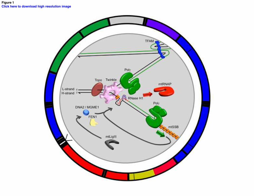
Figure 1Click here to download high resolution image

Figure 2Click here to download high resolution image

Figure 3Click here to download high resolution image