human immunodeficiency virus-like particles producedby vaccinia virus … · 2005-04-23 · proc....
TRANSCRIPT
Proc. Nail. Acad. Sci. USAVol. 86, pp. 8964-8967, November 1989Medical Sciences
Human immunodeficiency virus-like particles produced by avaccinia virus expression vector
(retrovirus/AIDS/virus assembly/reverse transcriptase)
VELISSARIos KARACOSTAS*, KUNIo NAGASHIMAt, MATTHEW A. GONDAt, AND BERNARD MOSS*t*Laboratory of Viral Diseases, National Institute of Allergy and Infectious Diseases, National Institutes of Health, Bethesda, MD 20892; and tLaboratory ofCell and Molecular Structure, Program Resources, Inc., National Cancer Institute, Frederick Cancer Research Facility, Frederick, MD 21701
Contributed by Bernard Moss, August 11, 1989
ABSTRACT Infectious retrovirus particles consist of acore structure containing RNA and gag-pol polypeptides sur-rounded by a lipid membrane studded with env proteins. Arecombinant vaccinia virus was designed to express the entiregag-pol precursor protein of the human immunodeficiencyvirus type 1. Synthesis and processing of gag proteins occurredin mammalian cells infected with this live recombinant virus,and reverse transcriptase was detected largely in the medium.Electron micrographs revealed immature retrovirus-like par-ticles budding from the plasma membrane and extracellularparticles with morphological characteristics of immature andmature human immunodeficiency virus. The latter containedfunctional reverse transcriptase as well as processed p24 andp17 gag polypeptides. Thus, assembly and maturation ofhuman immunodeficiency virus-like particles can occur in theabsence of either infectious RNA molecules or env proteins. Theproduction of noninfectious virus-like particles by expressionvectors should be useful for biochemical studies and couldprovide a safe source of material for the development ofvaccines.
Human immunodeficiency virus type 1 (HIV-1), the caus-ative agent of acquired immunodeficiency syndrome (AIDS),contains an RNA genome that encodes gag, pol, and envproteins, as well as additional regulatory proteins (1-4). Theprimary gag translation product is a 55-kDa precursor, p55,that is proteolytically processed to p24, p17, and p15, themajor core proteins. The pol open reading frame encodes theprotease, reverse transcriptase, and integrase (5-11).Expression of the protease, as well as other products of thepol gene, requires a relatively inefficient ribosomal frame-shifting event within the gag gene (12) that leads to theformation of small amounts ofthe putative gag-pol precursor.A myristic acid residue is present at the N terminus of p17 aswell as the gag precursor (13, 14) and by analogy with otherretroviruses is likely to be required for transport to theplasma membrane (15), into which the glycosylated envelopeproteins are inserted. Studies with defective avian and mu-rine retroviruses, however, have shown that neither infec-tious RNA nor env protein is required for particle assembly(16-19). In this context, Gheysen et al. (20) observed theformation of immature particles containing unprocessed p55in insect cells that were infected with a baculovirus contain-ing only the gag gene of HIV-1. In this communication, wedescribe HIV-like particle formation in mammalian cellsinfected with a recombinant vaccinia virus expressing theentire gag-pol gene under control of a vaccinia virus pro-moter. Production of noninfectious HIV-1 particles usingexpression vectors could be valuable both for studies ofassembly and for vaccine purposes.
MATERIALS AND METHODSPlasmid Construction. The plasmid sp64/HXB.2, contain-
ing an infectious cDNA copy of HIV-1 isolate HXB.2 (21)inserted into the Xba I site of plasmid sp64, was provided byF. Wong-Staal and R. C. Gallo (National Cancer Institute)and served as the source of the gag-pol gene. A 2300-base-pair Sac I-EcoRV fragment of sp64/HXB.2 was in-serted into the Sac I-Sma I sites of the replicative form ofbacteriophage M13mpl8. Oligonucleotide-directed mutagen-esis was carried out to place a Sal I site followed by aconsensus sequence for eukaryotic translation (22) before thestart codon of the gag gene. The gag-pol gene was insertedinto a modified form of the vector pSC11 (23) giving rise topVK3, the final plasmid used to obtain the recombinant virus(vVK1) expressing the HIV gag-pol genes. Restriction endo-nuclease analysis and DNA hybridization were used tocharacterize the plasmid pVK3.
Viruses and Cells. Vaccinia virus (strain WR) was originallyobtained from the American Type Culture Collection. Re-combinant vaccinia virus vVK1 was isolated using the plas-mid pVK3 and was purified as reported (23). CV-1 monkeykidney- cells were propagated as monolayers in minimalessential medium (Quality Biologicals, Gaithersburg, MD)supplemented with 2.5% (vol/vol) fetal bovine serum. Cellswere infected with 30 plaque-forming units of virus per cellfor 2 hr; the virus was then removed, and the cells werewashed and overlayed with fresh medium. The cells wereharvested at appropriate times after infection by scraping themonolayers in isotonic phosphate-buffered saline (PBS),washed with PBS, and lysed in 0.5% Nonidet P-40/PBS.
Immunoblotting. The detergent-disrupted cytoplasmic ex-tracts were treated with SDS and 2-mercaptoethanol prior topolyacrylamide gel electrophoresis. The electrophoreticallyseparated polypeptides were transferred by electroblottingonto a nitrocellulose membrane, blocked with 5% (wt/vol)nonfat dry milk/0.3% Tween 20 in PBS, incubated with theantibody in 0.3% Tween/PBS for 1 hr, washed three timeswith 0.3% Tween/PBS, incubated with 125I-labeled proteinA, washed three times with 0.3% Tween/PBS, dried, andsubjected to autoradiography.
Reverse Transcriptase Assay. Cells were infected as de-scribed above, and reverse transcriptase activity was mea-sured using 5 ,ul of cell extract or medium, poly(A), oligo(dT),and [a-32P]dTTP in 30 Al as described (24). After 2 hr at 37°C,10lO was spotted on DE81 ion-exchange chromatographypaper (Whatman), air-dried, and washed four times in 2Xstandard saline citrate (0.3 M NaCI/0.03 M sodium citrate,pH 7.0). The paper was dried and the radioactivity presentwas determined in a Beckman scintillation counter.
Purification of Particles. Medium, collected 15 hr afterinfection of CV-1 cells with vVK1, was clarified by centrif-
Abbreviation: HIV-1, human immunodeficiency virus type 1.tTo whom reprint requests should be addressed.
8964
The publication costs of this article were defrayed in part by page chargepayment. This article must therefore be hereby marked "advertisement"in accordance with 18 U.S.C. §1734 solely to indicate this fact.
Dow
nloa
ded
by g
uest
on
May
29,
202
0
Proc. Natl. Acad. Sci. USA 86 (1989) 8%5
ugation at 2000 X g, and layered on top of a 20% (wt/vol)sucrose cushion. The pellet, formed by centrifugation at27,000 rpm in an SW 27 rotor for 90 min, was suspended inPBS, and layered on top of a preformed 20-60% sucrosegradient. After centrifugation at 100,000 x g for 18 hr in anSW 41 rotor, 12 equal fractions were collected starting at thebottom.
Electron Microscopy. CV-1 cells infected with recombinantvaccinia virus expressing HIV-1 gag-pol genes were har-vested in PBS and centrifuged at 1500 x g. The pellets werefixed in 1.25% (vol/vol) gluteraldehyde and then in 1%osmium tetroxide, dehydrated in graded alcohols, and em-bedded in epoxy resins. Virus pellets were prepared bycentrifugation at 100,000 x g for 90 min and then processedfor electron microscopy as above. Thin sections were cut andstained with uranyl acetate and lead citrate.
RESULTSExpression and Processing ofHIV gag-pol. When cells were
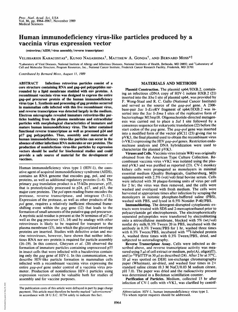
infected with the recombinant vaccinia virus, synthesis andprocessing of the gag-pol proteins occurred. In cell extractsprepared 4 hr after infection, the most prominent polypep-tides that reacted with HIV-1-specific antiserum were -55,46, and 41 kDa (Fig. 1). Also present were small amounts ofhigh molecular mass material (>100 kDa), presumably re-sulting from frame-shifting, as also shown by Gowda et al.(25). With time, three small polypeptides of 32, 24, and 17kDa increased in amount and the higher molecular masspolypeptides diminished. Pulse-chase experiments with[35S]methionine were consistent with precursor-product re-lationships between the higher and lower molecular massproteins (V.K., unpublished results). Specific antisera wereused to confirm the identities of the gag polypeptides p24 andp17 and the integrase polypeptide p32. In addition, the17-kDa polypeptide (as well as the 41- and 55-kDa polypep-tides) was labeled with [3H]myristic acid consistent with itsderivation from the N-terminal end of the gag precursor(V.K., unpublished results). Only a faint 66-kDa polypeptidewas detected with reverse transcriptase-specific antiserum(V.K., unpublished results).
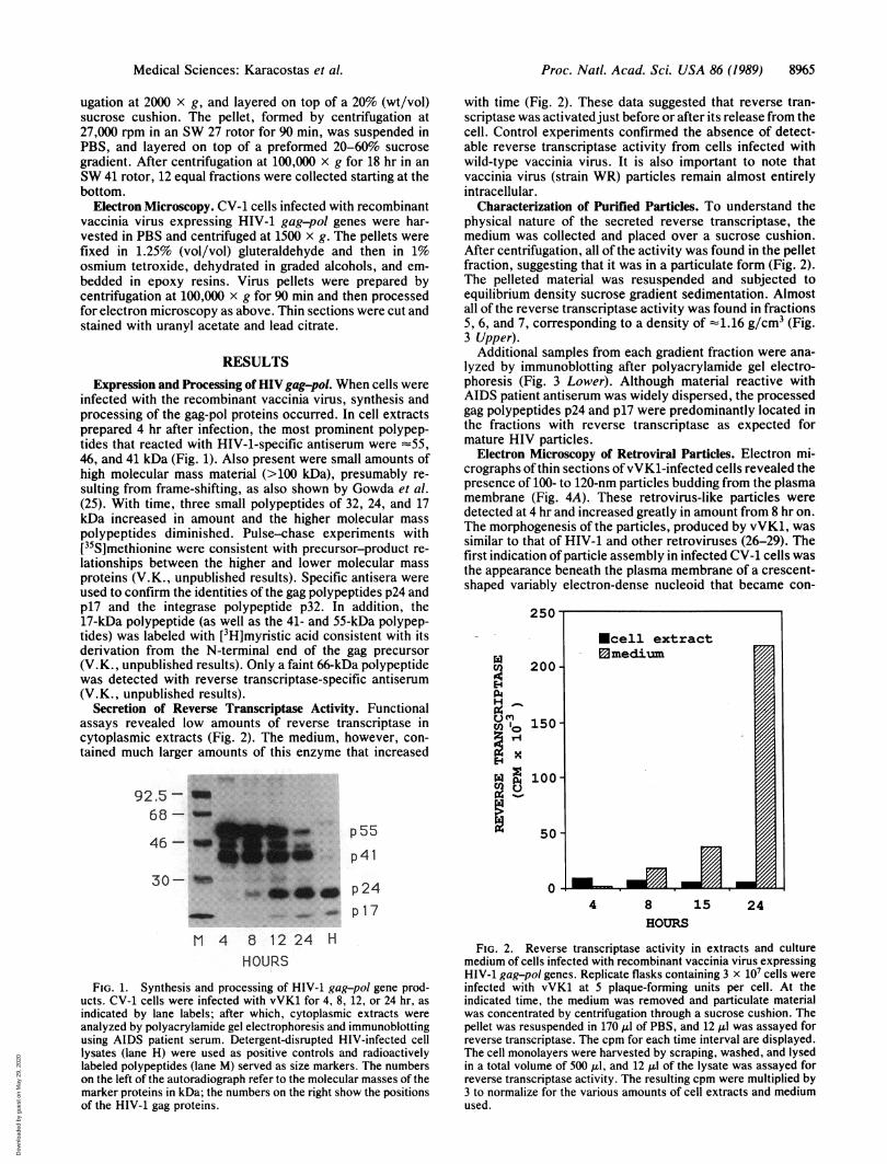
Secretion of Reverse Transcriptase Activity. Functionalassays revealed low amounts of reverse transcriptase incytoplasmic extracts (Fig. 2). The medium, however, con-tained much larger amounts of this enzyme that increased
92.5-68
46-
30-
p55p4l
p24s~. _w. am p 1 7
M 4 8 12 24 HHOURS
FIG. 1. Synthesis and processing of HIV-1 gag-pol gene prod-ucts. CV-1 cells were infected with vVK1 for 4, 8, 12, or 24 hr, asindicated by lane labels; after which, cytoplasmic extracts wereanalyzed by polyacrylamide gel electrophoresis and immunoblottingusing AIDS patient serum. Detergent-disrupted HIV-infected celllysates (lane H) were used as positive controls and radioactivelylabeled polypeptides (lane M) served as size markers. The numberson the left of the autoradiograph refer to the molecular masses of themarker proteins in kDa; the numbers on the right show the positionsof the HIV-1 gag proteins.
with time (Fig. 2). These data suggested that reverse tran-scriptase was activated just before or after its release from thecell. Control experiments confirmed the absence of detect-able reverse transcriptase activity from cells infected withwild-type vaccinia virus. It is also important to note thatvaccinia virus (strain WR) particles remain almost entirelyintracellular.
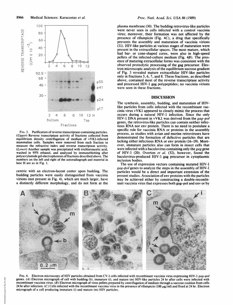
Characterization of Purified Particles. To understand thephysical nature of the secreted reverse transcriptase, themedium was collected and placed over a sucrose cushion.After centrifugation, all of the activity was found in the pelletfraction, suggesting that it was in a particulate form (Fig. 2).The pelleted material was resuspended and subjected toequilibrium density sucrose gradient sedimentation. Almostall of the reverse transcriptase activity was found in fractions5, 6, and 7, corresponding to a density of -1.16 g/cm3 (Fig.3 Upper).
Additional samples from each gradient fraction were ana-lyzed by immunoblotting after polyacrylamide gel electro-phoresis (Fig. 3 Lower). Although material reactive withAIDS patient antiserum was widely dispersed, the processedgag polypeptides p24 and p17 were predominantly located inthe fractions with reverse transcriptase as expected formature HIV particles.
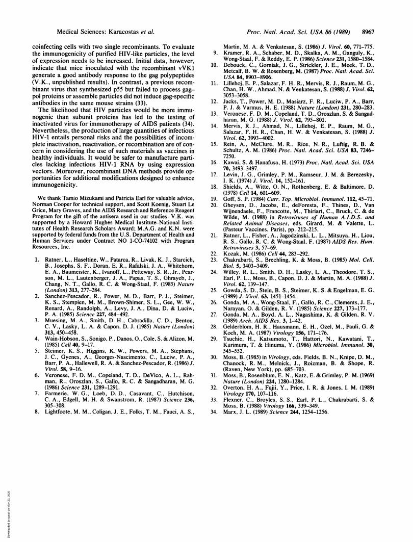
Electron Microscopy of Retroviral Particles. Electron mi-crographs of thin sections of vVK1-infected cells revealed thepresence of 100- to 120-nm particles budding from the plasmamembrane (Fig. 4A). These retrovirus-like particles weredetected at 4 hr and increased greatly in amount from 8 hr on.The morphogenesis of the particles, produced by vVK1, wassimilar to that of HIV-1 and other retroviruses (26-29). Thefirst indication of particle assembly in infected CV-1 cells wasthe appearance beneath the plasma membrane of a crescent-shaped variably electron-dense nucleoid that became con-
cn 200
H
C ^ncn 10 150E-i
p4
100
En
7:50,
0
4 8 15 24HOURS
FIG. 2. Reverse transcriptase activity in extracts and culturemedium of cells infected with recombinant vaccinia virus expressingHIV-1 gag-pol genes. Replicate flasks containing 3 x 10⁷ cells wereinfected with vVK1 at 5 plaque-forming units per cell. At theindicated time, the medium was removed and particulate materialwas concentrated by centrifugation through a sucrose cushion. Thepellet was resuspended in 170 ilI of PBS, and 12 1I was assayed forreverse transcriptase. The cpm for each time interval are displayed.The cell monolayers were harvested by scraping, washed, and lysedin a total volume of 500 ,ul, and 12 Al of the lysate was assayed forreverse transcriptase activity. The resulting cpm were multiplied by3 to normalize for the various amounts of cell extracts and mediumused.
Ecell extract0medium
I.mj,_FA
Medical Sciences: Karacostas et al.
Dow
nloa
ded
by g
uest
on
May
29,
202
0
8966 Medical Sciences: Karacostas et al.
100
roMD
x
CL
0_
80
60
40
20
92.5 -
68 -
46 -
2 4 6 8 10 12
.".ftmto- "ap55wl pel
li ___ p24
p27
2 4 6 8 12 H
Bottom TopFractions
FIG. 3. Purification of reverse transcriptase-containing particles.(Upper) Reverse transcriptase activity of fractions collected fromequilibrium density centrifugation of medium of vVK1-infectedmammalian cells. Samples were removed from each fraction tomeasure the refractive index and reverse transcriptase activity.(Lower) Another sample was precipitated with trichloroacetic acid,washed in 95% ethanol, and analyzed by immunoblotting afterpolyacrylamide gel electrophoresis offractions described above. Thenumbers on the left and right of the autoradiograph and material inlane H are as in Fig. 1.
centric with an electron-lucent center upon budding. Thebudding particles were easily distinguished from vacciniavirions (not present in Fig. 4), which are much larger, havea distinctly different morphology, and do not form at the
plasma membrane (30). The budding retrovirus-like particleswere never seen in cells infected with a control vacciniavirus; moreover, their formation was not affected by thepresence of rifampicin (Fig. 4C), a drug that specificallyprevents the assembly and maturation of vaccinia virions(31). HIV-like particles at various stages of maturation werepresent in the extracellular spaces. The most mature, whichhad bar- or cone-shaped cores, were also in high-speedpellets of the infected-culture medium (Fig. 4B). The pres-ence of maturing extracellular forms was consistent with theobserved proteolytic processing of the gag precursor. Elec-tron microscopic analysis of the equilibrium sucrose gradientof Fig. 3 revealed mature extracellular HIV-like particlesonly in fractions 5, 6, 7, and 8. These fractions, as describedabove, contained most of the reverse transcriptase activityand processed HIV-1 gag polypeptides; no vaccinia virionswere seen in these fractions.
DISCUSSIONThe synthesis, assembly, budding, and maturation of HIV-like particles from cells infected with the recombinant vac-cinia virus vVK1 appeared to closely mimic the process thatoccurs during a natural HIV-1 infection. Since the onlyHIV-1 DNA present in vVK1 was derived from the gag-polgenes, the retrovirus-like particles can contain neither infec-tious RNA nor env protein. There is no need to postulate aspecific role for vaccinia RNA or proteins in the assemblyprocess, as studies with avian and murine retroviruses havedemonstrated the formation of defective particles that arelacking either infectious RNA or env protein (16-19). More-over, immature particles also can form in insect cells thatwere infected with a baculovirus containing only the gag geneof HIV-1 (20). Overton et al. (32), however, found thebaculovirus-produced HIV-1 gag precursor in cytoplasmicinclusion bodies.The use of expression vectors containing mutated HIV-1
gag-pot genes to analyze the steps in the assembly of HIV-1particles would be a direct and important extension of thepresent studies. Association ofenv proteins with the particlesmay be achieved either by constructing a double-recombi-nant vaccinia virus that expresses both gag-pol and env or by
i.. VW.;4 siw,. '! ..-f `'-,
Z111- .:.A 1'..... Ar
%~~~~~~~~~~~~~~~~~'4
wAd is '''' i > X ; e R % </tt~~~~~~~~~~~~~~~~~~~~~~~~~~~~ld
0.
FIG. 4. Electron microscopy of HIV particles obtained from CV-1 cells infected with recombinant vaccinia virus expressing HIV-1 gag-polgenes. (A) Electron micrograph of cell with budding (b), immature (i), and mature (m) HIV-like particles 24 hr after cells were infected withrecombinant vaccinia virus. (B) Electron micrograph of virus pellets prepared by centrifugation of medium through a sucrose cushion from cells24 hr after infection. (C) Cells infected with the recombinant vaccinia virus in the presence of rifampicin (100 Ag/ml) and fixed at 24 hr. Electronmicrograph of a cell producing immature (i) and mature (m) HIV particles.
LUc)
CaL
LLX
U
cnp
z
Fy-LU
LUJ
LUJ
0
Pr
Proc. Natl. Acad. Sci. USA 86 (1989)
Dow
nloa
ded
by g
uest
on
May
29,
202
0
Proc. Natl. Acad. Sci. USA 86 (1989) 8967
coinfecting cells with two single recombinants. To evaluatethe immunogenicity of purified HIV-like particles, the levelof expression needs to be increased. Initial data, however,indicate that mice inoculated with the recombinant vVK1generate a good antibody response to the gag polypeptides(V.K., unpublished results). In contrast, a previous recom-binant virus that synthesized p55 but failed to process gag-pol proteins or assemble particles did not induce gag-specificantibodies in the same mouse strains (33).The likelihood that HIV particles would be more immu-
nogenic than subunit proteins has led to the testing ofinactivated virus for immunotherapy of AIDS patients (34).Nevertheless, the production of large quantities of infectiousHIV-1 entails personal risks and the possibilities of incom-plete inactivation, reactivation, or recombination are of con-cern in considering the use of such materials as vaccines inhealthy individuals. It would be safer to manufacture parti-cles lacking infectious HIV-1 RNA by using expressionvectors. Moreover, recombinant DNA methods provide op-portunities for additional modifications designed to enhanceimmunogenicity.
We thank Tamio Mizukami and Patricia Earl for valuable advice,Norman Cooper for technical support, and Scott Koenig, Stuart LeGrice, Mary Graves, and the AIDS Research and Reference ReagentProgram for the gift of the antisera used in our studies. V.K. wassupported by a Howard Hughes Medical Institute-National Insti-tutes of Health Research Scholars Award; M.A.G. and K.N. weresupported by federal funds from the U.S. Department of Health andHuman Services under Contract NO 1-CO-74102 with ProgramResources, Inc.
1. Ratner, L., Haseltine, W., Patarca, R., Livak, K. J., Starcich,B., Josephs, S. F., Doran, E. R., Rafalski, J. A., Whitehorn,E. A., Baumeister, K., Ivanoff, L., Petteway, S. R., Jr., Pear-son, M. L., Lautenberger, J. A., Papas, T. S., Ghrayeb, J.,Chang, N. T., Gallo, R. C. & Wong-Staal, F. (1985) Nature(London) 313, 277-284.
2. Sanchez-Pescador, R., Power, M. D., Barr, P. J., Steimer,K. S., Stempien, M. M., Brown-Shimer, S. L., Gee, W. W.,Renard, A., Randolph, A., Levy, J. A., Dina, D. & Luciw,P. A. (1985) Science 227, 484-492.
3. Muesing, M. A., Smith, D. H., Cabradilla, C. D., Benton,C. V., Lasky, L. A. & Capon, D. J. (1985) Nature (London)313, 450-458.
4. Wain-Hobson, S., Sonigo, P., Danos, O., Cole, S. & Alizon, M.(1985) Cell 40, 9-17.
5. Steimer, K. S., Higgins, K. W., Powers, M. A., Stephans,J. C., Gyenes, A., Georges-Nascimento, C., Luciw, P. A.,Barr, P. A., Hallewell, R. A. & Sanchez-Pescador, R. (1986) J.Virol. 58, 9-16.
6. Veronese, F. D. M., Copeland, T. D., DeVico, A. L., Rah-man, R., Oroszlan, S., Gallo, R. C. & Sangadharan, M. G.(1986) Science 231, 1289-1291.
7. Farmerie, W. G., Loeb, D. D., Casavant, C., Hutchison,C. A., Edgell, M. H. & Swanstrom, R. (1987) Science 236,305-308.
8. Lightfoote, M. M., Coligan, J. E., Folks, T. M., Fauci, A. S.,
Martin, M. A. & Venkatesan, S. (1986) J. Virol. 60, 771-775.9. Kramer, R. A., Schaber, M. D., Skalka, A. M., Ganguly, K.,
Wong-Staal, F. & Reddy, E. P. (1986) Science 231, 1580-1584.10. Debouck, C., Gorniak, J. G., Strickler, J. E., Meek, T. D.,
Metcalf, B. W. & Rosenberg, M. (1987) Proc. Natl. Acad. Sci.USA 84, 8903-8906.
11. Lillehoj, E. P., Salazar, F. H. R., Mervis, R. J., Raum, M. G.,Chan, H. W., Ahmad, N. & Venkatesan, S. (1988) J. Virol. 62,3053-3058.
12. Jacks, T., Power, M. D., Masiarz, F. R., Luciw, P. A., Barr,P. J. & Varmus, H. E. (1988) Nature (London) 231, 280-283.
13. Veronese, F. D. M., Copeland, T. D., Oroszlan, S. & Sangad-haran, M. G. (1988) J. Virol. 62, 795-801.
14. Mervis, R. J., Ahmad, N., Lillehoj, E. P., Raum, M. G.,Salazar, F. H. R., Chan, H. W. & Venkatesan, S. (1988) J.Virol. 62, 3993-4002.
15. Rein, A., McClure, M. R., Rice, N. R., Luftig, R. B. &Schultz, A. M. (1986) Proc. Natl. Acad. Sci. USA 83, 7246-7250.
16. Kawai, S. & Hanafusa, H. (1973) Proc. Natl. Acad. Sci. USA70, 3493-3497.
17. Levin, J. G., Grimley, P. M., Ramseur, J. M. & Berezesky,I. K. (1974) J. Virol. 14, 152-161.
18. Shields, A., Witte, 0. N., Rothenberg, E. & Baltimore, D.(1978) Cell 14, 601-609.
19. Goff, S. P. (1984) Curr. Top. Microbiol. Immunol. 112, 45-71.20. Gheysen, D., Jacobs, E., deForesta, F., Thines, D., Van
Wijnendaele, F., Francotte, M., Thiriart, C., Bruck, C. & deWilde, M. (1988) in Retroviruses of Human A.l.D.S. andRelated Animal Diseases, eds. Girard, M. & Valette, L.(Pasteur Vaccines, Paris), pp. 212-215.
21. Ratner, L., Fisher, A., Jagodzinski, L. L., Mitsuya, H., Liou,R. S., Gallo, R. C. & Wong-Staal, F. (1987) AIDS Res. Hum.Retroviruses 3, 57-69.
22. Kozak, M. (1986) Cell 44, 283-292.23. Chakrabarti, S., Brechling, K. & Moss, B. (1985) Mol. Cell.
Biol. 5, 3403-3409.24. Willey, R. L., Smith, D. H., Lasky, L. A., Theodore, T. S.,
Earl, P. L., Moss, B., Capon, D. J. & Martin, M. A. (1988) J.Virol. 62, 139-147.
25. Gowda, S. D., Stein, B. S., Steimer, K. S. & Engelman, E. G.-(1989) J. Virol. 63, 1451-1454.
26. Gonda, M. A., Wong-Staal, F., Gallo, R. C., Clements, J. E.,Narayan, 0. & Gilden, R. V. (1985) Science 227, 173-177.
27. Gonda, M. A., Boyd, A. L., Nagashima, K. & Gilden, R. V.(1989) Arch. AIDS Res. 3, 1-42.
28. Gelderblom, H. R., Hausmann, E. H., Ozel, M., Pauli, G. &Koch, M. A. (1987) Virology 156, 171-176.
29. Tsuchie, H., Katsumoto, T., Hattori, N., Kawatani, T.,Kurimura, T. & Hinuma, Y. (1986) Microbiol. Immunol. 30,545-552.
30. Moss, B. (1985) in Virology, eds. Fields, B. N., Knipe, D. M.,Chanock, R. M., Melnick, J., Roizman, B. & Shope, R.(Raven, New York), pp. 685-703.
31. Moss, B., Rosenblum, E. N., Katz, E. & Grimley, P. M. (1969)Nature (London) 224, 1280-1284.
32. Overton, H. A., Fujii, Y., Price, 1. R. & Jones, I. M. (1989)Virology 170, 107-116.
33. Flexner, C., Broyles, S. S., Earl, P. L., Chakrabarti, S. &Moss, B. (1988) Virology 166, 339-349.
34. Marx, J. L. (1989) Science 244, 1254-1256.
Medical Sciences: Karacostas et al.
Dow
nloa
ded
by g
uest
on
May
29,
202
0