human endogenous retrovirus-k contributes to motor … · human endogenous retrovirus-k contributes...
TRANSCRIPT

R E S EARCH ART I C L E
ALS
015
Human endogenous retrovirus-K contributes to motorneuron diseaseWenxue Li,1* Myoung-Hwa Lee,1* Lisa Henderson,1 Richa Tyagi,1 Muzna Bachani,2
Joseph Steiner,2 Emilie Campanac,3 Dax A. Hoffman,3 Gloria von Geldern,1 Kory Johnson,4
Dragan Maric,1 H. Douglas Morris,5 Margaret Lentz,6 Katherine Pak,7 Andrew Mammen,7
Lyle Ostrow,8 Jeffrey Rothstein,8 Avindra Nath1†
The role of human endogenous retroviruses (HERVs) in disease pathogenesis is unclear. We show that HERV-K isactivated in a subpopulation of patients with sporadic amyotrophic lateral sclerosis (ALS) and that its envelope(env) proteinmay contribute to neurodegeneration. The virus was expressed in cortical and spinal neurons of ALSpatients, but not in neurons from control healthy individuals. Expression of HERV-K or its env protein in humanneurons caused retraction and beading of neurites. Transgenic animals expressing the env gene developed pro-gressive motor dysfunction accompanied by selective loss of volume of the motor cortex, decreased synapticactivity in pyramidal neurons, dendritic spine abnormalities, nucleolar dysfunction, and DNA damage. Injuryto anterior horn cells in the spinal cord was manifested by muscle atrophy and pathological changes consistentwith nerve fiber denervation and reinnervation. Expression of HERV-K was regulated by TAR (trans-activation re-sponsive) DNA binding protein 43, which binds to the long terminal repeat region of the virus. Thus, HERV-K ex-pression within neurons of patients with ALS may contribute to neurodegeneration and disease pathogenesis.
, 2
onOct
ober
7D
ownl
oade
d fr
om
INTRODUCTION
Human endogenous retroviruses (HERVs) constitute nearly 8% of thehuman genome and have been termed junkDNA (1). These retroviralsequences are remnants of infections that occurred over several mil-lion years, resulting in the integration of provirus genomes into theDNA of germline cells. Most HERV proviruses have accumulated nu-merous nonsense mutations that have rendered them defective (1).However, it is becoming increasingly apparent that endogenous retro-viral sequences may get expressed under select pathological circum-stances. Multiple complete sequences of the most recently acquiredHERV-K are present in the human genome (2). HERV-K may be ex-pressed in the brain of patients with amyotrophic lateral sclerosis(ALS) (3) and reverse transcriptase activity can be found in the bloodand brain tissue of these patients (4–8), but the role of HERV-K in thepathophysiology of this disease remains unknown. ALS is a progres-sive neurodegenerative disease and is universally fatal, except in somepatients with HIV infection where an ALS-like syndrome can be re-versed by antiretroviral drugs (9). However, an extensive search forexogenous retroviruses in ALS has not been successful (5). Here, weexplored whether endogenous retroviral elements could be expressed
1Sectionof Infections of theNervous System,National Institute ofNeurological Disorders andStroke, National Institutes of Health, Bethesda, MD 20892, USA. 2Neurotherapeutics Unit,National Institute of Neurological Disorders and Stroke, National Institutes of Health,Bethesda, MD 20892, USA. 3Molecular Neurophysiology and Biophysics Section, EuniceKennedy Shriver National Institute of Child Health and Human Development, NationalInstitutes of Health, Bethesda, MD 20892, USA. 4Bioinformatics Unit, National Institute ofNeurological Disorders and Stroke, National Institutes of Health, Bethesda, MD 21042, USA.5Mouse Imaging Facility, National Institute of Neurological Disorders and Stroke, NationalInstitutes of Health, Bethesda,MD21042, USA. 6Integrated Research Facility, National Instituteof Allergy and Infectious Disease, National Institutes of Health, Fort Detrick, Frederick, MD21042, USA. 7Laboratory of Muscle Stem Cell and Gene Regulation, National Institute ofArthritis and Musculoskeletal and Skin Diseases, National Institutes of Health, Bethesda, MD21042 , USA. 8Department of Neurology, JohnsHopkins University, Baltimore,MD 28217, USA.*Contributed equally to the manuscript.†Corresponding author. E-mail: [email protected]
www.ScienceTra
in these patients and whether HERV-K could contribute to the patho-physiology of this disease.
RESULTS
HERV-K is expressed in the brain tissue of ALS patientsThe HERV-K genome similar to that of other retroviruses has threemajor structural genes, the gag, pol, and env genes that encode the cap-sid, reverse transcriptase, and envelope proteins, respectively. Primersets were used to amplify transcripts from each of these genes by re-verse transcription polymerase chain reaction (RT-PCR) (Fig. 1A).We found that transcripts for all three genes were elevated in postmor-tem brain tissue samples fromALS patients (Fig. 1B). There was goodcorrelation between the expression of each of these genes (Fig. 1C),confirming that the entire viral genomewas expressed in these patientsamples. The expression ofHERV-Kwas also compared to the expres-sion of several other HERVs. No significant elevation of these HERVswas noted (fig. S1A). Given that there are multiple loci that encode theHERV-K genome, we conducted RNA sequencing and analyzed thetranscripts of each of the loci. The loci at chromosomes 7C and 10Awere expressed in all three postmortem brain samples from patientswith sporadic ALS at higher levels compared to controls (fig. S1B andtable S1). No specific clinical phenotype was associated with the ex-pression of HERV-K in ALS patients. To determine the cell types inwhich HERV-Kwas expressed, we immunostained postmortem braintissue from patients with ALS and found expression of the env proteinin the cortex of ten individuals (Fig. 1D), with a strong expression inthe cytoplasmof large pyramidal neurons (Fig. 1E). Anterior hornneu-rons in the spinal cord also showed a similar pattern of immunostainingfor HERV-K env protein (Fig. 1F and fig. S1, C and D). No immuno-staining was seen in the lateral or posterior horns of the spinal cord(fig. S1D). No immunostaining was noted in the glial cells or in thewhite matter (Fig. 1G). Furthermore, no immunostaining was noted
nslationalMedicine.org 30 September 2015 Vol 7 Issue 307 307ra153 1

R E S EARCH ART I C L E
on O
ctob
er 7
, 201
5D
ownl
oade
d fr
om
in the cortex or white matter of brain tissue from healthy individuals(Fig. 1H) or in postmortembrain tissue from patients withAlzheimer’sdisease (Fig. 1I and fig. S1F). However, robust immunostaining for am-
www.ScienceTranslationalMedicine.org 30 Septe
yloid was present in postmortem braintissue from Alzheimer’s disease patients(fig. S1E).
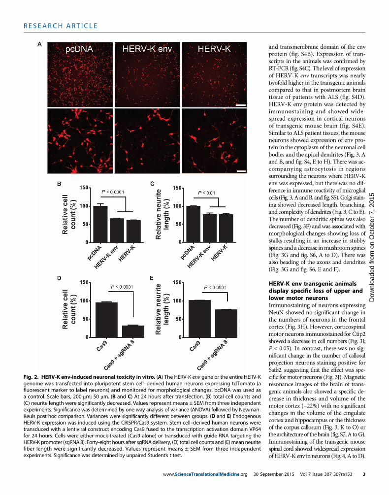
Expression of HERV-K in humanneurons in vitro causes toxicityTo determine the relevance of HERV-Kexpression in neurons, we transfectedthe HERV-K genome and the HERV-Kenv gene into human neuronal cultures.Both the entire genome and the env genecaused a similar decrease in cell numbersand retraction of neurites (Fig. 2, A toC) in a dose-dependent manner (fig.S2A). The expression of the genes wasconfirmed by RT-PCR (fig. S2C). Thissuggested that the env protein could con-tribute to neurotoxicity and neuronaldeath. To determine the effect of activa-tion of endogenousHERV-K in neurons,we used the gene-editing tool CRISPR/Cas9 (10) in which the nuclease activityof Cas9 [CRISPR (clustered regularly in-terspaced short palindromic repeats)–associated protein 9] was altered tocontain four copies of the transcriptionfactor VP16 (11). This was delivered tothe human neurons in culture by alentiviral vector. We designed a singleguide RNA (sgRNA 8) to direct thetranscription factor to the long terminalrepeat (LTR) region of HERV-K. Acti-vation of the endogenous HERV-Kthrough the LTR resulted in neuro-toxicity as evidenced by the loss of neu-rons (Fig. 2D) and the retraction ofneurites (Fig. 2E). Activation of the viralgenes was confirmed by RT-PCR, and atwofold increase in expression abovecontrols was observed (fig. S2D). To de-termine whether the process of neuronalinjury led to HERV-K activation, wetreated theneuronswith3-nitropropionicacid,N-methyl-D-aspartate, or hydrogenperoxide. No activation of HERV-K wasnoted as determined by measuring viraltranscripts (fig. S2B).
Expression of HERV-K env in vivocauses degeneration ofmotor neuronsThe findings were initially confirmedin vivo by in utero electroporation ofthe env gene into embryonic mousebrain, which resulted in dysmorphic
changes in neurons and punctate dilatation of neuronal processes(fig. S3). We next generated transgenic animals in which the env genewas expressed in neurons (fig. S4A). The gene produced the full length
Fig. 1. HERV-K expression in brain tissue from ALS patients. (A) HERV-K genome showing regions am-plified by PCR. (B) All HERV-K genes showed increased expression in ALS patients compared to healthy
controls (ALS, n = 11; control, n = 16). Brain tissue sample information is listed in table S1. Values representmeans ± SEM. Significance was determined by unpaired Student’s t test. Variances were significantly differ-ent between groups. (C) Pearson correlation analyses revealed positive correlations betweenmRNA expres-sion of HERV-K env, pol, and gag from autopsy brain cortical tissues. Pearson’s correlation coefficients wereused to quantify the linear relationship between two variables. (D) Representative images showHERV-K env-immunoreactive neurons in the frontal cortex of a patient with ALS. (E) A higher-magnification image of theboxed area in the image shows focal accumulation at the cell membrane of cortical neurons. (F) Anteriorhorn motor neurons in the lumbar spinal cord of an ALS patient were also immunoreactive for HERV-K env.(G) The white matter of a patient with ALS. (H and I) Cortical neurons of an individual with a normal brainwho died in a motor vehicle accident (H) and cortical neurons from a patient with Alzheimer’s diseaseshowing no immunoreactivity for HERV-K env (I). Scale bars, 50 mm (D to I).mber 2015 Vol 7 Issue 307 307ra153 2
R E S EARCH ART I C L E
www.ScienceTranslationalMedicine.org 30 Sep
on O
ctob
er 7
, 201
5D
ownl
oade
d fr
om
and transmembrane domain of the envprotein (fig. S4B). Expression of tran-scripts in the animals was confirmed byRT-PCR(fig. S4C).The level of expressionof HERV-K env transcripts was nearlytwofold higher in the transgenic animalscompared to that in postmortem braintissue of patients with ALS (fig. S4D).HERV-K env protein was detected byimmunostaining and showed wide-spread expression in cortical neuronsof transgenic mouse brain (fig. S4E).Similar toALS patient tissues, themouseneurons showed expression of env pro-tein in the cytoplasm of the neuronal cellbodies and the apical dendrites (Fig. 3, Aand B, and fig. S4, E to H). There was ac-companying astrocytosis in regionssurrounding the neurons where HERV-Kenv was expressed, but there was no dif-ference in immune reactivity of microglialcells (Fig. 3,AandB, and fig. S5).Golgi stain-ing showed decreased length, branching,and complexity of dendrites (Fig. 3, C toE).The number of dendritic spines was alsodecreased (Fig. 3F) andwas associatedwithmorphological changes showing loss ofstalks resulting in an increase in stubbyspines and a decrease inmushroom spines(Fig. 3G and fig. S6, A to D). There wasalso beading of the axons and dendrites(Fig. 3G and fig. S6, E and F).
HERV-K env transgenic animalsdisplay specific loss of upper andlower motor neuronsImmunostaining of neurons expressingNeuN showed no significant change inthe numbers of neurons in the frontalcortex (Fig. 3H). However, corticospinalmotor neurons immunostained for Ctip2showed a decrease in cell numbers (Fig. 3I;P < 0.05). In contrast, there was no sig-nificant change in the number of callosalprojection neurons staining positive forSatb2, suggesting that the effect was spe-cific for motor neurons (Fig. 3J). Magneticresonance images of the brain of trans-genic animals also showed a specific de-crease in thickness and volume of themotor cortex (~22%) with no significantchanges in the volume of the cingulatecortex and hippocampus or the thicknessof the corpus callosum (Fig. 3, K to O) orthe architectureof the brain (fig. S7,A toG).Immunostaining of the transgenic mousespinal cord showed widespread expressionofHERV-Kenv inneurons (Fig. 4,A toD).
Fig. 2. HERV-K env-induced neuronal toxicity in vitro. (A) The HERV-K env gene or the entire HERV-Kgenome was transfected into pluripotent stem cell–derived human neurons expressing tdTomato (a
fluorescent marker to label neurons) and monitored for morphological changes. pcDNA was used asa control. Scale bars, 200 mm; 50 mm. (B and C) At 24 hours after transfection, (B) total cell counts and(C) neurite length were significantly decreased. Values represent means ± SEM from three independentexperiments. Significance was determined by one-way analysis of variance (ANOVA) followed by Newman-Keuls post hoc comparison. Variances were significantly different between groups. (D and E) EndogenousHERV-K expression was induced using the CRISPR/Cas9 system. Stem cell–derived human neurons weretransduced with a lentiviral construct encoding Cas9 fused to the transcription activation domain VP64for 24 hours. Cells were either mock-treated (Cas9 alone) or transduced with guide RNA targeting theHERV-K promoter (sgRNA 8). Forty-eight hours after sgRNAdelivery, (D) total cell counts and (E)mean neuritefiber length were significantly decreased. Values represent means ± SEM from three independentexperiments. Significance was determined by unpaired Student’s t test.tember 2015 Vol 7 Issue 307 307ra153 3

R E S EARCH ART I C L E
on O
ctob
er 7
, 201
5D
ownl
oade
d fr
om
Fig. 3. HERV-K–inducedneuronal toxicity in vivo. (A) Coronal sections ofwild-type (WT) and HERV-K env transgenic (Tg) mice were immunostained
motor neurons (I), and Satb2 immunostaining as a marker for callosalprojection neurons (J) in layer V of the motor cortex of wild type (n = 4)
for HERV-K env (red) and glial fibrillary acidic protein (GFAP) (purple). DAPI,4′,6-diamidino-2-phenylindole (blue). (B) Enlarged images of cerebral cortex.(C to G) Golgi impregnated mouse pyramidal neurons show that (C) totaldendrite length, (D) mean branch number, (E) dendritic complexity by Shollanalysis, and (F) spine density were significantly reduced in transgenicmice with (G) extensive dendritic beading. Values represent means ±SEM. The number of animals used for quantification was three animalsper group. Significance was determined by unpaired Student’s t test. Scalebars, 500 mm (A); 50 mm (B and G). (H to J) Immunostaining for NeuN as amarker for neurons (H), Ctip2 immunostaining as a marker for corticospinal
www.ScienceTra
and transgenic (n = 3) mice. Values represent means ± SEM. Significancewas determined by unpaired Student’s t test. Scale bars, 50 mm. Magneticresonance images for wild type (n = 5) and transgenic (n = 5) mice wereacquired on a 14-Tesla magnetic resonance imaging scanner. (K and L) Re-gional analysis revealed a reduction in cortical thickness (K) and volume (L)of the rostral part of the motor cortex. (M to O) There was no significantdifference between wild-type and transgenic mice for the volume of the cin-gulate cortex (M), corpus callosum (N), andhippocampus (O). Values representmeans ± SEM. Significance was determined by unpaired Student’s t test. Thenumber of animals used for quantification was five animals in each group.
nslationalMedicine.org 30 September 2015 Vol 7 Issue 307 307ra153 4

R E S EARCH ART I C L E
on O
ctob
er 7
, 201
5D
ownl
oade
d fr
om
However, only raremotor neurons were present in the anterior horns,with near absence of motor neurons at some levels of the spinal cord(Fig. 4, E toH). Immunostaining of the quadriceps and tibialis anteriormuscles for type I and type II myosin isoforms showed fiber type
www.ScienceTranslationalMedicine.org 30 Septe
grouping and examples of grouped at-rophy suggestive of a chronic denervationand reinnervation process (Fig. 4I and fig.S8). There were no dystrophic changes inthe muscle fibers, and the nuclei were inthe periphery of the fibers, suggesting thatthere were no myopathic features.
Ongoing neuronal injury was also evi-dent by the presence of double-strandedDNA breaks as seen by immunostainingfor gH2A.X (12), which showed aggre-gated foci of the phosphorylated histoneprotein in the chromatin (Fig. 4J). Thenumber of gH2A.X foci was increased inneurons in the frontal cortex (Fig. 4J).Nucleolar dysfunction has been ob-served in several neurodegenerative dis-eases including Alzheimer’s disease andParkinson’s disease (13). We thereforeevaluated whether neurons from trans-genic mice showed signs of nucleolarstress. Immunostaining for the nucleolarmarker nucleophosmin showed trans-location from the nucleolus to the cyto-plasm of cortical neurons (Fig. 4K).Together, these data suggested that dis-ruption of nucleolar function may be akey mechanism by which HERV-Kleads to neuronal dysfunction (14).
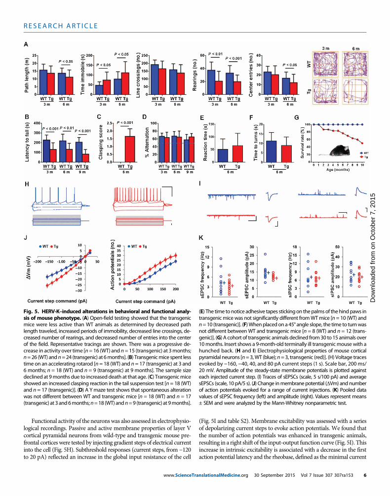
HERV-K env transgenic animalsdevelop motor dysfunctionTodetermine the functional consequencesof HERV-K env expression in neurons,we performed a panel of behavioral testson the animals. These tests showed thatthe animals developed progressivemotordysfunction. Inanopen field, they traveledshorter distances and rested for longerperiods (Fig. 5A). Transgenic mice fellfaster in a rotarod performance test (Fig.5B) and displayed evidence of spasticitywith increased clasping of the hindlimbs(Fig. 5C and movies S1A and S1B). Ymaze testing confirmed that these differ-ences were not due to an impairment ofworkingmemory (Fig. 5D). Sensory andvestibular functions were also unim-paired (Fig. 5, E and F). Motor functionin the transgenic mice showed a pro-gressive decline from 3 to 6 months ofage as evaluated in open-field testing(fig. S9, A to D) with 50% mortality by10months (Fig. 5G). In terminal stages,
the animals developed profound weakness of the limbs and spinalmuscles resulting in minimal movement and a hunched back causingdecreased movement of the thoracic cage, affecting the muscles ofrespiration (Fig. 5G).
Fig. 4. HERV-K env expression and injury to lower motor neurons. (A to C) Representative photomicro-graphs showHERV-K env immunoreactivity in the cervical (A), thoracic (B), and lumbar (C) spinal cord of trans-
genic mice. (D) This panel shows lack of HERV-K env immunoreactivity in the thoracic spinal cord of WTmice.(E to H) ChAT immunoreactivity is shown in the cervical (E), thoracic (F), and lumbar (G) spinal cord of trans-genic mice and in the thoracic spinal cord of WTmice at 6 months of age (H). Intense ChAT signal seen in themotor neurons of the ventral horn in the thoracic spinal cord of WT mice (H) contrasts with very few ChAT-positive cell bodies in the ventral horn of the thoracic spinal cord of transgenic mice (F). Scale bars, 50 mm(A to H). (I) Sections of the tibialis anterior muscle from 6-month-old animals show a normal mosaic distri-bution of type I (green), type IIb (red), and type IIa (unstained) fibers in WT mice and fiber type grouping inthe transgenic mice. Skeletal muscles from WT (n = 5) and transgenic (n = 6) mice were isolated and im-munostained. MyHC, myosin heavy chain. (J) gH2A.X-positive foci in immunostained entorhinal cortex from6-month-old WT (n = 4) and transgenic (n = 4) mice. Numbers of cells with gH2A.X-positive foci wereincreased in motor cortex of transgenic mice. Values represent means ± SEM. Significance was determinedby unpaired Student’s t test. Scale bar, 20 mm. (K) Fluorescence micrographs showing the localization ofnucleophosmin (NPM) in cells in the motor cortex of WT (n = 4) and transgenic (n = 3) mice. Numbers ofcells with nucleophosmin localized to the cytoplasmwere increased in themotor cortex of transgenic mice.Values represent means ± SEM. Significance was determined by unpaired Student’s t test. Scale bar, 10 mm.mber 2015 Vol 7 Issue 307 307ra153 5
R E S EARCH ART I C L E
on O
ctob
er 7
, 201
5D
ownl
oade
d fr
om
Functional activity of the neuronswas also assessed in electrophysio-logical recordings. Passive and active membrane properties of layer Vcortical pyramidal neurons from wild-type and transgenic mouse pre-frontal cortices were tested by injecting gradient steps of electrical currentinto the cell (Fig. 5H). Subthreshold responses (current steps, from −120to 20 pA) reflected an increase in the global input resistance of the cell
www.ScienceTra
(Fig. 5I and table S2). Membrane excitability was assessed with a seriesof depolarizing current steps to evoke action potentials. We found thatthe number of action potentials was enhanced in transgenic animals,resulting in a right shift of the input-output function curve (Fig. 5I). Thisincrease in intrinsic excitability is associated with a decrease in the firstaction potential latency and the rheobase, defined as theminimal current
Fig. 5. HERV-K–induced alterations in behavioral and functional analy-sis of mouse phenotype. (A) Open-field testing showed that the transgenic
(E) The time tonotice adhesive tapes stickingon thepalmsof thehindpaws intransgenicmicewas not significantly different fromWTmice [n= 10 (WT) and
mice were less active than WT animals as determined by decreased pathlength traveled, increased periods of immobility, decreased line crossings, de-creased number of rearings, and decreased number of entries into the centerof the field. Representative tracings are shown. There was a progressive de-crease in activity over time [n= 16 (WT) and n=15 (transgenic) at 3months;n=26 (WT) andn=24 (transgenic) at 6months]. (B) Transgenicmice spent lesstime on an accelerating rotarod [n = 18 (WT) and n= 17 (transgenic) at 3 and6 months; n = 18 (WT) and n = 9 (transgenic) at 9 months]. The sample sizedeclined at 9months due to increased death at that age. (C) Transgenicmiceshowed an increased clasping reaction in the tail suspension test [n = 18 (WT)and n = 17 (transgenic)]. (D) A Ymaze test shows that spontaneous alterationwas not different between WT and transgenic mice [n = 18 (WT) and n = 17(transgenic) at 3and6months;n=18 (WT) andn=9 (transgenic) at 9months].
n=10 (transgenic)]. (F)Whenplacedon a45° angle slope, the time to turnwasnot different between WT and transgenic mice [n = 8 (WT) and n = 12 (trans-genic)]. (G) A cohort of transgenic animals declined from30 to 15 animals over10months. Insert shows a 9-month-old terminally ill transgenicmousewith ahunched back. (H and I) Electrophysiological properties of mouse corticalpyramidal neurons [n=3,WT (blue);n=3, transgenic (red)]. (H) Voltage tracesevoked by −160, −40, 40, and 80 pA current steps (1 s). Scale bar, 200 ms/20 mV. Amplitude of the steady-state membrane potentials is plotted againsteach injected current step. (I) Traces of sEPSCs (scale, 5 s/100 pA) and averagesEPSCs (scale, 10 pA/5 s). (J) Change inmembranepotential (DVm) and numberof action potentials evoked for a range of current injections. (K) Pooled datavalues of sEPSC frequency (left) and amplitude (right). Values represent means± SEM and were analyzed by the Mann-Whitney nonparametric test.
nslationalMedicine.org 30 September 2015 Vol 7 Issue 307 307ra153 6
R E S EARCH ART I C L E
on O
ctob
er 7
, 201
5D
ownl
oade
d fr
om
to induce an action potential (table S2).Other action potential parameterssuch as threshold, amplitude, rise time, width, and after-hyper-polarization amplitude remained unchanged. Finally, we examined syn-aptic transmission by recording spontaneous excitatory and inhibitorypostsynaptic currents (sEPSCs and sIPSCs). We found that only thesEPSC amplitude was changed, with a significant decrease in the sEPSCobserved in transgenic animals compared to that in wild-type mice,which could be attributed to the decrease in spine density (Fig. 5, J andK). The increase in input resistance was consistent with the decrease inneurite number and branching.
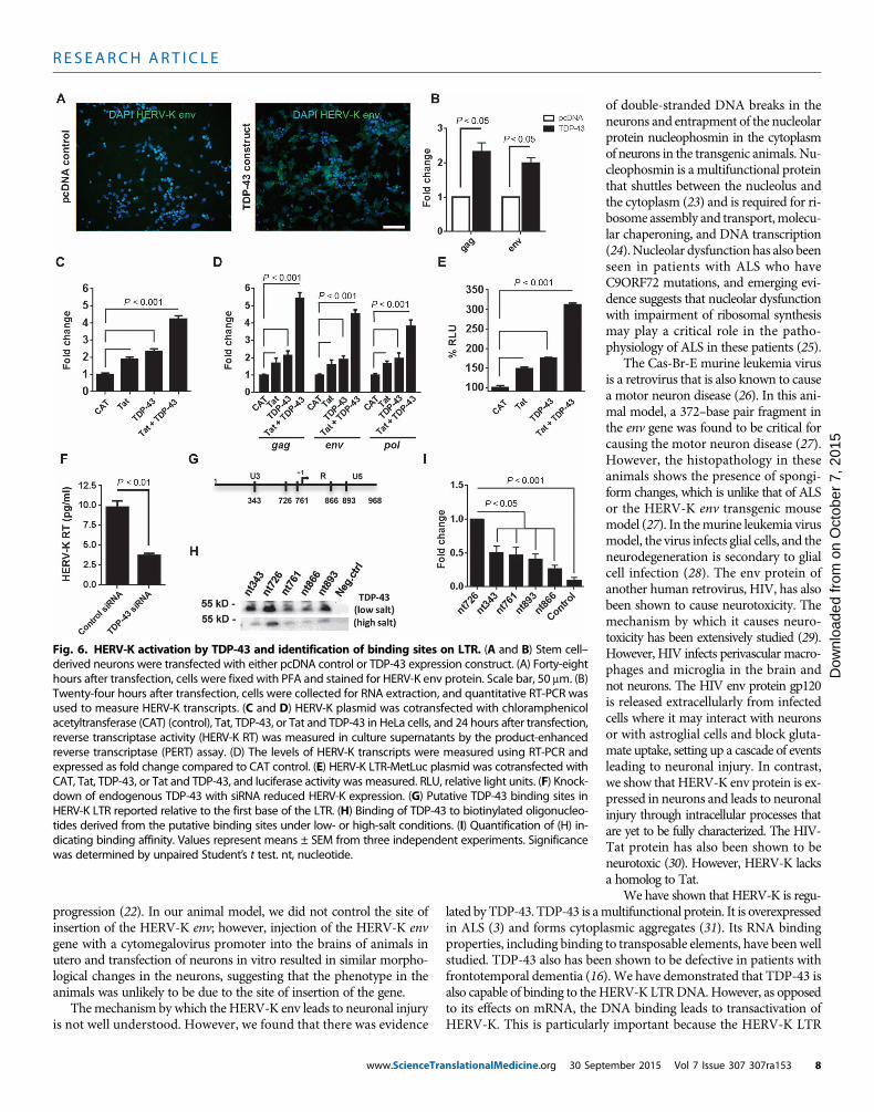
HERV-K expression is regulated by TAR DNA bindingprotein 43Previously, HERV-K pol gene expression was found to correlate withTAR (trans-activation-responsive) DNA binding protein 43 (TDP-43)mRNA in postmortem brain tissue from patients with ALS (3). TDP-43 has been shown to regulate the replication of HIV (15), and it alsobinds to transposable elements (16). Hence, we determined whetherTDP-43 could also regulateHERV-K expression.When a plasmidwithTDP-43 was transfected into human neurons, HERV-K expressionoccurred as demonstrated by immunostaining for the env proteinand measuring the viral transcripts (Fig. 6, A and B). When HERV-Kand TDP-43 were cotransfected into HeLa cells, there was an increasedreplication of HERV-K as evidenced by the reverse transcriptase activityin the culture supernatants (Fig. 6C) and increased viral transcripts in thecell extracts (Fig. 6D). HIV-transactivator of transcription (HIV-Tat)protein is known to increase HERV-K replication, and hence it was usedas a control (17). TDP-43 showed additive responses with Tat, suggestingthat theymay act on different sites. This was confirmed using aHERV-KLTR construct with a luciferase reporter gene (Fig. 6E). Knockdown ofendogenous TDP-43 with small interfering RNA (siRNA) also decreasedHERV-K expression (Fig. 6F). We next determined whether TDP-43–mediated induction of HERV-K involved direct association with theHERV-Kpromoter. The consensusHERV-KLTR sequencewas scannedto identify pyrimidine-rich motifs associated with TDP-43 DNA bind-ing. Putative TDP-43 binding sites, consisting of more than eight con-tiguous pyrimidine bases (16), were identified at five loci as indicatedand were labeled according to their position relative to the first base ofthe HERV-K LTR (Fig. 6G and fig. S10A). Binding of TDP-43 toHERV-K LTR was confirmed by chromatin immunoprecipitation (fig.S10B). Hence, we constructed biotinylated oligomers representing eachof these sites and incubated them with nuclear extracts from 293T cellsfollowedbywashingofDNA/protein complexes under low- andhigh-saltconditions and then analyzed the complexes by Western blots using anantibody to TDP-43. We found that TDP-43 bound to region 726–734(5′-CCCTCTCCC-3′) with the highest affinity, suggesting that it was thecritical binding site on the HERV-K LTR (Fig. 6, H and I). TDP-43binding to HERV-K LTR was associated with increased binding ofelongation-competent RNA polymerase II (fig. S10C). No effect wasseen on an unrelated genomic region (fig. S10D).
DISCUSSION
Here, we show that an endogenous retroviral protein contributes toneurodegeneration in mammalian brain and spinal cord. Expressionof HERV-K env protein in transgenic mouse neurons led to motordysfunction with accompanying DNA damage and morphological and
www.ScienceTra
functional changes consistent with axonal and dendritic injury. Thesefindings may have implications for a subgroup of ALS patients withincreased HERV-K expression. Because activation of HERV-K can leadto neurodegeneration, blocking its activation and replication may affectthe course of ALS.
It is becoming increasingly apparent thatmutations in several differ-ent genes can lead to the same phenotypic neurodegenerative disease.Several genetic mutations have been implicated in ALS. These includesuperoxide dismutase (SOD-1), TDP-43, fused in sarcoma (FUS), andrepeat expansions in C9ORF72 (18). However, none of thesemutationsexplain how the disease spreads along anatomical pathways. ALS typ-ically starts in a focal region in one of the limbs or the bulbar regionand then spreads to contiguous regions. Our finding that full-lengthtranscripts of HERV-K can be found activated in the brain of someALSpatients raises the possibility that the virus could spread from one neu-ron to the next and, in the process, lead to neurotoxicity through expres-sion of the env protein. Whereas the mechanism of spread needs to befurther investigated, the observation that the HERV-K env protein wasfound localized to the neuronal cell body suggests that the spread mayoccur laterally from neuron to neuron. This would be different fromthe transsynaptic spread seen with rabies virus (19), but would betterexplain the anatomical spread of ALS from one brain region to an ad-jacent region.
Because we studied only autopsy tissues, we considered the possi-bility that HERV-K was a response to neurodegeneration. However,HERV-K immunostaining could not be detected in brain tissue frompatients with Alzheimer’s disease. In a previous study, we showed thatHERV-K gene expression was absent in brain tissue from Parkinson’sdisease patients and in brains from patients who had died in accidents(3). We also showed that treatment of human neurons in culture withtoxins that induced mitochondrial or oxidative stress and excitotoxi-city (pathways that have been implicated in the pathophysiology ofALS) did not induce HERV-K expression. Further, there was low basal-level HERV-K expression in neurons in culture; however, endogenousHERV-K activation resulted in neuronal injury. Our findings contrastwith another study claiming thatHERV-Kmaybe neuroprotective (20).However, in this study, HERV-K was expressed in neuronal cell lines.Endogenous retroviruses includingHERV-K can be expressed in tumorcells where they may have cytoprotective effects (21). In terminally dif-ferentiated cells such as neurons, however, the expression of HERV-Kmay be cytotoxic.
We developed a transgenicmousemodel in which the HERV-K envprotein was expressed under a neuronal promoter leading to wide-spread expression in neurons. The amount of expression of the genewas comparable to that in brain tissue from patients with ALS. Despitethe widespread expression of the protein, the animals developed a pro-gressivemotor neuron disease affecting only the upper and lowermotorneurons. There were no developmental abnormalities in the brain, andthe cytoarchitecture of the brain appeared normal. The animals dis-played progressive motor dysfunction first noticeable at 2 to 3 monthsof age and developed selective atrophy of the motor cortex. The neu-rons showed morphological changes with abnormal processes andelectrophysiological abnormalities. There were changes consistent withdenervation and reinnervation of the muscles due to anterior horn cellinvolvement. This profound weakness of the limbs eventually involvedthe spinal muscles and thoracic cage resulting in 50% mortality by10 months. In contrast, the SOD-1 transgenic mousemodel shows pre-dominantly lower motor neuron involvement and a much more rapid
nslationalMedicine.org 30 September 2015 Vol 7 Issue 307 307ra153 7
R E S EARCH ART I C L E
on O
ctob
er 7
, 201
5D
ownl
oade
d fr
om
progression (22). In our animal model, we did not control the site ofinsertion of the HERV-K env; however, injection of the HERV-K envgene with a cytomegalovirus promoter into the brains of animals inutero and transfection of neurons in vitro resulted in similar morpho-logical changes in the neurons, suggesting that the phenotype in theanimals was unlikely to be due to the site of insertion of the gene.
Themechanism by which the HERV-K env leads to neuronal injuryis not well understood. However, we found that there was evidence
www.ScienceTranslationalMedicine.org 30 Sept
of double-stranded DNA breaks in theneurons and entrapment of the nucleolarprotein nucleophosmin in the cytoplasmof neurons in the transgenic animals. Nu-cleophosmin is a multifunctional proteinthat shuttles between the nucleolus andthe cytoplasm (23) and is required for ri-bosome assembly and transport,molecu-lar chaperoning, and DNA transcription(24). Nucleolar dysfunction has also beenseen in patients with ALS who haveC9ORF72 mutations, and emerging evi-dence suggests that nucleolar dysfunctionwith impairment of ribosomal synthesismay play a critical role in the patho-physiology of ALS in these patients (25).
The Cas-Br-E murine leukemia virusis a retrovirus that is also known to causea motor neuron disease (26). In this ani-mal model, a 372–base pair fragment inthe env gene was found to be critical forcausing the motor neuron disease (27).However, the histopathology in theseanimals shows the presence of spongi-form changes, which is unlike that of ALSor the HERV-K env transgenic mousemodel (27). In themurine leukemia virusmodel, the virus infects glial cells, and theneurodegeneration is secondary to glialcell infection (28). The env protein ofanother human retrovirus, HIV, has alsobeen shown to cause neurotoxicity. Themechanism by which it causes neuro-toxicity has been extensively studied (29).However, HIV infects perivascular macro-phages and microglia in the brain andnot neurons. The HIV env protein gp120is released extracellularly from infectedcells where it may interact with neuronsor with astroglial cells and block gluta-mate uptake, setting up a cascade of eventsleading to neuronal injury. In contrast,we show that HERV-K env protein is ex-pressed in neurons and leads to neuronalinjury through intracellular processes thatare yet to be fully characterized. The HIV-Tat protein has also been shown to beneurotoxic (30). However, HERV-K lacksa homolog to Tat.
We have shown that HERV-K is regu-
lated by TDP-43. TDP-43 is amultifunctional protein. It is overexpressedin ALS (3) and forms cytoplasmic aggregates (31). Its RNA bindingproperties, including binding to transposable elements, have beenwellstudied. TDP-43 also has been shown to be defective in patients withfrontotemporal dementia (16).We have demonstrated that TDP-43 isalso capable of binding to the HERV-K LTRDNA.However, as opposedto its effects on mRNA, the DNA binding leads to transactivation ofHERV-K. This is particularly important because the HERV-K LTRFig. 6. HERV-K activation by TDP-43 and identification of binding sites on LTR. (A and B) Stem cell–derived neurons were transfected with either pcDNA control or TDP-43 expression construct. (A) Forty-eight
hours after transfection, cells were fixed with PFA and stained for HERV-K env protein. Scale bar, 50 mm. (B)Twenty-four hours after transfection, cells were collected for RNA extraction, and quantitative RT-PCR wasused to measure HERV-K transcripts. (C and D) HERV-K plasmid was cotransfected with chloramphenicolacetyltransferase (CAT) (control), Tat, TDP-43, or Tat and TDP-43 in HeLa cells, and 24 hours after transfection,reverse transcriptase activity (HERV-K RT) was measured in culture supernatants by the product-enhancedreverse transcriptase (PERT) assay. (D) The levels of HERV-K transcripts were measured using RT-PCR andexpressed as fold change compared to CAT control. (E) HERV-K LTR-MetLuc plasmid was cotransfected withCAT, Tat, TDP-43, or Tat and TDP-43, and luciferase activity was measured. RLU, relative light units. (F) Knock-down of endogenous TDP-43 with siRNA reduced HERV-K expression. (G) Putative TDP-43 binding sites inHERV-K LTR reported relative to the first base of the LTR. (H) Binding of TDP-43 to biotinylated oligonucleo-tides derived from the putative binding sites under low- or high-salt conditions. (I) Quantification of (H) in-dicating binding affinity. Values represent means ± SEM from three independent experiments. Significancewas determined by unpaired Student’s t test. nt, nucleotide.ember 2015 Vol 7 Issue 307 307ra153 8

R E S EARCH ART I C L E
has five binding sites for TDP-43. Thus, TDP-43 may serve as an im-portant regulator of HERV-K expression and contribute to subsequentneurodegeneration.
Our study has several limitations. We studied only patients withsporadicALS, hence it remains unknownwhetherHERV-K activationcan also occur in patients with genetic causes of ALS. Our small samplesize also did not allow us to determine whether there was a clear pheno-type associatedwithHERV-Kactivation inpatientswithALS.Nonetheless,our study clearly demonstrates the activation ofHERV-K in patients withALS. This activation results in neuronal injury with the selective degen-eration of motor neurons. The expression of HERV-K is regulated byTDP-43. Thus, our study implicates HERV-K env in ALS pathogenesis.
on O
ctob
er 7
, 201
5D
ownl
oade
d fr
om
MATERIALS AND METHODS
Study designWe designed this study to determine whether HERV-K could play arole in the pathogenesis of ALS. We first showed the expression ofeach of the transcripts of HERV-K in the brains of 11 patients withALS. Brain tissue samples from 16 individuals with no known braindisease were used as controls. The samples werematched for sex, post-mortem interval, RNA integrity values, and the anatomical region ofthe brain studied. Immunohistochemistry showed that the expressionof the virus was localized to cortical neurons in brain tissue and ante-rior horn cells in spinal cord obtained from 10 patients with ALS.Brain tissue samples from 10 patients with Alzheimer’s disease wereused as controls. Sample sizes were based on the availability of tissuesand previous experience with such assays. To determine whether theexpression of the virus in neurons could cause neurotoxicity, we trans-fected neuronal cultures with plasmids containing the entire HERV-Kgenome,HERV-K env, or pcDNAandmonitored them for cell counts orchanges in neurite length. All experiments were done in replicates of 18and repeated twice. Similar amounts of toxicity were seen with HERV-KandHERV-K env. Hence, in a pilot experiment, we injected theHERV-Kenv plasmid (n = 11) or control plasmid (n = 9) into themouse brain inutero. Expression of HERV-K env reproduced the morphological ab-normalities in the neurons. We next generated transgenic animals inwhich HERV-K env was expressed under a neuronal promoter. Non-transgenic littermates were used as controls. The transgenic animals de-veloped progressive motor dysfunction over 6 months at which timenearly 50% of the animals died. Because of profound motor abnormal-ities in the animals, it was not possible to perform the testing in ablindedmanner. However, all histopathological and radiological assess-ments were performed by an investigator blinded to the genotype of theanimals. The transgenic animals had selective thinning of the motorcortex and morphological abnormalities in neurites of the motor neu-rons with DNA strand breaks and nucleolar abnormalities. There wasaccompanying astrocytosis. All behavioral experiments were done insample sizes of at least 15 animals in each group, and histological studieshad at least five animals in each group. The regulation of HERV-K ex-pression was studied in vitro. These data were analyzed from at leastthree separate experiments. The data showed that TDP-43 binds tothe LTR of HERV-K to regulate its expression.
Quantitative PCRAll human samples were obtained after the approval by the Office ofHuman Subjects Research Protection at the National Institutes of
www.ScienceTra
Health (NIH) and the Institutional Review Boards of Johns HopkinsUniversity. All samples were analyzed by investigators blinded to theclinical condition or identity of the patients. Total RNA was extractedfrom frozen brain tissue (table S1) with RNeasy PlusMini Kit (Qiagen).The RNA extracts were treated with ribonuclease-free deoxyribonuclease(Qiagen). The quality of RNA was evaluated with the Agilent 2100 Bio-analyzer.Only samples that had anRNA integrity number (RIN) >8wereused for this study to ensure that there was little or no RNA degradationin the samples. Reverse transcription was performed with 1 mg of RNAusing the Superscript III first-stand kit (Invitrogen). Quantitative PCRwas done usingApplied BiosystemsViiA 7. The amount of RNA in brainsamples was expressed as relative levels to control samples after normal-ization with glyceraldehyde-3-phosphate dehydrogenase (GAPDH)RNA. To confirm that there was noDNA contamination, control PCRswere performedwith reverse transcription product inwhich SuperscriptIII was omitted. Primers for quantitative PCR are listed in table S3. TheHERV-K full-length primer sequenceswere designed to amplify the un-spliced full-lengthHERV-K transcript without the LTRs and the splicedtranscripts representing env and rec. Primers for HERV-K env andmouse GAPDH were used to quantify the relative expression level ofenv in the transgenic mice (table S3).
DNA constructsTheHERV-Kwhole genome consensus sequence (32, 33)was synthesizedand cloned into pcDNA3.1 vector (Invitrogen). HERV-K 5′LTR wasPCR-cloned and inserted into pMetLuc reporter vector (Clontech).TDP-43 gene was obtained from Addgene and was cloned intopcDNA3.1 vector. HIV-Tat plasmids were reported previously (34).Codon-optimized HERV-K env gene was synthesized and cloned intopcDNA3.1 vector for transfection assays and in utero injections andinto the Xho I site of a Thy-1 expression cassette from Addgene forthe generation of transgenic mice.
Induction of endogenous HERV-K by lentiviral transductionLentiviral constructs encoding HERV-K LTR–targeting sgRNA ornuclease-null Cas9 linked to the transcription activator domain VP64were generated using the ViraPower Lentiviral PackagingMix accordingto the manufacturer’s protocol (Invitrogen). Lentivirus stocks obtainedfrom transfection of human embryonic kidney 293T cells were concen-trated using the Retro-X Concentrator (Clontech), and the copy numberwas determined using the Lenti-X qRT-PCR Titration Kit (Clontech).The dCas9-NLS-3×HA-VP64 vector (Addgene) served as the source ofinsert material for the Cas9 lentivirus. Briefly, neural stem cell–derivedneurons were incubated with Cas9-VP64 lentiviral vector with poly-brene (5 mg/ml) (Sigma). After 4 hours, the inoculum was diluted withan equal amount of completemedium, and the cells were incubated over-night. Twenty-four hours after transduction, the supernatant was re-moved, and the cells were incubated with the lentiviral vector expressingthe sgRNA 8, with polybrene (5 mg/ml) for an additional 24 hours.
Immunohistochemistry of human autopsy brain tissueWe analyzed human autopsy tissues of 10 patients with ALS from theALS Center at the University of Pittsburgh. Samples from the frontalcortex and the cervical spinal cord were examined. The mean age ofpatients at the time of death was 59.3 years (range, 45 to 73 years), andpatients were 50%male and 50% female. The autopsy was done on anaverage of 7.1 hours after death (range, 2 to 10 hours). Sections from10 Alzheimer’s patients served as controls. We obtained Alzheimer’s
nslationalMedicine.org 30 September 2015 Vol 7 Issue 307 307ra153 9

R E S EARCH ART I C L E
on O
ctob
er 7
, 201
5D
ownl
oade
d fr
om
patient samples from theDepartment of Pathology at theUniversity ofKentucky. The mean age of patients at the time of death was 84.5 years(range, 74 to 95 years), and patients were 50% male and 50% female.The autopsy was done on an average of 3.4 hours after death (range,2 to 5 hours). Autopsied brains were formalin-fixed and paraffin-embedded. Five-micrometer-thick sections were obtained and stainedas follows. The slides were deparaffinized and rehydrated using xyleneand graded ethanol. Antigen retrieval was done by steaming in citratebuffer for 20 min. Peroxidase blocking was achieved with dual enzymeblock solution (Dako), and protein blocking was done with protein blocksolution (Dako). Incubation with mouse anti–HERV-K env (1:500;Austral Biologicals) or anti–b amyloid (1:500; BioLegend) was doneovernight at room temperature. PowerVision polymeric horseradishperoxidase (HRP) anti-mouse (Leica Biosystems) or HRP-conjugatedanti-human immunoglobulinG (IgG)was applied as secondary antibo-dies for 2 hours at room temperature. Antibody binding was developedwith 3,3′-diaminobenzidine (DAB; Vector Laboratories). Sections werecounterstained with hematoxylin (Dako). Images were processed usingan Aperio whole slide scanner (Leica Biosystems).
Generation of transgenic miceThe Thy1–HERV-K env transgene cassette was excisedwith Eco RI andPvu I (fig. S4A). The purified fragmentwas injected into the pronuclei offertilized eggs from C57BL6 mice. Surviving embryos were implantedinto pseudopregnant C57BL6 mice. The transgenic founders werescreened by genotyping with the primer set GT-env-F2: 5′-ACCAGC-TGGCTGACCTGTAG-3′ and GT-env-R2: 5′-GGCAGCTTCATCTG-TTCCTC-3′. All experiments were performed on a single heterozygousline. HERV-K env transcripts were analyzed by gel electrophoresis andreal-time PCR (fig. S4, C and D). HERV-K env protein was confirmedbyWestern blot analysis (fig. S4B) after immunostaining for theHERV-Kenv, which showed staining in cortical neurons (fig. S4, E to H). For allstudies in this article, littermate animals without the transgene wereused as controls. All experiments involving mice were performedaccording to the recommendations in the Guide for the Care and Useof LaboratoryAnimals of theNIH.Micewere housed in a pathogen-freebarrier facility with a 12-hour light/12-hour dark cycle and ad libitumaccess to food and water. Both male and female animals of ages 6 weeksand 3 to 9 months were used. The sample size for each experiment wasdetermined on the basis of previous experiences with the transgenicanimal models. Quantification of data from all experiments involvingmice was done by an investigator blinded to the genotype of the animals.
Immunohistochemistry and confocal microscopy intransgenic animalsMice (6 to 9 months old) were deeply anesthetized with ketamine(100 mg/kg) and xylazine (10 mg/kg) intraperitoneally and perfusedtranscardially with saline followed by 4% (w/v) paraformaldehyde (PFA).After postfixing in PFA overnight, the brains and spinal cords wereimmersed in a 30% (v/v) sucrose solution. On the following day, thebrains and spinal cords were cryoprotected and cut in the coronal orhorizontal plane into 40-mm-thick sections on a sliding microtome.The sections were washed in tris-buffered saline (TBS) [10 mM tris-HCl (pH 7.5), 150 mM NaCl]. Endogenous peroxidase activity wasblocked with 3% (v/v) hydrogen peroxide before incubation inblocking solution [TBS with 0.5% (v/v) Triton X and 2.5% (v/v) don-key serum]. Mouse anti–HERV-K env (1:500; Austral Biologicals),mouse anti-NeuN (1:500; Millipore), rabbit anti-BCL11B (1:500; Novus
www.ScienceTran
Biologicals), or rabbit anti–choline acetyltransferase (anti-ChAT,1:1000; Millipore) was applied overnight at 4°C. Biotinylated goat anti-mouse IgG or goat anti-rabbit IgG was used as the secondary anti-body. Antibody binding was developed using the Vectastain EliteABCKit (Vector Laboratories) andvisualizedwithDAB(VectorLabora-tories). The sections were counterstained with hematoxylin (Dako).Images were processed using an Aperio whole slide scanner (Leica Bio-systems). For confocal microscopy, primary antibodies were diluted inblocking solution as follows: mouse anti–HERV-K env (1:500; AustralBiologicals), mouse anti-NeuN (1:500;Millipore), mouse anti-nucleophosmin(1:500;Millipore), mouse anti–phospho-histoneH2A.X (1:500;Millipore),and rabbit anti-GFAP (1:2000; Dako). Secondary antibodies were con-jugated with Alexa Fluor 488 or Alexa Fluor 594 (1:250; Invitrogen),followed by washing and counterstaining with DAPI to label all nuclei.All images were obtained using an LSM 510 META laser scanning con-focal microscope (Carl Zeiss). For quantification, positively labeledcells were counted in at least three 200 × 200-mm fields from the se-lected brain regions for each animal evaluated to assess protein expres-sion levels.
Multichannel fluorescence microscopyMultichannel wide-field fluorescence microscopy was performed on10-mm-thickmouse brain coronal sections. The sections were immuno-reacted for 1 hour at room temperature using amixture of the followingprimary antibodies: mouse IgG2a anti–HERV-K env (1:200; AustralBiologicals), guinea pig IgG anti-NeuN (1:500; Millipore), mouse IgG1anti-NeuN (1:500; Milipore), chicken IgG (IgY) anti-GFAP (1:500;Abcam), and rabbit IgG anti-Iba1 (1:200; Wako Chemicals). The sec-tions were then washed in phosphate-buffered saline and 0.5% bovineserum albumin and immunoreacted using a mixture of the followingfluorochrome-conjugated secondary antibodies: goat anti-mouse IgG2a–Alexa Fluor 647 (1:200; Invitrogen), goat anti–guinea pig IgG–Alexa Fluor488 (1:200; Invitrogen), donkey anti-chicken IgG (IgY)–IRDye 800CW(1:100; Li-Cor Biosciences), and goat anti-rabbit IgG–Alexa Fluor 594(1:200; Invitrogen). The sections were then washed in washing buffer,incubated for 5min at room temperature in DAPI (1mg/ml, Invitrogen)to stain the cell nuclei, rinsed in distilled water, air-dried, and cover-slipped using Immu-Mount medium (Thermo Fisher Scientific).All sections were imaged using an Axiovert 200M fluorescence mi-croscope (Carl Zeiss) equipped with a 20× Plan-Apochromat (Phase2) objective (Carl Zeiss), a high-resolution ORCA-ER cooled digitalcamera (Hamamatsu Photonics) sensitive to a wide spectrum ofemission wavelengths, including those approaching infrared, a100-Wmercury arc lamp (Carl Zeiss), and excitation/dichroic/emissionfilter sets (Semrock) optimized to detect the following fluorophores:DAPI, Alexa Fluor 488, Alexa Fluor 594, Alexa Fluor 647, and IRDye800CW.Each labeling reactionwas captured using filtered light throughan appropriate fluorescence filter set, and the images were individuallydigitized at 12-bit resolution using the Volocity imaging program (Im-provision). An appropriate color table was applied to each image ei-ther to match its emission spectrum or to set a distinguishing colorbalance. The pseudocolored images were then converted into TIFFfiles exported to Adobe Photoshop and overlaid as individual layersto create multicolored merged composites.
Statistical analysesNo statistical methods were used for predetermining the sample size.For animal studies, the sample size was based on previous experience
slationalMedicine.org 30 September 2015 Vol 7 Issue 307 307ra153 10

R E S EARCH ART I C L E
with transgenic animals. Because animal groupswere defined by geneticstatus, no randomization was used. The investigators were blinded togroup allocation during the experiment and when assessing the out-come. Because of profound effects in the transgenic animals, blindingfor analysis was not possible for motor tasks or for histological analysisof spinal cord and muscle. For this reason, the histological data forspinal cord and muscle are provided as descriptive analyses. All otherdata were analyzed with GraphPad Prism version 6 (GraphPad). TheShapiro-Wilk test of normality was applied to all data sets to deter-mine whether a data set is normally distributed. The F test or Bartlett’stest was used to determine equality of variances between groups. Dif-ferences between means were assessed by paired two-sided Student’st test or one-way ANOVA followed by post hoc testing. In cases wherethe data did not demonstrate a normal distribution, theMann-Whitneytest or Kruskal-Wallis test was used. Pearson’s correlation coefficientswere used to quantify the linear relationship between two variables. Allavailable samples or animals were included for statistical analysis.
on O
ctob
er 7
, 201
5D
ownl
oade
d fr
om
SUPPLEMENTARY MATERIALS
www.sciencetranslationalmedicine.org/cgi/content/full/7/307/307ra153/DC1Materials and MethodsFig. S1. HERV-K expression in autopsied human brain.Fig. S2. Induction of HERV-K expression in human neurons.Fig. S3. Effect of HERV-K env in postnatal neurons of mice.Fig. S4. Expression of HERV-K env in the brain of transgenic mice.Fig. S5. Distribution of cellular proteins in wild-type and HERV-K env transgenic mouse brains.Fig. S6. Changes in spine morphology and dendritic damage in pyramidal neurons in HERV-Kenv transgenic mice.Fig. S7. Magnetic resonance imaging and regional analysis of mouse brains.Fig. S8. Muscle pathology in HERV-K env transgenic mice.Fig. S9. Impairment of locomotor behavior in HERV-K env transgenic mice.Fig. S10. TDP-43 binds to the HERV-K LTR in vivo, and TDP-43 binding to HERV-K LTR correlateswith the association of a processive RNA polymerase II (phosphorylated Ser2) on the LTR.Table S1. Clinical data for the human brain samples.Table S2. Passive and active membrane properties of medial prefrontal cortex layer V pyramidalneurons.Table S3. Primer sequences.Movie S1. Hindlimb clasping in a tail suspension test. (A) Control. (B) Transgenic animal.References (35–48)
REFERENCES AND NOTES
1. J. F. Hughes, J. M. Coffin, A novel endogenous retrovirus-related element in the humangenome resembles a DNA transposon: Evidence for an evolutionary link? Genomics 80,453–455 (2002).
2. R. P. Subramanian, J. H. Wildschutte, C. Russo, J. M. Coffin, Identification, characterization,and comparative genomic distribution of the HERV-K (HML-2) group of human endoge-nous retroviruses. Retrovirology 8, 90 (2011).
3. R. Douville, J. Liu, J. Rothstein, A. Nath, Identification of active loci of a human endogenousretrovirus in neurons of patients with amyotrophic lateral sclerosis. Ann. Neurol. 69, 141–151(2011).
4. D. J. L. MacGowan, S. N. Scelsa, T. E. Imperato, K.-N. Liu, P. Baron, B. Polsky, A controlledstudy of reverse transcriptase in serum and CSF of HIV-negative patients with ALS. Neurology68, 1944–1946 (2007).
5. A. L. McCormick, R. H. Brown Jr., M. E. Cudkowicz, A. Al-Chalabi, J. A. Garson, Quantificationof reverse transcriptase in ALS and elimination of a novel retroviral candidate. Neurology70, 278–283 (2008).
6. A. J. Steele, A. Al-Chalabi, K. Ferrante, M. E. Cudkowicz, R. H. Brown Jr., J. A. Garson, Detectionof serum reverse transcriptase activity in patients with ALS and unaffected blood relatives.Neurology 64, 454–458 (2005).
7. W. D. Andrews, P. W. Tuke, A. Al-Chalabi, P. Gaudin, S. Ijaz, M. J. Parton, J. A. Garson, Detectionof reverse transcriptase activity in the serum of patients with motor neurone disease. J. Med.Virol. 61, 527–532 (2000).
www.ScienceTran
8. M. V. Viola, M. Frazier, L. White, J. Brody, S. Spiegelman, RNA-instructed DNA polymeraseactivity in a cytoplasmic particulate fraction in brains from Guamanian patients. J. Exp. Med.142, 483–494 (1975).
9. T. Alfahad, A. Nath, Retroviruses and amyotrophic lateral sclerosis. Antiviral Res. 99, 180–187(2013).
10. P. Horvath, R. Barrangou, CRISPR/Cas, the immune system of bacteria and archaea. Science327, 167–170 (2010).
11. P. Mali, K. M. Esvelt, G. M. Church, Cas9 as a versatile tool for engineering biology. Nat. Methods10, 957–963 (2013).
12. T. V. Pospelova, Z. N. Demidenko, E. I. Bukreeva, V. A. Pospelov, A. V. Gudkov,M. V. Blagosklonny,Pseudo-DNA damage response in senescent cells. Cell Cycle 8, 4112–4118 (2009).
13. J. A. Pfister, S. R. D’Mello, Insights into the regulation of neuronal viability by nucleophosmin/B23. Exp. Biol. Med. 240, 774–786 (2015).
14. L. E. Kerr, J.-L. Birse-Archbold,D.M. Short, A. L.McGregor, I. Heron, D. C.MacDonald, J. Thompson,G. J. Carlson, J. S. Kelly, J. McCulloch, J. Sharkey, Nucleophosmin is a novel Bax chaperone thatregulates apoptotic cell death. Oncogene 26, 2554–2562 (2007).
15. S.-H. I. Ou, F. Wu, D. Harrich, L. F. García-Martínez, R. B. Gaynor, Cloning and characterization ofa novel cellular protein, TDP-43, that binds to human immunodeficiency virus type 1 TAR DNAsequence motifs. J. Virol. 69, 3584–3596 (1995).
16. W. Li, Y. Jin, L. Prazak, M. Hammell, J. Dubnau, Transposable elements in TDP-43-mediatedneurodegenerative disorders. PLOS One 7, e44099 (2012).
17. M. J. Gonzalez-Hernandez, M. D. Swanson, R. Contreras-Galindo, S. Cookinham, S. R. King,R. J. Noel Jr., M. H. Kaplan, D. M. Markovitz, Expression of human endogenous retrovirustype K (HML-2) is activated by the Tat protein of HIV-1. J. Virol. 86, 7790–7805 (2012).
18. J. Finsterer, J.-M. Burgunder, Recent progress in the genetics of motor neuron disease. Eur.J. Med. Genet. 57, 103–112 (2014).
19. F. Osakada, E. M. Callaway, Design and generation of recombinant rabies virus vectors.Nat. Protoc. 8, 1583–1601 (2013).
20. R. K. Bhat, W. Rudnick, J. M. Antony, F. Maingat, K. K. Ellestad, B. M. Wheatley, R. R. Tönjes,C. Power, Human endogenous retrovirus-K(II) envelope induction protects neurons duringHIV/AIDS. PLOS One 9, e97984 (2014).
21. S. Fischer, N. Echeverria, G. Moratorio, A. I. Landoni, G. Dighiero, J. Cristina, P. Oppezzo,P. Moreno, Human endogenous retrovirus np9 gene is over expressed in chronic lymphocyticleukemia patients. Leuk. Res. Rep. 3, 70–72 (2014).
22. M. E. Gurney, H. Pu, A. Y. Chiu, M. C. Dal Canto, C. Y. Polchow, D. D. Alexander, J. Caliendo,A. Hentati, Y. W. Kwon, H. X. Deng, et al., Motor neuron degeneration in mice that expressa human Cu,Zn superoxide dismutase mutation. Science 264, 1772–1775 (1994).
23. M. Okuwaki, The structure and functions of NPM1/Nucleophsmin/B23, a multifunctionalnucleolar acidic protein. J. Biochem. 143, 441–448 (2008).
24. K. Murano, M. Okuwaki, M. Hisaoka, K. Nagata, Transcription regulation of the rRNA geneby a multifunctional nucleolar protein, B23/nucleophosmin, through its histone chaperoneactivity. Mol. Cell. Biol. 28, 3114–3126 (2008).
25. A. R. Haeusler, C. J. Donnelly, G. Periz, E. A. J. Simko, P. G. Shaw, M.-S. Kim, N. J. Maragakis,J. C. Troncoso, A. Pandey, R. Sattler, J. D. Rothstein, J. Wang, C9orf72 nucleotide repeatstructures initiate molecular cascades of disease. Nature 507, 195–200 (2014).
26. L. DesGroseillers, M. Barrette, P. Jolicoeur, Physical mapping of the paralysis-inducing de-terminant of a wild mouse ecotropic neurotropic retrovirus. J. Virol. 52, 356–363 (1984).
27. Y. Paquette, Z. Hanna, P. Savard, R. Brousseau, Y. Robitaille, P. Jolicoeur, Retrovirus-inducedmurine motor neuron disease: Mapping the determinant of spongiform degenerationwithin the envelope gene. Proc. Natl. Acad. Sci. U.S.A. 86, 3896–3900 (1989).
28. D. E. Dimcheff, L. G. Volkert, Y. Li, A. L. DeLucia, W. P. Lynch, Gene expression profiling ofmicroglia infected by a highly neurovirulent murine leukemia virus: Implications for neu-ropathogenesis. Retrovirology 3, 26 (2006).
29. M. Kaul, S. A. Lipton, Mechanisms of neuronal injury and death in HIV-1 associated dementia.Curr. HIV Res. 4, 307–318 (2006).
30. W. Li, G. Li, J. Steiner, A. Nath, Role of Tat protein in HIV neuropathogenesis. Neurotox. Res.16, 205–220 (2009).
31. T. J. Cohen, V. M. Y. Lee, J. Q. Trojanowski, TDP-43 functions and pathogenic mechanismsimplicated in TDP-43 proteinopathies. Trends Mol. Med. 17, 659–667 (2011).
32. Y. N. Lee, P. D. Bieniasz, Reconstitution of an infectious human endogenous retrovirus.PLOS Pathog. 3, e10 (2007).
33. Y. N. Lee, M. H. Malim, P. D. Bieniasz, Hypermutation of an ancient human retrovirus byAPOBEC3G. J. Virol. 82, 8762–8770 (2008).
34. L. Agoni, C. Guha, J. Lenz, Detection of human endogenous retrovirus K (HERV-K) transcriptsin human prostate cancer cell lines. Front. Oncol. 3, 180 (2013).
35. A. Flockerzi, A. Ruggieri, O. Frank, M. Sauter, E. Maldener, B. Kopper, B. Wullich, W. Seifarth,N. Müller-Lantzsch, C. Leib-Mösch, E. Meese, J. Mayer, Expression patterns of transcribedhuman endogenous retrovirus HERV-K(HML-2) loci in human tissues and the need for aHERV Transcriptome Project. BMC Genomics 9, 354 (2008).
36. A. Freund, A. V. Orjalo, P.-Y. Desprez, J. Campisi, Inflammatory networks during cellularsenescence: Causes and consequences. Trends Mol. Med. 16, 238–246 (2010).
slationalMedicine.org 30 September 2015 Vol 7 Issue 307 307ra153 11
R E S EARCH ART I C L E
r 7,
201
5
37. A. Haase, J. Frahm, D. Matthaei, W. Hänicke, K.-D. Merboldt, FLASH imaging: Rapid NMRimaging using low flip-angle pulses. J. Magn. Reson. 213, 533–541 (2011).
38. G. Paxinos, K. B. J. Franklin, The mouse brain in stereotaxic coordinates. (Elsevier AcademicPress, Amsterdam, ed. Compact 2nd, 2004).
39. A. Yamamoto, J. J. Lucas, R. Hen, Reversal of neuropathology and motor dysfunction in aconditional model of Huntington’s disease. Cell 101, 57–66 (2000).
40. M. Cyr, J.-M. Beaulieu, A. Laakso, T. D. Sotnikova, W.-D. Yao, L. M. Bohn, R. R. Gainetdinov,M. G. Caron, Sustained elevation of extracellular dopamine causes motor dysfunction andselective degeneration of striatal GABAergic neurons. Proc. Natl. Acad. Sci. U.S.A. 100,11035–11040 (2003).
41. G. A. Metz, M. E. Schwab, Behavioral characterization in a comprehensive mouse test batteryreveals motor and sensory impairments in growth-associated protein-43 null mutant mice.Neuroscience 129, 563–574 (2004).
42. D. D. Thiessen, G. Lindzey, Negative geotaxis in mice: Effect of balancing practice on inclinebehaviour in C57BL-6J male mice. Anim. Behav. 15, 113–116 (1967).
43. C. A. Schneider, W. S. Rasband, K. W. Eliceiri, NIH image to imageJ: 25 years of imageanalysis. Nat. Methods 9, 671–675 (2012).
44. E. Meijering, M. Jacob, J.-C. F. Sarria, P. Steiner, H. Hirling, M. Unser, Design and validationof a tool for neurite tracing and analysis in fluorescence microscopy images. Cytometry A58, 167–176 (2004).
45. M.-H. Lee, N. D. Amin, A. Venkatesan, T. Wang, R. Tyagi, H. C. Pant, A. Nath, Impaired neuro-genesis and neurite outgrowth in an HIV-gp120 transgenic model is reversed by exercise viaBDNF production and Cdk5 regulation. J. Neurovirol. 19, 418–431 (2013).
46. J. Vermeire, E. Naessens, H. Vanderstraeten, A. Landi, V. Iannucci, A. Van Nuffel, T. Taghon,M. Pizzato, B. Verhasselt, Quantification of reverse transcriptase activity by real-time PCR asa fast and accurate method for titration of HIV, lenti- and retroviral vectors. PLOS One 7,e50859 (2012).
47. M. J. Kowalczyk, A.Dánczak-Pazdrowska, B. Szramka-Pawlak, R.Żaba,W. Silny, A.Osmola-Mańkowska,Expression of selected human endogenous retroviral sequences in skin and peripheral bloodmononuclear cells in morphea. Arch. Med. Sci. 8, 819–825 (2012).
www.ScienceTran
48. K. Ahn, H.-S. Kim, Structural and quantitative expression analyses of HERV gene family inhuman tissues. Mol. Cells 28, 99–103 (2009).
Acknowledgments: We thank the Target ALS Human Postmortem Tissue Core at Johns Hopkins Uni-versity, the University of Pittsburgh ALS Research Center, the University of Kentucky Alzheimer’s ResearchCenter, and theNational Institute of ChildHealth andHumanDevelopment (NICHD)Brain andTissue BankforDevelopmentalDisorders atUniversity ofMaryland forprovidingpostmortemtissue samples.Funding:Thisworkwassupportedby funds fromtheNational InstituteofNeurologicalDisordersandStroke,NationalInstitute of Arthritis, Musculoskeletal and Skin Diseases, and the NICHD of the NIH and the Robert PackardCenter for ALS Research. Author contributions: W.L. and R.T. developed assays, analyzed HERV-K tran-scripts inhumanbrainandmouse tissues, anddeveloped themouse transgenic line.M.H.L. did all behaviorand histopathological experiments in the transgenic animals. L.H. did all experiments related to TDP-43regulation of HERV-K. G.v.G. did the immunostaining of the human brain tissues. M.B. and J.S. performedthe in vitro neurotoxicity experiments. E.C. andD.H. performed all the electrophysiology experiments.K.P. and A.M. analyzed the muscle pathology in the animal model. K.J. analyzed the RNA sequencedata. H.D.M. and M.L. performed magnetic resonance imaging of the mouse model. Each of theseauthors analyzed the data and wrote relevant parts of the manuscript. L.O. and J.R. collected andcharacterized brain samples from ALS patients and edited themanuscript. A.N. conceived, designed,and supervised the study;wrote themanuscript; andprovided laboratory space and financial support.Competing interests: The authors declare that they have no competing interests.
Submitted 17 June 2015Accepted 11 September 2015Published 30 September 201510.1126/scitranslmed.aac8201
Citation: W. Li, M.-H. Lee, L. Henderson, R. Tyagi, M. Bachani, J. Steiner, E. Campanac,D. A. Hoffman, G. von Geldern, K. Johnson, D. Maric, H. D. Morris, M. Lentz, K. Pak,A. Mammen, L. Ostrow, J. Rothstein, A. Nath, Human endogenous retrovirus-K contributesto motor neuron disease. Sci. Transl. Med. 7, 307ra153 (2015).
be
slationalMedicine.org 30 September 2015 Vol 7 Issue 307 307ra153 12
on O
cto
Dow
nloa
ded
from
DOI: 10.1126/scitranslmed.aac8201, 307ra153 (2015);7 Sci Transl Med
et al.Wenxue LiHuman endogenous retrovirus-K contributes to motor neuron disease
Editor's Summary
against this virus could potentially alter the course of the disease.neurons. Reactivation of the virus is regulated by the transcription factor TDP-43. Thus, therapeutic approachestransgenic animals expressing this protein develop an ALS-like syndrome caused by nucleolar dysfunction in motor progressive neurodegenerative disease. The envelope protein of this virus causes degeneration of neurons, andretrovirus-K, is expressed in neurons of a subpopulation of patients with amyotrophic lateral sclerosis (ALS), a
. now report that one such virus, human endogenouset alpathological conditions, these viruses can get expressed. Li A large number of viral sequences are present in the human genome but remain silent. However, under
A viral endgame
/content/7/307/307ra153.full.htmlcan be found at:
and other services, including high-resolution figures,A complete electronic version of this article
/content/suppl/2015/09/28/7.307.307ra153.DC1.html can be found in the online version of this article at: Supplementary Material
http://stm.sciencemag.org/content/scitransmed/7/307/307fs40.full.html can be found online at:Related Resources for this article
http://www.sciencemag.org/about/permissions.dtl in whole or in part can be found at: article
permission to reproduce this of this article or about obtaining reprintsInformation about obtaining
is a registered trademark of AAAS. Science Translational Medicinerights reserved. The title NW, Washington, DC 20005. Copyright 2015 by the American Association for the Advancement of Science; alllast week in December, by the American Association for the Advancement of Science, 1200 New York Avenue
(print ISSN 1946-6234; online ISSN 1946-6242) is published weekly, except theScience Translational Medicine
on O
ctob
er 7
, 201
5D
ownl
oade
d fr
om