human cell surface marker screening panel · 560747 rev.6 bd biosciences united states...
TRANSCRIPT

560747 Rev.6
BD Bioscienceswww.bdbiosciences.comUnited States877.232.8995
Canada888.259.0187
Europe32.53.720.211
Japan0120.8555.90
Asia Pacific65.6861.0633
Latin America/Caribbean55.11.5185.9995
For country-specific contact information, visit www.bdbiosciences.com/how_to_order/Conditions: The information disclosed herein is not to be construed as a recommendation to use the above product in violation of any patents. BD Biosciences will not be held responsible for patent infringement or other violations that may occur with the use of our products. Purchase does not include or carry any right to resell or transfer this product either as a stand-alone product or as a component of another product. Any use of this product other than the permitted use without the express written authorization of Becton Dickinson and Company is strictly prohibited.For Research Use Only. Not for use in diagnostic or therapeutic procedures. Not for resale.BD, BD Logo and all other trademarks are the property of Becton, Dickinson and Company. ©2011 BD
BD LyoplateTM
Technical Data Sheet
Human Cell Surface Marker Screening Panel Product Information
Material Number: 560747 Size: 5 tests
Component: 51-9006585AK Description: Human Cell Surface Marker Screening Panel – Part A Size: 5 tests (1 ea)
Component: 51-9006588BK Description: Human Cell Surface Marker Screening Panel – Part B Size: 5 tests (1 ea) Description The BD Lyoplate™ Human Cell Surface Marker Screening Panel contains 242 purified monoclonal antibodies to cell surface markers. The panel also contains both mouse and rat isotype controls for assessing isotype-specific background. The panel can be used for screening cell lines, primary cells or tissue, and is compatible with flow cytometry and bioimaging technology platforms. The panel contains three (3) 96 well plates, each well containing 2.75 µg of antibody, enough for five tests (0.5 µg/test) along with AlexaFluor® 647 conjugated goat anti-mouse Ig and goat anti-rat Ig secondary antibodies. This product is compatible with cells expressing fluorescent reporter genes, such as green fluorescent protein (GFP) and can be used with additional antibodies that recognize cell surface and intracellular molecules. Positive hits from screens can be followed-up with either purified or fluorochrome-conjugated antibodies offered by BD Biosciences. To access this content, you can search either the name of the clone and/or the name of the specificity on our website at bdbiosciences.com. It is important to note the antibodies present in this panel may not recognize all isoforms of each cell surface marker. In addition, antibody clones can behave differently on cell types depending on the availability of epitopes present, i.e., certain epitopes can be occluded by post-translational modifications. Results you obtain in this screen may only be relevant to the antibody clones tested. It has been reported (Maniecki et al., 2011) that the presence of calcium impacts the binding affinity of clone GHI/61 to CD163. There can be variation in detecting CD163 positive monocytes when the cells are prepared with different anticoagulants, where heparin was observed to have the highest inhibitory effect on clone GHI/61. Density gradient isolated monocytes may present very low levels of CD163 staining. Component 51-9006585AK - Human Cell Surface Marker Panel - Part A Human Cell Surface Marker Lyoplate Plate 1 (1 each) Human Cell Surface Marker Lyoplate Plate 2 (1 each) Human Cell Surface Marker Lyoplate Plate 3 (1 each) Store unopened plates at room temperature (18-25°C). Antibodies are lyophilized in an aqueous buffered solution containing BSA and ≤ 0.09% sodium azide. Component 51-9006588BK - Human Cell Surface Marker Screening Panel - Part B AlexaFluor® 647 Goat Anti-Mouse Ig (1.8 ml) AlexaFluor® 647 Goat Anti-Rat Ig (0.6 ml) Store the secondary antibodies at 4°C. The secondary antibodies are provided in an aqueous buffered solution containing ≤ 0.09% sodium azide.

560747 Rev.6
BD Bioscienceswww.bdbiosciences.comUnited States877.232.8995
Canada888.259.0187
Europe32.53.720.211
Japan0120.8555.90
Asia Pacific65.6861.0633
Latin America/Caribbean55.11.5185.9995
For country-specific contact information, visit www.bdbiosciences.com/how_to_order/Conditions: The information disclosed herein is not to be construed as a recommendation to use the above product in violation of any patents. BD Biosciences will not be held responsible for patent infringement or other violations that may occur with the use of our products. Purchase does not include or carry any right to resell or transfer this product either as a stand-alone product or as a component of another product. Any use of this product other than the permitted use without the express written authorization of Becton Dickinson and Company is strictly prohibited.For Research Use Only. Not for use in diagnostic or therapeutic procedures. Not for resale.BD, BD Logo and all other trademarks are the property of Becton, Dickinson and Company. ©2011 BD
Application Notes & Recommended Assay Procedure:
Important instructions before you begin:
Do not remove the plates from the foil bags until they are ready to be used. The foil bag is the primary moisture barrier. Once the plates are removed from the foil bags, the antibodies must be reconstituted.
Before removing the foil seal, be sure to spin plates and also use caution when removing foil seal. Please see "Reconstituting the antibody" section below for details.
After the foil seal is removed and prior to reconstitution, avoid placing the plastic lid or any cover on the plates or resealing the plates with
an adhesive-based plate seal. In each case, the resulting static can cause the cakes to dislodge and escape from the wells.
You may notice that not all lyophilized cakes have the same physical appearance. This is expected and will not affect performance of the antibodies.
Some cell surface markers are sensitive to enzymatic digestion. When possible use a non-enzymatic cell dissociation buffer for preparing
cells for flow cytometry. For enzymatic cell dissociation of cell lines we recommend using Accutase™ (Cat. No. 561527).
Ensure that cells are in a single cell suspension. A DNase treatment step and addition of EDTA to BD Pharmingen™ Stain Buffer (FBS) (Cat. No. 554656) will mitigate cell clumping.
Some antibodies to cell surface markers can produce artifacts (false positives and negatives) on fixed cells. If fixation is necessary,
staining live cells with subsequent fixation prior to analysis can help reduce these artifacts.
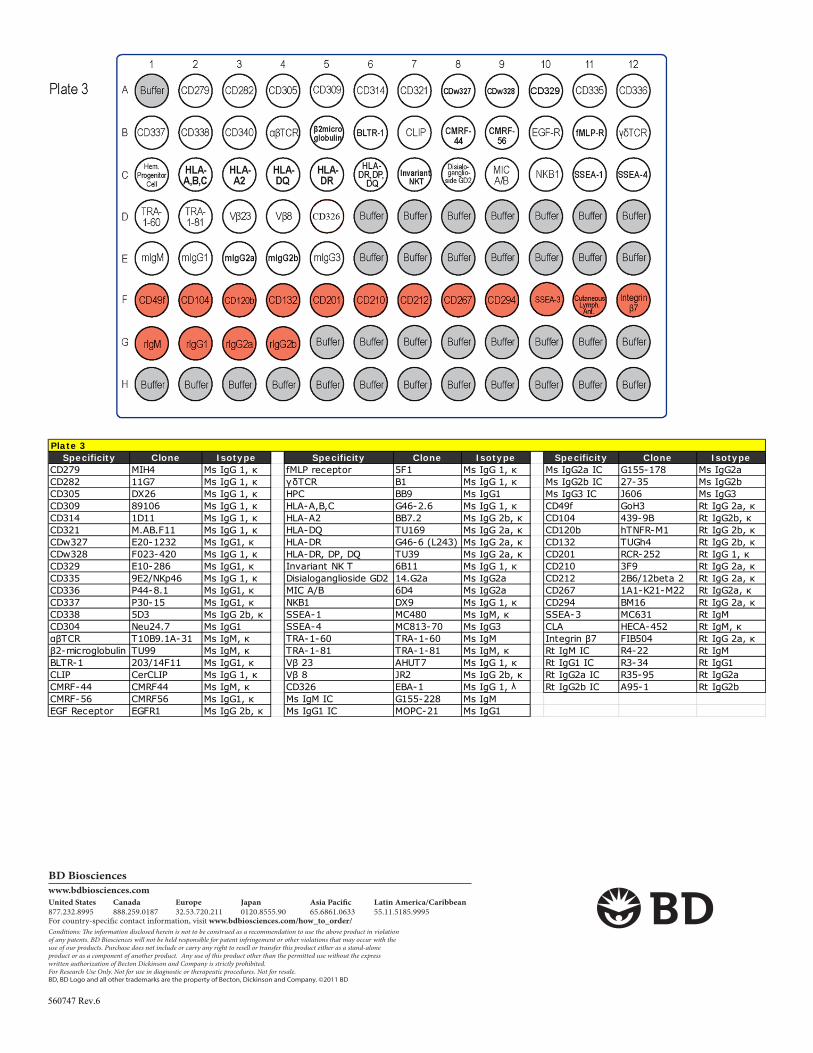
While the majority of the antibodies in the panel were raised in mouse, some of the antibodies were raised in rat; these are located on Plate 3 in wells F1-F12 and G1-G4 (red wells on map of Plate 3). Ensure that anti-rat Ig secondary antibody is used with cells stained with these antibodies.
Evaluating background staining on the cells by titrating the Alexa Fluor® 647 secondary antibodies before attempting a full screen is
recommended. Excess secondary antibody has been provided. Based on the cell types tested, we recommend using the anti-Mouse and anti-Rat secondary antibodies at 1.25µg/ml (100µl per well).
The following negative controls are recommended: (1) unstained cells, (2) cells + anti-mouse secondary antibody and (3) cells + anti-rat
secondary antibody. Blank wells (labeled as "buffer" on the plate maps) are provided for this purpose.
For flow cytometric analysis, running 500,000 to 1,000,000 cells per well is recommended for best results. However, we have been successful in running as few as 250,000 cells per well.
For flow cytometric analysis, using a 96-well plate loader is recommended. However, we have also been successful transferring stained
cells from 96-well plates into Falcon® 12 X 75 mm round bottom tubes (Cat. No. 352008) and using a manual loader to run samples. Reconstituting the antibody:
After removing BD Lyoplate™ Human Cell Surface Marker Screening Panel plates from foil bags, centrifuge at 300 X g for 5 minutes.
Hold the plate firmly on the work bench and gently remove the foil seal starting from one end and pulling across the plate to completely remove the seal. Once the foil seal is removed, all lyophilized antibodies must be immediately reconstituted. Do not replace the lid on the plate prior to reconstitution.
Using a multi-channel pipette, reconstitute lyophilized antibodies in 110 µl of 1X sterile PBS. This results in an antibody solution that
contains five tests (20 µl/test). Be sure to use fresh pipette tips for each row to prevent well-to-well contamination. Allow antibodies to reconstitute for five minutes at room temperature.
Store the reconstituted antibodies at 4°C until the cells are prepared for experiments. Reconstituted antibodies can be stored in plates with lids at 4°C for at least 10 days. Seal the plate edges (with lid on) with Parafilm "M"® laboratory film to prevent loss of reconstituted antibody due to evaporation.

560747 Rev.6
BD Bioscienceswww.bdbiosciences.comUnited States877.232.8995
Canada888.259.0187
Europe32.53.720.211
Japan0120.8555.90
Asia Pacific65.6861.0633
Latin America/Caribbean55.11.5185.9995
For country-specific contact information, visit www.bdbiosciences.com/how_to_order/Conditions: The information disclosed herein is not to be construed as a recommendation to use the above product in violation of any patents. BD Biosciences will not be held responsible for patent infringement or other violations that may occur with the use of our products. Purchase does not include or carry any right to resell or transfer this product either as a stand-alone product or as a component of another product. Any use of this product other than the permitted use without the express written authorization of Becton Dickinson and Company is strictly prohibited.For Research Use Only. Not for use in diagnostic or therapeutic procedures. Not for resale.BD, BD Logo and all other trademarks are the property of Becton, Dickinson and Company. ©2011 BD
Screening cells by flow cytometry:
1. Prepare a single cell suspension of live cells from a cell line, tissue or a three dimensional culture. For adherent cell lines, using either a mild enzyme such as Accutase™ or a non-enzymatic dissociation buffer is recommended.
2. Wash the cells in two to four volumes of 1X PBS. Centrifuge at 300 X g for 5 minutes.
3. Remove any clumps by passing the cells through a Falcon® 40 or 70 µm cell strainer (Cat. No. 352340, Cat. No. 352350).
4. Determine the cell concentration and total number of cells. If you are dissociating tissue or a three dimensional culture, we recommend
treating the single cells with DNase to prevent cell clumping. Resuspend cells in the recommended growth media or 1X PBS with calcium and magnesium with the addition of 100 units/ml DNase at 10 million cells per ml. Incubate for 15 minutes at room temperature.
5. Wash the cells in two to four volumes of 1X PBS. Centrifuge at 300 X g for 5 minutes.
6. Prepare 275 ml of BD Pharmingen Stain Buffer (FBS) with the addition of 5 mM EDTA (final concentration) for subsequent steps.
7. Resuspend the sample in BD Pharmingen Stain Buffer + EDTA. You will need 135 to 270 million cells (in approximately 27 ml total volume) to fill the antibody containing wells of the three plates (500,000-1,00,000 cells per well). The minimum number of cells per well
will depend on the cytometer and/or loss of cells during washing. We have been successful in running as few as 250,000 cells per well.
8. Label three Falcon® round bottom 96 well plates (Cat No. 353910) plates 1, 2 and 3 for your sample plates.
9. Using a multi-channel pipette aliquot 100 µl of cell solution to required wells of the three labeled round-bottom 96-well plates.
a. If you have a limited number of cells, you can omit buffer only wells from plate 3. Please refer to the Plate 3 map to identify wells that can be excluded taking into consideration unstained cells and secondary antibody controls.
10. Using a multi-channel pipette, pipette up and down 2-3 times to fully mix the reconstituted antibody from the first row of wells from the BD Lyoplate™ Screening Panel Plate 1. Add 20 µl to the cells in the corresponding wells of sample plate 1. Continue to add reconstituted antibody to the corresponding sample wells for all remaining wells of each plate. Use fresh tips for every well. Incubate on ice for 20-30 minutes.
11. To wash, add 100 µl of BD Pharmingen Stain Buffer + EDTA to each well. Centrifuge at 300 X g for 5 minutes.
12. Remove supernatant carefully and wash cells with an additional 200 µl of BD Pharmingen Stain Buffer + EDTA. Centrifuge at 300 X g
for 5 minutes.
13. During the centrifugation step of the final wash, dilute the secondary antibody 1:200 dilution (1.25 µg/ml) in BD Pharmingen Stain Buffer + EDTA. You will need about 26 ml of dilute anti-mouse secondary antibody and about 3 ml of diluted anti-rat secondary antibody.
14. Remove supernatant and apply 100 µl of the appropriate secondary antibody directly to cells in each well and incubate for 20-30 minutes on ice in the dark.
a. Add anti-mouse secondary antibody to all wells of the first two labeled round-bottom 96-well sample plates. For sample plate 3,
please refer to the Plate maps and add anti-mouse secondary antibody to the appropriate sample wells (white wells: all wells in rows A, B and C; wells D1-D5 and E1-E5) and add anti-rat secondary antibody to the appropriate wells (red wells: wells F1-F12 and G1-G4).
b. Use remaining wells in sample plate 3 that do not contain antibody (gray wells) to set up unstained cells and anti-rat secondary
antibody controls.
15. To wash, add 100 µl of BD Pharmingen Stain Buffer + EDTA to each well. Centrifuge at 300 X g for 5 minutes.
16. Remove supernatant and wash cells with an additional 200µl of BD Pharmingen Stain Buffer + EDTA. Centrifuge at 300 x g for 5 minutes.
17. At this point you may wish to fix your cells prior to analysis. To fix, remove supernatant and add 100 µl of 4% paraformaldehyde in 1X PBS or BD Cytofix™ Fixation Buffer (Cat. No. 554655) per well and incubate for 10 minutes. If you do not wish to fix your cells go to step 19.

560747 Rev.6
BD Bioscienceswww.bdbiosciences.comUnited States877.232.8995
Canada888.259.0187
Europe32.53.720.211
Japan0120.8555.90
Asia Pacific65.6861.0633
Latin America/Caribbean55.11.5185.9995
For country-specific contact information, visit www.bdbiosciences.com/how_to_order/Conditions: The information disclosed herein is not to be construed as a recommendation to use the above product in violation of any patents. BD Biosciences will not be held responsible for patent infringement or other violations that may occur with the use of our products. Purchase does not include or carry any right to resell or transfer this product either as a stand-alone product or as a component of another product. Any use of this product other than the permitted use without the express written authorization of Becton Dickinson and Company is strictly prohibited.For Research Use Only. Not for use in diagnostic or therapeutic procedures. Not for resale.BD, BD Logo and all other trademarks are the property of Becton, Dickinson and Company. ©2011 BD
18. Wash cells twice with 1X PBS. Centrifuge at 300 X g for 5 minutes.
19. Remove supernatant and resuspend cells in 150 µl of BD Pharmingen Stain Buffer + EDTA per well. Analyze your samples on a flow
cytometer. We recommend collecting at least 10,000 events per well. While the first plate is being read, store the other plates on ice in the dark.
Screening cells by bioimaging:
1. Seed the cells in appropriate culture medium at an appropriate cell density in a Falcon® 96-well Imaging Plate (Cat. No. 353219), and culture cells to an appropriate density. We recommend 70-80% confluency for imaging screens.
2. BD Lyoplate surface staining should be performed on live cells as fixation can cause artifacts (false positive and negative signals) with
some cell surface markers. In cases where cells must be fixed prior to staining, we recommend confirming any positive hits with a live sample stain using imaging or flow cytometry.
3. Using a multi-channel pipette add 20 µl of each reconstituted antibody to the corresponding wells of your sample plates and incubate on ice for 20-30 minutes. Stain cells directly in 50 to 100 µl of fresh growth media. If staining fixed cells, stain cells in 1X PBS.
4. Wash cells twice in 100 µl 1X PBS.
5. Dilute secondary antibodies to 1:100 (2.5 µg/mL) in growth media and apply 100 µl of the appropriate secondary antibody directly to cells in each well of the sample plates and incubate for 20-30 minutes on ice in the dark. You will need about 26 ml of diluted anti-mouse secondary antibody and about 3 ml of diluted anti-rat secondary antibody.
a. Add anti-mouse secondary antibody to all wells of the first two labeled imaging 96-well sample plates. For sample plate 3, please refer to the Plate maps and add anti-mouse secondary antibody to the appropriate sample wells (white wells: all wells in rows A, B and C; wells D1-D5 and E1-E5) and add anti-rat secondary antibody to the appropriate wells (red wells: wells F1-F12 and G1-G4).
b. Use remaining wells in sample plate 3 that do not contain antibody (grey wells) to set up unstained cells and anti-rat
secondary antibody controls.
6. Remove supernatant and wash cells twice in 100 µl 1X PBS.
7. At this point you may wish to fix your cells prior to analysis. To fix, remove supernatant and add 100 µl of 4% paraformaldehyde in 1X PBS or BD Cytofix Fixation Buffer per well and incubate for 10 minutes. If you do not wish to fix your cells go to step 9.
8. Remove the fixative from the wells, and wash the wells twice with 100 µl of 1X PBS.
9. Add 100 µl 1X PBS with a cell-permeable nucleic acid stain, such as Hoechst 33342 Solution (Cat. No. 561908).
10. Analyze your samples on a high content bioimager.
Suggested Companion Products Description Size Catalog Number BD Pharmingen™ Stain Buffer (FBS) 500 ml 554656 BD Cytofix™ Fixation Buffer 100 ml 554655 BD Accutase™ 100 ml 561527 BD Pharmingen™ Hoechst 33342 Solution 1 mg/ml 561908 Related Products Description Size Catalog number Falcon® 96-well Microplates, Black/Clear With Lid, for High-Content Imaging Assays 32/case 353219 Falcon® 96-well Microplates, Round Bottom, No Lid, for High-Throughput Flow Cytometry Analysis 50/case 353910 Falcon® Low Evaporation Lids for Falcon® 96-well Microplates 50/case 353071

560747 Rev.6
BD Bioscienceswww.bdbiosciences.comUnited States877.232.8995
Canada888.259.0187
Europe32.53.720.211
Japan0120.8555.90
Asia Pacific65.6861.0633
Latin America/Caribbean55.11.5185.9995
For country-specific contact information, visit www.bdbiosciences.com/how_to_order/Conditions: The information disclosed herein is not to be construed as a recommendation to use the above product in violation of any patents. BD Biosciences will not be held responsible for patent infringement or other violations that may occur with the use of our products. Purchase does not include or carry any right to resell or transfer this product either as a stand-alone product or as a component of another product. Any use of this product other than the permitted use without the express written authorization of Becton Dickinson and Company is strictly prohibited.For Research Use Only. Not for use in diagnostic or therapeutic procedures. Not for resale.BD, BD Logo and all other trademarks are the property of Becton, Dickinson and Company. ©2011 BD
Product Notices
1. Falcon® is a registered trademark of Corning Incorporated. 2. Alexa Fluor® is a registered trademark of Life Technologies Corporation. 3. This product is provided under an intellectual property license between Life Technologies Corporation and BD Businesses. The purchase
of this product conveys to the buyer the non-transferable right to use the purchased amount of the product and components of the product in research conducted by the buyer (whether the buyer is an academic or for-profit entity). The buyer cannot sell or otherwise transfer (a) this product (b) its components or (c) materials made using this product or its components to a third party or otherwise use this product or its components or materials made using this product or its components for Commercial Purposes. Commercial Purposes means any activity by a party for consideration and may include, but is not limited to: (1) use of the product or its components in manufacturing; (2) use of the product or its components to provide a service, information, or data; (3) use of the product or its components for therapeutic, diagnostic or prophylactic purposes; or (4) resale of the product or its components, whether or not such product or its components are resold for use in research. For information on purchasing a license to this product for any other use, contact Life Technologies Corporation, Cell Analysis Business Unit Business Development, 29851 Willow Creek Road, Eugene, OR 97402, USA, Tel: (541) 465-8300. Fax: (541) 335-0504.
4. Alexa Fluor® 647 fluorochrome emission is collected at the same instrument settings as for allophycocyanin (APC). 5. This product may be covered by US Patent No. 5,543,320. 6. US Patent No. 5,994,515, University of Pennsylvania. 7. Source of all serum proteins is from USDA inspected abattoirs located in the United States. 8. Caution: Sodium azide yields highly toxic hydrazoic acid under acidic conditions. Dilute azide compounds in running water before
discarding to avoid accumulation of potentially explosive deposits in plumbing.
References
1. Maniecki MB, Etzerodt A, Moestrup S, Møller J, Graversen J. Comparative assessment of the recognition of domain-specific CD163
monoclonal antibodies in human monocytes explains wide discrepancy in reported levels of cellular surface CD163 expression. Immunobiology. 2011: 216 (8):882-890.
560747 Rev.6
BD Bioscienceswww.bdbiosciences.comUnited States877.232.8995
Canada888.259.0187
Europe32.53.720.211
Japan0120.8555.90
Asia Pacific65.6861.0633
Latin America/Caribbean55.11.5185.9995
For country-specific contact information, visit www.bdbiosciences.com/how_to_order/Conditions: The information disclosed herein is not to be construed as a recommendation to use the above product in violation of any patents. BD Biosciences will not be held responsible for patent infringement or other violations that may occur with the use of our products. Purchase does not include or carry any right to resell or transfer this product either as a stand-alone product or as a component of another product. Any use of this product other than the permitted use without the express written authorization of Becton Dickinson and Company is strictly prohibited.For Research Use Only. Not for use in diagnostic or therapeutic procedures. Not for resale.BD, BD Logo and all other trademarks are the property of Becton, Dickinson and Company. ©2011 BD
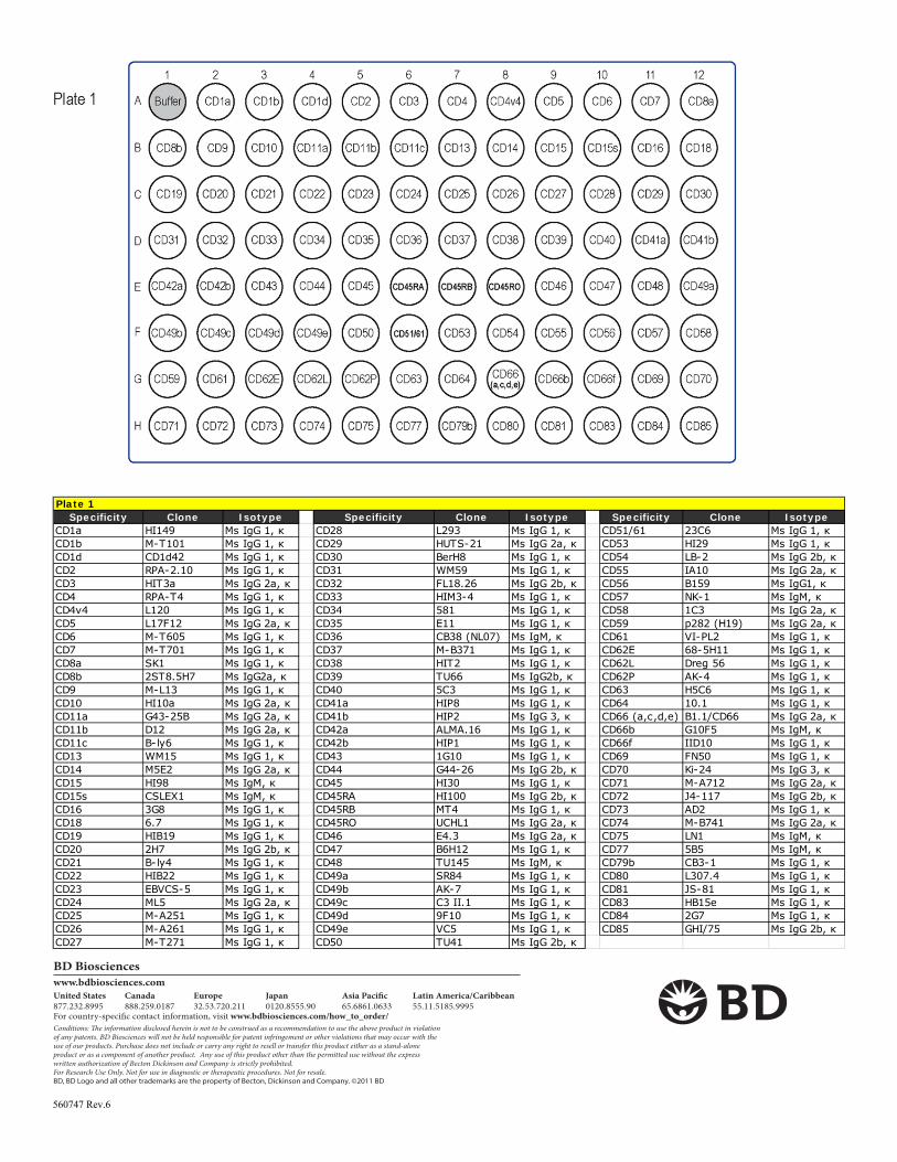
Plate 1Specificity Clone Isotype Specificity Clone Isotype Specificity Clone Isotype
CD1a HI149 Ms IgG 1, κ CD28 L293 Ms IgG 1, κ CD51/61 23C6 Ms IgG 1, κ CD1b M-T101 Ms IgG 1, κ CD29 HUTS-21 Ms IgG 2a, κ CD53 HI29 Ms IgG 1, κ CD1d CD1d42 Ms IgG 1, κ CD30 BerH8 Ms IgG 1, κ CD54 LB-2 Ms IgG 2b, κ CD2 RPA-2.10 Ms IgG 1, κ CD31 WM59 Ms IgG 1, κ CD55 IA10 Ms IgG 2a, κ CD3 HIT3a Ms IgG 2a, κ CD32 FL18.26 Ms IgG 2b, κ CD56 B159 Ms IgG1, κ CD4 RPA-T4 Ms IgG 1, κ CD33 HIM3-4 Ms IgG 1, κ CD57 NK-1 Ms IgM, κ CD4v4 L120 Ms IgG 1, κ CD34 581 Ms IgG 1, κ CD58 1C3 Ms IgG 2a, κ CD5 L17F12 Ms IgG 2a, κ CD35 E11 Ms IgG 1, κ CD59 p282 (H19) Ms IgG 2a, κ CD6 M-T605 Ms IgG 1, κ CD36 CB38 (NL07) Ms IgM, κ CD61 VI-PL2 Ms IgG 1, κ CD7 M-T701 Ms IgG 1, κ CD37 M-B371 Ms IgG 1, κ CD62E 68-5H11 Ms IgG 1, κ CD8a SK1 Ms IgG 1, κ CD38 HIT2 Ms IgG 1, κ CD62L Dreg 56 Ms IgG 1, κ CD8b 2ST8.5H7 Ms IgG2a, κ CD39 TU66 Ms IgG2b, κ CD62P AK-4 Ms IgG 1, κ CD9 M-L13 Ms IgG 1, κ CD40 5C3 Ms IgG 1, κ CD63 H5C6 Ms IgG 1, κ CD10 HI10a Ms IgG 2a, κ CD41a HIP8 Ms IgG 1, κ CD64 10.1 Ms IgG 1, κ CD11a G43-25B Ms IgG 2a, κ CD41b HIP2 Ms IgG 3, κ CD66 (a,c,d,e) B1.1/CD66 Ms IgG 2a, κ CD11b D12 Ms IgG 2a, κ CD42a ALMA.16 Ms IgG 1, κ CD66b G10F5 Ms IgM, κ CD11c B-ly6 Ms IgG 1, κ CD42b HIP1 Ms IgG 1, κ CD66f IID10 Ms IgG 1, κ CD13 WM15 Ms IgG 1, κ CD43 1G10 Ms IgG 1, κ CD69 FN50 Ms IgG 1, κ CD14 M5E2 Ms IgG 2a, κ CD44 G44-26 Ms IgG 2b, κ CD70 Ki-24 Ms IgG 3, κ CD15 HI98 Ms IgM, κ CD45 HI30 Ms IgG 1, κ CD71 M-A712 Ms IgG 2a, κ CD15s CSLEX1 Ms IgM, κ CD45RA HI100 Ms IgG 2b, κ CD72 J4-117 Ms IgG 2b, κ CD16 3G8 Ms IgG 1, κ CD45RB MT4 Ms IgG 1, κ CD73 AD2 Ms IgG 1, κ CD18 6.7 Ms IgG 1, κ CD45RO UCHL1 Ms IgG 2a, κ CD74 M-B741 Ms IgG 2a, κ CD19 HIB19 Ms IgG 1, κ CD46 E4.3 Ms IgG 2a, κ CD75 LN1 Ms IgM, κ CD20 2H7 Ms IgG 2b, κ CD47 B6H12 Ms IgG 1, κ CD77 5B5 Ms IgM, κ CD21 B-ly4 Ms IgG 1, κ CD48 TU145 Ms IgM, κ CD79b CB3-1 Ms IgG 1, κ CD22 HIB22 Ms IgG 1, κ CD49a SR84 Ms IgG 1, κ CD80 L307.4 Ms IgG 1, κ CD23 EBVCS-5 Ms IgG 1, κ CD49b AK-7 Ms IgG 1, κ CD81 JS-81 Ms IgG 1, κ CD24 ML5 Ms IgG 2a, κ CD49c C3 II.1 Ms IgG 1, κ CD83 HB15e Ms IgG 1, κ CD25 M-A251 Ms IgG 1, κ CD49d 9F10 Ms IgG 1, κ CD84 2G7 Ms IgG 1, κ CD26 M-A261 Ms IgG 1, κ CD49e VC5 Ms IgG 1, κ CD85 GHI/75 Ms IgG 2b, κ CD27 M-T271 Ms IgG 1, κ CD50 TU41 Ms IgG 2b, κ

560747 Rev.6
BD Bioscienceswww.bdbiosciences.comUnited States877.232.8995
Canada888.259.0187
Europe32.53.720.211
Japan0120.8555.90
Asia Pacific65.6861.0633
Latin America/Caribbean55.11.5185.9995
For country-specific contact information, visit www.bdbiosciences.com/how_to_order/Conditions: The information disclosed herein is not to be construed as a recommendation to use the above product in violation of any patents. BD Biosciences will not be held responsible for patent infringement or other violations that may occur with the use of our products. Purchase does not include or carry any right to resell or transfer this product either as a stand-alone product or as a component of another product. Any use of this product other than the permitted use without the express written authorization of Becton Dickinson and Company is strictly prohibited.For Research Use Only. Not for use in diagnostic or therapeutic procedures. Not for resale.BD, BD Logo and all other trademarks are the property of Becton, Dickinson and Company. ©2011 BD
Plate 2 Specificity Clone Isotype Specificity Clone Isotype Specificity Clone Isotype
CD86 2331 (FUN-1) Ms IgG 1, κ CD123 9F5 Ms IgG 1, κ CD172b B4B6 Ms IgG 1, κ CD87 VIM5 Ms IgG 1, κ CD124 hIL4R-M57 Ms IgG 1, κ CD177 MEM-166 Ms IgG 1, κ CD88 D53-1473 Ms IgG 1, κ CD126 M5 Ms IgG1, κ CD178 NOK-1 Ms IgG 1CD89 A59 Ms IgG 1, κ CD127 hIL-7R-M21 Ms IgG 1, κ CD180 G28-8 Ms IgG 1, κ CD90 5E10 Ms IgG 1, κ CD128b 6C6 Ms IgG 1, λ CD181 5A12 Ms IgG 2b, κ CD91 A2MR-alpha 2 Ms IgG 1, κ CD130 AM64 Ms IgG 1, κ CD183 1C6/CXCR3 Ms IgG 1, κ CDw93 R139 Ms IgG 2b, κ CD134 ACT35 Ms IgG 1, κ CD184 12G5 Ms IgG 2a, κ CD94 HP-3D9 Ms IgG 1, κ CD135 4G8 Ms IgG 1, κ CD193 5E8 Ms IgG 2b, κ CD95 DX2 Ms IgG 1, κ CD137 4B4-1 Ms IgG 1, κ CD195 2D7/CCR5 Ms IgG 2a, κ CD97 VIM3b Ms IgG 1, κ CD137 Ligand C65-485 Ms IgG 1, κ CD196 11A9 Ms IgG 1, κ CD98 UM7F8 Ms IgG 1, κ CD138 Mi15 Ms IgG 1, κ CD197 2H4 Ms IgM, κ CD99 TU12 Ms IgG 2a, κ CD140a alpha R1 Ms IgG 2a, κ CD200 MRC OX-104 Ms IgG 1, κ CD99R HIT4 Ms IgM, κ CD140b 28D4 Ms IgG 2a, κ CD205 MG38 Ms IgG 2bCD100 A8 Ms IgG 1, κ CD141 1A4 Ms IgG 1, κ CD206 19.2 Ms IgG 1, κ CD102 CBR-1C2/2.1 Ms IgG 2a, κ CD142 HTF-1 Ms IgG 1, κ CD209 DCN46 Ms IgG 2b, κ CD103 Ber-ACT8 Ms IgG 1, κ CD144 55-7H1 Ms IgG 1, κ CD220 3B6/IR Ms IgG 1, κ CD105 266 Ms IgG 1, κ CD146 P1H12 Ms IgG 1, κ CD221 3B7 Ms IgG 1, κ CD106 51-10C9 Ms IgG 1, κ CD147 HIM6 Ms IgG 1, κ CD226 DX11 Ms IgG 1, κ CD107a H4A3 Ms IgG 1, κ CD150 A12 Ms IgG 1, κ CD227 HMPV Ms IgG 1, κ CD107b H4B4 Ms IgG 1, κ CD151 14A2.H1 Ms IgG 1, κ CD229 HLy9.1.25 Ms IgG 1, κ CD108 KS-2 Ms IgG 2a, κ CD152 BNI3 Ms IgG 2a, κ CD231 M3-3D9 (SN1a) Ms IgG 1, κ CD109 TEA 2/16 Ms IgG 1, κ CD153 D2-1173 Ms IgG 1, κ CD235a GA-R2 (HIR2) Ms IgG 2b, κ CD112 R2.525 Ms IgG 1, κ CD154 TRAP1 Ms IgG 1, κ CD243 17F9 Ms IgG 2b, κ CD114 LMM741 Ms IgG 1, κ CD158a HP-3E4 Ms IgM, κ CD244 2-69 Ms IgG 2a, κ CD116 M5D12 Ms IgM, κ CD158b CH-L Ms IgG 2b, κ CD255 CARL-1 Ms IgG3CD117 YB5.B8 Ms IgG 1, κ CD161 DX12 Ms IgG 1, κ CD268 11C1 Ms IgG 1, κ CD118 12D3 Ms IgG1, κ CD162 KPL-1 Ms IgG 1, κ CD271 C40-1457 Ms IgG 1, κ CD119 GIR-208 Ms IgG 1, κ CD163 GHI/61 Ms IgG 1, κ CD273 MIH18 Ms IgG 1, κ CD120a MABTNFR1-A1 Ms IgG 1 CD164 N6B6 Ms IgG 2a, κ CD274 MIH1 Ms IgG 1, κ CD121a HIL1R-M1 Ms IgG1, κ CD165 SN2 Ms IgG 1, κ CD275 2D3/B7-H2 Ms IgG 2b, κ CD121b MNC2 Ms IgG 1, κ CD166 3A6 Ms IgG 1, κ CD278 DX29 Ms IgG 1CD122 Mik-beta 3 Ms IgG 1, κ CD171 5G3 Ms IgG2 a
560747 Rev.6
BD Bioscienceswww.bdbiosciences.comUnited States877.232.8995
Canada888.259.0187
Europe32.53.720.211
Japan0120.8555.90
Asia Pacific65.6861.0633
Latin America/Caribbean55.11.5185.9995
For country-specific contact information, visit www.bdbiosciences.com/how_to_order/Conditions: The information disclosed herein is not to be construed as a recommendation to use the above product in violation of any patents. BD Biosciences will not be held responsible for patent infringement or other violations that may occur with the use of our products. Purchase does not include or carry any right to resell or transfer this product either as a stand-alone product or as a component of another product. Any use of this product other than the permitted use without the express written authorization of Becton Dickinson and Company is strictly prohibited.For Research Use Only. Not for use in diagnostic or therapeutic procedures. Not for resale.BD, BD Logo and all other trademarks are the property of Becton, Dickinson and Company. ©2011 BD
Plate 3 Specificity Clone Isotype Specificity Clone Isotype Specificity Clone Isotype
CD279 MIH4 Ms IgG 1, κ fMLP receptor 5F1 Ms IgG 1, κ Ms IgG2a IC G155-178 Ms IgG2aCD282 11G7 Ms IgG 1, κ γδTCR B1 Ms IgG 1, κ Ms IgG2b IC 27-35 Ms IgG2bCD305 DX26 Ms IgG 1, κ HPC BB9 Ms IgG1 Ms IgG3 IC J606 Ms IgG3CD309 89106 Ms IgG 1, κ HLA-A,B,C G46-2.6 Ms IgG 1, κ CD49f GoH3 Rt IgG 2a, κ CD314 1D11 Ms IgG 1, κ HLA-A2 BB7.2 Ms IgG 2b, κ CD104 439-9B Rt IgG2b, κCD321 M.AB.F11 Ms IgG 1, κ HLA-DQ TU169 Ms IgG 2a, κ CD120b hTNFR-M1 Rt IgG 2b, κ CDw327 E20-1232 Ms IgG1, κ HLA-DR G46-6 (L243) Ms IgG 2a, κ CD132 TUGh4 Rt IgG 2b, κ CDw328 F023-420 Ms IgG 1, κ HLA-DR, DP, DQ TU39 Ms IgG 2a, κ CD201 RCR-252 Rt IgG 1, κ CD329 E10-286 Ms IgG1, κ Invariant NK T 6B11 Ms IgG 1, κ CD210 3F9 Rt IgG 2a, κ CD335 9E2/NKp46 Ms IgG 1, κ Disialoganglioside GD2 14.G2a Ms IgG2a CD212 2B6/12beta 2 Rt IgG 2a, κ CD336 P44-8.1 Ms IgG1, κ MIC A/B 6D4 Ms IgG2a CD267 1A1-K21-M22 Rt IgG2a, κCD337 P30-15 Ms IgG1, κ NKB1 DX9 Ms IgG 1, κ CD294 BM16 Rt IgG 2a, κ CD338 5D3 Ms IgG 2b, κ SSEA-1 MC480 Ms IgM, κ SSEA-3 MC631 Rt IgMCD304 Neu24.7 Ms IgG1 SSEA-4 MC813-70 Ms IgG3 CLA HECA-452 Rt IgM, κ αβTCR T10B9.1A-31 Ms IgM, κ TRA-1-60 TRA-1-60 Ms IgM Integrin β7 FIB504 Rt IgG 2a, κ β2-microglobulin TU99 Ms IgM, κ TRA-1-81 TRA-1-81 Ms IgM, κ Rt IgM IC R4-22 Rt IgMBLTR-1 203/14F11 Ms IgG1, κ Vβ 23 AHUT7 Ms IgG 1, κ Rt IgG1 IC R3-34 Rt IgG1CLIP CerCLIP Ms IgG 1, κ Vβ 8 JR2 Ms IgG 2b, κ Rt IgG2a IC R35-95 Rt IgG2aCMRF-44 CMRF44 Ms IgM, κ CD326 EBA-1 Ms IgG 1, λ Rt IgG2b IC A95-1 Rt IgG2bCMRF-56 CMRF56 Ms IgG1, κ Ms IgM IC G155-228 Ms IgMEGF Receptor EGFR1 Ms IgG 2b, κ Ms IgG1 IC MOPC-21 Ms IgG1