human anatomy lab: thoracic and abdominal regionspdarcey/bio 4/fall 2011/lab /labthorabd.pdf ·...
TRANSCRIPT

Bio 4 Lab: Thoracic and Abdominal Cavities 1
Human Anatomy Lab: Thoracic and Abdominal Regions Table of Contents:
• Expected Learning Outcomes . . . . 1 • Introduction . . . . . . 1 • Structures of the Thoracic Cavity. . . . 2 • Structures of the Abdominal and Pelvic Regions . 3 • Blood Vessels . . . . . . 5
Expected Learning Outcomes At the end of the labs where you examine internal sructures, you will be able to • locate and write the names of structures in the thoracic cavity; • locate and write the names of structures in the abdominal and pelvic cavities; • observe pathologies and variation among individuals: and • identify and write the names of major blood vessels that are located on the cat.
Introduction
The opportunity to look inside the body is rare and enlightening. The thoracic and abdominopelvic cavities have been opened for you by instructors, who used a bone saw to cut through the bony thoracic components—the ribs, sternum, and parts of the clavicles. The anterior wall of the abdomen was cut with a scalpel. Once this front covering is removed, we can explore the contents of the thoracic and abdominal cavities.
The cadavers are elderly individuals who typically died from the leading causes of death in our society—heart disease, cancer, stroke, and COPD (chronic obstructive pulmonary disorder). They also may have suffered from diabetes, hypertension, atherosclerosis, dementia, or other long-term conditions that are common as people age. In addition to identifying structures that all of us share, you will have the opportunity to explore variation among individuals and pathologies. What are you required to know for the test?
1. Locate and write down the name of all bulleted structures. (Spelling counts!) 2. Be able to identify and name obvious pathologies. 3. Be able to answer the questions in the Check Your Understanding boxes.
Fig. 1 Anterior view of the dissected body

Bio 4 Lab: Thoracic and Abdominal Cavities 2
How does one learn the information listed above?
1. Consult the illustrations in chapters 23 and 24 of Saladin’s Human Anatomy textbook.
2. Review the torso models in lab.
Additional Resources: These websites review internal anatomy of cadavers. <ect.downstate.edu/courseware/haonline/index.htm> <www.anatomy.wisc.edu/courses/gross/> Cat Circulatory System: homes.bio.psu.edu/faculty/strauss/anatomy/circ/circulat.htm STRUCTURES OF THE THORACIC CAVITY Note the position of the lungs and heart. The heart is typically slightly displaced toward the left, and nestles in the cardiac impression of the left lung. Locate the following bulleted structures: • diaphragm Relative to the lungs: • lungs
o Right Lung: Superior, Middle, and Inferior Lobes
o Left Lung: Superior and Inferior Lobes, Cardiac Impression
• parietal pleura • visceral pleura • mediastinum • Bronchi • Pulmonary artery/vein (it may not be possible to
differentiate between the two) Relative to the heart: • parietal pericardium • visceral pericardium • aorta • pulmonary trunk • superior vena cava • inferior vena cava • right ventricle • left ventricle • right and left atria If the heart wall has been opened: • bicuspid valve
Fig. 2 The Respiratory System

Bio 4 Lab: Thoracic and Abdominal Cavities 3
• tricuspid valve • chordae tendineae • papillary muscles Examine the heart for any pathologies. Note the size of the heart. Is it enlarged or average? There may be a pacemaker implanted between the pectoralis major and the skin of the chest. If so, trace the leads (wires) that snake through the superior vena cava to the right atrium and right ventricle. Note evidence of surgical procedures such as sutures or staples that may be associated with coronary bypass. Make notes here about the observed individual characteristics: Cad aver #:_______ Notes on individual characteristics: Cadaver #:________ Notes on individual characteristics:
Check Your Understanding Try to answer the following questions without looking at your notes. 1. List and explain two differences between the right and left lungs. 2. List at least two structures found in the mediastinum. 3. Name the layers that surround the heart. STRUCTURES OF THE ABDOMINAL AND PELVIC REGIONS
The abdominal cavity has been opened. Observe the layers of the abdominal wall—the skin, the abdominal muscles, and the inner lining, the shiny parietal peritoneum. Inferior to the diaphragm, you should be able to see the liver, and the intestines covered by the greater omentum, which hangs off the greater curvature of the stomach. Locate the following bulleted structures:
• parietal peritoneum • visceral peritoneum
Relative to the stomach:
• greater omentum • lesser omentum • cardiac region • fundus • body • pyloric region • greater curvature • lesser curvature
Relative to the small intestine: • mesentery • duodenum • jejunum/ileum (it is not possible to determine where one ends and the other begins)
Fig. 3 The Stomach
Bio 4 Lab: Thoracic and Abdominal Cavities 4
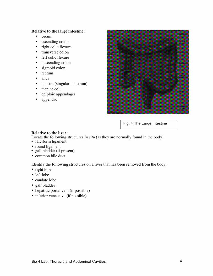
Relative to the large intestine: • cecum • ascending colon • right colic flexure • transverse colon • left colic flexure • descending colon • sigmoid colon • rectum • anus • haustra (singular haustrum) • taeniae coli • epiploic appendages • appendix
Relative to the liver: Locate the following structures in situ (as they are normally found in the body): • falciform ligament • round ligament • gall bladder (if present) • common bile duct Identify the following structures on a liver that has been removed from the body: • right lobe • left lobe • caudate lobe • gall bladder • hepatitic portal vein (if possible) • inferior vena cava (if possible)
Fig. 4 The Large Intestine

Bio 4 Lab: Thoracic and Abdominal Cavities 5
Other organs: Locate the following organs. • pancreas • spleen • kidney • urinary bladder Check Your Understanding Try to answer the following questions without looking at your notes. 4. Draw a simple diagram of the stomach and label the regions and curvatures. 5. Where would you look to locate the following structures: the cecum; the duodenum;
the greater omentum; the mesentery? 6. Identify the lobes of the liver. BLOOD VESSELS
We will examine selected vessels on the cat, which has easily identifiable arteries and veins because they are injected with colored latex: Red in the arteries and Blue in the veins. Locate and write the names of all of the bulleted vessels in the following list.
Veins: In the Thorax: • Superior vena cava • Brachiocephalic vein • External jugular vein • Subclavian vein • Inferior vena cava In the Abdomen: • Inferior vena cava
Fig. 5 The Liver

Bio 4 Lab: Thoracic and Abdominal Cavities 6
• Renal vein • External iliac vein In the Lower Limb: • Femoral vein • Greater saphenous vein Arteries: In the Thorax: • Aortic arch • Brachiocephalic artery • Left and right common carotid arteries • Left subclavian artery • Right subclavian artery In the Abdomen: • Abdominal aorta • Celiac artery • Superior mesenteric artery • Renal artery • Inferior mesenteric artery • External iliac artery • Internal iliac artery In the Lower Limb: • Femoral artery Check Your Understanding Try to answer the following questions without looking at your notes. 7. What vein is found traveling superiorly from the heart until it branches into the
brachiocephalic veins? 8. List four major branches of the abdominal aorta. 9. What is the difference between the pathways of the right and left subclavian arteries? 10. Draw simple diagrams of the pattern of arteries and veins in the cat. On your drawing,
label the vessels from the bulleted list.