ht29-mtx/caco-2 cocultures as an in vitro model for the intestinal epithelium: in vitro–in vivo...
TRANSCRIPT
HT29-MTX/Caco-2 Cocultures as an in Vitro Model for the IntestinalEpithelium: In Vitro −in Vivo Correlation with Permeability Data from Rats andHumans
ELKE WALTER*, SONIA JANICH†, BLAKE J. ROESSLER†, JOHN M. HILFINGER‡, AND GORDON L. AMIDON*X
Received March 4, 1996, from the *College of Pharmacy, The University of Michigan, Ann Arbor, MI 48109-1065, †Department ofInternal Medicine, Division of Rheumatology, University of Michigan Medical Center, Ann Arbor, MI 48109, and ‡TSRL, Incorporated,Ann Arbor, MI 48108. Final revised manuscript received June 7, 1996. Accepted for publication June 18, 1996X.
Abstract 0 The diverse secretory and absorptive functions of the intestinalepithelium are conducted by a mixed population of absorptive cells andmucus-producing goblet cells as the major cell types. In order to approachthe main characteristics in an in vitro model, a coculture system ofabsorptive Caco-2 cells and mucus-secreting HT29-MTX cells wasdeveloped and the permeability of a range of different drugs was tested.Variable goblet cell frequency can be achieved, preserving a significantbarrier to drug transport and maintaining the differentiated features ofboth cell types. Absorption rates for actively transported drugs are ratherunderestimated in the cell culture model when compared to in vivo data.However, a good correlation with fraction absorbed in humans was attainedseparating the range of passively transported drugs into two groups ofwell-absorbable compounds with Peff g 10 × 10-6 cm/s and drugs thatare absorbed 40−70% with Peff ) 0.1−1 × 10⁻⁶ cm/s. A permeability ofPeff < 0.1 × 10⁻⁶ cm/s is suggested for low absorbable drugs.
IntroductionThe estimation of human oral drug absorption based on in
vitro and in vivo measurements is of considerable utility inthe design of efficacious oral drug products.1-6 The experi-mental models are designed to isolate the barrier of interestto permit relatively rapid and mechanistic evaluation of drugcandidates. The Caco-2 cell line, a well-differentiated humanintestinal cell line, has been investigated as a potential in vitromodel for oral drug absorption and metabolism studies.2,4,7-10
When Caco-2 cells are grown on microporous membranes, theyform a confluent monolayer with several properties charac-teristic of differentiated enterocytes in the small intestine.11,12The monolayers are morphologically polar and develop brushborders at their apical surface. Furthermore, Caco-2 cells areknown to express carrier-mediated transport systems forvitamin B12, D-glucose, amino acids, and dipeptides.13-16
However, the intestinal epithelium consists of a variety ofdifferent cell types with two major cell phenotypes representedby enterocytes and goblet cells. Therefore, cocultures ofCaco-2 cells and mucus-producing goblet cells would providea drug absorption model incorporating the mucus barrier todrug absorption. Recently, a goblet cell clone (HT29-MTX)with a large proportion of mature goblet cells has beenestablished from the human intestinal cell line HT29.17 Atlate confluence, HT29-MTX monolayers show a dense mucusgel with numerous mucus buds on their apical surface.18However, the mucus gel layer is labile and could be removedby extensive washings.The aim of this study was to develop a Caco-2/HT29-MTX
coculture system and to characterize it in regard to occurrenceof mucus production, morphological features, and barrierfunction. We compared the penetration of a range of different
drugs through Caco-2/HT29-MTX monolayers with theirintestinal permeability in rats and humans. The transportof passively as well as actively transported drugs was evalu-ated to assess whether the model was predictive for drugcandidates and additionally for those with enhanced absorp-tion through a carrier-mediated transport mechanism.
Experimental Section
MaterialssTissue culture reagents were obtained from Gibco(Grand Island, NY), and tissue culture articles were from Corning(Corning, NY). All other chemicals were purchased from Sigma (St.Louis, MO).Cell CulturesCaco-2 cells (ATCC HTB37) were routinely main-
tained in Dulbecco’s modified Eagle’s medium (DMEM) with 25 mMD-glucose, containing 10% fetal bovine serum (FBS), 1% nonessentialamino acids, 1 mM sodium pyruvate, 1% L-glutamine, and penicillin(100 units/mL)/streptomycin (100 µg/mL), referred to as Caco-2medium. Caco-2 cells grown in 100-mm tissue culture Petri disheswere passaged every 5 days at a split ratio of 1:5. The mucus-secreting clone HT29-MTX-6 was kindly provided by Dr. T. Lesuffleur,INSERM U178, Villejuif Cedex, France.17,19 HT29-MTX-6 wasmaintained in DMEM with 25 mM glucose supplemented with 10%inactivated FBS (30 min, 56 °C), referred to as HT29 medium. Cellswere passaged weekly and seeded at a density of 2 × 10⁴ cells/cm2.The mycoplasma free cells were used at passage numbers 58-77(Caco-2) and 25-32 (HT29-MTX-6, passage in drug-free medium afteradaptation to 10-6 M methotrexate). All cells were maintained inan atmosphere of 5% CO2 and 90% relative humidity at 37 °C. Forcharacterization and transport studies, cells were seeded in six-wellculture plates or in cell culture inserts, 24.5-mm diameter, 0.4- or3-µm pores (Falcon poly(ethylene terephthalate) (PET) membranes,Becton Dickinson, Franklin Lakes, NJ, or Transwell tissue-treatedpolycarbonate membranes (PC), Costar, Cambridge, MA), at a densityof 2 × 10⁴ cells/cm2 and cultivated with medium change every secondday.CharacterizationsHistological staining for mucus-secreting cells
with alcian blue (pH 2.5) and periodic acid Schiff was performeddirectly on the cell layers as described previously.17 For transmission(TEM) and scanning (SEM) electron microscopy, cells were grown oncell culture inserts. Cells were fixed with 1.6% formaldehyde and2% glutaraldehyde in 0.1 M cacodylate buffer (pH 7.4) at 4 °C for 30min. After three washes with cacodylate buffer, cells were postfixedfor 30 min with 2% osmium tetraoxide (only TEM), washed threetimes with cacodylate buffer, and dehydrated in a graded series (70-100%) of ethanol. SEM samples were treated with hexamethylsila-zane for 10 min, dried overnight under vacuum, and coated with gold(Polaron E5100). The samples were then examined with a scanningISI-DS 130 electron microscope. TEM samples were embedded inEpon, and perpendicular thin sections were stained with uranylacetate and lead citrate. The specimens were studied in a PhilipsCM-10 electron microscope operated at 60 kV.Transepithelial Electrical Resistance (TEER)Measurementss
The integrity of the cell monolayers grown on permeable membranesin cell culture inserts was checked at different time points afterseeding by measurement of the transmembrane resistance using avoltohmmeter designed for this purpose (EVOM, World PrecisionInstruments, Sarasota, FL). The intrinsic resistance (membranealone) was subtracted from the total resistance (cell monolayers +X Abstract published in Advance ACS Abstracts, August 15, 1996.
S0022-3549(96)00110-4 CCC: $12.001070 / Journal of Pharmaceutical Sciences © 1996, American Chemical Society andVol. 85, No. 10, October 1996 American Pharmaceutical Association
+ +

membrane) to give the monolayer resistance. The resistance wascorrected for surface area and expressed as Ω‚cm2.Transport StudiessCocultures of 50% Caco-2 and HT29-MTX-6
on PET 3-µm filter inserts were used for transport studies. Experi-ments were performed directly in the cell culture inserts at 37 °C.Medium was removed, and the basolateral chamber was rinsed withtransport buffer (145 mM NaCl, 3 mM KCl, 1 mM CaCl2, 0.5 mMMgCl2, 1 mMNaH2PO4, 5 mM D-glucose, and 5 mM 2-(N-morpholino)-ethanesulfonic acid (pH 6.0) or HEPES (pH 6.8 and pH 7.4),respectively. Transport buffer (1.5 mL) containing 1 mg/mL of therespective drug was carefully added to the apical side of the mono-layers, and transport buffer without drug was added to the basolateralchamber (2.5 mL). Samples were withdrawn from the receiverchamber at different time intervals and replaced by fresh buffer. Theintegrity of the monolayers was checked at the beginning and at theend of the experiments by determination of the transepithelialelectrical resistance.HPLC ProceduressThe instrumentation consists of a pump
(Model Spectroflow 400, Kratos) and automatic sampler (Model 712WISP, Waters) and diode array detector (Model 996, Waters) all fromBeckman. Data acquisition and integration were carried out usingMillennium software. All compounds were separated on a BeckmanUltrasphere (5 µm, 4.6 × 250 mm) C-18 reversed-phase column witha flow rate of 1 mL/min under the following conditions (mobile phase(v/v), wavelength): propranolol, 60% methanol plus 40% 0.01 M pH3.5 sodium phosphate buffer, 215 nm; atenolol, 15% methanol plus85% 0.01 M pH 3.5 sodium phosphate buffer, 201 nm; cephalexin,30% methanol plus 70% 0.03 M pH 7.0 sodium phosphate buffer, 201nm; naproxen, 50% acetonitrile plus 49% water plus 1% glacial aceticacid, 243 nm; piroxicam, 70% methanol plus 30% 0.01 M sodiumphosphate (Na2HPO4) buffer containing 0.01 M citric acid, 205 nm;cimetidine, 20% methanol plus 80% water containing 0.03% phos-phoric acid, 201 nm; ranitidine, 30% methanol plus 67.5% 0.02 M pH6.0 sodium phosphate buffer plus 2.5% tetrahydrofuran, 318 nm;ampicillin, 8% acetonitrile plus 1% 1 M KH2PO4 plus 0.1% 1 M aceticacid plus 90.9% water, 201 nm; amoxicillin, 95% 0.01 M potassiumphosphate buffer pH 6.1 plus 5% acetonitrile, 201 nm; furosemide,59% water plus 1% glacial acetic acid plus 40% tetrahydrofuran, 276nm; L-DOPA and R-methyldopa, 95% 0.03 M pH 2.8 sodium phosphatebuffer plus 5% methanol, 201 nm. Quantification of the compoundswas carried out by measuring peak areas in relation to those ofstandards chromatographed under the same conditions.Data TreatmentsPermeability coefficients (Peff) were calculated
using the following equation:
where dc/dt is the flux across the monolayer (mM/mL), V the volumein the receiver chamber (mL), A the surface area of the monolayer(cm2), and c0 the initial concentration (mM) in the donor compartment.The flux across the monolayer describes the amount transportedversus time and was calculated from the slope of the regression lineobtained from the linear part of the curve.
ResultsCharacterization of HT29-MTX/Caco-2 CoculturessIn
order to simulate the intestinal epithelium in vitro, we grewmixed cultures of absorptive Caco-2 cells and mucus-secretingHT29-MTX on permeable supports. Transmission electronmicroscopy of 26-day-old monolayers revealed three morpho-logically different cell types. Cultures generally formed mixedmonolayers of well-differentiated absorptive and goblet cells,although some multilayered regions were observed containingundifferentiated and predifferentiated cells mixed with ma-ture cells. Cells with typical differentiated goblet cell mor-phology are shown in Figure 1A. Sparse microvilli that areirregular in both size and shape are developed at the apicalmembrane, and the apical cytoplasm is filled with closelypacked secretory granules. Adjacent goblet-shaped cellsdisplay junctional complexes, and the nucleus is locatedbasolaterally. Examination of the apical part of the goblet
cells in a higher magnification illustrates the fusion of mucusgranules with the apical cell membrane and release of mucusinto the luminal compartment (Figure 1B,C). Furthermore,a few cells with ultrastructural features similar to those ofundifferentiated and predifferentiated cells in the humanintestinal epithelium were found (Figure 1D). It is believedthat the predifferentiated so-called “oligomucous cells” arecapable of proliferation and serve as a precursor for gobletcells.20 The presence of absorptive enterocyte cells is dem-onstrated in Figure 1E. The tall columnar-shaped cells arepolarized with a row of straight dense microvilli on their apicalsurface and a basolaterally located nucleus. The presence oftight junctional complexes at the apical membrane boundariesis clearly visible at higher magnifications.Mixed cultures of HT29-MTX and Caco-2 in different
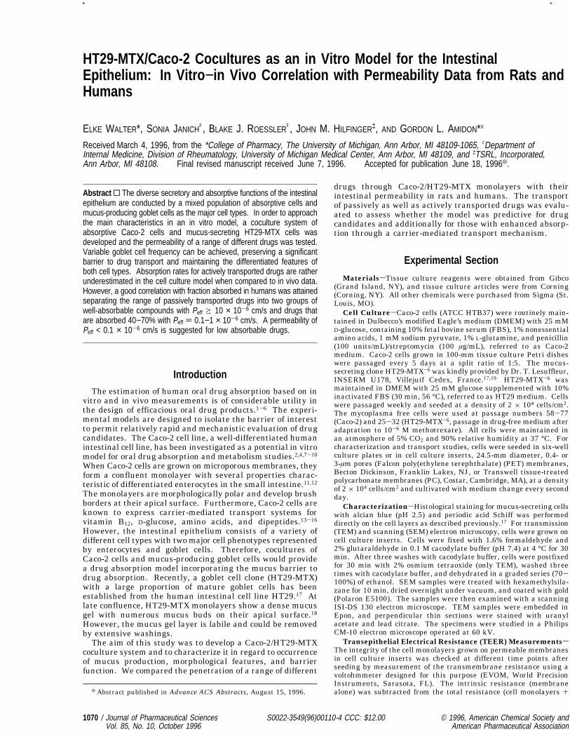
concentrations (25, 50, and 75%) were grown for 3-4 weeksin plastic culture dishes. At late confluence a mucous geladhering loosely to the monolayers could be observed in allcocultures. The percentage of positive cells after staining withperiodic acid Schiff (PAS) for detection of neutral and someacid mucosubstances and alcian blue for acid glycoconjugatesreflects the amount of HT29-MTX seeded in relation to Caco-2(Figure 2). Similar results were achieved with cocultures onPET membranes.Visualization of the cell surface of HT29-MTX/Caco-2 co-
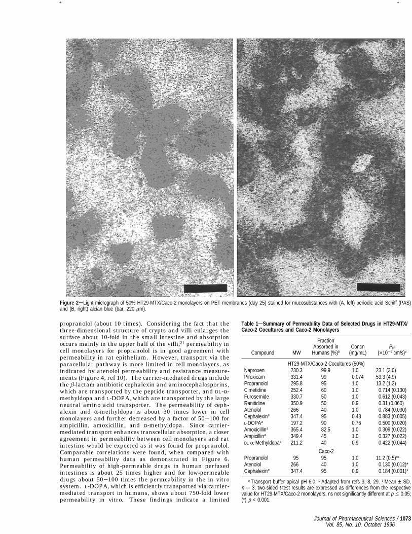
cultures at late confluence (26 days of culture) by scanningelectron microscopy shows a mucus gel with numerous mucusbuds on top of enterocyte-like cells with an apical brush borderand goblet cells with sparse microvilli that secrete mucus blebs(Figure 3). The mucus gel is labile, and the different washingand fixation steps required for SEM sample preparationremoved parts of it from the monolayer surface.An in vitro model for the study of oral drug transport needs
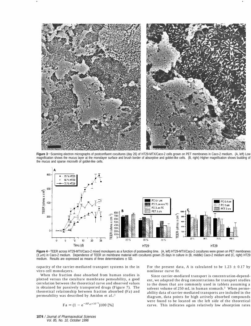
to perform a significant absorption barrier function. Theincrease in TEER over a cell monolayer during cell growthindicates the development of tight junctions between polarizeddifferentiated cells. When grown on permeable membranesthe cocultures showed a typical pattern of increase of theTEER over time (Figure 4A). After a lag phase of about 5days, TEER increased and reached a plateau depending onthe relation of Caco-2 and HT29-MTX in the monolayers. Ahigher amount of Caco-2 cells as the differentiated absorptivecell type leads to higher TEER values and a “tighter”epithelium probably due to more intensely formed tightjunctions. Membrane material and pore size as well as thechoice of culture medium (Caco-2 or HT29 medium; thecomposition is given in the Experimental Section) did notsignificantly influence the TEER, as shown in Figure 4B,C.Permeability Studies and Correlation with in Vivo
Data fromRats and HumanssWe studied the permeabilityof a range of different drugs in cocultures of 50% HT29-MTX/Caco-2 grown on PET membranes in Caco-2 medium (Table1). The relatively high amount of mucus-producing cells waschosen to yield significant differences in cell composition,which should make it possible to study the influence of mucuson drug transport. Figure 5 compares the permeabilities ofpassively and nonpassively transported drugs in Caco-2 andcoculture monolayers as well as in perfused rats. For pas-sively transported drugs, propranolol was chosen as a lipo-philic compound that is mainly absorbed by the transcellularroute, and atenolol and furosemide were chosen as hydrophilicparacellularly transported compounds.10 Mixed monolayersof HT29-MTX and Caco-2 cells are more permeable for alltested drugs than pure Caco-2 cultures. Permeabilities in thecell culture systems are generally lower compared to ratperfusion data. Regarding the coculture system, the differencein permeability for passively transported compounds is moresignificant for the low-permeability drugs atenolol and fur-osemide (about 30 times) than for the high-permeability drug
Peff ) dc Vdt Ac0
[cm/s]
Journal of Pharmaceutical Sciences / 1071Vol. 85, No. 10, October 1996
+ +

Figure
1sTransm
ission
electronmicroscopyof
postconfluentc
ocultures(day
26)of
HT29-MTX
/Caco-2
cells
grow
non
PETmem
branes
inCaco-2medium.(A,top
left)
Differentiatedgoblet
cells
with
sparse
microvillion
theapica
lsurface,closelypacked
secretorygranules
intheapica
lcytoplasm
,and
abasolaterally
locatednucleus(bar,3
.1µm
).(B,top
right,a
ndC,b
ottom
left)
Mucingranules
fuse
with
theapicalcell
mem
braneandreleasetheircontents(bar,0.6
µm(B)and
0.3
µm(C)).
(D,bottommiddle)Undifferentiated
cells
lackingtight
junctions
andmucin-type
secretorygranules
andpredifferentiatedcells
with
polarized
epithelialfeaturesfoundinmultilayered
regions.
Theleftcorner
atthebottom
show
sthefilterm
embrane
(bar,4.2
µm).
(E,bottomright)C
olum
nara
bsorptive-likecells
with
brushbordersattheira
picalm
embrane
andtight
junctions
betweenadjacent
cells
(bar,1
.6µm
).
1072 / Journal of Pharmaceutical SciencesVol. 85, No. 10, October 1996
+ +
propranolol (about 10 times). Considering the fact that thethree-dimensional structure of crypts and villi enlarges thesurface about 10-fold in the small intestine and absorptionoccurs mainly in the upper half of the villi,21 permeability incell monolayers for propranolol is in good agreement withpermeability in rat epithelium. However, transport via theparacellular pathway is more limited in cell monolayers, asindicated by atenolol permeability and resistance measure-ments (Figure 4, ref 10). The carrier-mediated drugs includethe â-lactam antibiotic cephalexin and aminocephalosporins,which are transported by the peptide transporter, and DL-R-methyldopa and L-DOPA, which are transported by the largeneutral amino acid transporter. The permeability of ceph-alexin and R-methyldopa is about 30 times lower in cellmonolayers and further decreased by a factor of 50-100 forampicillin, amoxicillin, and R-methyldopa. Since carrier-mediated transport enhances transcellular absorption, a closeragreement in permeability between cell monolayers and ratintestine would be expected as it was found for propranolol.Comparable correlations were found, when compared withhuman permeability data as demonstrated in Figure 6.Permeability of high-permeable drugs in human perfusedintestines is about 25 times higher and for low-permeabledrugs about 50-100 times the permeability in the in vitrosystem. L-DOPA, which is efficiently transported via carrier-mediated transport in humans, shows about 750-fold lowerpermeability in vitro. These findings indicate a limited
Figure 2sLight micrograph of 50% HT29-MTX/Caco-2 monolayers on PET membranes (day 25) stained for mucosubstances with (A, left) periodic acid Schiff (PAS)and (B, right) alcian blue (bar, 220 µm).
Table 1sSummary of Permeability Data of Selected Drugs in HT29-MTX/Caco-2 Cocultures and Caco-2 Monolayers
Compound MW
FractionAbsorbed inHumans (%)b
Concn(mg/mL)
Peff
(×10-6 cm/s)c
HT29-MTX/Caco-2 Cocultures (50%)Naproxen 230.3 99.9 1.0 23.1 (3.0)Piroxicam 331.4 99 0.074 53.3 (4.9)Propranolol 295.8 95 1.0 13.2 (1.2)Cimetidine 252.4 60 1.0 0.714 (0.130)Furosemide 330.7 50 1.0 0.612 (0.043)Ranitidine 350.9 50 0.9 0.31 (0.060)Atenolol 266 40 1.0 0.784 (0.030)Cephalexina 347.4 95 0.48 0.883 (0.005)L-DOPAa 197.2 90 0.76 0.500 (0.020)Amoxicillina 365.4 82.5 1.0 0.309 (0.022)Ampicillina 349.4 45 1.0 0.327 (0.022)DL-R-Methyldopaa 211.2 40 0.9 0.422 (0.044)
Caco-2Propranolol 95 95 1.0 11.2 (0.5)ns
Atenolol 266 40 1.0 0.130 (0.012)*Cephalexina 347.4 95 0.9 0.184 (0.001)*
a Transport buffer apical pH 6.0. b Adapted from refs 3, 8, 29. c Mean ± SD,n ) 3, two-sided t-test results are expressed as differences from the respectivevalue for HT29-MTX/Caco-2 monolayers, ns not significantly different at p e 0.05;(*) p < 0.001.
Journal of Pharmaceutical Sciences / 1073Vol. 85, No. 10, October 1996
+ +
capacity of the carrier-mediated transport systems in the invitro cell monolayers.When the fraction dose absorbed from human studies is
plotted versus the coculture membrane pemeability, a goodcorrelation between the theoretical curve and observed valuesis obtained for passively transported drugs (Figure 7). Thetheoretical relationship between fraction absorbed (Fa) andpermeability was described by Amidon et al.:1
For the present data, A is calculated to be 1.23 ( 0.17 bynonlinear curve fit.Since carrier-mediated transport is concentration-depend-
ent, we adapted the drug concentrations for transport studiesto the doses that are commonly used in tablets assuming asolvent volume of 250 mL in human stomach.3 When perme-ability data of carrier-mediated transports are included in thediagram, data points for high actively absorbed compoundswere found to be located on the left side of the theoreticalcurve. This indicates again relatively low absorption rates
Figure 3sScanning electron micrographs of postconfluent cocultures (day 26) of HT29-MTX/Caco-2 cells grown on PET membranes in Caco-2 medium. (A, left) Lowmagnification shows the mucus layer at the monolayer surface and brush border of absorptive and goblet-like cells. (B, right) Higher magnification shows budding ofthe mucus and sparse microvilli of goblet-like cells.
Figure 4sTEER across HT29-MTX/Caco-2 mixed monolayers as a function of postseeding time. (A, left) HT29-MTX/Caco-2 cocultures were grown on PET membranes(3 µm) in Caco-2 medium. Dependence of TEER on membrane material with cocultures grown 25 days in culture in (B, middle) Caco-2 medium and (C, right) HT29medium. Results are expressed as means of three determinations ± SD.
Fa ) (1 - e-APeff×10-6)100 [%]
1074 / Journal of Pharmaceutical SciencesVol. 85, No. 10, October 1996
+ +

in the cell culture model compared to in vivo data. The extentof in vivo absorption for high actively transported drugs wouldbe underestimated when compared to compounds that areabsorbed primarily by simple passive diffusion.
Discussion
The diverse secretory and absorptive functions of theintestinal epithelium are conducted by a mixed population ofabsorptive cells displaying a brush border membrane andmucus-producing goblet cells as the major cell types.21,22 Inorder to approach the main characteristics of the intestinalepithelium in an in vitro model to study intestinal absorptionprocesses, we investigated a mixed-cell coculture system ofmucus-producing HT29-MTX cells and absorptive Caco-2 cells.Goblet cells form a quantitatively significant component ofthe gastrointestinal tract, comprising approximately 10% ofthe duodenal epithelium and increasing about 24% of the totalepithelial cell population in the distal colon.20 Due to aconstant release of mucin from goblet cells, the gastrointes-tinal epithelium is covered by a viscoelastic, lubricant layerof mucus.20 With the HT29-MTX/Caco-2 coculture system,variable goblet cell frequency can be achieved, preserving asignificant barrier to drug transport as shown by TEERmeasurements and transport experiments. HT29-MTX cellsmaintain mucus-producing features in cocultures with Caco-2and when grown in Caco-2 medium.It has already been demonstrated that drug permeabilities
across Caco-2 monolayers are lower compared to intestinesof various species and more similar to colon permeability.5,6,23Transport rates for low absorbable hydrophilic drugs aregenerally higher in cocultures of Caco-2 and HT29-MTX thanin pure Caco-2 monolayers, designating higher paracellularpermeability in the coculture system. HT29-MTX/Caco-2cocultures are still 5-30-fold lower compared to in situ ratperfusion studies. This could be explained by a reducedabsorption area due to the lack of villi (indicated by lowerpermeability of propranolol) together with lower paracellularpermeability (indicated by lower permeability of atenolol).However, a good correlation with fraction absorbed in humanswas achieved, clearly separating the range of different drugsinto two groups, with well-absorbable compounds (Peff g 10× 10-6 cm/s) and drugs that are absorbed 40-70% (Peff )0.1-1 × 10⁻⁶ cm/s). The separation between the differentgroups is well defined, and the permeability of 40-70%absorbable drugs encompasses a relatively narrow range (Peff
) 0.3-0.8 × 10⁻⁶ cm/s). Therefore, we estimate a perme-ability of Peff < 0.1 × 10⁻⁶ cm/s for low absorbable drugs.Peptide and amino acid transporters are expressed in
Caco-2 cell monolayers, and affinity constants for the differentsubstrates seem to correlate with data in animals andhumans.15,16,24-26 However, the capacities of the carriers forthe studied substrates are reduced and lead to lower absorp-tion rates across the cell layers. The demonstration of carrier-mediated transport mechanisms in vitro on Caco-2 monolayershas been shown in many studies,15,26-28 but fraction absorbedafter oral administration of well actively transported com-pounds may be underestimated from the cell culture model.To overcome this problem, overexpression of the intestinalpeptide transporter in Caco-2 cells is currently under inves-tigation. This will allow us to search for drugs with enhancedpermeability due to an active transport component in additionto the screening of passively transported drugs.In summary, we developed a simulated intestinal epithe-
lium in vitro which consists of absorptive and mucus-secretingcell types as well as a small amount of undifferentiated orpredifferentiated cells. The coculture system contributes asignificant barrier to drug transport, and permeability coef-ficients for passively absorbed drugs correlate well withfractions absorbed in humans. It provides a potential tool tostudy the influence of mucin on oral drug absorption.
Figure 5sComparison of drug permeabilities in cell culture systems and ratperfusion experiments. Results are expressed as means of three determinations± SD. Permeabilities in rat jejunum are adapted from refs 1, 29−30, 32.
Figure 6sComparison of drug permeabilities in HT29/Caco-2 monolayers withpermeability in humans. Human permeabilities are calculated from perfusionexperiments.32
Figure 7sPlot of the fraction dose absorbed (%) in humans versus the meanpermeability in HT29-MTX/Caco-2 monolayers. Fraction doses absorbed areadapted from refs 1, 3, 8. The theoretical line was calculated for passivelytransported drugs by nonlinear regression as described in the text.
Journal of Pharmaceutical Sciences / 1075Vol. 85, No. 10, October 1996
+ +

References and Notes1. Amidon, G. L.; Sinko, P. J.; Fleisher, D. Pharm. Res. 1988, 10,
651-654.2. Artursson, P.; Karlsson, J. Biochem. Biophys. Res. Commun.
1991, 175, 880-885.3. Amidon, G. L.; Lennernas, H.; Shah, V. P.; Crison, J. R. Pharm.
Res. 1995, 12, 413-420.4. Walter, E.; Kissel, T.; Reers, M.; Dickneite, G.; Hoffmann, D.;
Stueber, W. Pharm. Res. 1995, 12, 360-365.5. Stewart, B. H.; Chan, O. H.; Lu, R. H.; Reyner, E. L.; Schmid,
H. L.; Hamilton, H. W.; Steinbaugh, B. A.; Taylor, M. D. Pharm.Res. 1995, 12, 693-699.
6. Rubas, W.; Jezyk, N.; Grass, G. M. Pharm. Res. 1993, 10, 113-118.
7. Walter, E.; Kissel, T. Pharm. Res. 1994, 11, 1575-1580.8. Gan, L. S.; Hsyu, P. H.; Pritchard, J. F.; Thakker, D. Pharm.
Res. 1993, 10, 1722-1725.9. Hilgers, A. R.; Conradi, R. A.; Burton, P. S. Pharm. Res. 1990,
9, 902-910.10. Artursson, P. J. Pharm. Sci. 1990, 79, 476-482.11. Hidalgo, I. J.; Raub, T. J.; Borchardt, R. T. Gastroenterology
1989, 96, 736-749.12. Artursson, P. Crit. Rev. Ther. Drug Carrier Syst. 1991, 8, 305-
330.13. Dix, C. J.; Hassan, I. F.; Obray, H. Y.; Shah, R.; Wilson, G.
Gastroenterology 1990, 98, 1272-1279.14. Bissonnette, P.; Blais, A.; Berteloot, A. Fed. Proc. 1987, 46, 1273.15. Hu, M.; Borchardt, R. T. Biochim. Biophys. Acta 1992, 1135,
233-244.16. Dantzig, A. H.; Bergin, L. Biochim. Biophys. Acta 1990, 1027,
211-217.17. Lesuffleur, T.; Babat, A.; Luccioni, C.; Beaumatin, J.; Clair, M.;
Kornowski, A.; Dussaulx, E.; Dutrillaux, B.; Zweibaum, A. J.Cell Biol. 1991, 115, 1409-1418.
18. Kerneis, S.; Bernet, M. F.; Coconnier, M. H.; Servin, A. L. Gut1994, 35, 1449-1454.
19. Lesuffleur, T.; Barbat, A.; Dussaulx, E.; Zweibaum, A. CancerRes. 1990, 50, 6334-6343.
20. Forstner, J. F.; Forstner, G. G. Gastrointestinal mucus. InPhysiology of the gastrointestinal tract, 3rd ed.; Johnson, L. R.,Ed.; Raven Press: New York, 1994; pp 1255-1283.
21. Johnson, L. R., Ed. Physiology of the gastrointestinal tract, 3rded.; Raven Press: New York, 1994.
22. Specian, R. D.; Oliver, M. G. Am. J. Physiol. 1991, 260, C183-C193.
23. Artursson, P.; Ungell, A. L.; Lofroth, J. E. Pharm. Res. 1993,10, 1123-1129.
24. Liang, R.; Fei, Y. J.; Prasad, P. D.; Ramamoorthy, S.; Han, H.;Yamg-Feng, T. L.; Hedinger, M. A.; Ganapathy, V.; Leibach, F.H. J. Biol. Chem. 1995, 270, 6456-6463.
25. Gochoco, C. H.; Ryan, F. M.; Miller, J.; Smith, P. L.; Hidalgo, I.J. Int. J. Pharm. 1994, 104, 187-202.
26. Dantzig, A. H.; Duckworth, D. C.; Tabas, L. B. Biochim. Biophys.Acta 1994, 1191, 7-13.
27. Dantzig, A. H.; Tabas, L. B.; Bergin, L. Biochim. Biophys. Acta1992, 1112, 167-173.
28. Hu, M.; Chen, J.; Zhu, Y.; Dantzig, A. H.; Stratford, R. E.;Kuhfeld, M. T. Pharm. Res. 1994, 11, 1405-1413.
29. Amidon, G. L.; Lee, H. J. Annu. Rev. Pharmacol. Toxicol. 1994,34, 321-341.
30. Oh, D. M.; Sinko, P. J.; Amidon, G. L. Pharm. Res. 1989, 6, S-91.31. Amidon, G. L.; Merfeld, A. E.; Dressman, J. B. J. Pharm.
Pharmacol. 1986, 38, 363-368.32. Amidon, G. L., et al. Unpublished results.
AcknowledgmentsThis work was supported by NIH/SBIR Grant R43 GM 53850 and
Deutsche Forschungsgemeinschaft Fellowship Wa 1016/1-1 (E.W.).We would also like to thank B. Donohoe for skillful technicalassistance in the SEM experiments.
JS960110X
1076 / Journal of Pharmaceutical SciencesVol. 85, No. 10, October 1996
+ +