hsiao et al., j proteomics bioinform 2012, 5:1 … et al., j proteomics bioinform 2012, 5:1 ......
TRANSCRIPT
Volume 5(1) : 015-023 (2012) - 0015 J Proteomics Bioinform ISSN:0974-276X JPB, an open access journal
Research Article Open Access
Hsiao et al., J Proteomics Bioinform 2012, 5:1http://dx.doi.org/10.4172/jpb.1000208
Research Article Open Access
Proteomics & Bioinformatics
Proteomics Investigation Reveals Apoptosis-Associated Proteins in Aryl Hydrocarbon Receptor-Deficient Human Lung Cells Treated with 2,3,7,8-Tetrachlorobenzo-p-dioxinEric S. L. Hsiao1, Yin-Wei Chang1, Pinpin Lin2* and Pao-Chi Liao1,3,4*1Department of Environmental and Occupational Health, College of Medicine, National Cheng Kung University, Tainan, Taiwan2Division of Environmental Health and Occupational Medicine, National Health Research Institutes, Zhunan, Taiwan3Sustainable Environment Research Center, National Cheng Kung University, Tainan, Taiwan4Center for Micro/Nano Science and Technology (CMNST), National Cheng Kung University, Tainan, Taiwan
*Corresponding authors: Pao-Chi Liao, Department of Environmental Occupational Health, National Cheng Kung University, College of Medicine, 138 Sheng-Li Road, Tainan 70428, Taiwan, Fax: 886-6-2743748; E-mail: [email protected]
Pinpin Lin, Division of Environmental Health and Occupational Medicine, National Health Research Institutes, No. 35 Keyan Road, Zhunan Town, Miaoli County 350, Taiwan, Fax: 886-37-587406; E-mail: [email protected]
Received November 25, 2011; Accepted January 09, 2012; Published January 20, 2012
Citation: Hsiao ESL, Chang YW, Lin P, Liao PC (2012) Proteomics Investigation Reveals Apoptosis-Associated Proteins in Aryl Hydrocarbon Receptor-Deficient Human Lung Cells Treated with 2,3,7,8-Tetrachlorobenzo-p-dioxin. J Proteomics Bioinform 5: 015-023. doi:10.4172/jpb.1000208
Copyright: © 2012 Hsiao ESL, et al. This is an open-access article distributed under the terms of the Creative Commons Attribution License, which permits unrestricted use, distribution, and reproduction in any medium, provided the original author and source are credited.
Keywords: 2,3,7,8-tetrachlorodibenzo-p-dioxin; Aryl hydrocarbon receptor; Proteomics; Apoptosis; GRB10; 2DE
Introduction 2,3,7,8-Tetrachlorodibenzo-p-dioxin (TCDD), a prototype of
Polychlorinated dibenzo-p-dioxins and dibenzofurans (PCDD/Fs), is one of most dangerous pollutants in the environment due to its resistance to degradation in animal and human fats [1-3]. By industrialization, TCDD and other dioxin-like congeners as a by-product are unintentionally produced during the combustion of fossil fuels and woodland during the incineration of industrial wastes. In 1997, TCDD is classified as a group 1 carcinogen by the International Agency for Research on Cancer (IARC) according to limited evidence of carcinogenicity in humans and sufficient evidence in animals [4]. In epidemiological studies, people with TCDD exposure increase the risk of various cancers, such as liver cancer, non-Hodgkin’s lymphoma, soft-tissue sarcoma, rectal cancer and lung cancer [5-7]. Some carcinogenic researches report that experimental animals treated different doses of TCDD have been applied comprehensively to uncover the mechanisms of TCDD-induced tumor promotion [8]. In our previous study, one of the mechanisms is oxidative stress induced by TCDD to lead to DNA mispairings [9]. These mutations cause the perturbation of oncogenes or tumor suppressor genes to result in the uncontrolled signal transduction pathways, i.e., NF-κB, AP-1, c-fos, c-jun, or p53 [10-11]. Therefore the tumor progression represents from hyperplasia to carcinoma. This indicates that oxidative stress plays a role in the mechanisms of carcinogenesis by TCDD. In the past decade, we continually monitored the PCDD/F levels of dietary intake, formula/breast milk, eggs and serum in Taiwanese [12-14]. A “hot spot” has been discovered in Taiwan to may help the future epidemiologic studies when the relationships between PCDD/F levels and health effects [15]. Therefore, it is important to understand the pathogenic mechanism of TCDD or other congeners.
Up to date, the molecular mechanisms of TCDD-mediated responses have been slightly cleared. The characterization of TCDD is as a high affinity ligand to interact with aryl hydrocarbon receptor (AhR), which is a cytosolic transcription factor of the basic helix-loop-helix/Per-Arnt-Sim family. Upon TCDD binding, the AhR complex translocates into nucleus and acts a concert with AhR nuclear translocator (ARNT). The activated AhR/ARNT heterodimer binds to DNA at xenobiotic-responsive elements (XREs) located and induced upstream the target genes of many drug-metabolizing enzymes including cytochrome P450 enzyme 1A1 (CYP1A1), CYP1B1and CYP1A2 [4, 16-17]. In previous study, we have established the interactive effects between CYP1A1 genotypes and PCDD/Fs exposure containing TCDD on liver function profile [18]. Furthermore, we also utilized the mouse model to establish the casual relationship of AhR expression, AhR activation (CYP1A2 expression up-regulation) and hepatic toxicity [19]. Consequently, AhR plays an important role for detoxification pathway depended on the high-affinity of environmental contaminants. Although the AhR in human is widely expressed in liver, kidney, thymus and lung, the
Abstract2,3,7,8-Tetrachlorodibenzo-p-dioxin (TCDD), the most potent dioxin, binds to aryl hydrocarbon receptor (AhR), a
ligand-activated transcription factor, which subsequently alters gene expression. Epidemiological studies suggested that exposure to TCDD highly enhanced the risk of pulmonary diseases. In this study, we adopted a comparative proteomic approach using two-dimensional gel electrophoresis (2DE) to identify the differential expression of proteins between TCDD-treated control and AhR-deficient lung adenocarcinoma cells. Fifteen proteins from seventeen differential protein spots were identified that four of which were down-regulated and eleven were up-regulated. Western blotting analysis confirmed differential expression for five identified proteins, including GRB10, YWHAG, HSP27, LGALS1 and ACTB. Further protein-protein interaction network analysis of the identified proteins illustrated that twelve proteins were linked to a network containing caspase-3, p53, MDM2 and Akt/PKB, which is known to be associated with apoptosis pathway. Among the identified proteins, GRB10 may play a critical role to regulate the development of apoptosis in human lung cells by TCDD exposure. According to our study, it is expected to lead to new insights into the apoptosis pathway in lung adenocarcinoma by TCDD exposure.

Citation: Hsiao ESL, Chang YW, Lin P, Liao PC (2012) Proteomics Investigation Reveals Apoptosis-Associated Proteins in Aryl Hydrocarbon Receptor-Deficient Human Lung Cells Treated with 2,3,7,8-Tetrachlorobenzo-p-dioxin. J Proteomics Bioinform 5: 015-023. doi:10.4172/jpb.1000208
Volume 5(1) : 015-023 (2012) - 0016 J Proteomics Bioinform ISSN:0974-276X JPB, an open access journal
research core of AhR mediated with TCDD focus on liver development, which is high expression of drug-metabolizing enzymes. Besides central role of detoxification, many studies demonstrated that AhR also participates in other signal transduction pathways to influence the biological processes of cell cycle regulation, oxidative stress response and apoptosis [20-22]. However, only few studies emphasized that the TCDD-mediated AhR influences the mechanisms associated with lung diseases [23-24]. In recent years, exposure to TCDD that accompanied over-expression of CYP1A1 and CYP1B1 has been implicated in the pathogenesis of lung adenocarcinoma [25]. In our previous studies, the expression level of AhR in the lung adenocarcinoma is up-regulated [26]. In further analysis, the AhR-deficient lung adenocarcinoma H1355 cell line by RNA interference technique is constructed to investigate the regulation of intracellular oxidative stress and cell growth [27]. However, the relationship of AhR and TCDD exposure in lung cancer is not clear until now. The objective in this study is to utilize the proteomic approach to research various molecular mechanisms and potential proteins involved in TCDD-mediated AhR signaling pathway of lung adenocarcinoma cells.
At present, the comparative proteomic approach is considered to be important for the system-based understanding of the molecular function of each identified protein between different treatments at the same conditional background [28]. The differently expression of
proteins are expected to provide insights into the various molecular mechanisms. Previously, the major proteomic studies are emphasized that the aspect of liver lesion is underlying TCDD-induced toxicity according to the modulation of drug-metabolizing enzymes [29-31]. Although the lung lesion and cancer are considered to associate with TCDD exposure, the systemic proteomic analysis has not been reported. In this study, we performed that one of a control vector (mock) was transfected into H1355 cells to compare with the other of a stably expressed short hairpin RNA vector for AhR (shAhR) using two-dimensional gel electrophoresis (2DE) coupled with mass spectrometry (MS). Figure 1 contains the experimental design developed for this study. The differential expression of identified proteins in mock and shAhR cells are considered to be involved in some biological processes and then the candidate proteins may be as the potential targets for therapy or the possible markers for diagnosis.
Materials and MethodsCell culture and TCDD treatment
There were two human lung adenocarcinama cell types to be constructed as previous described [27]. One was H1355 cell transfected with a control vector (mock) and the other was transfected a stably expressed short hairpin RNA for aryl hydrocarbon receptor vector (shAhR) and then all maintained in a RPMI-1640 medium (Gibco BRL, Gaithersbug, MD). The medium was supplemented with 5% heat-
Figure 1: Scheme of the sample processes and the analytic strategy used for comparative control and AhR-deficient human lung cells with TCDD exposure. The independent variable of AhR expression is to investigate the effects of TCDD toxicity in human lung adenocarcinoma. The various experiment steps of the analytic approach describes in this report.

Citation: Hsiao ESL, Chang YW, Lin P, Liao PC (2012) Proteomics Investigation Reveals Apoptosis-Associated Proteins in Aryl Hydrocarbon Receptor-Deficient Human Lung Cells Treated with 2,3,7,8-Tetrachlorobenzo-p-dioxin. J Proteomics Bioinform 5: 015-023. doi:10.4172/jpb.1000208
Volume 5(1) : 015-023 (2012) - 0017 J Proteomics Bioinform ISSN:0974-276X JPB, an open access journal
inactivated fetal bovine serum at 37°C under 5% CO2. A total 5×10⁵ cells of mock and shAhR, plated on respective 10 cm cell culture dishes, were treated with 10 nM TCDD when cultures were 70-80% confluent. After incubation for 48 h, these cell lines were collected and then lysed in NP-40 lysis buffer for 30 min at 4°C. The cell lysates were centrifuged at 12,000 g for 30 min. The resulting supernatants were collected and stored at -20°C.
Protein preparation and 2DE
Cell extracts were subjected to 10% thichloroacetic acid in ice cold acetone at -20°C overnight and then precipitated proteins were collected by centrifugation (16,000 g, 4°C, 20 min). The protein pellet was washed with 100% ice cold acetone and allowed to air dry. Protein concentration was evaluated using the Bradford protein assay (Bio-Rad, Hercules, CA) according to according to the manufacturer’s instructions. The 100 μg protein amounts were dissolved in rehydration buffer for isoelectric focusing (IEF) analysis. Immobilized pH gradient (IPG) strips of pH 4-7 and length 18 cm (IPGphor, GE health, Uppsala, Sweden) were focused on a stepwise incremental voltage program: 30V for 12h, 500V for 1h, 1000 V for 1h and 8000 V for 5h, with a total power of 42 kVh. After IEF, the strips were subjected to a two-step equilibration in equilibration buffer I (6 M urea, 30% glycerol, 2% SDS and 50 mM Tris-HCl; pH 8.8) containing 1% w/v dithiothreitol (DTT) followed by equilibration buffer II containing 2.5% w/v iodoacetamide (IAA) for 15 min, respectively. Proteins were separated in gradient (8-16%) polyacrylamide gels using a PROTEAN II xi system (Bio-Rad, Hercules, CA) at 15°C. Following the gels into a fixation solution, the proteins in gels were detected by silver staining [32].
Gel image analysis
The stained gels were scanned with resolution of 300 dpi using ImageScanner and LabScan software (GE health, Piscataway, NJ). The resulting gel images were analyzed with ImageMaster 2D Platinum software. The detection of protein spots, subtraction of background and spot matching were performed automatically and manually. After normalization for spot intensity in each 2DE-gel by computerized software, the spot intensity ratio of shAhR cells is divided by mock cells. The 95% confidence interval (CI) of spot intensity ratio was calculated from six replicates. The lower confidence limit was greater than 1 and the upper confidence limit was smaller than 1 to be determined as up-regulated and down-regulated, respectively. Each experimental ratio from six replicate 2DE analyses of mock and shAhR cells was performed with t-test. The p < 0.05 was considered statistically significant. The protein spots with significantly altered levels were excised for identification by mass spectrometry.
Tryptic In-Gel digestion
Excised protein spots were destained with 1:1 solution of 30mM potassium ferricyanide and 100 mM sodium thiosulfate until colorless at room temperature for 15 min. The destained spots were washed with 1:1 solution of acetonitrile (ACN) and 25 mM ammonium bicarbonate. The spots were reduced with 10 mM DTT for 1 h at 56°C and alkylated with 55 mM IAA for 45 min in the dark at room temperature. Subsequent digestion was performed using 0.1 μg of TPCK-treated modified porcine trypsin (Promega, Madison, WI) overnight at 37°C. The supernatant containing tryptic peptides was combined with two more extracts of the pieces by 50% ACN/5% formic acid (FA) and
then dried using a centrifugal evaporation instrument (Vacufuge, Eppendorf, Hauppauge, NY).
MS/MS analysis and database searching
The dried peptides suspended with 1% FA/5% ACN were analyzed by nano-HPLC-ESI-MS/MS. The peptides were separated by a nano-HPLC system (LC Packings, Amsterdam, Netherlands) connected to an ion trap mass spectrometer (LCQ DECA XP Plus, ThermoFinnigan, San Jose, CA) equipped with an electrospray ionization source. Peptide mixtures were loaded onto a 75 μm I.D., 150 mm length C18 microcapillary column packed with 3 μm particles with a pore of 100Å and separated using a segmented gradient in 40 min from 0% to 60% solvent B (80% ACN with 0.1% FA) in solvent A (5% ACN with 0.1% FA) at a flow rate of 200 nl/min. Peptides were electrosprayed into the LCQ with the application of a distal 1.3 kV spraying voltage. Each cycle of one full scan MS spectrum (m/z 450-2000) was followed by three tandem mass spectra with collision energy set at 35%.
All MS and MS/MS raw files were processed by Bioworks Browser 3.1 (ThermoFinnigan, San Jose, CA) and searched against the human protein sequence database (Swiss-Prot) via an in-house Mascot server (Matrix Science Ltd, London, UK). Carboxyamidomethylation on cysteine and oxidation on methionine residues were set as variable modification. Search criteria used were trypsin digestion and allowing up to 2 missed cleavages. The mass tolerances of parental and fragment ion were all set to 1 Da. MOWSE scores of greater than 38 were considered significant (p < 0.05).
Bioinformatics analysis
To functionally annotate the identified proteins, the cellular components and biological processes were described in the Human Protein Reference Database (HPRD) [33]. For pathway analysis, the accession numbers of identified proteins were uploaded and analyzed for evidence of a protein-protein interaction network using an integrated knowledge and software suite, MetaCore (GeneGo, Inc., St. Joseph, MI). The existing canonical map was generated by an add-on tool, MapEditor, which integrated with MetaCore.
Western blotting
Proteins extracted from the cell lysates of mock and shAhR cells with 10 nM TCDD were resolved by SDS-PAGE using 10% polyacrylamide. The proteins were mixed with an equal volume of 2× sample buffer according to the suggestion in the Bio-Rad instruction manual and the mixtures were boiled for 10 min. After electrophoresis, the separating gels were transferred onto PDVF membranes (Pall Corp., East Hills, NY) in a Bio-Rad Trans-Blot system (Bio-Rad, Hercules, CA) according to the manufacturer’s instructions. These were blocked with TBST buffer (20 mM Tris-HCl, 0.5 M NaCl, 0.1% Tween 20, pH 7.6) containing 5% skim milk for 1 h at room temperature. The membranes were incubated with primary antibodies against the proteins of cell lysates (GRB10, YWHAG, HSP27, LGALS1, ACTB and GAPDH) overnight at 4°C, washed three times with TBST buffer and incubated with secondary antibodies conjugated with horseradish peroxidase (HRP) for 1 h at room temperature. After the membranes had been washed three times with TBST buffer, the protein bands were developed with enhanced chemiluminescence (ECL) detection.
Statistical analysis
The relationship of significantly altered protein levels between mock and shAhR cells were normalized to GAPDH levels. Differences

Citation: Hsiao ESL, Chang YW, Lin P, Liao PC (2012) Proteomics Investigation Reveals Apoptosis-Associated Proteins in Aryl Hydrocarbon Receptor-Deficient Human Lung Cells Treated with 2,3,7,8-Tetrachlorobenzo-p-dioxin. J Proteomics Bioinform 5: 015-023. doi:10.4172/jpb.1000208
Volume 5(1) : 015-023 (2012) - 0018 J Proteomics Bioinform ISSN:0974-276X JPB, an open access journal
in mean values between the two groups were determined using t-test. The p < 0.05 was considered statistically significant.
ResultsComparative protein quantitation of mock and shAhR cells treated with TCDD by 2DE
H1355 cells, a kind of lung adenocarcinoma cell line, were transfected with a control vector (mock) and a stably expressed short hairpin RNA for aryl hydrocarbon receptor vector (shAhR). The protein expression level of aryl hydrocarbon receptor (AhR) in shAhR cells was reduced to 30% of mock cells as previous described [27]. The protein
expression profiles in mock and shAhR cells with TCDD treatment were obtained by 2DE with linear IPG ranging from pH 4-7 (Figure 2). The six replicates of samples from mock and shAhR cell lysates were performed, respectively. The relative intensities of the normalized spot volumes were analyzed and compared between mock and shAhR cells using ImageMaster 2D Platinum software. Approximately 500 spots were detected on each gel with molecular weight ranging from 10 to 150 kDa and pI value between 4.5 and 7. After software process, spot intensity presented in gels was calculated. A ratio was determined that the spot intensity of the calculation of shAhR cells was divided by mock cells,he calculation of shAhR cells was divided by mock cells and then the confidence interval (CI) of ratio was calculated from the six replicated pairs. The spot variation between these two cell lines was determined when the lower confidence was greater than 1 as up-regulated or the upper confidence was smaller than 1 as down-regulated. Consequently, there were seventeen differential protein spots to be identified that thirteen protein spots were up-regulated and four was down-regulated. The protein spots differentially expressed in mock and shAhR cells were excised and then subjected to further analysis.
Identification of variant protein spots in 2DE maps of mock and shAhR cells
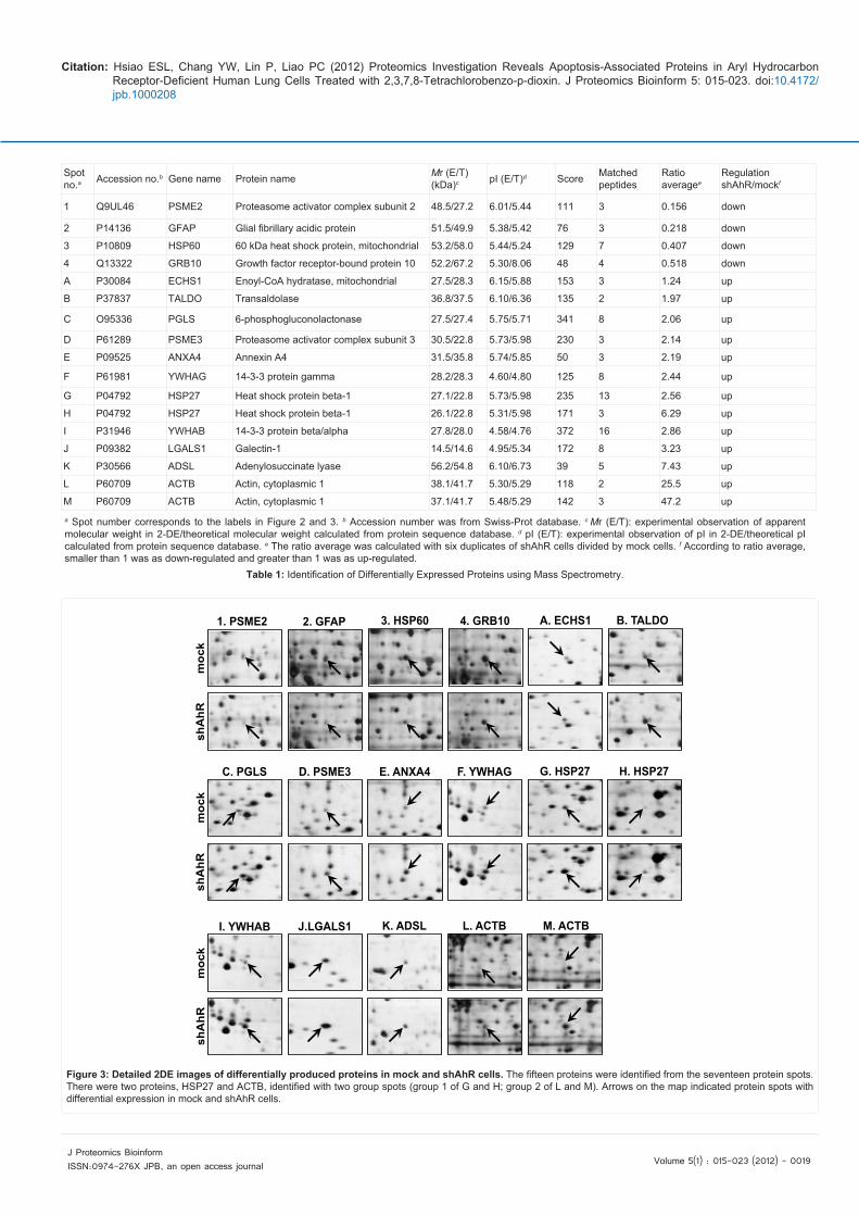
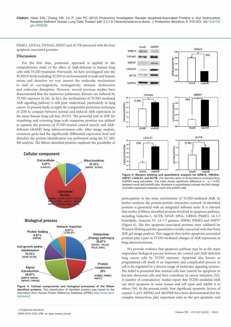
After MS/MS analysis and database searching, a total of fifteen proteins in seventeen protein spots were identified that four proteins were as down-regulated and eleven proteins were as up-regulated in mock cells compared with shAhR cells (Table 1). The two spot numbers, G and H, were identified as heat shock protein beta-1 (HSP27) and the other two, L and M, were also identified as beta-actin (ACTB). The close-up images of seventeen identified protein spots with differentially expression in mock and shAhR cells were presented in Figure 3. For further functionally annotation, the fifteen identified proteins were categorized to cellular components and biological processes according to the HPRD website (http://www.hprd.org/query) (Figure 4). The largest proportion of identified proteins was to be located in the cytoplasm. In the classification of biological processes, many of the identified proteins were functioned into metabolism and signal transduction.
Validation and Interaction Network Analysis of Identified Proteins
Regarding the result of proteomic analysis, the five identified proteins revealed by Western blotting and the result concurred with comparative protein quantization in 2DE (Figure 5). There were one protein, GRB10, higher expression in mock cells and the other four proteins, YWHAG, HSP27, LGALS1 and ACTB, higher in shAhR cells. Band intensity was measured and the relative intensity ratio to corresponding GAPDH band was calculated. After statistical analysis of triplicate, the results of fold change showed a consistent trend in protein abundance in comparative 2DE gels. The description of protein-protein interaction was analyzed by analytic software, MetaCore, loading the Swiss-Prot accession numbers of fifteen identified proteins with altered expression level between mock and shAhR cells. The interactive map was illustrated by the software, MapEditor, integrated into MetaCore according to proteins’ cellular localizations (Figure 6). The processed result showed that the twelve of fifteen identified proteins were involved in the generated network containing 29 proteins. The interactive network also includes the significant apoptosis-associated proteins such as caspase-3, p53, MDM2 and Akt/PKB. The map illustrated that the eight identified proteins of GFAP, HSP60, GRB10,
kDa 4 5 7pI
16%
shAhR
mock
21
A
34
M BCD
EF
GH
I
J
K
L
250
150
100
75
50
37
2520
1510
6
2
1
A
34
M BCD
EF
GH
I
J
K
L
250
150
100
75
50
37
2520
1510
Figure 2: Representative 2DE gel maps for mock and shAhR cells. A total of 100 μg of proteins were separated by 2DE using the first dimension of 18 cm pH 4-7 linear strips and the second dimension of 8-16% gradient SDS-PAGE. Separated proteins were visualized by silver staining. The spots were analyzed with ImageMaster software and identified by MS/MS sequencing. Down-regulated and up-regulated proteins are labeled as Arabic numerals and English alphabet, respectively.
Citation: Hsiao ESL, Chang YW, Lin P, Liao PC (2012) Proteomics Investigation Reveals Apoptosis-Associated Proteins in Aryl Hydrocarbon Receptor-Deficient Human Lung Cells Treated with 2,3,7,8-Tetrachlorobenzo-p-dioxin. J Proteomics Bioinform 5: 015-023. doi:10.4172/jpb.1000208
Volume 5(1) : 015-023 (2012) - 0019 J Proteomics Bioinform ISSN:0974-276X JPB, an open access journal
Spot no.a Accession no.b Gene name Protein name Mr (E/T)
(kDa)c pI (E/T)d Score Matched peptides
Ratio averagee
RegulationshAhR/mockf
1 Q9UL46 PSME2 Proteasome activator complex subunit 2 48.5/27.2 6.01/5.44 111 3 0.156 down
2 P14136 GFAP Glial fibrillary acidic protein 51.5/49.9 5.38/5.42 76 3 0.218 down
3 P10809 HSP60 60 kDa heat shock protein, mitochondrial 53.2/58.0 5.44/5.24 129 7 0.407 down
4 Q13322 GRB10 Growth factor receptor-bound protein 10 52.2/67.2 5.30/8.06 48 4 0.518 down
A P30084 ECHS1 Enoyl-CoA hydratase, mitochondrial 27.5/28.3 6.15/5.88 153 3 1.24 up
B P37837 TALDO Transaldolase 36.8/37.5 6.10/6.36 135 2 1.97 up
C O95336 PGLS 6-phosphogluconolactonase 27.5/27.4 5.75/5.71 341 8 2.06 up
D P61289 PSME3 Proteasome activator complex subunit 3 30.5/22.8 5.73/5.98 230 3 2.14 up
E P09525 ANXA4 Annexin A4 31.5/35.8 5.74/5.85 50 3 2.19 up
F P61981 YWHAG 14-3-3 protein gamma 28.2/28.3 4.60/4.80 125 8 2.44 up
G P04792 HSP27 Heat shock protein beta-1 27.1/22.8 5.73/5.98 235 13 2.56 up
H P04792 HSP27 Heat shock protein beta-1 26.1/22.8 5.31/5.98 171 3 6.29 up
I P31946 YWHAB 14-3-3 protein beta/alpha 27.8/28.0 4.58/4.76 372 16 2.86 up
J P09382 LGALS1 Galectin-1 14.5/14.6 4.95/5.34 172 8 3.23 up
K P30566 ADSL Adenylosuccinate lyase 56.2/54.8 6.10/6.73 39 5 7.43 up
L P60709 ACTB Actin, cytoplasmic 1 38.1/41.7 5.30/5.29 118 2 25.5 up
M P60709 ACTB Actin, cytoplasmic 1 37.1/41.7 5.48/5.29 142 3 47.2 up
a Spot number corresponds to the labels in Figure 2 and 3. b Accession number was from Swiss-Prot database. c Mr (E/T): experimental observation of apparent molecular weight in 2-DE/theoretical molecular weight calculated from protein sequence database. d pI (E/T): experimental observation of pI in 2-DE/theoretical pI calculated from protein sequence database. e The ratio average was calculated with six duplicates of shAhR cells divided by mock cells. f According to ratio average, smaller than 1 was as down-regulated and greater than 1 was as up-regulated.
Table 1: Identification of Differentially Expressed Proteins using Mass Spectrometry.
moc
ksh
AhR
1. PSME2 2. GFAP 3. HSP60 4. GRB10 A. ECHS1 B. TALDO
C. PGLS
shA
hRm
ock
D. PSME3 E. ANXA4 F. YWHAG G. HSP27 H. HSP27
I. YWHAB
moc
ksh
AhR
J.LGALS1 K. ADSL L. ACTB M. ACTB
Figure 3: Detailed 2DE images of differentially produced proteins in mock and shAhR cells. The fifteen proteins were identified from the seventeen protein spots. There were two proteins, HSP27 and ACTB, identified with two group spots (group 1 of G and H; group 2 of L and M). Arrows on the map indicated protein spots with differential expression in mock and shAhR cells.
Citation: Hsiao ESL, Chang YW, Lin P, Liao PC (2012) Proteomics Investigation Reveals Apoptosis-Associated Proteins in Aryl Hydrocarbon Receptor-Deficient Human Lung Cells Treated with 2,3,7,8-Tetrachlorobenzo-p-dioxin. J Proteomics Bioinform 5: 015-023. doi:10.4172/jpb.1000208
Volume 5(1) : 015-023 (2012) - 0020 J Proteomics Bioinform ISSN:0974-276X JPB, an open access journal
Cellular component
(HSP60, ECHS1)(LGALS1)
(PSME2, GFAP, GRB10, TALDO,PGLS, PSME3, ANXA4, YWHAG,HSP27, YWHAB, ADSL, ACTB)
Biological process
(PSME2, PSME3, HSP27)
(GFAP, ACTB)
(HSP60)
(GRB10, ANXA4, YWHAG, YWHAB)
(ECHS1, TALDO, PGLS, ADSL)
(LGALS1)
Figure 4: Cellular components and biological processes of the fifteen identified proteins. The classification of identified proteins was based on the information from Human Protein Reference Database (HPRD) (http://www.hprd.org/query).
Figure 5: Western blotting and quantitative analysis for GRB10, YWHAG, HSP27, LGALS1, and ACTB. The intensity ratios of all samples to corresponding GAPDH were calculated. The t-test shows significant difference (* : p < 0.05) between mock and shAhR cells. Numbers in parentheses indicate the fold change of protein expression between mock and shAhR cells.
PSME3, ANXA4, YWHAG, HSP27 and ACTB interacted with the four apoptosis-associated proteins.
DiscussionFor the first time, proteomic approach is applied to the
comprehensive study of the effect of AhR-deficient in human lung cells with TCDD treatment. Previously, we have investigated into the PCDD/F levels including TCDD in environmental victuals and human serum and therefore we very interest the molecular mechanisms to lead to carcinogenicity, teratogenicity, immune dysfunction and endocrine disruption. However, several previous studies have demonstrated that the numerous pulmonary diseases are induced by TCDD exposure [6,34]. In fact, the mechanisms of TCDD-mediated AhR signaling pathway is still poor understood, particularly in lung cancer. In present study, to apply the comparative proteomic technique of 2DE to compare between normal and reduced AhR expression in the same human lung cell line, H1355. The powerful tool of 2DE for visualizing and screening large-scale numerous proteins was utilized to separate the proteins of TCDD-treated control (mock) and AhR-deficient (shAhR) lung adenocarcinoma cells. After image analysis, seventeen spots had the significantly differential expression level and therefore the protein identification was performed using the LC-MS/MS analysis. The fifteen identified proteins implicate the possibility of
participation in the some mechanisms of TCDD-mediated AhR. In further analysis, the protein-protein interactive network of identified proteins is generated with an integrated software suite. It is relevant that twelve of fifteen identified proteins involved in apoptosis pathway, including Galectin-1, ACTB, GFAP, 6PGL, GRB10, PSME3, 14-3-3 beta/alpha, Annexin IV, 14-3-3 gamma, HSP60, PSME2 and HSP27 (Figure 6). The five apoptosis-associated proteins were validated by Western blotting and the quantitative results concurred with that from 2DE gel image analysis. This suggests that twelve apoptosis-associated proteins play a part in TCDD-mediated changes of AhR expression in lung adenocarcinoma.
We provide evidence that apoptosis pathway may be as the main respondent biological process between the control and AhR-deficient lung cancer cells by TCDD exposure. Apoptosis also known as programmed cell death is an important and complicated process in cell to be regulated by a diverse range of molecular signaling systems. The belief is presented that normal cells lose control for apoptosis to become abnormal cells and then contribute to cancer initiation [35]. A number of contradictory studies report that TCDD-mediated AhR can elicit apoptosis in some tissues and cell types and inhibit it in others [36]. In the present study, four significant apoptotic factors of caspase-3, p53, MDM2 and Akt/PKB have been demonstrated that the complex interactions play important roles in the pro-apoptotic and

Citation: Hsiao ESL, Chang YW, Lin P, Liao PC (2012) Proteomics Investigation Reveals Apoptosis-Associated Proteins in Aryl Hydrocarbon Receptor-Deficient Human Lung Cells Treated with 2,3,7,8-Tetrachlorobenzo-p-dioxin. J Proteomics Bioinform 5: 015-023. doi:10.4172/jpb.1000208
Volume 5(1) : 015-023 (2012) - 0021 J Proteomics Bioinform ISSN:0974-276X JPB, an open access journal
anti-apoptotic signals [37-39]. One of the understanding pathways is that Akt/PKB kinase phosphorylates MDM2, which is an E3 ubiquitin ligase and increases its activity and then MDM2 ubiquitinates p53 to lead to p53 proteasomal degradation [40-41]. As a feedback loop, p53 targets to MDM2 gene promoter and induces MDM2 expression [42]. In a recent study, TCDD can increase MDM2 protein level and inhibit p53 response to influence the liver carcinogenesis [43]. It is also verified that TCDD increases Akt phosphorylation by inducing PI3K activity to lead to suppression of cell death [44]. In Figure 6, the three identified proteins of GRB10, PSME3 and HSP27 interact to the Akt/PKB-MDM2-p53 signaling axis following the alteration of TCDD-mediated AhR pathway in lung adenocarcinoma cells.
On the other hand, a largest proportion of identified proteins interact with caspase-3 in the interactive network. Caspase-3, which is a cysteine-aspartic acid protease, is indispensable for apoptosis in mammals, particularly in oxidative stress-induced [45]. According to our previous study, TCDD induces oxidative stress in human breast carcinoma cell lines [9]. We suggest that TCDD may induce oxidative stress to modulate the caspase-3-mediated apoptosis in human lung cells. In chondrocytes, the other study also demonstrates that TCDD induces ROS-mediated caspase-3 activity to modulate apoptosis process [46]. Consequently, our study unambiguous indicated that the most of identified proteins, which are the differential expression level between the TCDD-treated mock and shAhR cells, are involved in two
major apoptosis-related signaling pathways of Akt/PKB-MDM2-p53 and caspase-3-mediated axes. Furthermore, some of identified proteins in Figure 6 have been demonstrated to be regulated by TCDD exposure such as HSP60, GFAP, HSP27 and LGASL1 in different investigations [30, 47-49]. Herein, these proteins may respond to apoptosis via TCDD-mediated AhR pathway. There are still many potential targets remaining to be investigated.
In addition, we utilized human cancer pathway microarray assay to investigate the gene expression changes between control and AhR-deficient cells by TCDD exposure (Supplementary Table 1). In the results of differential gene expression levels, seven apoptosis-associated genes were significant changes (shAhR/mock ratio3 or 0.33), including, APAF1, BAD, BCL1L2, CFLAR, NFKB1, PIK3CB and FAS. The seven genes were a relatively high percentage in 20 significant changed genes. These data combined with proteomics results would strongly support the hypothesis that TCDD-mediated AhR in H1355 cells takes place mainly through an apoptotic process.
In our present result, the higher expression level of GRB10 in mock cells compared to shAhR cells is considered to quantification of 2DE images and Western blotting (Figure 3 & 5). For the first time, it is demonstrated that GRB10 is reduced by TCDD treatment in AhR-deficient lung adenocarcinoma cell line. The molecular function of GRB10 is as an adaptor protein that interacts with some tyrosine kinase receptors and signaling molecules, including EGFR, Ret and IGF-R
Figure 6: The interaction network analysis of proteins of altered expression level between mock and shAhR cells. The interactive map was illustrated by the MapEditor program appended to MetaCore. Proteins identified in this study (Galectin-1 (LGALS1), ACTB, GFAP, 6PGL (PGLS), GRB10, PSME3, 14-3-3 beta/alpha (YWHAB), Annexin IV (ANXA4), 14-3-3 gamma (YWHAG), HSP60, PSME2 and HSP27) were marked with a white arrow on top. The symbols and links shown on the network illustration include: generic binding protein ( ), receptor ligand ( ), transporter ( ), generic receptor ( ), generic channel ( ), generic enzyme ( ), generic protease ( ), protein kinase ( ), protein phosphatase ( ), Ras- superfamily ( ), protein ( ), transcription factor ( ), activation ( ), inhibition ( ), and unspecified ( ).

Citation: Hsiao ESL, Chang YW, Lin P, Liao PC (2012) Proteomics Investigation Reveals Apoptosis-Associated Proteins in Aryl Hydrocarbon Receptor-Deficient Human Lung Cells Treated with 2,3,7,8-Tetrachlorobenzo-p-dioxin. J Proteomics Bioinform 5: 015-023. doi:10.4172/jpb.1000208
Volume 5(1) : 015-023 (2012) - 0022 J Proteomics Bioinform ISSN:0974-276X JPB, an open access journal
[50]. The SH2 domain in GRB10 is as a trigger for phosphorylation of tyrosine kinase that has been reported to regulate cell proliferation and apoptosis and also implicates in tumor progression [51]. Furthermore, GRB10, which is as a positive regulator, interacts with a synergistic factor of c-kit to activate Akt/PKB [52]. Therefore, these evidences reveal that GRB10 is involved in the mechanism of apoptosis. It is further studied that GRB10 possesses an anti-apoptotic activity through interaction with Raf-1 to contribute in cell survival underlying tumorigenesis [53-54]. Other researchers have indicated that TCDD treatment in B cell leads to alteration of phosphorylation level of kinases such as Akt/PKB, ERK and JNK [55]. Nevertheless, the mechanism of regulation of Akt/PKB in TCDD-mediated AhR pathway is still unclear.
Taken together, we hypothesize that GRB10 is regulated by the complex of TCDD-liganded AhR involved in the apoptosis pathway of Akt/PKB-MDM2-p53 signaling axis. In detail, we integrate with our previous and present study to suggest that GRB10-mediated apoptosis leads to the reduced AhR expression in lung adenocarcinoma cell to decrease intracellular oxidative stress and suppress cell growth [26]. To verify the hypothesis, the expression level of GRB10 compared between normal AhR-expressed (mock) and AhR-deficient (shAhR) cells are examined by Western blotting (Figure 5). The GRB10 expression in shAhR cells is significantly decreased two-fold (p < 0.05), compared to mock cells. Therefore, our data demonstrate that disrupting AhR expression with TCDD treatment reduces the expression of GRB10.
In conclusion, we have provided strategy that protein expression in comparative proteomic approach of TCDD toxicity. The strategy has often been applied that only one gene over-expressed or silenced in the same cell line decreases the variables and perturbation in proteomic study. We apply the lung adenocarcinoma cell line with reduction of AhR expression to recognize the differential expression of proteins and analyze those involved in particular biological mechanisms. In present study, we find that the most of identified proteins are involved in apoptosis pathway. GRB10 has a critical role to regulate the cell apoptosis and proliferation by TCDD-liganded AhR in lung adenocarcinoma cell. It may lead to be as a potential target to prevent the development of lung adenocarcinoma.Acknowledgement
The authors thank the National Cheng-Kung University Proteomics Research Core Laboratory for assistance in mass spectrometry analysis for protein identification. The study was supported by a grant from the National Science Council of Taiwan (NSC 99-2923-M-006-001-MY3, NSC 100-2113-M-006-002-MY3 and NSC 100-2325-B-006 -004) and the National Cheng-Kung University Project of Promoting Academic Excellence & Developing World Class Research Centers from the Ministry of Education of Taiwan. Pathway analysis was performed using the system provided by the Bioinformatics Core for Genomic Medicine and Biotechnology Development at the National Cheng-Kung University and were supported by National Science Council grant (NSC97-3112-B-006-011).
References
1. Jensen AA (1987) Polychlorobiphenyls (PCBs), polychlorodibenzo-p-dioxins (PCDDs) and polychlorodibenzofurans (PCDFs) in human milk, blood and adipose tissue. Sci Total Environ 64: 259-293.
2. Hirako M (2008) Distribution of PCDDs, PCDFs and dioxin-like PCBs in the blood, testis and adipose tissue of suckling beef calves. Chemosphere 71: 219-226.
3. Pelclova D, Urban P, Preiss J, Lukas E, Fenclova Z, et al. (2006) Adverse health effects in humans exposed to 2,3,7,8-tetrachlorodibenzo-p-dioxin (TCDD). Rev Environ Health 21: 119-138.
4. Steenland K, Bertazzi P, Baccarelli A, Kogevinas M (2004) Dioxin revisited:
developments since the 1997 IARC classification of dioxin as a human carcinogen. Environ Health Perspect 112: 1265-1268.
5. Ngaon LT, Yoshimura T (2001) Liver Cancer in Viet Nam: Risk Estimates of Viral Infections and Dioxin Exposure in the South and North Populations. Asian Pac J Cancer Prev 2: 199-202.
6. Bertazzi PA, Consonni D, Bachetti S, Rubagotti M, Baccarelli A, et al. (2001) Health effects of dioxin exposure: a 20-year mortality study. Am J Epidemiol 153: 1031-1044.
7. Martinez JM, Afshari CA, Bushel PR, Masuda A, Takahashi T, et al. (2002) Differential toxicogenomic responses to 2,3,7,8-tetrachlorodibenzo-p-dioxin in malignant and nonmalignant human airway epithelial cells. Toxicol Sci 69: 409-423.
8. Knerr S, Schrenk D (2006) Carcinogenicity of 2,3,7,8-tetrachlorodibenzo-p-dioxin in experimental models. Mol Nutr Food Res 50: 897-907.
9. Lin PH, Lin CH, Huang CC, Chuang MC, Lin P (2007) 2,3,7,8-Tetrachlorodibenzo-p-dioxin (TCDD) induces oxidative stress, DNA strand breaks, and poly (ADP-ribose) polymerase-1 activation in human breast carcinoma cell lines. Toxicol Lett 172: 146-158.
10. Vallyathan V, Shi X, Castranova V (1998) Reactive oxygen species: their relation to pneumoconiosis and carcinogenesis. Environ Health Perspect 104 Suppl 5: 1151-1155.
11. Yoshida R, Ogawa Y (2000) Oxidative stress induced by 2,3,7,8-tetrachlorodibenzo-p-dioxin: an application of oxidative stress markers to cancer risk assessment of dioxins. Ind Health 38: 5-14.
12. Chen HL, Lee CC, Liao PC, Guo YL, Chen CH, et al. (2003) Associations between dietary intake and serum polychlorinated dibenzo-p-dioxin and dibenzofuran (PCDD/F) levels in Taiwanese. Environ Res 91: 172-178.
13. Hsu JF, Guo YL, Liu CH, Hu SC, Wang JN, et al. (2007) A comparison of PCDD/PCDFs exposure in infants via formula milk or breast milk feeding. Chemosphere 66: 311-319.
14. Hsu JF, Chen C, Liao PC (2010) Elevated PCDD/F levels and distinctive PCDD/F congener profiles in free range eggs. J Agric Food Chem 58: 7708-7714.
15. Lee CC, Guo YL, Kuei CH, Chang HY, Hsu JF, et al. (2006) Human PCDD/PCDF levels near a pentachlorophenol contamination site in Tainan, Taiwan. Chemosphere 65: 436-448.
16. Lin P, Chang YC, Chen CH, Yang WJ, Cheng YH, et al. (2004) A comparative study on the effects of 2,3,7,8,-tetrachlorodibenzo-p-dioxin polychlorinated biphenyl126 and estrogen in human bronchial epithelial cells. Toxicol Appl Pharmacol 195: 83-91.
17. Williamson MA, Gasiewicz TA, Opanashuk LA (2005) Aryl hydrocarbon receptor expression and activity in cerebellar granule neuroblasts: implications for development and dioxin neurotoxicity. Toxicol Sci 83: 340-348.
18. Chen HL, Su HJ, Wang YJ, Guo YL, Liao PC, et al. (2006) Interactive effects between CYP1A1 genotypes and environmental polychlorinated dibenzo-p-dioxins and dibenzofurans exposures on liver function profile. J Toxicol Environ Health A 69: 269-281.
19. Chang H, Wang YJ, Chang LW, Lin P (2005) A histochemical and pathological study on the interrelationship between TCDD-induced AhR expression, AhR activation, and hepatotoxicity in mice. J Toxicol Environ Health A 68: 1567-1579.
20. Nebert DW, Roe AL, Dieter MZ, Solis WA, Yang Y, et al. (2000) Role of the aromatic hydrocarbon receptor and [Ah] gene battery in the oxidative stress response, cell cycle control, and apoptosis. Biochem Pharmacol 59: 65-85.
21. Matsumura F (2003) On the significance of the role of cellular stress response reactions in the toxic actions of dioxin. Biochem Pharmacol 66: 527-540.
22. Puga A, Ma C, Marlowe JL (2009) The aryl hydrocarbon receptor cross-talks with multiple signal transduction pathways. Biochem Pharmacol 77: 713-722.
23. Tritscher AM, Mahler J, Portier CJ, Lucier GW, Walker NJ (2000) Induction of lung lesions in female rats following chronic exposure to 2,3,7,8-tetrachlorodibenzo-p-dioxin. Toxicol Pathol 28: 761-769.
Citation: Hsiao ESL, Chang YW, Lin P, Liao PC (2012) Proteomics Investigation Reveals Apoptosis-Associated Proteins in Aryl Hydrocarbon Receptor-Deficient Human Lung Cells Treated with 2,3,7,8-Tetrachlorobenzo-p-dioxin. J Proteomics Bioinform 5: 015-023. doi:10.4172/jpb.1000208
Volume 5(1) : 015-023 (2012) - 0023 J Proteomics Bioinform ISSN:0974-276X JPB, an open access journal
24. Martinez JM, Baek SJ, Mays DM, Tithof PK, Eling TE, et al. (2004) EGR1 is a novel target for AhR agonists in human lung epithelial cells. Toxicol Sci 82: 429-435.
25. Hukkanen J, Lassila A, Paivarinta K, Valanne S, Sarpo S, et al. (2000) Induction and regulation of xenobiotic-metabolizing cytochrome P450s in the human A549 lung adenocarcinoma cell line. Am J Respir Cell Mol Biol 22: 360-366.
26. Lin P, Chang H, Tsai WT, Wu MH, Liao YS, et al. (2003) Overexpression of aryl hydrocarbon receptor in human lung carcinomas. Toxicol Pathol 31: 22-30.
27. Chang JT, Chang H, Chen PH, Lin SL, Lin P (2007) Requirement of aryl hydrocarbon receptor overexpression for CYP1B1 up-regulation and cell growth in human lung adenocarcinomas. Clin Cancer Res 13: 38-45.
28. Listgarten J, Emili A (2005) Statistical and computational methods for comparative proteomic profiling using liquid chromatography-tandem mass spectrometry. Mol Cell Proteomics 4: 419-434.
29. Lee DH, Chung K, Song JA, Kim TH, Kang H, et al. (2010) Proteomic identification of paclitaxel-resistance associated hnRNP A2 and GDI 2 proteins in human ovarian cancer cells. J Proteome Res 9: 5668-5676.
30. Pastorelli R, Carpi D, Campagna R, Airoldi L, Pohjanvirta R, et al. (2006) Differential expression profiling of the hepatic proteome in a rat model of dioxin resistance: correlation with genomic and transcriptomic analyses. Mol Cell Proteomics 5: 882-894.
31. Kim JH, In YJ, Kim WK, Bae KH, Kang S, et al. (2008) Differential signatures of protein glycosylation and phosphorylation in human Chang liver cells induced by TCDD treatment. Toxicol Lett 178: 20-28.
32. Tyan YC, Wu HY, Su WC, Chen PW, Liao PC (2005) Proteomic analysis of human pleural effusion. Proteomics 5: 1062-1074.
33. Keshava Prasad TS, Goel R, Kandasamy K, Keerthikumar S, Kumar S, et al. (2009) Human Protein Reference Database--2009 update. Nucleic Acids Res 37: D767-772.
34. Johnson ES (1993) Important aspects of the evidence for TCDD carcinogenicity in man. Environ Health Perspect 99: 383-390.
35. Schulze-Bergkamen H, Krammer PH (2004) Apoptosis in cancer--implications for therapy. Semin Oncol 31: 90-119.
36. Marlowe JL, Puga A (2005) Aryl hydrocarbon receptor, cell cycle regulation, toxicity, and tumorigenesis. J Cell Biochem 96: 1174-1184.
37. Mayo LD, Donner DB (2002) The PTEN, Mdm2, p53 tumor suppressor-oncoprotein network. Trends Biochem Sci 27: 462-467.
38. Soussi T, Wiman KG (2007) Shaping genetic alterations in human cancer: the p53 mutation paradigm. Cancer Cell 12: 303-312.
39. Parcellier A, Tintignac LA, Zhuravleva E, Hemmings BA (2008) PKB and the mitochondria: AKTing on apoptosis. Cell Signal 20: 21-30.
40. Fayard E, Tintignac LA, Baudry A, Hemmings BA (2005) Protein kinase B/Akt at a glance. J Cell Sci 118: 5675-5678.
41. Hu B, Gilkes DM, Chen J (2007) Efficient p53 activation and apoptosis by simultaneous disruption of binding to MDM2 and MDMX. Cancer Res 67: 8810-8817.
42. Zhao LY, Liu J, Sidhu GS, Niu Y, Liu Y, et al. (2004) Negative regulation of p53 functions by Daxx and the involvement of MDM2. J Biol Chem 279: 50566-50579.
43. Paajarvi G, Viluksela M, Pohjanvirta R, Stenius U, Hogberg J (2005) TCDD activates Mdm2 and attenuates the p53 response to DNA damaging agents. Carcinogenesis 26: 201-208.
44. Davis JW 2nd, Melendez K, Salas VM, Lauer FT, Burchiel SW (2000) 2,3,7,8-Tetrachlorodibenzo-p-dioxin (TCDD) inhibits growth factor withdrawal-induced apoptosis in the human mammary epithelial cell line, MCF-10A. Carcinogenesis 21: 881-886.
45. Chandra J, Samali A, Orrenius S (2000) Triggering and modulation of apoptosis by oxidative stress. Free Radic Biol Med 29: 323-333.
46. Lee HG, Yang JH (2010) PKC-delta mediates TCDD-induced apoptosis of
chondrocyte in ROS-dependent manner. Chemosphere 81: 1039-1044.
47. Takanaga H, Kunimoto M, Adachi T, Tohyama C, Aoki Y (2001) Inhibitory effect of 2,3,7,8-tetrachlorodibenzo-p-dioxin on cAMP-induced differentiation of rat C6 glial cell line. J Neurosci Res 64: 402-409.
48. Ishimura R, Ohsako S, Kawakami T, Sakaue M, Aoki Y, et al. (2002) Altered protein profile and possible hypoxia in the placenta of 2,3,7,8-tetrachlorodibenzo-p-dioxin-exposed rats. Toxicol Appl Pharmacol 185: 197-206.
49. Jeon CH, Kim HL, Park JH (2009) Induction of S100A4, S100A6, and galectin-1 during the lineage commitment of CD4+CD8+ thymocyte cell line is suppressed by 2,3,7,8-tetrachlorodibenzo-p-dioxin. Toxicol Lett 187: 157-163.
50. Brazil DP, Park J, Hemmings BA (2002) PKB binding proteins. Getting in on the Akt. Cell 111: 293-303.
51. Han DC, Shen TL, Guan JL (2001) The Grb7 family proteins: structure, interactions with other signaling molecules and potential cellular functions. Oncogene 20: 6315-6321.
52. Jahn T, Seipel P, Urschel S, Peschel C, Duyster J (2002) Role for the adaptor protein Grb10 in the activation of Akt. Mol Cell Biol 22: 979-991.
53. Nantel A, Huber M, Thomas DY (1999) Localization of endogenous Grb10 to the mitochondria and its interaction with the mitochondrial-associated Raf-1 pool. J Biol Chem 274: 35719-35724.
54. Kebache S, Ash J, Annis MG, Hagan J, Huber M, et al. (2007) Grb10 and active Raf-1 kinase promote Bad-dependent cell survival. J Biol Chem 282: 21873-21883.
55. North CM, Crawford RB, Lu H, Kaminski NE (2010) 2,3,7,8-tetrachlorodibenzo-p-dioxin-mediated suppression of toll-like receptor stimulated B-lymphocyte activation and initiation of plasmacytic differentiation. Toxicol Sci 116: 99-112.
Submit your next manuscript and get advantages of OMICS Group submissionsUnique features:
• Userfriendly/feasiblewebsite-translationofyourpaperto50world’sleadinglanguages• AudioVersionofpublishedpaper• Digitalarticlestoshareandexplore
Special features:
• 200OpenAccessJournals• 15,000editorialteam• 21daysrapidreviewprocess• Qualityandquickeditorial,reviewandpublicationprocessing• IndexingatPubMed(partial),Scopus,DOAJ,EBSCO,IndexCopernicusandGoogleScholaretc• SharingOption:SocialNetworkingEnabled• Authors,ReviewersandEditorsrewardedwithonlineScientificCredits• Betterdiscountforyoursubsequentarticles
Submityourmanuscriptat:http://www.editorialmanager.com/proteomics