how daring to be different works for staphylococcus lugdunensis
TRANSCRIPT

CLINICAL MICROBIOLOGY REVIEWS, Jan. 2008, p. 111–133 Vol. 21, No. 10893-8512/08/$08.00�0 doi:10.1128/CMR.00036-07Copyright © 2008, American Society for Microbiology. All Rights Reserved.
From Clinical Microbiology to Infection Pathogenesis: How Daring ToBe Different Works for Staphylococcus lugdunensis
Kristi L. Frank,1,2† Jose Luis del Pozo,2 and Robin Patel2,3*Department of Biochemistry and Molecular Biology,1 Infectious Diseases Research Laboratory, Division of Infectious Diseases,
Department of Medicine,2 and Division of Clinical Microbiology, Department of Laboratory Medicine and Pathology,3
Mayo Clinic College of Medicine, Rochester, Minnesota
INTRODUCTION .......................................................................................................................................................112CLINICAL DISEASE .................................................................................................................................................112
Characteristics of Colonizing and Infectious Isolates .......................................................................................112Bloodstream Infection, Sepsis, and Toxic Shock Syndrome.............................................................................113Skin and Soft Tissue Infection..............................................................................................................................113Oral Infection ..........................................................................................................................................................113Ocular Infection ......................................................................................................................................................113Peritonitis.................................................................................................................................................................114UTI............................................................................................................................................................................114Infections of the Central Nervous System...........................................................................................................114Endocarditis.............................................................................................................................................................114
Native valve endocarditis ...................................................................................................................................114Prosthetic valve endocarditis ............................................................................................................................117Pacemaker-associated endocarditis ..................................................................................................................118
Bone and Joint Infection .......................................................................................................................................118Vertebral osteomyelitis and disk space infection ...........................................................................................118Prosthetic joint infection ...................................................................................................................................118
CLINICAL MICROBIOLOGY..................................................................................................................................118Species Description and Microbiological Characteristics.................................................................................118
Hemolysis .............................................................................................................................................................119Colony variation..................................................................................................................................................119
Identification in the Clinical Microbiology Laboratory ....................................................................................119Biochemical profile for identification...............................................................................................................119Variable presence of clumping factor ..............................................................................................................119Identification with commercial kits or automated systems ..........................................................................120Species identification by molecular methods ..................................................................................................120
ANTIMICROBIAL SUSCEPTIBILITY....................................................................................................................121Prevalence of Antimicrobial Resistance in S. lugdunensis .................................................................................121Frequencies of �-Lactamase and mecA ...............................................................................................................121Breakpoints for Oxacillin Resistance...................................................................................................................122Vancomycin Tolerance............................................................................................................................................123Response of S. lugdunensis Biofilms to Antimicrobial Treatment....................................................................123Cadmium Resistance ..............................................................................................................................................123
PATHOGENESIS AND VIRULENCE FACTORS .................................................................................................124Toxins, Hemolysins, and the agr Locus ...............................................................................................................124
agr locus ...............................................................................................................................................................124slush locus ............................................................................................................................................................125
Resistance to Lysozyme..........................................................................................................................................126Experimental Animal Studies ...............................................................................................................................126Adherence Proteins.................................................................................................................................................126
vWf-binding protein............................................................................................................................................126Fibrinogen-binding protein................................................................................................................................126
Biofilm Formation...................................................................................................................................................127ica locus................................................................................................................................................................128Composition of the biofilm matrix ...................................................................................................................128
* Corresponding author. Mailing address: Division of Clinical Mi-crobiology, Department of Laboratory Medicine and Pathology, MayoClinic College of Medicine, 200 First Street S.W., Rochester, MN55905. Phone: (507) 538-0579. Fax: (507) 284-4272. E-mail: [email protected].
† Present address: Department of Microbiology, University of Min-nesota Medical School, 420 Delaware St., S.E., Minneapolis, MN 55455.
111
on Novem
ber 16, 2018 by guesthttp://cm
r.asm.org/
Dow
nloaded from

Phenotypic responses of biofilm formation to changing environmental conditions..................................128CONCLUDING REMARKS......................................................................................................................................129ACKNOWLEDGMENTS ...........................................................................................................................................129REFERENCES ............................................................................................................................................................129
INTRODUCTION
A large number of Staphylococcus species distinct fromStaphylococcus aureus comprise the group known as coagulase-negative staphylococci (CNS), so named for their inability toclot plasma due to the lack of production of the secretedenzyme coagulase (5). CNS, which often occur as skin com-mensals, were historically considered innocuous or, rarely, op-portunistic pathogens of low virulence (84). However, the im-portant role of CNS as pathogens, with particular regard toinfections associated with indwelling medical devices, is be-coming increasingly appreciated (44, 61, 140). CNS are now aleading cause of bacteremia in the United States, Canada,Latin America, and Europe (61, 140), and many of these CNSare resistant to multiple classes of antimicrobial agents (44).Despite this, and in contrast to the case for S. aureus, infectionscaused by CNS typically manifest as less severe and subacutediseases that are infrequently associated with mortality.
In 1988, Freney et al. described two new coagulase-negativespecies, Staphylococcus schleiferi and Staphylococcus lugdunen-sis, isolated from human clinical specimens (68). Since thattime, S. lugdunensis, named after Lyon, France, the city wherethe organism was initially isolated (68), has emerged as animportant human pathogen with notable clinical and microbi-ological characteristics that stand out among those of otherCNS. Described previously as “surreptitious” (171) and a “wolfin sheep’s clothing,” S. lugdunensis behaves more like S. aureusthan other CNS in many respects, including exhibiting an ele-vated degree of virulence. S. lugdunensis is both a skin com-mensal and a pathogen responsible for nosocomial and com-munity-acquired infections that may proceed aggressively andwith a level of severity reminiscent of that of S. aureus infec-tions. Unlike S. aureus, S. lugdunensis does not possess secretedcoagulase. However, some isolates produce a membrane-bound form of the enzyme (clumping factor) that yields apositive result in slide coagulase and/or rapid latex agglutina-tion tests, potentially leading to misidentification of the organ-ism as S. aureus in the clinical laboratory. S. lugdunensis has apropensity to cause native valve endocarditis with a fulminantand highly destructive clinical course that is quite remarkablefor a coagulase-negative species, which are otherwise morefrequently the etiologic agents of prosthetic valve endocarditis(84). Equally surprising, compared to CNS, most S. lugdunensisisolates remain susceptible to a large number of antimicrobialagents (67).
With little doubt, S. lugdunensis cannot be considered a“typical” coagulase-negative Staphylococcus species, and itssuccessful position as an unusually virulent pathogen deservesattention. In this review, we aim to provide a comprehensiveoverview of the body of literature on S. lugdunensis since itsdescription 20 years ago, with particular emphasis on the as-pects of clinical infection, clinical microbiology, antimicrobialsusceptibility, and virulence that are unique to this organism.
CLINICAL DISEASE
In 1989, Etienne et al. described two cases of native valveendocarditis and one case of prosthetic valve endocarditis inwhich S. lugdunensis was retrospectively identified as being thecausative agent (53). In each case, the infection was aggressive,involving valve destruction and abscess formation, and ulti-mately resulted in the deaths of two of the three patients. Theresemblance of the clinical course of S. lugdunensis infectionsto that of S. aureus was apparent in that early report. The fullpathogenic potential of and the wide spectrum of diseasescaused by S. lugdunensis are now unmistakable (Table 1). Inthis section, we review the role of S. lugdunensis as a constit-uent of the human normal flora as well as a human pathogen.
Characteristics of Colonizing and Infectious Isolates
S. lugdunensis is a constituent of the human normal skinflora and an infrequent, but not rare, human pathogen. Earlystudies established S. lugdunensis as a skin commensal (82,183). The organism preferentially colonizes the perineal region(111, 183, 186) and has been rarely found in the anterior naresor nasal cavities of hemodialysis patients (97), cardiothoracicpatients (9), and healthy subjects (147). Akiyama et al. re-ported that five of nine S. lugdunensis isolates (among a total of162 CNS of colonizing or infective origin) collected from pa-tients with skin lesions in Japan were considered to be colo-nizing isolates, although the source of isolation was not re-ported (1). Van der Mee-Marquet et al. (186) cultured theinguinal folds of 140 consecutive patients in an emergencydepartment over three months and found that S. lugdunensiscolonized 23% of women and 19% of men, providing support-ing data that S. lugdunensis is frequently found in the perinealarea.
Herchline and Ayers reported the occurrence of 229 S. lug-dunensis isolates, comprising 10% of all non-S. aureus, non-S.epidermidis isolates, during a 63-month study at a U.S. tertiary-care hospital and its associated outpatient clinics (82). Themajority of isolates (55%) originated from outpatients. In areview of 50 consecutive cases from which S. lugdunensis wasisolated, both colonizing and infectious isolates were identi-fied; infectious isolates derived predominantly from skin-asso-ciated infections and, to a lesser extent, from vascular-relatedinfections (82). S. lugdunensis was often found as part of amixed culture (82). Although S. lugdunensis is commonly de-scribed in patients with underlying illnesses or undergoing im-munosuppressive therapies (82, 121), it can cause both super-ficial and serious infections in otherwise healthy people (24, 82,99, 196).
In addition to Herchline and Ayers’ study (82), severalgroups from multiple countries have reported the frequency ofisolation of clinical S. lugdunensis isolates. In contrast to the10% rate of S. lugdunensis isolation reported by Herchline andAyers (82), S. lugdunensis accounted for fewer than 3% ofnon-S. aureus, non-S. epidermidis strains from human clinical
112 FRANK ET AL. CLIN. MICROBIOL. REV.
on Novem
ber 16, 2018 by guesthttp://cm
r.asm.org/
Dow
nloaded from

sources collected in six Japanese hospitals (91). Overall, in thatstudy, S. lugdunensis accounted for 1.3% of the 1,230 staphy-lococci (including S. aureus) isolated. S. lugdunensis repre-sented only 1 of 499 (0.2%) CNS recovered from blood cul-tures in several counties in Denmark during a 2-month period(86). Other rates of isolation of clinically significant S. lug-dunensis among CNS range from 0.8% in Korea (165) to 3% inthe United States (92) to 6% in Argentina (43).
Bloodstream Infection, Sepsis, and Toxic Shock Syndrome
S. lugdunensis accounted for only 4 of 1,256 (0.3%) CNScausing community-acquired or nosocomially acquired blood-stream infection during a 12-month period in a worldwidenetwork of hospitals, making it the seventh most commoncoagulase-negative species isolated (140). A retrospective re-view of the occurrence of clinically significant cases of S. lug-dunensis bacteremia at a hospital in the United States over a 12year period revealed a total of six patients with S. lugdunensis
bacteremia (51). In each case, the patient had an underlyingdisease and the illness manifested as fever. Most patients hadintravascular catheters that were reportedly infected. A mecA-positive S. lugdunensis isolate causing catheter-associatedbloodstream infection in a premature neonate has been de-scribed (178).
Several instances of S. lugdunensis-induced septicemia andseptic shock, including one that occurred as a result of receiv-ing contaminated platelets, have been documented (25, 60). Afemale patient developed S. lugdunensis bacteremia and toxicshock syndrome 48 h after tooth extraction with foam packing(133).
Skin and Soft Tissue Infection
Skin and soft tissue infections account for a prominent num-ber of the total infections caused by S. lugdunensis. In a 63-month study examining the occurrence of S. lugdunensis inconsecutive cultures, 55% of the 155 specimens collected from143 patients originated from wounds, abscesses, or cellulitis(82). Others have reported that S. lugdunensis represents 5 to6% of CNS isolated from skin lesions (1, 83). The organism ismore likely than S. epidermidis or other CNS to be clinicallysignificant when isolated from superficial infections, and itcauses suppurative lesions, including furuncles, felons, and se-baceous cysts, at a higher frequency than other CNS (1, 177).Many S. lugdunensis skin infections, particularly abscesses, oc-cur in the perineal, inguinal, or pelvic girdle region (10, 81, 131,177, 186).
There are three reports describing a total of seven cases ofbreast abscesses caused by S. lugdunensis in nonlactatingwomen (4, 108, 194). Abscesses developed in two patientsshortly after surgical procedures (108), whereas the remainderpresented spontaneously.
Oral Infection
S. lugdunensis, as well as S. aureus and other CNS, has beenisolated from patients with acute oral infections, includingabscesses and osteomyelitis (200). S. lugdunensis, but not S.aureus, was isolated more frequently from patients with oralinfection than from healthy subjects, suggesting that S. lug-dunensis may be a pathogen when isolated from oral infections(200).
Ocular Infection
S. lugdunensis is an infrequent but significant pathogen inocular infections. Nearly 6% (31/524) of CNS isolates recov-ered from the eyelids, anterior chamber fluid, or vitreous fluidof patients with postoperative endophthalmitis were S. lug-dunensis, which was second only to S. epidermidis in terms offrequency of isolation (6). All pairs of S. lugdunensis intraoc-ular isolates and eyelid isolates from single patients had iden-tical pulsed-field gel electrophoresis profiles, indicating thatthe source of infection in these cases was the patient’s skinflora (6). Similar results were observed in a more recent study(29), in which there were five cases (5.7%) of S. lugdunensispostoperative endophthalmitis among 87 documented infec-tions. S. lugdunensis endophthalmitis is associated with a high
TABLE 1. Reported infections caused byStaphylococcus lugdunensis
Infection type Reference(s)
Cardiovascular infectionNative valve endocarditis ...........................1, 2, 7, 17, 19, 26, 38, 42,
50, 53, 55, 58, 60, 70,71, 87, 89, 95, 98, 99,104–106, 109, 116, 134,139, 144, 148, 149, 155,156, 160, 161, 166,169–171, 180, 183, 187,189, 193, 195–197
Prosthetic valve endocarditis .....................2, 37, 53, 60, 96, 164, 183Pacemaker-related endocarditis ................2, 15, 101, 162Myocarditis...................................................142
Skin and soft tissue infection.........................1, 60, 83, 177Abscesses or wounds...................................10, 60, 82, 131, 177, 186Nonpuerperal breast abscess .....................4, 108, 194
Bloodstream infection, sepsis, orseptic shock ..............................................25, 51, 60, 178
Toxic shock syndrome.....................................133
Acute oral infection (abscesses,osteomyelitis) ...........................................200
UTI....................................................................24, 79
Bone and joint infection.................................167Infective arthritis or osteomyelitis.............60, 98, 118, 132, 181Vertebral osteomyelitis...............................77, 98, 121, 198Disk space infection....................................18, 20, 36, 78Prosthetic joint infection ............................154, 158, 182, 198
Central nervous system infectionBrain abscess................................................60, 71Meningitis.....................................................88Ventriculoperitoneal shunt infection ........52, 157
Peritonitis .........................................................60, 111, 159
Ocular infectionSuppurative keratitis ...................................141Postoperative endophthalmitis ..................6, 29
VOL. 21, 2008 MICROBIOLOGY AND PATHOGENESIS OF S. LUGDUNENSIS 113
on Novem
ber 16, 2018 by guesthttp://cm
r.asm.org/
Dow
nloaded from

rate of complications and a poor functional prognosis (29). S.lugdunensis has also been reported as a cause of suppurativekeratitis (141).
Peritonitis
S. lugdunensis peritonitis after Caesarean section (60) or inpatients undergoing continuous ambulatory peritoneal dialysishas been noted (111, 159). S. lugdunensis accounted for 2.3%of CNS recovered from 106 cases of continuous ambulatoryperitoneal dialysis-associated peritonitis in 46 patients over a2-year period (111). S. lugdunensis peritonitis closely resemblesperitonitis caused by S. aureus, both of which may involvecatheter tunnel abscesses (159).
UTI
Urinary tract infections (UTIs) caused by S. lugdunensishave been observed infrequently in adult and pediatric patients(24, 79). Haile et al. conducted a 3-month prospective study of4,652 consecutive urine cultures to determine the frequency ofdetection of S. lugdunensis (79). Of 500 CNS cultured, 31 (6%)grew S. lugdunensis as part of mixed cultures; it was unclear tothe authors whether these isolates were uropathogens or con-taminants. S. lugdunensis isolated in pure culture was deemedthe causative agent of UTI in a child (24).
Infections of the Central Nervous System
S. lugdunensis brain abscesses have been described in pa-tients with dental infection and embolic native valve endocar-ditis (60, 71). A case of S. lugdunensis meningitis in a childsubsequent to ventriculostomy was reported (88). Three bloodcultures grew S. lugdunensis, leading the authors to suggest thatcommunity-acquired bacteremia may have resulted in theseeding of the cerebrospinal fluid.
Three S. lugdunensis ventriculoperitoneal shunt infections inpediatric and adult patients, of both early and late onset, havebeen reported (52, 157). In all three cases, the infected shuntwas surgically removed. Shunt infections caused by S. lug-dunensis may present like a shunt infection caused by S. aureusrather than like one caused by S. epidermidis (157).
Endocarditis
In 1993, Vandenesch et al. reported 11 cases of S. lugdunen-sis endocarditis, 8 of which involved native mitral and/or aorticvalves (183). This report brought the number of documentedcases of S. lugdunensis endocarditis in the literature to 20during the 5 years following the original species description(183). Based on those cases, those authors concluded that S.lugdunensis is an aggressive pathogen when causing endocar-ditis, based on the findings that most patients had symptomsfor less than 3 weeks, that there was a high degree of valvedestruction commonly associated with abscess formation, thatvalve replacement was often required, and that the mortalityrate was 70% (14/20 patients died) (183). A further review ofseveral published cases of S. lugdunensis endocarditis also re-vealed a pattern of embolus formation (95).
In a prospective study of 912 consecutive infective endocar-
ditis cases from a Madrid, Spain, hospital occurring between1990 and 2003, S. lugdunensis accounted for 1.1% of casesoverall or 1.5% of cases excluding endocarditis in intravenousdrug users in whom there were no cases of S. lugdunensisendocarditis (2). Four cases of native valve endocarditis(0.8%), two cases of prosthetic valve endocarditis (1.5%), andfour cases of pacemaker lead endocarditis (7.8%) were ob-served (2). Significantly more patients with S. lugdunensis en-docarditis underwent surgery than did patients with S. aureusendocarditis (70% versus 37%, respectively; P � 0.04); surgicalrates for S. epidermidis endocarditis were similar to those for S.lugdunensis. The mortality rate associated with S. lugdunensisendocarditis (50%) was significantly higher than those associ-ated with S. aureus (14.5%; P � 0.01) and S. epidermidis (20%;P � 0.04). In a univariate analysis of data from 69 cases re-ported in the literature between 1988 and 2003, a diagnosis ofS. lugdunensis endocarditis after 1995 was associated with de-creased mortality (2).
More than 80 cases of endocarditis attributable to S. lug-dunensis have been reported to date; these are summarized inTable 2. Further characteristics of S. lugdunensis native valve,prosthetic valve, and pacemaker-associated endocarditis arediscussed separately below. It is noteworthy that, in addition toendocarditis, S. lugdunensis myocarditis has been described(142).
Native valve endocarditis. S. lugdunensis accounted fornearly 5% of 89 staphylococcal endocarditis isolates recoveredfrom patients at our institution between 1980 and 1999 (134).All S. lugdunensis isolates originated from native valves, com-prising 44% of the nine CNS in the collection causing nativevalve endcoarditis. Upon review of 69 published reports of S.lugdunensis endocarditis from 1988 through 2003, Anguera etal. reported that native valve endocarditis accounted for 77%of all cases (2). Of the native valve cases, the mitral valve wasinvolved 55% of the time. The disease was characterized byacute onset (54% of cases) with cardiac failure, abscess forma-tion, and embolization arising at rates of 45%, 19%, and 30%,respectively. Fifty-one percent of patients underwent surgery,which was associated with a mortality rate of 29%; the overallmortality rate was 42%.
Many reports of S. lugdunensis endocarditis describe casesoccurring following surgical procedures or skin trauma in thepelvic region. There have been five reports of S. lugdunensisnative valve endocarditis that developed 1 to 3 months follow-ing vasectomy in men ranging in age from 32 to 45 years (58,104, 195). Significant valve damage occurred in all cases. Fourpatients underwent urgent valve replacement (104, 195), whilevalvular reconstruction was performed on the fifth patient fol-lowing successful antimicrobial therapy (58). These cases sug-gest that S. lugdunensis endocarditis may be a rare complica-tion of vasectomy. In addition, native valve infection hasdeveloped in patients following a scrotal wound (134), kidneytransplantation (134), and femoral angiography or angioplasty(17, 116, 144, 189).
Three patients on chronic hemodialysis have developed S.lugdunensis endocarditis (89, 160, 166). In each case, the arte-riovenous fistula or venous catheter was suspected to be theorigin of infection. One patient developed native valve endo-carditis of the pulmonary valve (89).
114 FRANK ET AL. CLIN. MICROBIOL. REV.
on Novem
ber 16, 2018 by guesthttp://cm
r.asm.org/
Dow
nloaded from

TABLE 2. Reported cases of Staphylococcus lugdunensis endocarditis
Authors, yr Age (yr)/gendera Comorbidity(ies) Valve(s) Suspected source Outcome Reference
Smyth et al., 1988 67/F None Native aortic Not stated Recovered 169
Etienne et al., 1989 72/F None Native aortic,mitral,tricuspid
Not stated Died 53
65/F None Native mitral Cutaneous finger lesion Recovered64/M None Prosthetic aortic Not stated Died
Fleurette et al., 1989 70/F None Not stated Not stated Died 6064/M None Prosthetic aortic Not stated Recovered
Walsh and Mounsey,1990
32/M None Native aortic Vasectomy 3 mo prior Recovered 195
Barker et al., 1991 77/M Congestive cardiac failure;rheumatic feveraffecting mitral, aortic,and tricuspid valves
Native mitral Not stated Died 7
Cormican et al., 1992 42/F Mastectomy Prosthetic aortic Not stated Died 37
Sheppard andJankowski, 1992
59/M Rheumatic heart disease13 yr prior
Prosthetic mitral,native aortic
Not stated Died 164
Shuttleworth andColby, 1992
60/M Atherosclerotic disease,aortic aneurysm, end-stage renal disease
Native mitral Hemodialysis catheter Died 166
Schonheyder et al.,1993
55/F Hypertension, mitral valveregurgitation, end-stagerenal disease
Native mitral Not stated Died 160
Vandenesch et al., 54/M Not stated Prosthetic aortic Cheek infection Died 1831993 71/F Not stated Native mitral Not stated Recovered
57/F Not stated Native mitral,aortic
Arm lymphangitis Recovered
81/F Not stated Native mitral Toe infection Died66/F Not stated Prosthetic aortic Not stated Recovered65/M Not stated Native mitral Not stated Died77/M Not stated Native mitral Not stated Died23/M Not stated Native mitral,
aorticHickman catheter Died
69/F Not stated Native aortic Not stated Died37/M Not stated Native aortic Not stated Died74/M Not stated Prosthetic mitral Pacemaker Died
Breen and Karchner,1994
72/F Coronary artery disease Native aortic Coronary angioplasty Recovered 17
Costello et al., 1995 76/M Lymphoma Native mitral Not stated Recovered 38
Kralovic et al., 1995 26/M None Native aortic Dental procedure 3 moprior
Recovered 99
Dupont et al., 1996 88/F None Native mitral Not stated Died 50
Koh et al., 1996 52/F None Native mitral Not stated Recovered 95
Lessing et al., 1996 34/M None Native aortic Vasectomy 30 days prior Recovered 10437/M None Native aortic Vasectomy 21 days prior Recovered42/M None Native aortic Vasectomy 29 days prior Recovered45/F None Native aortic,
tricuspidInguinal furuncle 30 days
priorRecovered
De Hondt et al., 1997 33/F Bicuspid aortic valve Native aortic,mitral
Not stated Recovered 42
Continued on following page
VOL. 21, 2008 MICROBIOLOGY AND PATHOGENESIS OF S. LUGDUNENSIS 115
on Novem
ber 16, 2018 by guesthttp://cm
r.asm.org/
Dow
nloaded from

TABLE 2—Continued
Authors, yr Age (yr)/gendera Comorbidity(ies) Valve(s) Suspected source Outcome Reference
Waterer et al., 1997 62/M Rheumatoid arthritis,mitral valveregurgitation
Native mitral Not stated Died 197
Celard et al., 1997 41/M None Native tricuspid Cardioverter defibrillator-associated infectionsfor 5 yr
Recovered 26
Laguno et al., 1998 68/F Pacemaker Endocarditis onpacemakerleads
Pocket infection 1 yearprior
Recovered 101
Llinares et al., 1998 70/F None Native mitral Not stated Died 10966/M None Native mitral Not stated Died60/M None Native aortic Not stated Died
Sanchis-BayarriVaillant et al., 1999
65/M Rheumatic fever affectingmitral valve
Native mitral Not stated Recovered 156
Bobin et al., 1999 62/M Pacemaker Native tricuspid Pacemaker insertion 1mo prior
Recovered 15
65/M Diabetes mellitus,pacemaker
Endocarditis onpacemakerleads
Cutaneous effraction of atoenail 3 days prior
Recovered
Burgert et al., 1999 33/M Bicuspid aortic valve Native aortic Not stated Recovered 19
Fervenza et al., 1999 39/M None Native mitral Vasectomy 2.5 mo prior Recovered 58
Kamaraju et al., 1999 65/F Type 2 diabetes mellitus,hypertension, end-stagerenal disease, multiplevascular accessinfections
Nativepulmonary
Not stated Recovered 89
Wasserman et al.,1999
27/F Pelvic inflammatorydisease
Native mitral Pelvic inflammatorydisease 1 wk prior
Recovered 196
Kragsbjerg et al.,2000
79/M Psoriasis, hypertension,rheumatoid arthritis,prosthetic knee, aorticand mitral valveregurgitation
Native mitral,aortic
Prosthetic joint infection1 yr prior
Died 98
Patel et al., 2000 49/M Renal transplant, Mitralvalve prolapse
Native mitral Not stated Recovered 134
85/F Total knee arthroplasty Native mitral Prosthetic joint infection16 mo prior
Recovered
67/M Rheumatic heart disease,cryptogenic cirrhosis
Native mitral Not stated Recovered
Polenakovik et al.,2000
55/M Dyslipemia Not stated Arteriography via rightinguinal area 2 wkprior
Recovered 144
Renzulli et al., 2000 51/M None Native aortic,mitral
Not stated Recovered 148
Teong et al., 2000 22/M None Native aortic Not stated Recovered 180
Sanchez et al., 2000 71/F Breast cancer, prosthetichip failure
Native aortic Not stated Recovered 155
Farrag et al., 2001 78/F None Native mitral Not stated Died 55
Jones et al., 2002 16/M Congenital aortic stenosis Native aortic Not stated Recovered 87
Continued on facing page
116 FRANK ET AL. CLIN. MICROBIOL. REV.
on Novem
ber 16, 2018 by guesthttp://cm
r.asm.org/
Dow
nloaded from

Prosthetic valve endocarditis. Like other CNS, S. lugdunen-sis also causes prosthetic valve endocarditis (2, 37, 53, 60, 96,164, 183). Thirteen percent of 69 cases of S. lugdunensis endo-carditis cases between 1988 and 2003 involved prosthetic valves
(2). The aortic valve was affected in over three-quarters ofcases (2). Over half of patients underwent surgery; S. lugdunen-sis prosthetic valve endocarditis was associated with a 78%mortality rate (2). Abscess formation, pus, and significant tis-
TABLE 2—Continued
Authors, yr Age (yr)/gendera Comorbidity(ies) Valve(s) Suspected source Outcome Reference
Watchler et al., 2002 22/F None Native mitral Not stated Recovered 193
Sotutu et al., 2002 7/M Congenital aortic stenosis Native aortic Not stated Recovered 171
Garcia Fernandezet al., 2003
65/F Breast cancer Native mitral Not stated Died 70
Seenisavan and Yu,2003
36/F Cocaine abuse Native mitral Not stated Recovered 161
Rodriguez-Gasconet al., 2003
77/F Hypertension, diabetesmellitus, vulvarcarcinoma
Native mitral Not stated Recovered 149
Petzsch et al., 2004 68/M None Native aortic Not stated Recovered 139
Anguera et al., 2005 77/F Liver cirrhosis Native mitral Not stated Died 282/F Ischemic heart disease Not stated Not stated Died68/F Pacemaker Endocarditis on
pacemakerleads
Not stated Relapse 1 yearlater
66/M Pacemaker Endocarditis onpacemakerleads
Not stated Died
78/M Pacemaker Endocarditis onpacemakerleads
Not stated Recovered
70/M None Prosthetic aortic Not stated Died77/M None Prosthetic aortic Not stated Recovered43/F Congenital pulmonary
stenosisNative
pulmonaryNot stated Recovered
37/M None Native aortic Not stated Died63/M Pacemaker Endocarditis on
pacemakerleads
Not stated Recovered
Van Hoovels et al., 66/M Pulmonary lobectomy Native mitral Not stated Recovered 1872005 78/F Heart failure, gastric
ulcersNative mitral Not stated Died
19/M Aortic and mitral valveinsufficiency
Native mitraland tricuspid
Not stated Died
Seifert et al., 2005 61/M Nephrectomy for cancer,pacemaker
Endocarditis onpacemakerleads
Battery replacement 3 moprior
Recovered 162
Gianella et al., 2006 49/F Bicuspid aortic valve Native aortic Not stated Recovered 71
Sorli Redo et al.,2006
66/M Chronic obstructivepulmonary disease
Native mitral Not stated Recovered 170
Kouberti et al., 2007 33/M Congenital aortic bicuspidvalve
Prosthetic aortic Aortic valve replacement40 days prior
Died 96
Viganego et al., 2007 75/M Aortic valve sclerosis, type2 diabetes mellitus,hypertension
Native aortic Femoral endoarterectomyand femoral-poplitealbypass 7 mo prior
Died 189
Matthews et al., 2007 77/F Coronary artery disease Native mitral Cardiac catheterization3 wk prior
Recovered 116
a F, female; M, male.
VOL. 21, 2008 MICROBIOLOGY AND PATHOGENESIS OF S. LUGDUNENSIS 117
on Novem
ber 16, 2018 by guesthttp://cm
r.asm.org/
Dow
nloaded from
sue destruction commonly occur in S. lugdunensis prostheticvalve endocarditis (2, 37, 164, 183).
Pacemaker-associated endocarditis. S. lugdunensis endocar-ditis due to infected pacemaker systems has been described (2,15, 101, 162). Pacemaker-associated endocarditis was respon-sible for 10% of 69 S. lugdunensis endocarditis cases reportedin the literature between 1988 to 2003 (2). Symptom onset isreported to be acute, and metastatic infection is common (15,101). Infection with S. lugdunensis small-colony variants(SCVs) reportedly caused recurrent symptoms in one patient(162). Surgical removal of infected pacemaker systems, in ad-dition to antimicrobial therapy, has been commonly used (2,15, 101, 162) and is associated with a mortality rate of 14% (2).
Bone and Joint Infection
S. lugdunensis is a noteworthy cause of bone and joint infec-tion. In a 4-year prospective study of the occurrence of CNS inpatients undergoing orthopedic surgery for bone and jointinfection, S. lugdunensis was isolated at a frequency of 3% in212 CNS derived from 119 surgeries in 104 patients (167).Another study revealed that during a 40-month period, S.lugdunensis accounted for 1% of the 601 CNS obtained frompatients with orthopedic clinical infections, including surgicalwounds and infected prostheses (3). Temporal bone osteomyelitisand three cases of S. lugdunensis infective arthritis following sur-gical procedures have been reported (60, 118, 132, 181).
Vertebral osteomyelitis and disk space infection. Multiplereports describe vertebral osteomyelitis due to S. lugdunensis(77, 121, 198). In one case, the source of infection was notobvious, although the patient was immunosuppressed (121).Infection in immunocompetent hosts has also been reported(77, 198). S. lugdunensis disk space infection was reported in apatient receiving chemotherapy for multiple myeloma (20), ina patient with osteoarthritis and bacteremia 1 month followingfoot surgery, and in an immunocompetent patient followingdisk surgery (78). A patient who developed S. lugdunensisspondylodiscitis several months after the replacement of apacemaker battery (thought to be the source of infection) wassuccessfully treated with antimicrobial therapy alone (18).Clinical manifestations of S. lugdunensis spine-related infec-tions can be severe (20).
Prosthetic joint infection. S. lugdunensis should be considereda pathogen when isolated from patients with prosthetic joint in-fection (63). In a prospective study, S. lugdunensis was the caus-ative agent in 4% (3/79) of all prosthetic joint infection cases frompatients undergoing hip or knee arthroplasty revision or resection(182). Four other cases of S. lugdunensis prosthetic joint infectionhave been communicated in the literature (154, 158, 198). S.lugdunensis prosthetic joint infection has been reported to presentfrom 6 weeks to 4 years after implantation (154). Acute onsetoccurred in three of the four cases (154, 198).
CLINICAL MICROBIOLOGY
Species Description and Microbiological Characteristics
S. lugdunensis is a gram-positive, nonmotile, catalase-posi-tive coccus (68). Cells are 0.8 to 1.0 �m in diameter and occursingly or in pairs, clusters, or short chains (68). S. lugdunensis
is a facultative anaerobe that grows well in rich media attemperatures of between 30 and 45°C. Growth is sustained on10% or 15% sodium chloride after incubation for 24 or 96 h,respectively (68). Colony morphology, pigmentation, and he-molysis vary among strains (60, 68). The species is coagulasenegative (Fig. 1A), but a variable percentage of strains arepositive for clumping factor (bound coagulase) when tested ina slide agglutination test with human plasma or various com-mercial latex agglutination kits (discussed further below) (5,68). S. lugdunensis does not possess protein A (68, 135). Iso-lates are positive for acetoin production, nitrate reduction, andornithine decarboxylase and pyrrolidonyl arylamidase (PYR)activities (Fig. 1B and C, respectively) but negative for oxidaseand alkaline phosphatase (5, 68). S. lugdunensis is susceptible
FIG. 1. Key biochemical characteristics of S. lugdunensis. (A) Tubecoagulase (long coagulase) test. S. aureus RN4220 (bottom) clotsplasma (BBL rabbit coagulase plasma with EDTA; BD, FranklinLakes, NJ), whereas S. lugdunensis IDRL-5258 (top) is unable to clotplasma. (B) Ornithine decarboxylase test. S. aureus RN4220 (left) andS. lugdunensis IDRL-5258 (right) were used to inoculate decarboxylasemedium agar (BBL Moeller decarboxylase broth base; BD) containingornithine and overlaid with mineral oil. S. lugdunensis demonstratesornithine decarboxylase activity, causing a change in the medium’s pHindicator (bromocresol purple) to a purple color, signifying a positiveresult. In contrast, a negative result (yellow) is obtained with S. aureus.(C) Rapid PYR test. S. lugdunensis IDRL-5258 (bottom) producesPYR, exhibiting a positive (pink) reaction in a rapid test (Remel,Lenexa, KS). S. aureus RN4220 (top) is PYR negative. (D) Synergistichemolysis. The �-like hemolysin of S. lugdunensis, mediated by threesmall peptides produced upon expression of the slush locus (see textfor details), acts synergistically with the �-hemolysin of S. aureus toproduce a zone of complete hemolysis on sheep red blood cells. S.lugdunensis IDRL-5258 (left) and S. lugdunensis IDRL-2414 (right,streaked in triplicate) are streaked perpendicular to, but not touching,S. aureus RN4220, which is streaked vertically. In panels A, B, and D,photographs were taken 18 h after inoculation and incubation in am-bient air at 37°C. Panel C was photographed approximately 5 minutesafter the rapid test was performed.
118 FRANK ET AL. CLIN. MICROBIOL. REV.
on Novem
ber 16, 2018 by guesthttp://cm
r.asm.org/
Dow
nloaded from

to novobiocin and variably resistant to polymyxin B (5, 80).Acid is produced under aerobic growth conditions from D-glucose, D-fructose, D-mannose, maltose, D-trehalose, �-lac-tose, sucrose, N-acetylglucosamine, and glycerol (5, 68). Thecell wall peptidoglycan is composed of L-lysine-glycine5–6, andthe cell wall teichoic acids contain glycerol, glucose, and glu-cosamine (68).
To date, the S. lugdunensis genome has not been sequenced.However, the base composition of the genome of the typestrain ATCC 43809 (accession no. N860297) has been esti-mated by thermal denaturation experiments to be 32% G�C(68), which is comparable to the G�C compositions of se-quenced S. aureus and S. epidermidis strains (5). Many strainscarry one or more plasmids, some of which confer resistance tocadmium (54, 143). Genomic analyses by restriction endonu-clease digestion and pulsed-field gel electrophoresis suggest arelatively low level of genomic variability among S. lugdunensisstrains (54, 81, 107, 186).
Hemolysis. S. lugdunensis may demonstrate hemolysis onblood agar containing rabbit erythrocytes and weak hemolysisafter 2 days or more on blood agar containing sheep erythro-cytes (60, 68). Additionally, a synergistic hemolytic phenotyperesembling the activity of the S. aureus �-hemolysin was ob-served in 73% of S. lugdunensis strains when streaked perpen-dicular to a �-hemolysin-producing staphylococcal strain (e.g.,Staphylococcus intermedius) on blood agar containing rabbiterythrocytes (185). Similarly, in a different study, 95% ofstrains demonstrated a zone of complete hemolysis whenstreaked in proximity with a beta-hemolytic staphylococcalstrain on blood agar containing sheep erythrocytes (Fig. 1C)(80).
Colony variation. Variation in colony morphology and pig-mentation among S. lugdunensis strains has been described (60,68, 106, 162). In the original description of the species, colonydiameters ranged from 1 to 4 mm (68). Colonies may haveyellow to gold pigmentation after 3 to 5 days of incubation ormay remain cream-colored or unpigmented (60, 68). Three ofthe 11 originally described strains exhibited two distinct colonymorphotypes, displaying both smooth and glossy or rough anddull morphologies (68). Several colonial morphotypes subcul-tured from an apparent mixed staphylococcal culture takenfrom a patient with native valve endocarditis were subse-quently all identified as S. lugdunensis (106). Subsequent sub-culturing of single colonies demonstrated colonial variationthrough three serial passages (106). In a collection of nine S.lugdunensis isolates, three displayed colony variation after 48 h,as well as after being subcultured and incubated for 24 h (106).
SCVs are a slow-growing, nonpigmented, and nonhemolyticsubpopulation of staphylococci, particularly common in S. au-reus, that may arise upon culturing of clinical specimens (190).SCVs often grow to only 1/10th of the size expected of anormal colony and present difficulty in identification and sus-ceptibility testing due to their fastidious and auxotrophicgrowth characteristics (5, 190). S. aureus SCVs may have adefect in the electron transport chain (190). Hemin-auxotro-phic SCVs of S. lugdunensis were isolated from thromboticmaterial scraped from pacemaker leads in a reported case ofpacemaker-related endocarditis (162). In addition to theSCVs, the infecting strain produced at least three other colonymorphologies that persisted upon serial subculturing (162).
Identification in the Clinical Microbiology Laboratory
CNS are not routinely identified to the species level in mostclinical microbiology laboratories. Typically, cultures positivefor staphylococci are tested to identify S. aureus, which in manycases can be simply determined with a slide or latex aggluti-nation test for clumping factor and, depending if a commercialkit is used, protein A or other S. aureus-specific cell surfaceantigens. A negative result in a slide agglutination test may befollowed with a tube coagulase test, which can confirm whetherthe organism is tube coagulase negative and therefore classi-fiable as a CNS. The enhanced virulence and destructive na-ture of S. lugdunensis are compelling reasons for the promptidentification of this organism to the species level when it issuspected during infection, especially when isolated from ster-ile sites. As discussed below, S. lugdunensis is easily identifiablewith a relatively few biochemical tests, namely, tests for PYRactivity, ornithine decarboxylase activity, and secreted coagu-lase (via a tube coagulase test). Caution should be exercisedwhen testing staphylococci for clumping factor using rapidmethods, as some isolates are clumping factor positive.
Biochemical profile for identification. S. lugdunensis can bepositively differentiated from other CNS by a negative tubecoagulase test, a positive PYR reaction, and positive ornithinedecarboxylase activity (Fig. 1). S. lugdunensis, along with S.haemolyticus, S. schleiferi, and S. intermedius, is PYR positive(5). While the reference method for staphylococcal PYR test-ing is with PYR broth, Fig. 1 shows testing of S. lugdunensisand S. aureus for PYR using the disk test that is commonlyused for the identification of streptococci. Due to the pigmen-tation of some staphylococcal species, the PYR disk test maybe difficult to interpret. In our experience, however, we havenot encountered difficulties when identifying S. lugdunensiswith this method. Positive and negative Staphylococcus controlstrains should be included when performing the PYR disk test.
S. lugdunensis is the only Staphylococcus species for which�90% of isolates are positive for ornithine decarboxylase; apositive result can be obtained in as soon as 8 h (5). It shouldbe noted that a small number of S. epidermidis strains report-edly decarboxylate ornithine (5). S. lugdunensis can also beidentified by the production of acid from trehalose, mannose,maltose, and sucrose but not from mannitol (5).
Several biochemical identification schemes to differentiatemultiple CNS species, including other clumping factor-positivestaphylococci, incorporate tests to accurately identify S. lug-dunensis (43, 105, 159). Readers are referred to those publi-cations for further information.
Variable presence of clumping factor. In S. aureus, cellwall-associated clumping factor A is a protein adhesin forfibrinogen that mediates bacterial aggregation upon cominginto contact with plasma (5). The initial description of 11 S.lugdunensis strains reported that all were positive for clumpingfactor when mixed with human plasma (68). However, only58% were clumping factor positive with human plasma in asubsequent study of 31 isolates (60).
In addition to human plasma, there exist a large number ofcommercially available latex agglutination kits containing fi-brinogen-coated particles to facilitate visible clumping reac-tions mediated by clumping factor (5). Some commercial kitshave improved their sensitivity of detection of S. aureus by
VOL. 21, 2008 MICROBIOLOGY AND PATHOGENESIS OF S. LUGDUNENSIS 119
on Novem
ber 16, 2018 by guesthttp://cm
r.asm.org/
Dow
nloaded from

incorporating monoclonal antibodies that detect protein A,capsular polysaccharides, or other cell-associated antigens. Ithas been repeatedly shown that positive results for clumpingfactor vary significantly, depending on the type of kit used (67,80, 183). While we detected clumping factor in only 13% (2/15)of S. lugdunensis isolates with the Staphaurex kit (Remel, Le-nexa, KS) (67), other investigators have reported positive re-sults ranging from 79% (30/38) with the BBL Staphyloslide kit(BD, Franklin Lakes, NJ) (80) to 82% (9/11) with the Staphys-lide kit (bioMerieux, Marcy-l’Etoile, France) (183).
Side-by-side evaluations of S. lugdunensis strain collectionsin multiple commercial latex agglutination systems have beenperformed (135, 138). Paulsson et al. tested 11 strains of in-fectious or colonizing S. lugdunensis with human plasma andthree commercial systems (135). Six of 11 strains were positiveunder all four conditions tested. The remaining five isolateswere negative in all tests. A second study evaluated 30 S.lugdunensis isolates in six commercial agglutination kits (138).Positive results ranged from 7% to 60%, depending upon theability of each kit to detect clumping factor, protein A, andother S. aureus-specific antigens.
The Manual of Clinical Microbiology states that latex agglu-tination methods for the detection of clumping factor in S.lugdunensis are not as reliable as detection with human plasma(5). However, the use of human plasma in clinical laboratoriesis discouraged unless it has been determined that it lacks in-fectious agents and is capable of clotting (5). Overall, thisphenotype has varied widely among strains in published stud-ies; results may depend on the testing method used. Collec-tively, the results of published studies indicate that testing forclumping factor in S. lugdunensis is not a dependable methodfor the identification of this species.
Identification with commercial kits or automated systems.Many clinical laboratories employ commercial identificationkits or automated instruments that allow quick determinationof bacterial species. Numerous systems exist, and manufactur-ers continuously update their database systems to improve theaccuracy of organism identification as new information be-comes available. As would be expected, in the years immedi-ately following the description of S. lugdunensis, several iden-tification kits or systems were unable to accurately identify thisorganism due to incomplete or a general lack of information inthe system databases (76, 80, 93, 166). Interestingly, the Mi-crobial Identification System (MIDI, Newark, DE), whichidentifies organisms based on the analysis of their microbialcellular fatty acid compositions, correctly identified 26/26 S.lugdunensis strains as early as 1994 (172). The Manual of Clin-ical Microbiology gives the following list of systems that includeS. lugdunensis in their databases (23): API-Staph, version 4.0(bioMerieux); Vitek 2 GP, version 4.01 (bioMerieux) (theolder Vitek Legacy did not include S. lugdunensis); MicroScanConventional Pos ID (LabPro version 1.5) and Rapid Pos ID(LabPro version 1.6) (Dade MicroScan, Inc., West Sacra-mento, CA); BBL Crystal Gram-Pos ID and Rapid Gram-PosID (BD); Microbact Staph 12S (Oxoid); Biolog CP2 version6.11/6.12 (Biolog, Hayward, CA); MIDI version 5.0 (MIDI);and Phoenix-100 GPID PMIC/II-100 (BD Diagnostics). In ad-dition, the RapID Staph Plus (Remel, Lenexa, KS) correctlyidentifies S. lugdunensis. The databases of other systems thatare not listed here may also include S. lugdunensis; users are
advised to consult the manufacturer of their automated iden-tification system for further information on this topic.
Species identification by molecular methods. Methods todifferentiate microorganisms by unique nucleic acid sequencesare becoming more commonplace in the clinical microbiologylaboratory due in part to increasing technological advances,including real-time PCR and high-throughput DNA sequenc-ing systems. Many efforts particularly focus on the timely iden-tification of pathogenic CNS. Several promising nucleic acidtargets that provide accurate identification of S. lugdunensishave been identified and are discussed below.
The sequence diversity of the 16S rRNA genes of staphylo-cocci enables species-level identification. The 16S rRNA generepresents a ubiquitous, highly conserved gene in which certainregions have accumulated changes during the evolution ofindividual species. By comparing the 16S rRNA sequence ofan unknown organism to a sequence repository, such as theGenBank database maintained by the National Center for Bio-technology Information (http://www.ncbi.nlm.nih.gov/GenBank/index.html), identification can usually be obtained. Thus, PCRamplification and sequencing of this gene have become anoption for molecular identification of pathogenic bacteria inthe clinical microbiology laboratory (33), and this techniquewas used to confirm the identity of a mecA-positive S. lug-dunensis isolate upon its isolation from a bloodstream infectionin a pediatric patient (178). Real-time PCR assays utilizingfluorescent resonance energy transfer probes that bind to re-gions of the staphylococcal 16S rRNA gene following amplifi-cation with broad-range primers have also been developed(168). Such methods may enable identification of organisms ina shorter time than is needed to amplify and sequence the 16SrRNA gene or other genes.
The internal transcribed spacer (ITS) regions of the pro-karyotic rRNA gene locus, which show considerable variabilityamong genera and species, serve to separate the 16S, 23S, and5S genes and may contain coding sequences for tRNA genes.Staphylococci possess several nonidentical copies of the rRNAlocus, and the ITS region of each copy may vary in length andsequence composition. PCR amplification of the ITS regionsof a particular species’ genome thus yields a polymorphic,species-specific banding pattern. In a study demonstrating theutility of PCR amplification of the ITS region to identify staph-ylococcal species, two S. lugdunensis strains produced bandingpatterns that were dissimilar from those of the other 28 staph-ylococcal species examined (39). In this regard, ITS-PCR pro-vides a method that discriminates S. lugdunensis isolates fromother CNS yet does not require sequencing. ITS-PCR bandingpatterns must be compared to banding patterns from referencestrains in order to obtain a species identification, so inclusionof an adequate variety of reference strains is necessary forsuccessful interpretation. Using this method, coupled with mi-crochip gel electrophoresis for rapid analysis of PCR amplifi-cation products, S. lugdunensis and other staphylococci weresuccessfully identified from blood culture bottles (69).
Ribotyping, the analysis of rRNA restriction fragmentlength polymorphisms, provides an alternative method formolecular differentiation of bacterial species. This tech-nique analyzes differences over the entire rRNA locus andmay be useful for epidemiological studies. An automated
120 FRANK ET AL. CLIN. MICROBIOL. REV.
on Novem
ber 16, 2018 by guesthttp://cm
r.asm.org/
Dow
nloaded from

ribotyping system is commercially available (5) and has beenused to evaluate collections of CNS isolated from blood ororthopedic prosthesis infections to determine its ability tocorrectly identify individual species (21, 22). Ribotyping cor-rectly identified all 11 S. lugdunensis strains included in thetwo published studies (21, 22), indicating the validity of thistechnique for identification of this species. Interestingly, inboth reports, S. lugdunensis strains displayed three uniqueribotype patterns.
Sequences of several other genes, including hsp60, sodA,rpoB, and tuf, have proven useful as targets for molecularidentification of CNS. A 600-base-pair region of the S. lug-dunensis heat shock protein gene, hsp60 (also called groEL),amplified with universal primers, identified S. lugdunensis iso-lates with 100% accuracy in hybridization experiments (72, 73).Over 40 clinical staphylococcal isolates were tested in theseexperiments; none of the other species tested were falselyidentified as S. lugdunensis (72, 73). Additionally, restrictionfragment length polymorphism analysis of groEL followingPCR amplification permitted identification of S. lugdunensisand 11 other Staphylococcus species (8). The sodA gene, en-coding manganese-dependent superoxide dismutase, is presentin and has been sequenced from approximately 40 CNS, in-cluding S. lugdunensis (145). The sodA sequences of CNS shareless similarity with each other than do their 16S rRNA genesequences, providing an alternative target for classifyingclosely related species (145). Sivadon et al. used this target toprospectively identify S. lugdunensis and other CNS causingbone and joint infections (167). Amplification and sequencingof a 751-base-pair region of the RNA polymerase � subunitgene rpoB and an 881-base-pair span of the elongation factorTu (EF-Tu) gene tuf have also served to differentiate S. lug-dunensis from other CNS (47, 113). Sequencing of the rpoBand 16S rRNA genes of several isolates recovered from apediatric patient with meningitis facilitated identification of S.lugdunensis as the causative organism (88).
ANTIMICROBIAL SUSCEPTIBILITY
Prevalence of Antimicrobial Resistance in S. lugdunensis
S. lugdunensis, unlike most CNS, has remained remarkablysusceptible to a wide array of antimicrobial agents. In 2007, wereported that 14 clinical isolates from a variety of sources weresusceptible to 10 antistaphylococcal antimicrobial agents rep-resenting different drug classes (67). A 15th organism wasresistant to trimethoprim-sulfamethoxazole but remained sus-ceptible to the other nine agents tested. The distribution ofantimicrobial susceptibilities of these isolates is summarized inTable 3.
The trend that S. lugdunensis is generally susceptibility tomultiple agents, including pencillins, does not change based onthe source of infection. Eleven endocarditis isolates were sus-ceptible to the following 19 antimicrobial agents: penicillin,oxacillin, streptomycin, kanamycin, gentamicin, tobramycin,erythromycin, lincomycin, pristinamycin, tetracycline, minocy-cline, chloramphenicol, perfloxacin, fusidic acid, vancomycin,teicoplanin, fosfomycin, rifampin, and co-trimoxazole (183).Fifteen bloodstream isolates exhibited susceptibility to manycommon antimicrobials, including penicillin (51). Fifteen S.
lugdunensis isolates from diabetic foot infections were suscep-tible to eight out of nine agents tested, including ceftobiprole,a new broad-spectrum cephalosporin (74).
In a study of non-S. aureus, non-S. epidermidis staphylococcifrom orthopedic infections, S. hominis, S. capitis, S. haemolyti-cus, and S. warneri exhibited high rates of resistance (51 to 66%of isolates) to penicillin, ampicillin, cefazolin, and cefamandole(3). In contrast, of eight S. lugdunensis isolates studied, onlythree were resistant to the same �-lactams. In addition, one S.lugdunensis isolate was resistant to erythromycin. There havealso been single reports of S. lugdunensis isolates resistant tostreptomycin (86), tetracycline (183), penicillin (183), gen-tamicin (83), ceftazidime (74), and aminoglycosides andmacrolides (60). A single case report described the emer-gence of resistance to rifampin and ciprofloxacin that cor-related with treatment of a persistent S. lugdunensis infec-tion that manifested over 3 years as septic arthritis of bothknees, vertebral osteomyelitis, and aortic and mitral valveendocarditis (98).
There has been only one reported observation of S. lug-dunensis isolates that are resistant to multiple antimicrobialagents (200). Six S. lugdunensis oral infection isolates collectedin Korea were resistant to ampicillin, penicillin, cephalothin,oxacillin, and clindamycin (200). Three of the isolates werealso resistant to erythromycin and/or gentamicin, and threeharbored plasmids (although it was not determined whetherthe plasmids contributed to the antimicrobial resistance phe-notypes). These organisms are unusual in comparison withother reported collections of S. lugdunensis.
Some S. lugdunensis strains exhibit resistance to the transla-tion-inhibiting antimicrobial agent fusidic acid, which preventsribosomal release of EF-G. Fusidic acid-resistant S. lugdunen-sis strains have acquired a chromosomal fusB resistant deter-minant that encodes an EF-G-binding protein located down-stream of groEL in the genome (130).
Frequencies of �-Lactamase and mecA
The frequency of �-lactamase in S. lugdunensis is reported todiffer between isolates from North American and Europeancountries. This percentage ranges from 7 to 24% in publica-tions originating from French laboratories (60, 186). In con-trast, three separate collections of S. lugdunensis isolates fromlaboratories in the United States showed rates of 24%, 29%, and40% (67, 80, 82). A Spanish study and a Swedish study reportedrates of 12% and 15% �-lactamase-positive S. lugdunensis iso-lates, respectively (81). As expected, �-lactamase-positive S. lug-dunensis isolates demonstrate resistance to penicillin and otherrelated antimicrobial agents.
In accordance with the overwhelming antimicrobial suscep-tibility exhibited by S. lugdunensis isolates, this organism hasgenerally remained susceptible to oxacillin. PCR screening formecA in large collections of S. lugdunensis has repeatedlyyielded negative results (57, 67, 81, 85, 110). In addition toscreening S. lugdunensis for oxacillin resistance by mecA PCR,a slide latex agglutination test to detect the presence of PBP2aor Mueller-Hinton agar supplemented with 4% NaCl contain-ing 6 �g/ml oxacillin may also be used (110, 115, 199).
Only two descriptions of mecA in S. lugdunensis presentlyexist in the English literature (9, 178). A mecA-positive
VOL. 21, 2008 MICROBIOLOGY AND PATHOGENESIS OF S. LUGDUNENSIS 121
on Novem
ber 16, 2018 by guesthttp://cm
r.asm.org/
Dow
nloaded from

strain identified as S. lugdunensis by conventional pheno-typic, automated, and molecular methods was reported ascausing a bloodstream infection in a premature neonatewith an intravascular catheter in Singapore in 2003 (178).No zone of inhibition was observed with a 5-�g methicillindisk, and the oxacillin Etest MIC was �256 �g/ml. ThemecA gene was detected by PCR, and the MRSA-Screenlatex agglutination test was positive after induction of ex-pression of the oxacillin resistance gene. The patient wassuccessfully treated with vancomycin. Additionally, Beckeret al. reported the isolation of a mecA PCR-positive S.lugdunensis nasal colonizing strain, but no further charac-terization was performed (9).
Breakpoints for Oxacillin Resistance
In 1999, NCCLS (now the Clinical Laboratory and Stan-dards Institute [CLSI]) lowered the oxacillin resistance break-points for CNS from �4 �g/ml to �0.5 �g/ml in order toimprove the sensitivity of identification of mecA-positive iso-lates as oxacillin resistant (122). Subsequently, Hussain et al.demonstrated that while the revised breakpoints accuratelyclassified several mecA-positive species of CNS (i.e., S. epider-midis, S. hominis, and S. haemolyticus) as oxacillin resistant, S.lugdunensis isolates with MICs of 0.5 to 2 �g/ml but lacking themecA gene, as detected by PCR, were falsely categorized asbeing oxacillin resistant (85). A patient with native valve en-
TABLE 3. Distribution of antimicrobial susceptibilities of Staphylococcus lugdunensis clinical isolates
Antimicrobialagent and
parametera
% of isolates with MIC or MBC at or below the following concn (�g/ml)b:
0.03 0.06 0.125 0.25 0.5 1 2 4 8 16 32 64 128
CFZMIC 20c 67 100MBC 7c 40 73 93 100
DAPMIC 21 50 93 100MBC 14 29 36 79 100
LZDMIC 13 100MBC 100d
MXFMIC 87 100MBC 60 80 93 100
NAFMIC 47 100MBC 20 73 87 100
Q-DMIC 7 27 60 80 87 100MBC 7 13 53 67 73 100e
RIFMIC 100f
MBC 20 40 47 53 100e
TETMIC 20 27 67 80 100MBC 100e
SXTg
MIC 47 67 87 93 100h
MBC 20 27 40 100h
VANMIC 27 87 100MBC 7 20 100d
a CFZ, cefazolin; DAP, daptomycin; LZD, linezolid; MXF, moxifloxacin; NAF, nafcillin; RIF, rifampin; Q-D, quinupristin-dalfopristin; TET, tetracycline; SXT,trimethoprim-sulfamethoxazole; VAN, vancomycin.
b Percentage of clinical S. lugdunensis isolates (n was 15 for all drugs except daptomycin, for which n was 14). S. lugdunensis isolates and the methods used forsusceptibility testing were described previously (67).
c MIC or MBC � 0.125.d MBC � 128.e MBC � 32.f MIC � 0.03.g SXT susceptibility values correspond to trimethoprim concentrations.h MIC or MBC � 16.
122 FRANK ET AL. CLIN. MICROBIOL. REV.
on Novem
ber 16, 2018 by guesthttp://cm
r.asm.org/
Dow
nloaded from

docarditis caused by an S. lugdunensis mecA-negative isolate,which was classified as oxacillin resistant due to its oxacillinMIC of 1 �g/ml, was successfully treated with ceftriaxone,suggesting that the breakpoints for oxacillin resistance in CNSwere not appropriate for S. lugdunensis (134). NCCLS laterrecommended that S. lugdunensis isolates should be screenedfor oxacillin resistance by detection of PBP2a by latex agglu-tination or mecA by PCR (123). Finally, in 2005, the oxacillinbreakpoints for S. lugdunensis were revised once more to fol-low those set for S. aureus, which should more accuratelypredict mecA-negative S. lugdunensis isolates (34). Currently,S. lugdunensis strains showing oxacillin MICs of �2 �g/ml areconsidered susceptible, whereas those showing oxacillin MICsof �4 �g/ml are classified as resistant (34).
Vancomycin Tolerance
Vancomycin, a glycopeptide antimicrobial agent, exerts bac-tericidal activity against staphylococci. It is generally recom-mended that vancomycin be reserved for situations in whichother antimicrobials are not viable treatment options. A phe-nomenon known as vancomycin tolerance, in which organismswith MICs indicating susceptibility are refractory to killing inbactericidal killing assays, has been documented in S. aureus(117, 136) and has recently been recognized among S. lug-dunensis isolates (16, 67). In our collection of vancomycin-susceptible (MIC range, 0.5 to 2 �g/ml) S. lugdunensis clinicalisolates, 93% (n � 15) demonstrated tolerance to vancomycin,as defined by a minimal bactericidal concentration (MBC)/MIC ratio of �32 (67). Vancomycin MBCs for 12 isolates were�128 �g/ml (67). Bourgeois et al. reported similar findingswhen the bactericidal activities of vancomycin and teicoplanin,a related glycopeptide, were tested against clinically significantS. lugdunensis isolates using time-kill curve methodology (16).They found that 6/13 S. lugdunensis isolates were tolerant tovancomycin or teicoplanin, including three organisms thatwere tolerant to both antimicrobial agents (16). The killingcapacities of vancomycin and teicoplanin against the 13 S.lugdunensis isolates were reduced compared to those against 77other CNS, despite the susceptibility of the isolates to bothantimicrobial agents (16).
Optimal treatment of bacterial endocarditis is considered torequire a bactericidal antimicrobial regimen. Cases of S. lug-dunensis native valve endocarditis have been successfullytreated with vancomycin, usually in combination with amino-glycosides or rifampin (2, 161, 183). The general susceptibilityof S. lugdunensis to the �-lactams usually precludes the neces-sity to rely on vancomycin therapy. The observation that S.lugdunensis glycopeptide tolerance may be widespread is con-cerning and warrants further in vitro and in vivo studies todelineate whether this is a clinically significant finding.
Response of S. lugdunensis Biofilms toAntimicrobial Treatment
Most infections caused by CNS are attributable to the for-mation of biofilms, surface-associated multicellular communi-ties of microorganisms that surround themselves in a self-produced extracellular polymeric matrix, on host tissues orindwelling medical devices (191). Bacterial biofilms exhibit
high levels of resistance to antimicrobial therapies and evadehost immune defenses, making biofilm-related infections ex-tremely difficult to treat. Several types of documented S. lug-dunensis infection derive from a biofilm etiology, and the clin-ical features and pathogenesis of these infections are coveredelsewhere in this review. Here we discuss our present under-standing of the response of S. lugdunensis biofilms to antimi-crobial agents.
We investigated the ability of 10 antistaphylococcal agents atconcentrations traditionally tested in MIC assays to signifi-cantly reduce the number of bacteria recovered from biofilmsof 15 S. lugdunensis isolates (67). The biofilm bactericidal con-centration (BBC) assay, which measures the amount of re-growth in recovery media following the exposure of biofilms toantimicrobial agents, revealed that the BBC90s for all drugstested were considered to indicate resistance by the break-points for planktonic organisms set forth by the CLSI. Sevendrugs had BBCs of �128 �g/ml, the highest concentrationtested in the assay. Interestingly, the BBC range of the quin-olone moxifloxacin was �0.125 to 2 �g/ml, and BBCs of 73%of biofilms could be considered to indicate susceptibility by theplanktonic breakpoints for this drug.
The activity of sodium metabisulfite, a commonly encoun-tered preservative and antioxidant in intravenously adminis-tered pharmaceuticals, was tested against staphylococcal bio-films using an in vitro model of biofilm formation (64). Sodiummetabisulfite (0.72 mg/ml) caused only a 1.4 log10 drop in S.lugdunensis viable biofilm cell counts following 24-hour treat-ment of established biofilms. However, the same concentrationof sodium metabisulfite prevented S. lugdunensis biofilm for-mation in an in vitro microtiter plate biofilm assay, suggestingthat sodium metabisulfite may be effective at preventing, butnot treating, S. lugdunensis biofilms.
Since subinhibitory concentrations of various antimicrobialagents enhance or impair biofilm formation by S. epidermidis(49, 146), we performed similar studies using a microtiter platebiofilm formation assay to define the effects of 10 antistaphy-lococcal antimicrobials at subinhibitory concentrations on S.lugdunensis biofilms (67). Tetracycline, which enhances S. epi-dermidis biofilm formation (146), exerted a negative effect onbiofilm formation by 93% of the S. lugdunensis isolates tested.In contrast, the �-lactam nafcillin significantly increased bio-film formation by 93% of the organisms. Considering the wide-spread susceptibility of S. lugdunensis strains to �-lactamagents, which makes these drugs preferred options for treatingS. lugdunensis infections, this result is concerning and deserv-ing of further investigation with in vivo studies. Linezolid alsocaused a decrease in biofilm formation by 80% of the isolates.The effects of several of the other antimicrobial agents variedamong strains, although cefazolin, daptomycin, and rifampindid not substantially affect biofilm formation by most of theisolates.
Cadmium Resistance
Widespread resistance to cadmium and arsenate was re-ported soon after the description of S. lugdunensis (60). In acollection of 35 S. lugdunensis strains from different Europeancities, 20 were found to carry plasmids that conferred resis-tance to cadmium at concentrations of �125 �g/ml (143). A
VOL. 21, 2008 MICROBIOLOGY AND PATHOGENESIS OF S. LUGDUNENSIS 123
on Novem
ber 16, 2018 by guesthttp://cm
r.asm.org/
Dow
nloaded from

3.2-kb plasmid called pLUG10 is the most frequently occurringplasmid in cadmium-resistant S. lugdunensis strains (28, 143).In S. aureus, the plasmid pOX6 carries a cadmium resistancegene, cadB, which is hypothesized to encode a membrane-associated cadmium-binding protein (137), in association withcadX, which encodes a putative transcriptional activator (28).Probes generated from the S. aureus cadB gene hybridized topLUG10 DNA in Southern blot analyses, suggesting that cad-mium resistance mechanisms were similar in these two species(143). Subsequent sequence analysis of pLUG10 revealed thatthe 3,117-base-pair plasmid contains two open reading frameswith predicted amino acid sequences that share high degrees ofidentity with CadB and CadX from pOX6 (28). Inactivation ofeither cadB or cadX in pLUG10 decreases bacterial resistanceto cadmium, indicating that both gene products are necessaryfor full expression of cadmium resistance (28). When trans-formed into S. aureus strain RN4220, pLUG10 replicates andconfers resistance to cadmium at levels commensurate withthose when the plasmid is present in S. lugdunensis (28). In-terestingly, despite the regions of homologous cadmium resis-tance between the two plasmids, pLUG10 is a member of theplasmid pT181 family (127) and pOX6 belongs to the distinctfamily of pC194 plasmids.
The S. aureus plasmid pRW001 also carries a cadmium re-sistance cassette composed of two genes, cadD and cadX* (41).The predicted CadD protein is 84% identical to CadB frompLUG10, and CadX* represents a truncated peptide that is86% identical to the positive regulatory protein CadX frompLUG10. The cadmium MIC for S. aureus carrying pRW001alone was 20 �g/ml, but introduction of cadX from pLUG10 ona second plasmid was found to increase the cadmium MICto �150 �g/ml, indicating that full-length cadX complementedthe nonfunctional cadX* found on pRW001 (41). The cadD-cadX* resistance determinant of pRW001 is located on a3.9-kb DNA fragment flanked by direct repeats of the insertionsequence element IS257, leading to the hypothesis that thisgenetic element passed from pLUG10 to pRW001 through arecombination event that resulted in truncation of the C-ter-minal region of cadX (41).
Although the clinical relevance of cadmium resistance in S.lugdunensis is unclear, in S. aureus, genes for cadmium resis-tance and �-lactamase have been found together on plasmids(114, 128). In particular, genes related to cadD and cadX ofplasmid pRW001 are present in several S. aureus �-lactamaseresistance plasmids that demonstrate cadmium resistance(114). In contrast to the case for pRW001, however, this cadXhomologue is present as a full-length gene and IS257 sequenceelements are absent, suggesting that the recently describedfamily of plasmids acquired the cadmium resistance markersthrough an alternate evolutionary pathway (114).
PATHOGENESIS AND VIRULENCE FACTORS
Toxins, Hemolysins, and the agr Locus
Efforts to identify toxins in S. lugdunensis similar to thoseproduced by S. aureus, including enterotoxins A to E, toxicshock syndrome toxin 1, and exfoliatin A, have been unsuc-cessful (60, 133), despite a case of S. lugdunensis toxic shocksyndrome having been reported (133).
Hemolytic activity due to �-, �-, or -hemolysins has notbeen detected in S. lugdunensis (185). Further, probes for S.aureus hemolysins did not hybridize to S. lugdunensis genomicDNA in Southern blotting experiments (185). Most S. lug-dunensis isolates produce a heat-stable �-like hemolysin thatshares phenotypic properties with the S. aureus delta-toxin,which is encoded by the hld gene (185). In S. aureus, hld is partof the agr locus; hybridization experiments have demonstratedthe presence of a sequence similar to hld in S. lugdunensis(185).
agr locus. The staphylococcal agr (accessory gene regulator)locus is a quorum-sensing system that acts as a global regulatorof virulence factors, particularly secreted exoproteins, includ-ing enterotoxins, hemolysins, and numerous host protein-mod-ifying enzymes (126). Regions of homology to S. aureus agrhave been identified by PCR or Southern blotting in a largenumber staphylococcal species, including S. lugdunensis (46,48). agr loci from S. lugdunensis, S. epidermidis, S. saprophyti-cus, and the coagulase-positive veterinary pathogen S. interme-dius, have been characterized in detail (Table 4) (153, 175, 184,188). The locus is comprised of two divergent transcripts,RNAII and RNAIII, which are expressed from promoters P2and P3, respectively (126). RNAII encodes four genes, agrB,agrD, agrC, and agrA, whose protein products are the machin-ery of a two-component signal transduction system (126). Thepropeptide AgrD is processed into a small peptide thatserves as the two-component system autoinducer by themembrane protein AgrB. AgrB secretes the autoinducerpeptide (AIP), which in turn binds to AgrC, the transmem-brane histidine kinase signal transduction component of thetwo-component system. Upon AIP binding, AgrC phosphor-ylates the DNA-binding protein AgrA. Extracellular accu-mulation of AIP to a critical quorum threshold leads toAgrA activation of expression of promoters P2 and P3. Inthe S. aureus agr locus, promoter P3 directs transcription ofa 517-nucleotide transcript called RNAIII, which acts as anintracellular effector to upregulate transcription of extracel-lular protein genes and downregulate that of surface proteingenes, and contains the hld gene, encoding the 26-amino-acid delta-toxin (126). With the exception of the nonhemo-lytic S. saprophyticus and S. lugdunensis (discussed below),the hld locus is located within the RNAIII transcription unit(153, 175, 184, 188).
The S. lugdunensis agr locus is an actively transcribedgenomic region that was initially identified by its homology toS. aureus RNAIII in S. lugdunensis isolates demonstrating syn-ergistic hemolytic activity (184). Sequence analysis of the S.lugdunensis agr gene shows it to be 63% identical to the S.aureus agr locus, with a similar genomic organization, includingpromoters P2 and P3, agr-rnaIII, a highly conserved intergenicregion, and open reading frames matching agrB, agrD, agrC,and agrA (184) (GenBank accession no. AF173933). Despitethe synergistic hemolytic phenotype frequently associated withS. lugdunensis isolates, which was expected to be mediated bya �-hemolysin homologue, no open reading frames with simi-larity to the S. aureus hld gene are located in the S. lugdunensissequence for RNAIII. Further, in contrast to the observationthat RNAIII sequences of other CNS are highly homologouswith S. aureus RNAIII (179), the homology between theRNAIIIs of S. aureus and S. lugdunensis is rather low, with the
124 FRANK ET AL. CLIN. MICROBIOL. REV.
on Novem
ber 16, 2018 by guesthttp://cm
r.asm.org/
Dow
nloaded from

highest regions of conservation occurring at the 5� and 3� ends(11). Nonetheless, full-length S. lugdunensis RNAIII wasshown to be transcribed at a level equivalent to its nativebackground when heterologously expressed in S. aureus fromits endogenous promoter (11). This expression pattern wasshown to be agr dependent, and S. lugdunensis RNAIII wascapable of transcriptional and phenotypic complementation ofseveral agr-regulated exoproteins in an agr-null S. aureus strain(11). These studies indicate that the absence of the hld genedoes not impair the ability of S. lugdunensis RNAIII to func-tion as a regulatory molecule.
slush locus. Following the finding that the S. lugdunensis�-like hemolysin is not encoded within the agr locus (184),Donvito et al. purified three small exoproteins that exhibitedsynergistic hemolytic activity with S. aureus on sheep bloodagar (45). Sequencing of the peptide fragments following tryp-sin digestion generated highly related 10- or 23-amino-acidsequences for each of the three peptides. A degenerate DNAprobe based on the amino acid sequence of one of the trypticfragments was hybridized to S. lugdunensis restriction enzyme-digested genomic DNA, and the corresponding hybridizingfragment was subsequently cloned and sequenced in order to
determine the genetic locus responsible the synergistic hemo-lytic peptides. The locus, denoted slush for S. lugdunensissynergistic hemolysin, contains three open reading frames en-coding 43-amino-acid peptides of high similarity, calledSLUSH-A, SLUSH-B, and SLUSH-C, which are identical tothe sequenced hemolytic peptide fragments (Table 4). An ad-ditional open reading frame, ORF-X, encoding a predicted24-amino-acid peptide of unknown function and lacking se-quence similarity to the slush open reading frames, lies up-stream of slush-A. The slush locus was detected in 14/14 S.lugdunensis strains by Southern blotting, indicating geneticconservation of the locus among strains of this species (46).The same probe also hybridized to DNAs from 4/4 strains of S.haemolyticus, S. cohnii subsp. cohnii, and S. cohnii subsp. urea-lyticum and to those of some strains of S. caprae (3/4) and S.xylosus (1/4) (46). No sequences bound to the slush probe whenhybridized against S. aureus and 11 other CNS species or sub-species (46). Peptides with high degrees of identity toSLUSH-A (45 to 65%) are found in the genomes of otherpathogenic CNS species, S. saprophyticus and S. haemolyticus(100, 176). Transcriptional regulation of the slush locus may beunder control of the agr locus (46).
TABLE 4. Staphylococcus lugdunensis virulence factors
Virulence factor Gene name Description Homologue(s) in other species Reference(s)
Accessory gene regulator system(agr) and RNAIII
agr locus Quorum-sensing system thatacts as a global regulatorof virulence factors
S. aureus, S. epidermidis, S.saprophyticus, S. intermediusagr locus
11, 46, 48, 126,153, 175,184, 188
SLUSH-A, SLUSH-B, SLUSH-Chemolytic peptides
slush locus Hemolytic peptides withdelta-toxin-like activity
S. haemolyticus, S. cohnii subsp.cohnii, S. cohnii subsp.urealyticum, S. caprae, S.xylosus slush-like sequences
45, 46, 100, 176
OatA peptidoglycan O-acetyltransferase
oatA Membrane-bound enzymethat confers resistance tolysozyme by O-acetylatingcell wall N-acetylmuramicacid and preventinglysozyme binding
S. aureus oatA 12, 13
vWf-binding protein vWbl vwbl Mediates interaction withvWf-expressing host cells,including platelets andendothelial cells; containsan RGD motif
No sequence similarity withknown proteins
125
Fibrinogen-binding protein Fbl fbl Facilitates binding tofibrinogen in the host,member of the Sdr (SDrepeat) family ofStaphylococcus surfaceproteins
S. aureus clumping factor A(cflA)
120, 124
Biofilm formationPNAG/PIA extracellular
matrix synthesis genesicaADBC locus Biosynthetic enzymes of a
�-1,6-linked N-acetylglucosaminepolysaccharide polymercommonly found in theextracellular matrix ofstaphylococcal biofilms
S. aureus, S. epidermidis, S.caprae icaADBC
30, 66
Biofilm extracellular matrixprotein(s)
Unknown Components of the biofilmextracellular matrix
Unknown 27, 66, 94, 152
VOL. 21, 2008 MICROBIOLOGY AND PATHOGENESIS OF S. LUGDUNENSIS 125
on Novem
ber 16, 2018 by guesthttp://cm
r.asm.org/
Dow
nloaded from

Resistance to Lysozyme
Lysozyme is an essential enzymatic component of the humaninnate immune system that defends against microbial infection.As reported in the species description, S. lugdunensis is resis-tant to 400 mg/ml lysozyme (68). Several other pathogenicstaphylococci, including S. aureus, display resistance to highconcentrations of lysozyme, whereas nonpathogenic staphylo-cocci remain sensitive or display hypersensitive reactions (12).In S. aureus, lysozyme resistance is mediated by expression ofa membrane-bound peptidoglycan O-acetyltransferase (OatA),which O acetylates cell wall N-acetylmuramic acid, preventingbinding of lysozyme (13). S. lugdunensis possesses an oatAhomologue in its genome and O-acetylated peptidoglycan in itscell wall (Table 4), indicating that the mechanism of lysozymeresistance in S. lugdunensis is likely the same as that in S.aureus (12).
Experimental Animal Studies
Lambe et al. tested the ability of six strains of S. lugdunensisto induce abscess formation in a mouse model of foreign-bodyinfection (102). Pieces of silicone rubber catheter preadheredwith each test strain were subcutaneously implanted in micethat subsequently received a subcutaneous injection with thetest strain. After 7 days, the six S. lugdunensis strains collec-tively induced abscess formation in 76% of mice. S. lugdunensiswas cultured from 97% of the explanted foreign bodies and/orthe surrounding tissues. The results obtained with S. lugdunen-sis (76% � 19% abscess formation) did not statistically varyfrom the collective results obtained from nine S. epidermidisstrains (91% � 13% abscess formation) in the same model,suggesting that the ability of S. lugdunensis to infect foreignbodies is similar to that of S. epidermidis.
To further investigate the relationship of the foreign body toS. lugdunensis virulence, Ferguson et al. used the same mousemodel described above but compared abscess formation withand without the presence of the foreign body (56). Abscessformation and positive culture of the surrounding tissues werestatistically higher when a foreign body was present for 4/5 S.lugdunensis strains tested. Similar results were obtained with3/5 S. epidermidis strains examined. S. epidermidis induced ab-scess formation in the absence of a foreign body at a rate of 11to 88%, whereas abscess formation in the absence of a foreignbody occurred in 8 to 46% of infections induced by the five S.lugdunensis strains, attesting to the ability of S. lugdunensis tocause diseases on par with those of other pathogenic CNS.Virulence was enhanced with most tested strains when a for-eign body was involved, suggesting that biofilm formation onimplanted foreign devices is a virulence mechanism used by S.lugdunensis.
The pathogenicity of a single S. lugdunensis strain was alsotested in a murine model of implant-associated peritoneal in-fection (151). Although the strain was essentially unable toadhere to the surface of the implant material in vitro, it wasfound to persist in the peritoneums of normal or neutropenicmice with peritoneal implants at statistically higher numbersthan in normal mice without implants. As observed with twostrains of S. epidermidis and single strains of S. haemolyticusand S. scheiferi, high numbers of S. lugdunensis organisms dis-
seminated to the liver and kidneys in neutropenic mice withimplants, further confirming the generally virulent nature of S.lugdunensis.
Adherence Proteins
The ability of S. lugdunensis to cause endocarditis and for-eign-body-associated infections suggests that the organism isproficient at interacting with host tissues and proteins that maycoat foreign surfaces following implantation. Paulsson et al.undertook an investigation to identify cell surface factors of S.lugdunensis that facilitate binding to proteins commonly en-countered in host tissues (135). Of the soluble proteins tested,S. lugdunensis strains exhibited the highest percentage of bind-ing to purified collagen types I and IV and immunoglobulin G,although less than 40% of the total protein was bound in eachcase (135). S. lugdunensis strains bound fibrinogen, laminin,vitronectin, fibronectin, thrombospondin, and plasminogen atlower levels (8 to 19% bound protein). Strains variablybound to immobilized forms of collagen type I, fibrinogen, andvitronectin. Primers designed to anneal to the S. aureus fi-bronectin-binding protein gene (fbn) were used to successfullyamplify products of the expected size, based on the S. aureusfbn sequence, in three clinical isolates of S. lugdunensis,thereby suggesting the genomic presence of a fibronectin-bind-ing protein gene similar to that found in S. aureus (119).
vWf-binding protein. A protein that specifically binds vonWillebrand factor (vWf) was identified from the affinity selec-tion of a S. lugdunensis phage display library against vWf (Ta-ble 4) (125). vWf is a blood plasma glycoprotein produced byplatelets and endothelial cells that is involved in several facetsof coagulation, including binding to platelets and subendothe-lial collagen following vascular injury and stabilizing factorVIII of the clotting cascade. The vwbl gene, found in all clinicalisolates tested, encodes the 2,060-amino-acid S. lugdunensisvWf-binding protein (vWbl). The protein sequence contains apredicted N-terminal signal peptide that, upon cleavage, re-sults in a 226-kilodalton, 2,013-amino-acid mature protein(125). The 1,255 amino acids following the signal peptide,termed the A region, contain an arginine-glycine-asparagine(RGD) motif. RGD motifs are most often found in integrin-binding proteins, although the physiological relevance of themotif in this protein is unknown. The A region is followed bya series of 10 67-amino-acid repeats that are similar to oneanother and house the minimal vWf-binding domain of theprotein. The C-terminal portion of vWbl likely anchors theprotein in the cell membrane. Although S. aureus also pos-sesses a vWf-binding protein (14), vWbl shares no similaritywith any other known proteins (125). Interestingly, binding ofvWbl to recombinant vWf could be inhibited by the S. aureusvWf-binding protein (125), suggesting that both proteins inter-act with vWf at the same site.
Fibrinogen-binding protein. Two concurrent reports de-scribed and characterized a fibrinogen-binding protein calledFbl in S. lugdunensis (Table 4) (120, 124). Staphylococcal bind-ing to fibrinogen, the precursor molecule of fibrin, is welldocumented (62). Fbl is an 881-amino-acid protein encoded byfbl, which was found in all 30 S. lugdunensis strains screened(120, 124). The full-length polypeptide contains an N-terminalsignal sequence followed by the N1, N2, and N3 domains that
126 FRANK ET AL. CLIN. MICROBIOL. REV.
on Novem
ber 16, 2018 by guesthttp://cm
r.asm.org/
Dow
nloaded from

share significant homology (about 60% identity) with S. aureusclumping factor A (ClfA) (120, 124). The fibrinogen-bindingregion is located within the N2 and N3 domains between res-idues 40 and 534 (120). Fbl also contains a repetitive SD repeatregion between the N3 domain and the C-terminal cell wallanchoring sequence, indicating that Fbl belongs to the largeSdr (SD repeat) family of structurally related staphylococcalsurface-associated proteins (62). S. lugdunensis Fbl, whetherrecombinantly purified from Escherichia coli (124) or ex-pressed on the cell surface of Lactococcus lactis (120), specif-ically binds fibrinogen, and this interaction can be disruptedwith antibodies raised against S. aureus ClfA, confirming thehigh degree of similarity between ClfA and Fbl and the im-portance of Fbl in mediating S. lugdunensis binding to fibrin-ogen. Further, Fbl and ClfA interact with the same generalregion of fibrinogen, although ClfA binds fibrinogen with a10-fold-greater affinity than Fbl (120).
Taken together, these studies provide evidence that S. lug-dunensis possesses surface-associated proteins that enable bac-terial adherence to a variety of host cells and tissues, as well asforeign body surfaces, a critical step in the process of staphy-lococcal biofilm formation (75). The ability of S. lugdunensis tobind fibrinogen and vWf may contribute to this organism’ssuccess in causing endocarditis.
Biofilm Formation
Biofilm formation by CNS, referred to as “slime production”in early reports, has long been associated with virulence (31),although it is now understood that commensal or environmen-tal isolates can also form biofilm. Adherence capabilities ofCNS and S. aureus can be quickly and easily assessed in thelaboratory by evaluating whether organisms produce a stain-able film on the walls of the growth chamber following incu-bation (31, 32). Initial studies revealed “slime production” onpolystyrene conical tubes by 8/31 S. lugdunensis colonizing,infectious, or environmental isolates (60) and on glass tubes byonly 4/38 S. lugdunensis clinical isolates (80). Despite the rel-atively low frequency of in vitro biofilm formation originallyobserved in these two collections of organisms, these studiesindicated that some S. lugdunensis isolates have the potentialto adhere to inert surfaces. We confirmed that most organismsin a collection of clinical isolates form in vitro biofilm onpolystyrene surfaces (66). Scanning electron microscopy of a24-h biofilm formed by an S. lugdunensis folliculitis isolate ona silicone elastomer surface revealed a complex architecture ofcells that appeared to be embedded in an extracellular sub-stance (Fig. 2A). When these data are coupled with the knowntypes of infections caused by S. lugdunensis and the knowledgethat host surface adherence proteins are present in S. lug-dunensis, it is reasonable to propose that, like for most otherpathogenic staphylococci, biofilm formation is one of the vir-ulence mechanisms employed by this organism (Table 4).
Staphylococcal biofilm formation, characterized in detail forS. epidermidis and S. aureus, proceeds through an orderedseries of events in which cells first attach to a surface and beginto form layers as they replicate and accumulate, followed bythe production of extracellular matrix components that lead tointercellular adhesion (150). A large number of S. aureus andS. epidermidis adherence proteins mediate primary surface at-
tachment, which is critical to biofilm formation (150). Onceestablished, the elaboration of a polymeric matrix, often com-posed of polysaccharides, ensues. The matrix serves to connectcells within the structured architecture of the biofilm, as well asto provide protection from harsh conditions encountered in
FIG. 2. Microscopic visualization of S. lugdunensis biofilms.(A) Scanning electron micrograph (magnification, 11,000) of S. lug-dunensis IDRL-2640 biofilm formed on a silicone elastomer disk (Ben-tec Medical, Woodland, CA) after 24 h. Nonadherent cells were rinsedfrom the disk following incubation, and the disk was placed in Trumpsfixative (4% formaldehyde and 1% glutaraldehyde in phosphate buffer,pH 7.3). Images were acquired on a Hitachi S-4700 instrument (Hita-chi Ltd., Tokyo, Japan) by cold-field emission microscopy followingcritical-point drying and gold-palladium sputter coating. Bar, 5 �m.(B) Confocal scanning laser microscopy image of an S. lugdunensisIDRL-2640 biofilm grown in a chambered coverglass well (Lab-Tek II;Nalge Nunc International, Rochester, NY) for 24 h. Cells (green) werestained with the nucleic acid stain Syto-9 (Molecular Probes, Eugene,OR), and extracellular proteins (red) were visualized with Sypro Ruby(Molecular Probes). Fluorophores were excited with an argon laser at458 nm (Sypro Ruby) and 488 nm (Syto-9). Fluorescence was detectedfrom Sypro Ruby with an LP 650 filter and from Syto-9 with a BP505-530 filter. Images were collected on an LSM410 unit equippedwith an Axiovert 100 M inverted microscope using a Plan-Apochromat100 /1.4 NA oil immersion objective (Carl Zeiss, Inc., Thornwood,NY). Bar, 5 �m.
VOL. 21, 2008 MICROBIOLOGY AND PATHOGENESIS OF S. LUGDUNENSIS 127
on Novem
ber 16, 2018 by guesthttp://cm
r.asm.org/
Dow
nloaded from

the environment, including interfering with attempts of thehost immune system to clear the infection.
Soon after the description of S. lugdunensis, Lambe et al.showed that a glycocalyx, described as a negatively chargedpolysaccharide matrix surrounding bacterial microcolonies,could be visualized by transmission electron microscopy whencells were stained with ruthenium red, a stain with affinity forpolyanionic structures (102, 103). Various amounts of glyco-calyx were produced by different S. lugdunensis strains (103).That group further showed that a carbohydrate-containing gly-cocalyx purified from S. lugdunensis strains effectively stimu-lated monocyte prostaglandin E2 production, which leads tothe inhibition of T-cell proliferation, thus having an immuno-modulatory effect on the immune system (173). A later studyverified that purified glycocalyx from two distinct S. lugdunensisstrains stimulated murine peritoneal macrophages to synthe-size prostaglandin E2 and the proinflammatory cytokine inter-leukin-1. In addition, glycocalyx modulated macrophage pro-duction of tumor necrosis factor alpha and nitric oxide (174).This work provides mechanistic evidence that a polymeric car-bohydrate-containing material produced by S. lugdunensis pro-tects surface-attached cells from host immune defenses byimpeding the normal functions of monocytes and macrophages.
ica locus. A polysaccharide polymer of �-1,6-linked N-acetyl-glucosamine residues (PNAG), also referred to as the polysac-charide intercellular adhesin (PIA), is the most well-character-ized component comprising the staphylococcal biofilm matrix(75, 129). The genes of the icaADBC locus encode the biosyn-thetic machinery required to assemble (icaAD), export (icaC),and deacetylate (icaB) PNAG/PIA (75, 192). A negative reg-ulatory protein gene, icaR, is located upstream and in theopposite direction of icaA (35). The genomic organization andthe regulation of the icaADBC genes have been described indetail for S. aureus and S. epidermidis (129), and homologueshave been detected in several other CNS species, including S.lugdunensis (40, 63). PCR amplification and sequencing of icagenes in S. lugdunensis were first reported by Sandoe andLongshaw in 2001 following characterization of biofilm forma-tion by an isolate causing ventriculoperitoneal shunt infection(157); however, the authors did not submit the sequence to apublic sequence repository. We subsequently attempted to am-plify icaA from S. lugdunensis isolates causing prosthetic jointinfection but were unable to do so with primers designed fromconserved areas in the S. aureus and S. epidermidis icaA se-quences, suggesting a marked difference in the sequence of theS. lugdunensis ica locus (63). We eventually amplified an icaAhomologue using the same primers as Sandoe and Longshaw(157) and used a primer-walking strategy to acquire sequencefor the entire region surrounding the S. lugdunensis ica locus(66). More recently, Chokr et al. reported PCR amplificationof icaA and/or icaD in only 6/11 S. lugdunensis strains fromimplant-associated infections (30), although we showed thaticaADBC genes were present in all 15 S. lugdunensis isolatesthat we tested (66). Similar to the case for other staphylococcalica loci, the S. lugdunensis icaADBC homologues are 30 to 60%identical to their related genes in S. aureus, S. epidermidis, andS. caprae (Table 4). Unexpectedly, icaR was not detected in thegenome of S. lugdunensis; rather, a novel open reading framewith putative glycosyl hydrolase activity and which shares ahigh degree of similarity to the biofilm-degrading enzyme dis-
persin B from Actinobacillus species was found upstream oficaA. The S. lugdunensis ica genes and the adjacent open read-ing frame span a region of 4.75 kilobases that is flanked on the5� and 3� ends by homologues of the yycJ and yycI genes,respectively, from other staphylococci.
Composition of the biofilm matrix. Chokr et al. reportedthat of 11 S. lugdunensis isolates, only one icaA-positive strainthat concomitantly formed biofilm was detected (30). Theywere unable to detect PIA in biofilm extracts from this strainusing an anti-PIA antiserum. PIA was also not detected instatic cultures of the remaining five icaA-positive, biofilm-neg-ative S. lugdunensis strains in their collection. Two other or-ganisms formed biofilm but lacked both PIA and the icaAlocus, signifying that alternative mechanisms for biofilm for-mation exist in these strains. In subsequent studies, these in-vestigators showed that biofilms formed by the two ica-nega-tive S. lugdunensis strains contained proteins and extracellularteichoic acids composed of glycerol, glucose, and glucosaminebut lacked prominent levels of PNAG (94, 152). Proteinase Kand other proteases, but not the carbohydrate-degrading re-agent sodium metaperiodate or the biofilm-degrading enzymedispersin B (90), released preformed ica-negative S. lugdunen-sis biofilms (27, 94, 152), suggesting that protein componentsstabilize the PNAG-negative biofilms formed by these twostrains (Table 4).
In our own studies with 15 icaADBC-positive S. lugdunensisclinical isolates that form in vitro biofilm to various degrees, wewere surprised to find that none of the isolates produced de-tectable levels of PNAG in biofilms or static-phase cultures(66). Similar to the results described above for the reportedica-negative strains, treatment of ica-positive S. lugdunensisbiofilms with proteinase K, trypsin, thermolysin, and chymo-trypsin led to the overwhelming detachment of nearly all iso-lates (66). Visualization of the S. lugdunensis extracellularmatrix by fluorescence confocal scanning laser microscopy re-vealed abundant amounts of extracellular protein (Fig. 2B).Despite the presence of an intact icaADBC locus in all isolatesof S. lugdunensis that we examined, the extracellular matrix ofin vitro biofilms formed by this species appears to be composedpredominantly of proteins (Table 4).
Phenotypic responses of biofilm formation to changing en-vironmental conditions. The presence of exogenous factors inthe growth environment directly influences the staphylococcalbiofilm formation phenotype (59). Such changes include ac-centuation of S. epidermidis and S. aureus biofilm formation inresponse to increasing glucose concentrations, heightenedosmolarity, and ethanol (129). Like for other staphylococci,increasing concentrations of glucose stimulate S. lugdunensisbiofilms. However, opposite to what is observed for otherstaphylococci, the presence of sodium chloride and ethanol inthe growth medium prevents most S. lugdunensis isolates testedfrom adhering to polystyrene (66).
Intravenously administered catecholamine inotropes andheparin also enhance biofilm formation by S. epidermidis (112)and S. aureus (163). When S. lugdunensis isolates were grownin medium containing epinephrine, biofilm production de-creased in 47% (7/15) of isolates (65). Dopamine decreasedbiofilm formation in some isolates but enhanced biofilm for-mation in the majority of the S. lugdunensis isolates. Dobu-tamine demonstrated the most pronounced effect on S.
128 FRANK ET AL. CLIN. MICROBIOL. REV.
on Novem
ber 16, 2018 by guesthttp://cm
r.asm.org/
Dow
nloaded from

lugdunensis biofilms by preventing adherence in 87% of iso-lates tested. The biofilm phenotype when bacteria were grownin the presence of heparin was identical to the results obtainedwith dobutamine, which is in direct contrast to the resultspreviously reported for S. aureus (163). Further, treatment ofpreformed biofilms with concentrations of heparin used clini-cally to flush catheters resulted in biofilm detachment of 73%of S. lugdunensis isolates, suggesting a mechanism throughwhich organisms colonizing and infecting intravascular cathe-ter surfaces may metastasize to alternate sites in the host.
In nearly all environmental situations examined for biofilmsformed by S. lugdunensis isolates thus far, the phenotypic re-sponse is opposite to what would be predicted based on priorstudies with S. aureus or S. epidermidis. The phenotypic differ-ences observed for S. lugdunensis biofilms under these condi-tions are likely due to the as-yet-uncharacterized mechanism ofPNAG-independent, proteinaceous biofilm formation em-ployed by these isolates.
CONCLUDING REMARKS
Although S. lugdunensis does not cause infection at the samefrequency as S. aureus or S. epidermidis, its pathogenic poten-tial should not be underestimated. In 20 years, the plethora ofcase reports published on S. lugdunensis (Table 1) reveal thesignificance of this organism as a pathogen in a large numberof infections, including particularly virulent cases of endocar-ditis. The relative lack of genomic diversity and the prevalentsusceptibility to numerous antimicrobial agents suggest that S.lugdunensis has evolved along a different path than the otherpathogenic staphylococci. In particular, in comparison withwhat is known about other staphylococci, the substantial dif-ferences in S. lugdunensis biofilm formation phenotypes, thecomposition of the biofilm matrix, and the genomic organiza-tion of the ica locus serve as examples of the distinctive qual-ities that this organism has acquired during evolution. Furtherinvestigations of S. lugdunensis biofilm formation should yieldadditional insight into other aspects of the properties thatcontribute to this organism’s enhanced virulence. In thepresent age of readily available genomic sequencing technol-ogy, we anticipate that any S. lugdunensis genome sequencingprojects that may commence in the future will provide moreclarity in understanding the exceptional traits that make thisorganism such a successful pathogen.
ACKNOWLEDGMENTS
We gratefully acknowledge Jose Miro for sharing his data on S.lugdunensis endocarditis. We thank Prerana Bhatia for photography,Scott Gambe of the Mayo Clinic Electron Microscopy core facility forexcellent technical assistance, and Jim Tarara of the Mayo Clinic FlowCytometry/Optical Morphology core facility for expert confocal mi-croscopy assistance.
This work was supported, in part, by the Anderson Gift.
REFERENCES
1. Akiyama, H., H. Kanzaki, J. Tada, and J. Arata. 1998. Coagulase-negativestaphylococci isolated from various skin lesions. J. Dermatol. 25:563–568.
2. Anguera, I., A. Del Rio, J. M. Miro, X. Matinez-Lacasa, F. Marco, J. R.Guma, G. Quaglio, X. Claramonte, A. Moreno, C. A. Mestres, E. Mauri, M.Azqueta, N. Benito, C. Garcia-de la Maria, M. Almela, M. J. Jimenez-Exposito, O. Sued, E. De Lazzari, and J. M. Gatell. 2005. Staphylococcuslugdunensis infective endocarditis: description of 10 cases and analysis ofnative valve, prosthetic valve, and pacemaker lead endocarditis clinicalprofiles. Heart 91:e10.
3. Arciola, C. R., D. Campoccia, Y. H. An, L. Baldassarri, V. Pirini, M. E.Donati, F. Pegreffi, and L. Montanaro. 2006. Prevalence and antibioticresistance of 15 minor staphylococcal species colonizing orthopedic im-plants. Int. J. Artif. Organs. 29:395–401.
4. Asnis, D. S., S. St. John, R. Tickoo, and A. Arora. 2003. Staphylococcuslugdunensis breast abscess: is it real? Clin. Infect. Dis. 36:1348.
5. Bannerman, T. L., and S. J. Peacock. 2007. Staphylococcus, Micrococcus,and other catalase-positive cocci, p. 390–411. In P. R. Murray, E. J. Baron,J. H. Jorgensen, M. L. Landry, and M. A. Pfaller (ed.), Manual of clinicalmicrobiology, 9th ed., vol. 1. ASM Press, Washington, DC.
6. Bannerman, T. L., D. L. Rhoden, S. K. McAllister, J. M. Miller, and L. A.Wilson. 1997. The source of coagulase-negative staphylococci in theEndophthalmitis Vitrectomy Study. A comparison of eyelid and intraocularisolates using pulsed-field gel electrophoresis. Arch. Ophthalmol. 115:357–361.
7. Barker, K. F., J. C. O’Driscoll, and A. Bhargava. 1991. Staphylococcuslugdunensis. J. Clin. Pathol. 44:873–874.
8. Barros, E. M., N. L. Iorio, M. D. Bastos, K. R. Dos Santos, and M.Giambiagi-Demarval. 2007. Species-level identification of clinical staphy-lococcal isolates based on polymerase chain reaction-restriction fragmentlength polymorphism analysis of a partial groEL gene sequence. Diagn.Microbiol. Infect. Dis. 59:251–257.
9. Becker, K., I. Pagnier, B. Schuhen, F. Wenzelburger, A. W. Friedrich, F.Kipp, G. Peters, and C. von Eiff. 2006. Does nasal cocolonization bymethicillin-resistant coagulase-negative staphylococci and methicillin-sus-ceptible Staphylococcus aureus strains occur frequently enough to representa risk of false-positive methicillin-resistant S. aureus determinations bymolecular methods? J. Clin. Microbiol. 44:229–231.
10. Bellamy, R., and T. Barkham. 2002. Staphylococcus lugdunensis infectionsites: predominance of abscesses in the pelvic girdle region. Clin. Infect.Dis. 35:E32–E34.
11. Benito, Y., G. Lina, T. Greenland, J. Etienne, and F. Vandenesch. 1998.trans-complementation of a Staphylococcus aureus agr mutant by Staphylo-coccus lugdunensis agr RNAIII. J. Bacteriol. 180:5780–5783.
12. Bera, A., R. Biswas, S. Herbert, and F. Gotz. 2006. The presence of peptido-glycan O-acetyltransferase in various staphylococcal species correlates withlysozyme resistance and pathogenicity. Infect. Immun. 74:4598–4604.
13. Bera, A., S. Herbert, A. Jakob, W. Vollmer, and F. Gotz. 2005. Why arepathogenic staphylococci so lysozyme resistant? The peptidoglycanO-acetyltransferase OatA is the major determinant for lysozyme resistanceof Staphylococcus aureus. Mol. Microbiol. 55:778–787.
14. Bjerketorp, J., M. Nilsson, A. Ljungh, J. I. Flock, K. Jacobsson, and L.Frykberg. 2002. A novel von Willebrand factor binding protein expressed byStaphylococcus aureus. Microbiology 148:2037–2044.
15. Bobin, S., A. Durand-Dubief, D. Bouhour, G. Kirkorian, F. Vandenesch, J.Etienne, M. Ballet-Mechain, and D. Peyramond. 1999. Pacemaker endo-carditis due to Staphylococcus lugdunensis: report of two cases. Clin. Infect.Dis. 28:404–405.
16. Bourgeois, I., M. Pestel-Caron, J. F. Lemeland, J. L. Pons, and F. Caron.2007. Tolerance to the glycopeptides vancomycin and teicoplanin in coag-ulase-negative staphylococci. Antimicrob. Agents Chemother. 51:740–743.
17. Breen, J. D., and A. W. Karchner. 1994. Usefulness of pulsed-field gelelectrophoresis in confirming endocarditis due to Staphylococcus lugdunen-sis. Clin. Infect. Dis. 19:985–986.
18. Bucher, E., A. Trampuz, L. Donati, and W. Zimmerli. 2000. Spondylodis-citis associated with bacteraemia due to coagulase-negative staphylococci.Eur. J. Clin. Microbiol. Infect. Dis. 19:118–120.
19. Burgert, S. J., M. T. LaRocco, and S. Wilansky. 1999. Destructive nativevalve endocarditis caused by Staphylococcus lugdunensis. South. Med. J.92:812–814.
20. Camacho, M., S. Guis, J. P. Mattei, R. Costello, and J. Roudier. 2002.Three-year outcome in a patient with Staphylococcus lugdunensis discitis.Joint Bone Spine. 69:85–87.
21. Campoccia, D., L. Baldassarri, Y. H. An, Q. K. Kang, V. Pirini, S. Gam-berini, F. Pegreffi, L. Montanaro, and C. R. Arciola. 2006. Automatedribotyping to distinguish the different nonSau/ nonSep staphylococcal emerg-ing pathogens in orthopedic implant infections. Int. J. Artif. Organs 29:421–429.
22. Carretto, E., D. Barbarini, I. Couto, D. De Vitis, P. Marone, J. Verhoef, H.De Lencastre, and S. Brisse. 2005. Identification of coagulase-negativestaphylococci other than Staphylococcus epidermidis by automated ribotyp-ing. Clin. Microbiol. Infect. 11:177–184.
23. Carroll, K. C., and M. P. Weinstein. 2007. Manual and automatedsystems for detection and identification of microorganisms, p. 192–244.In P. R. Murray, E. J. Baron, J. H. Jorgensen, M. L. Landry, and M. A.Pfaller (ed.), Manual of clinical microbiology, 9th ed., vol. 1. ASM Press,Washington, DC.
24. Casanova-Roman, M., A. Sanchez-Porto, and M. Casanova-Bellido. 2004.Urinary tract infection due to Staphylococcus lugdunensis in a healthy child.Scand. J. Infect. Dis. 36:149–150.
25. Castro, J. G., and L. Dowdy. 1999. Septic shock caused by Staphylococcuslugdunensis. Clin. Infect. Dis. 28:681–682.
VOL. 21, 2008 MICROBIOLOGY AND PATHOGENESIS OF S. LUGDUNENSIS 129
on Novem
ber 16, 2018 by guesthttp://cm
r.asm.org/
Dow
nloaded from

26. Celard, M., H. Lelievre, J. F. Obadia, P. Chevalier, F. Forey, F. Vanden-esch, and J. Etienne. 1997. Long-standing bacteremia and endocarditiscaused by Staphylococcus lugdunensis in a patient with an implantablecardioverter defibrillator. Clin. Microbiol. Infect. 3:387–388.
27. Chaignon, P., I. Sadovskaya, C. Ragunah, N. Ramasubbu, J. B. Kaplan,and S. Jabbouri. 2007. Susceptibility of staphylococcal biofilms to enzy-matic treatments depends on their chemical composition. Appl. Microbiol.Biotechnol. 75:125–132.
28. Chaouni, L. B., J. Etienne, T. Greenland, and F. Vandenesch. 1996. Nucleicacid sequence and affiliation of pLUG10, a novel cadmium resistance plas-mid from Staphylococcus lugdunensis. Plasmid 36:1–8.
29. Chiquet, C., A. Pechinot, C. Creuzot-Garcher, Y. Benito, J. Croize, S.Boisset, J. P. Romanet, G. Lina, and F. Vandenesch. 2007. Acute post-operative endophthalmitis caused by Staphylococcus lugdunensis. J. Clin.Microbiol. 45:1673–1678.
30. Chokr, A., D. Watier, H. Eleaume, B. Pangon, J. C. Ghnassia, D. Mack, andS. Jabbouri. 2006. Correlation between biofilm formation and productionof polysaccharide intercellular adhesin in clinical isolates of coagulase-negative staphylococci. Int. J. Med. Microbiol. 296:381–388.
31. Christensen, G. D., W. A. Simpson, A. L. Bisno, and E. H. Beachey. 1982.Adherence of slime-producing strains of Staphylococcus epidermidis tosmooth surfaces. Infect. Immun. 37:318–326.
32. Christensen, G. D., W. A. Simpson, J. J. Younger, L. M. Baddour, F. F.Barrett, D. M. Melton, and E. H. Beachey. 1985. Adherence of coagulase-negative staphylococci to plastic tissue culture plates: a quantitative modelfor the adherence of staphylococci to medical devices. J. Clin. Microbiol.22:996–1006.
33. Clarridge, J. E., III. 2004. Impact of 16S rRNA gene sequence analysis foridentification of bacteria on clinical microbiology and infectious diseases.Clin. Microbiol. Rev. 17:840–862.
34. CLSI. 2005. Performance standards for antimicrobial susceptibility testing;Fifteenth informational supplement. CLSI/NCCLS document M100-S15.Clinical and Laboratory Standards Institute, Wayne, PA.
35. Conlon, K. M., H. Humphreys, and J. P. O’Gara. 2002. icaR encodes atranscriptional repressor involved in environmental regulation of icaoperon expression and biofilm formation in Staphylococcus epidermidis. J.Bacteriol. 184:4400–4408.
36. Cooke, R. P., S. E. James, and D. F. Sallomi. 2003. Infective discitis due toStaphylococcus lugdunensis—a case of missed opportunity. Br. J. Biomed.Sci. 60:162–164.
37. Cormican, M. G., K. el Bouri, G. Corbett-Feeney, J. Flynn, and K. Daly.1992. Staphylococcus lugdunensis endocarditis. J. Infect. 24:335–336.
38. Costello, R., M. Miquel, J. A. Gastaut, and R. Bouabdallah. 1995. Nativevalve Staphylococcus lugdunensis endocarditis in a patient with lymphoma.Ann. Med. Intern. (Paris). 146:369.
39. Couto, I., S. Pereira, M. Miragaia, I. S. Sanches, and H. de Lencastre. 2001.Identification of clinical staphylococcal isolates from humans by internaltranscribed spacer PCR. J. Clin. Microbiol. 39:3099–3103.
40. Cramton, S. E., C. Gerke, N. F. Schnell, W. W. Nichols, and F. Gotz. 1999.The intercellular adhesion (ica) locus is present in Staphylococcus aureusand is required for biofilm formation. Infect. Immun. 67:5427–5433.
41. Crupper, S. S., V. Worrell, G. C. Stewart, and J. J. Iandolo. 1999. Cloningand expression of cadD, a new cadmium resistance gene of Staphylococcusaureus. J. Bacteriol. 181:4071–4075.
42. De Hondt, G., M. Ieven, C. Vandermersch, and J. Colaert. 1997. Destruc-tive endocarditis caused by Staphylococcus lugdunensis. Case report andreview of the literature. Acta Clin. Belg. 52:27–30.
43. De Paulis, A. N., S. C. Predari, C. D. Chazarreta, and J. E. Santoianni.2003. Five-test simple scheme for species-level identification of clinicallysignificant coagulase-negative staphylococci. J. Clin. Microbiol. 41:1219–1224.
44. Diekema, D. J., M. A. Pfaller, F. J. Schmitz, J. Smayevsky, J. Bell, R. N.Jones, and M. Beach. 2001. Survey of infections due to Staphylococcusspecies: frequency of occurrence and antimicrobial susceptibility of isolatescollected in the United States, Canada, Latin America, Europe, and theWestern Pacific region for the SENTRY Antimicrobial Surveillance Pro-gram, 1997–1999. Clin. Infect. Dis. 32:S114–132.
45. Donvito, B., J. Etienne, L. Denoroy, T. Greenland, Y. Benito, and F.Vandenesch. 1997. Synergistic hemolytic activity of Staphylococcus lug-dunensis is mediated by three peptides encoded by a non-agr genetic locus.Infect. Immun. 65:95–100.
46. Donvito, B., J. Etienne, T. Greenland, C. Mouren, V. Delorme, and F.Vandenesch. 1997. Distribution of the synergistic haemolysin genes hld andslush with respect to agr in human staphylococci. FEMS Microbiol. Lett.151:139–144.
47. Drancourt, M., and D. Raoult. 2002. rpoB gene sequence-based identifica-tion of Staphylococcus species. J. Clin. Microbiol. 40:1333–1338.
48. Dufour, P., S. Jarraud, F. Vandenesch, T. Greenland, R. P. Novick, M. Bes,J. Etienne, and G. Lina. 2002. High genetic variability of the agr locus inStaphylococcus species. J. Bacteriol. 184:1180–1186.
49. Dunne, W. M., Jr. 1990. Effects of subinhibitory concentrations of vanco-
mycin or cefamandole on biofilm production by coagulase-negative staph-ylococci. Antimicrob. Agents Chemother. 34:390–393.
50. Dupont, C., L. Turner, O. Pinguet, V. Leflon, E. Rouveix, M. H. Nicolas-Chanoine, and M. Dorra. 1996. Staphylococcus lugdunensis endocarditis. Anew case. Ann. Med. Intern. (Paris) 147:374–375.
51. Ebright, J. R., N. Penugonda, and W. Brown. 2004. Clinical experience withStaphylococcus lugdunensis bacteremia: a retrospective analysis. Diagn. Mi-crobiol. Infect. Dis. 48:17–21.
52. Elliott, S. P., R. Yogev, and S. T. Shulman. 2001. Staphylococcus lugdunen-sis: an emerging cause of ventriculoperitoneal shunt infections. Pediatr.Neurosurg. 35:128–130.
53. Etienne, J., B. Pangon, C. Leport, M. Wolff, B. Clair, C. Perronne, Y. Brun,and A. Bure. 1989. Staphylococcus lugdunensis endocarditis. Lancet i:390.
54. Etienne, J., F. Poitevin-Later, F. Renaud, and J. Fleurette. 1990. Plasmidprofiles and genomic DNA restriction endonuclease patterns of 30 inde-pendent Staphylococcus lugdunensis strains. FEMS Microbiol. Lett. 55:93–97.
55. Farrag, N., P. Lee, R. Gunney, and G. M. Viagappan. 2001. Staphylococcuslugdunensis endocarditis. Postgrad. Med. J. 77:259–260.
56. Ferguson, K. P., D. W. Lambe, Jr., J. L. Keplinger, and J. H. Kalbfleisch.1991. Comparison of the pathogenicity of three species of coagulase-neg-ative Staphylococcus in a mouse model with and without a foreign body.Can. J. Microbiol. 37:722–724.
57. Ferreira, R. B., N. L. Iorio, K. L. Malvar, A. P. Nunes, L. S. Fonseca, C. C.Bastos, and K. R. Santos. 2003. Coagulase-negative staphylococci: compar-ison of phenotypic and genotypic oxacillin susceptibility tests and evaluationof the agar screening test by using different concentrations of oxacillin.J. Clin. Microbiol. 41:3609–3614.
58. Fervenza, F. C., G. E. Contreras, K. N. Garratt, and J. M. Steckelberg.1999. Staphylococcus lugdunensis endocarditis: a complication of vasectomy?Mayo Clin. Proc. 74:1227–1230.
59. Fitzpatrick, F., H. Humphreys, and J. P. O’Gara. 2005. The genetics ofstaphylococcal biofilm formation—will a greater understanding of patho-genesis lead to better management of device-related infection? Clin. Mi-crobiol. Infect. 11:967–973.
60. Fleurette, J., M. Bes, Y. Brun, J. Freney, F. Forey, M. Coulet, M. E.Reverdy, and J. Etienne. 1989. Clinical isolates of Staphylococcus lugdunen-sis and S. schleiferi: bacteriological characteristics and susceptibility to an-timicrobial agents. Res. Microbiol. 140:107–118.
61. Fluit, A. C., J. Verhoef, and F. J. Schmitz. 2001. Frequency of isolation andantimicrobial resistance of gram-negative and gram-positive bacteria frompatients in intensive care units of 25 European university hospitals partic-ipating in the European arm of the SENTRY Antimicrobial SurveillanceProgram 1997–1998. Eur. J. Clin. Microbiol. Infect. Dis. 20:617–625.
62. Foster, T. J., and M. Hook. 1998. Surface protein adhesins of Staphylococ-cus aureus. Trends Microbiol. 6:484–488.
63. Frank, K. L., A. D. Hanssen, and R. Patel. 2004. icaA is not a usefuldiagnostic marker for prosthetic joint infection. J. Clin. Microbiol. 42:4846–4849.
64. Frank, K. L., and R. Patel. 2007. Activity of sodium metabisulfite againstplanktonic and biofilm Staphylococcus species. Diagn. Microbiol. Infect.Dis. 57:355–359.
65. Frank, K. L., and R. Patel. 2007. Intravenously administered pharmaceu-ticals impact biofilm formation and detachment of Staphylococcus lug-dunensis and other staphylococci. Diagn. Microbiol. Infect. Dis. doi:10.1016/j.diagmicrobio.2007.07.008.
66. Frank, K. L., and R. Patel. 2007. Poly-N-acetylglucosamine is not a majorcomponent of the extracellular matrix in biofilms formed by icaADBC-positive Staphylococcus lugdunensis isolates. Infect. Immun. 75:4728–4742.
67. Frank, K. L., E. J. Reichert, K. E. Piper, and R. Patel. 2007. In vitro effectsof antimicrobial agents on planktonic and biofilm forms of Staphylococcuslugdunensis clinical isolates. Antimicrob. Agents Chemother. 51:888–895.
68. Freney, J., Y. Brun, M. Bes, H. Meugnier, F. Grimont, P. A. D. Grimont, C.Nervi, and J. Fleurette. 1988. Staphylococcus lugdunensis sp. nov. and Staph-ylococcus schleiferi sp. nov., two species from human clinical specimens. Int.J. Syst. Bacteriol. 38:168–172.
69. Fujita, S., Y. Senda, T. Iwagami, and T. Hashimoto. 2005. Rapid identifi-cation of staphylococcal strains from positive-testing blood culture bottlesby internal transcribed spacer PCR followed by microchip gel electrophore-sis. J. Clin. Microbiol. 43:1149–1157.
70. Garcia Fernandez, F. J., J. Berjon Reyero, V. Ruiz Quevedo, and E. ArcosLage El. 2003. Staphylococcus lugdunensis endocarditis. Case report andreview of the literature. Rev. Clin. Esp. 203:98–99.
71. Gianella, S., S. Ulrich, B. Huttner, and R. Speich. 2006. Conservativemanagement of a brain abscess in a patient with Staphylococcus lugdunensisendocarditis. Eur. J. Clin. Microbiol. Infect. Dis. 25:476–478.
72. Goh, S. H., S. Potter, J. O. Wood, S. M. Hemmingsen, R. P. Reynolds, andA. W. Chow. 1996. HSP60 gene sequences as universal targets for microbialspecies identification: studies with coagulase-negative staphylococci. J. Clin.Microbiol. 34:818–823.
73. Goh, S. H., Z. Santucci, W. E. Kloos, M. Faltyn, C. G. George, D. Driedger,and S. M. Hemmingsen. 1997. Identification of Staphylococcus species and
130 FRANK ET AL. CLIN. MICROBIOL. REV.
on Novem
ber 16, 2018 by guesthttp://cm
r.asm.org/
Dow
nloaded from

subspecies by the chaperonin 60 gene identification method and reversecheckerboard hybridization. J. Clin. Microbiol. 35:3116–3121.
74. Goldstein, E. J., D. M. Citron, C. V. Merriam, Y. A. Warren, K. L. Tyrrell,and H. T. Fernandez. 2006. In vitro activity of ceftobiprole against aerobicand anaerobic strains isolated from diabetic foot infections. Antimicrob.Agents Chemother. 50:3959–3962.
75. Gotz, F. 2002. Staphylococcus and biofilms. Mol. Microbiol. 43:1367–1378.76. Grant, C. E., D. L. Sewell, M. Pfaller, R. V. Bumgardner, and J. A. Wil-
liams. 1994. Evaluation of two commercial systems for identification ofcoagulase-negative staphylococci to species level. Diagn. Microbiol. Infect.Dis. 18:1–5.
77. Greig, J. M., and M. J. Wood. 2003. Staphylococcus lugdunensis vertebralosteomyelitis. Clin. Microbiol. Infect. 9:1139–1141.
78. Guttmann, G., S. Garazi, and D. Van Linthoudt. 2000. Spondylodiscitis dueto Staphylococcus lugdunensis. Clin. Exp. Rheumatol. 18:271–272.
79. Haile, D. T., J. Hughes, E. Vetter, P. Kohner, R. Snyder, R. Patel, and F. R.Cockerill III. 2002. Frequency of isolation of Staphylococcus lugdunensis inconsecutive urine cultures and relationship to urinary tract infection.J. Clin. Microbiol. 40:654–656.
80. Hebert, G. A. 1990. Hemolysins and other characteristics that help differ-entiate and biotype Staphylococcus lugdunensis and Staphylococcus schleiferi.J. Clin. Microbiol. 28:2425–2431.
81. Hellbacher, C., E. Tornqvist, and B. Soderquist. 2006. Staphylococcus lug-dunensis: clinical spectrum, antibiotic susceptibility, and phenotypic andgenotypic patterns of 39 isolates. Clin. Microbiol. Infect. 12:43–49.
82. Herchline, T. E., and L. W. Ayers. 1991. Occurrence of Staphylococcuslugdunensis in consecutive clinical cultures and relationship of isolation toinfection. J. Clin. Microbiol. 29:419–421.
83. Higaki, S., T. Kitagawa, M. Morohashi, and T. Yamagishi. 1999. Distribu-tion and antimicrobial susceptibility of coagulase-negative staphylococcifrom skin lesions. J. Int. Med. Res. 27:191–195.
84. Huebner, J., and D. A. Goldmann. 1999. Coagulase-negative staphylococci:role as pathogens. Annu. Rev. Med. 50:223–236.
85. Hussain, Z., L. Stoakes, V. Massey, D. Diagre, V. Fitzgerald, S. El Sayed,and R. Lannigan. 2000. Correlation of oxacillin MIC with mecA genecarriage in coagulase-negative staphylococci. J. Clin. Microbiol. 38:752–754.
86. Jarlov, J. O., T. Hojbjerg, C. Busch-Sorensen, J. Scheibel, J. K. Moller,H. J. Kolmos, and D. A. Wandall. 1996. Coagulase-negative staphylococci inDanish blood cultures: species distribution and antibiotic susceptibility. J.Hosp. Infect. 32:217–227.
87. Jones, R. M., M. A. Jackson, C. Ong, and G. K. Lofland. 2002. Endocarditiscaused by Staphylococcus lugdunensis. Pediatr. Infect. Dis. J. 21:265–268.
88. Kaabia, N., D. Scauarda, G. Lena, and M. Drancourt. 2002. Molecularidentification of Staphylococcus lugdunensis in a patient with meningitis.J. Clin. Microbiol. 40:1824–1825.
89. Kamaraju, S., K. Nelson, D. N. Williams, W. Ayenew, and K. S. Modi. 1999.Staphylococcus lugdunensis pulmonary valve endocarditis in a patient onchronic hemodialysis. Am. J. Nephrol. 19:605–608.
90. Kaplan, J. B., C. Ragunath, K. Velliyagounder, D. H. Fine, and N. Rama-subbu. 2004. Enzymatic detachment of Staphylococcus epidermidis biofilms.Antimicrob. Agents Chemother. 48:2633–2636.
91. Kawamura, Y., X. G. Hou, F. Sultana, K. Hirose, M. Miyake, S. E. Shu, andT. Ezaki. 1998. Distribution of Staphylococcus species among human clin-ical specimens and emended description of Staphylococcus caprae. J. Clin.Microbiol. 36:2038–2042.
92. Kleeman, K. T., T. L. Bannerman, and W. E. Kloos. 1993. Species distri-bution of coagulase-negative staphylococcal isolates at a community hospi-tal and implications for selection of staphylococcal identification proce-dures. J. Clin. Microbiol. 31:1318–1321.
93. Kloos, W. E., and C. G. George. 1991. Identification of Staphylococcusspecies and subspecies with the MicroScan Pos ID and Rapid Pos ID panelsystems. J. Clin. Microbiol. 29:738–744.
94. Kogan, G., I. Sadovskaya, P. Chaignon, A. Chokr, and S. Jabbouri. 2006.Biofilms of clinical strains of Staphylococcus that do not contain polysac-charide intercellular adhesin. FEMS Microbiol. Lett. 255:11–16.
95. Koh, T. W., S. J. Brecker, and C. A. Layton. 1996. Successful treatment ofStaphylococcus lugdunensis endocarditis complicated by multiple emboli: acase report and review of the literature. Int. J. Cardiol. 55:193–197.
96. Kourbeti, I. S., D. E. Alegakis, G. E. Roditakis, and G. Samonis. 2007.Staphylococcus lugdunensis early prosthetic valve endocarditis in a youngHIV positive patient. Infection 35:40–42.
97. Koziol-Montewka, M., A. Szczepanik, I. Baranowicz, L. Jozwiak, A. Ksi-azek, and D. Kaczor. 2006. The investigation of Staphylococcus aureus andcoagulase-negative staphylococci nasal carriage among patients undergoinghaemodialysis. Microbiol. Res. 161:281–287.
98. Kragsbjerg, P., J. Bomfim-Loogna, E. Tornqvist, and B. Soderquist. 2000.Development of antimicrobial resistance in Staphylococcus lugdunensis dur-ing treatment—report of a case of bacterial arthritis, vertebral osteomyelitisand infective endocarditis. Clin. Microbiol. Infect. 6:496–499.
99. Kralovic, S. M., H. Melin-Aldana, K. K. Smith, and C. C. Linnemann, Jr.1995. Staphylococcus lugdunensis endocarditis after tooth extraction. Clin.Infect. Dis. 20:715–716.
100. Kuroda, M., A. Yamashita, H. Hirakawa, M. Kumano, K. Morikawa, M.Higashide, A. Maruyama, Y. Inose, K. Matoba, H. Toh, S. Kuhara, M.Hattori, and T. Ohta. 2005. Whole genome sequence of Staphylococcussaprophyticus reveals the pathogenesis of uncomplicated urinary tract in-fection. Proc. Natl. Acad. Sci. USA 102:13272–13277.
101. Laguno, M., O. Miro, C. Font, and A. de la Sierra. 1998. Pacemaker-relatedendocarditis. Report of 7 cases and review of the literature. Cardiology90:244–248.
102. Lambe, D. W., Jr., K. P. Ferguson, J. L. Keplinger, C. G. Gemmell, andJ. H. Kalbfleisch. 1990. Pathogenicity of Staphylococcus lugdunensis, Staph-ylococcus schleiferi, and three other coagulase-negative staphylococci in amouse model and possible virulence factors. Can. J. Microbiol. 36:455–463.
103. Lambe, D. W., Jr., C. Jeffery, K. P. Ferguson, and M. D. Cooper. 1994.Examination of the glycocalyx of four species of Staphylococcus by trans-mission electron microscopy and image analysis. Microbios 78:133–143.
104. Lessing, M. P., D. W. Crook, I. C. Bowler, and B. Gribbin. 1996. Native-valve endocarditis caused by Staphylococcus lugdunensis. Q. J. Med. 89:855–858.
105. Leung, M. J., N. Nuttall, M. Mazur, T. L. Taddei, M. McComish, and J. W.Pearman. 1999. Case of Staphylococcus schleiferi endocarditis and a simplescheme to identify clumping factor-positive staphylococci. J. Clin. Micro-biol. 37:3353–3356.
106. Leung, M. J., N. Nuttall, T. M. Pryce, G. W. Coombs, and J. W. Pearman.1998. Colony variation in Staphylococcus lugdunensis. J. Clin. Microbiol.36:3096–3098.
107. Lina, B., F. Vandenesch, J. Etienne, B. Kreiswirth, and J. Fleurette. 1992.Comparison of coagulase-negative staphylococci by pulsed-field electro-phoresis. FEMS Microbiol. Lett. 71:133–138.
108. Lina, B., F. Vandenesch, M. E. Reverdy, T. Greenland, J. Fleurette, and J.Etienne. 1994. Non-puerperal breast infections due to Staphylococcus lug-dunensis. Eur. J. Clin. Microbiol. Infect. Dis. 13:686–687.
109. Llinares, P., R. Moure, J. Cerqueiro, M. Abalde, E. Miguez, A. Echaniz,and A. Guerrero. 1998. Endocarditis caused by Staphylococcus lugdunensis.Hospital incidence. Enferm. Infecc. Microbiol. Clin. 16:233–236.
110. Louie, L., A. Majury, J. Goodfellow, M. Louie, and A. E. Simor. 2001.Evaluation of a latex agglutination test (MRSA-Screen) for detection ofoxacillin resistance in coagulase-negative staphylococci. J. Clin. Microbiol.39:4149–4151.
111. Ludlam, H., and I. Phillips. 1989. Staphylococcus lugdunensis peritonitis.Lancet ii:1394.
112. Lyte, M., P. P. Freestone, C. P. Neal, B. A. Olson, R. D. Haigh, R. Bayston,and P. H. Williams. 2003. Stimulation of Staphylococcus epidermidis growthand biofilm formation by catecholamine inotropes. Lancet 361:130–135.
113. Martineau, F., F. J. Picard, D. Ke, S. Paradis, P. H. Roy, M. Ouellette, andM. G. Bergeron. 2001. Development of a PCR assay for identification ofstaphylococci at genus and species levels. J. Clin. Microbiol. 39:2541–2547.
114. Massidda, O., M. Mingoia, D. Fadda, M. B. Whalen, M. P. Montanari, andP. E. Varaldo. 2006. Analysis of the beta-lactamase plasmid of borderlinemethicillin-susceptible Staphylococcus aureus: focus on bla complex genesand cadmium resistance determinants cadD and cadX. Plasmid 55:114–127.
115. Mateo, M., J. R. Maestre, L. Aguilar, F. Cafini, P. Puente, P. Sanchez, L.Alou, M. J. Gimenez, and J. Prieto. 2005. Genotypic versus phenotypiccharacterization, with respect to susceptibility and identification, of 17 clin-ical isolates of Staphylococcus lugdunensis. J. Antimicrob. Chemother. 56:287–291.
116. Matthews, P. C., C. G. Missouris, J. Jordaan, and M. P. Lessing. 2007.Staphylococcus lugdunensis endocarditis following cardiac catheterisation.Int. J. Cardiol. doi:10.1016/j.ijcard.2007.06.068.
117. May, J., K. Shannon, A. King, and G. French. 1998. Glycopeptide tolerancein Staphylococcus aureus. J. Antimicrob. Chemother. 42:189–197.
118. Mei-Dan, O., G. Mann, G. Steinbacher, S. J. Ballester, R. B. Cugat, andP. D. Alvarez. 2007. Septic arthritis with Staphylococcus lugdunensis follow-ing arthroscopic ACL revision with BPTB allograft. Knee Surg. SportsTraumatol. Arthrosc. doi:10.1007/s00167-007-0379-8.
119. Minhas, T., H. A. Ludlam, M. Wilks, and S. Tabaqchali. 1995. Detection byPCR and analysis of the distribution of a fibronectin-binding protein gene(fbn) among staphylococcal isolates. J. Med. Microbiol. 42:96–101.
120. Mitchell, J., A. Tristan, and T. J. Foster. 2004. Characterization of thefibrinogen-binding surface protein Fbl of Staphylococcus lugdunensis. Mi-crobiology 150:3831–3841.
121. Murdoch, D. R., R. J. Everts, S. T. Chambers, and I. A. Cowan. 1996.Vertebral osteomyelitis due to Staphylococcus lugdunensis. J. Clin. Micro-biol. 34:993–994.
122. NCCLS. 1999. Performance standards for antimicrobial susceptibility test-ing. Ninth informational supplement M100-S9. NCCLS, Wayne, PA.
123. NCCLS. 2002. Performance standards for antimicrobial susceptibility test-ing. Twelfth informational supplement M100-S12. NCCLS, Wayne, PA.
124. Nilsson, M., J. Bjerketorp, B. Guss, and L. Frykberg. 2004. A fibrinogen-binding protein of Staphylococcus lugdunensis. FEMS Microbiol. Lett. 241:87–93.
125. Nilsson, M., J. Bjerketorp, A. Wiebensjo, A. Ljungh, L. Frykberg, and B.
VOL. 21, 2008 MICROBIOLOGY AND PATHOGENESIS OF S. LUGDUNENSIS 131
on Novem
ber 16, 2018 by guesthttp://cm
r.asm.org/
Dow
nloaded from

Guss. 2004. A von Willebrand factor-binding protein from Staphylococcuslugdunensis. FEMS Microbiol. Lett. 234:155–161.
126. Novick, R. P. 2003. Autoinduction and signal transduction in the regulationof staphylococcal virulence. Mol. Microbiol. 48:1429–1449.
127. Novick, R. P. 1989. Staphylococcal plasmids and their replication. Annu.Rev. Microbiol. 43:537–565.
128. O’Brien, F. G., C. I. Botterill, T. G. Endersby, R. L. Lim, W. B. Grubb, andJ. E. Gustafson. 1998. Heterogeneous expression of fusidic acid resistancein Staphylococcus aureus with plasmid or chromosomally encoded fusidicacid resistance genes. Pathology 30:299–303.
129. O’Gara, J. P. 2007. ica and beyond: biofilm mechanisms and regulation inStaphylococcus epidermidis and Staphylococcus aureus. FEMS Microbiol.Lett. 270:179–188.
130. O’Neill, A. J., F. McLaws, G. Kahlmeter, A. S. Henriksen, and I. Chopra.2007. Genetic basis of resistance to fusidic acid in staphylococci. Antimi-crob. Agents Chemother. 51:1737–1740.
131. Ortiz de la Tabla, V., F. Gutierrez-Rodero, C. Martin, A. Zorraquino, andI. Belinchon. 1996. Staphylococcus lugdunensis as a cause of abscesses in theperineal area. Eur. J. Clin. Microbiol. Infect. Dis. 15:405–407.
132. Palazzo, E., J. Pierre, and N. Besbes. 1992. Staphylococcus lugdunensisarthritis: a complication of arthroscopy. J. Rheumatol. 19:327–328.
133. Pareja, J., K. Gupta, and H. Koziel. 1998. The toxic shock syndrome andStaphylococcus lugdunensis bacteremia. Ann. Intern. Med. 128:603–604.
134. Patel, R., K. E. Piper, M. S. Rouse, J. R. Uhl, F. R. Cockerill, 3rd, and J. M.Steckelberg. 2000. Frequency of isolation of Staphylococcus lugdunensisamong staphylococcal isolates causing endocarditis: a 20-year experience.J. Clin. Microbiol. 38:4262–4263.
135. Paulsson, M., A. C. Petersson, and A. Ljungh. 1993. Serum and tissueprotein binding and cell surface properties of Staphylococcus lugdunensis.J. Med. Microbiol. 38:96–102.
136. Perry, J. D., A. L. Jones, and F. K. Gould. 1999. Glycopeptide tolerance inbacteria causing endocarditis. J. Antimicrob. Chemother. 44:121–124.
137. Perry, R. D., and S. Silver. 1982. Cadmium and manganese transport inStaphylococcus aureus membrane vesicles. J. Bacteriol. 150:973–976.
138. Personne, P., M. Bes, G. Lina, F. Vandenesch, Y. Brun, and J. Etienne.1997. Comparative performances of six agglutination kits assessed by usingtypical and atypical strains of Staphylococcus aureus. J. Clin. Microbiol.35:1138–1140.
139. Petzsch, M., W. Leber, B. Westphal, S. Crusius, and E. C. Reisinger. 2004.Progressive Staphylococcus lugdunensis endocarditis despite antibiotic treat-ment. Wien. Klin. Wochenschr. 116:98–101.
140. Pfaller, M. A., R. N. Jones, G. V. Doern, H. S. Sader, K. C. Kugler, M. L.Beach, et al. 1999. Survey of blood stream infections attributable to gram-positive cocci: frequency of occurrence and antimicrobial susceptibility ofisolates collected in 1997 in the United States, Canada, and Latin Americafrom the SENTRY Antimicrobial Surveillance Program. Diagn. Microbiol.Infect. Dis. 33:283–297.
141. Pinna, A., S. Zanetti, M. Sotgiu, L. A. Sechi, G. Fadda, and F. Carta. 1999.Identification and antibiotic susceptibility of coagulase negative staphylo-cocci isolated in corneal/external infections. Br. J. Ophthalmol. 83:771–773.
142. Pirila, L., K. O. Soderstrom, M. Hietarinta, J. Jalava, V. Kyto, and A.Toivanen. 2006. Fatal myocardial necrosis caused by Staphylococcus lug-dunensis and cytomegalovirus in a patient with scleroderma. J. Clin. Micro-biol. 44:2295–2297.
143. Poitevin-Later, F., F. Vandenesch, K. Dyke, J. Fleurette, and J. Etienne.1992. Cadmium-resistance plasmid in Staphylococcus lugdunensis. FEMSMicrobiol. Lett. 78:59–63.
144. Polenakovik, H., T. Herchline, C. Bacheller, and J. Bernstein. 2000. Staph-ylococcus lugdunensis endocarditis after angiography. Mayo Clin. Proc. 75:656–657.
145. Poyart, C., G. Quesne, C. Boumaila, and P. Trieu-Cuot. 2001. Rapid andaccurate species-level identification of coagulase-negative staphylococci byusing the sodA gene as a target. J. Clin. Microbiol. 39:4296–4301.
146. Rachid, S., K. Ohlsen, W. Witte, J. Hacker, and W. Ziebuhr. 2000. Effect ofsubinhibitory antibiotic concentrations on polysaccharide intercellular ad-hesin expression in biofilm-forming Staphylococcus epidermidis. Antimi-crob. Agents Chemother. 44:3357–3363.
147. Rasmussen, T. T., L. P. Kirkeby, K. Poulsen, J. Reinholdt, and M. Kilian.2000. Resident aerobic microbiota of the adult human nasal cavity. APMIS108:663–675.
148. Renzulli, A., A. Della Corte, M. Torella, G. Dialetto, and M. Cotrufo. 2000.Mitral and aortic valve endocarditis due to Staphylococcus lugdunensis. Tex.Heart Inst. J. 27:67–69.
149. Rodriguez-Gascon, M., P. Roig, J. B. Montagud, and J. Merino. 2003.Acute Staphylococcus lugdunensis endocarditis with septic cerebral andpulmonary emboli, showing favorable evolution. Enferm. Infecc. Microbiol.Clin. 21:465–467.
150. Rohde, H., D. Mack, M. Christner, C. Burdelski, G. Franke, and J. K.Knobloch. 2006. Pathogenesis of staphylococcal device-related infections:from basic science to new diagnostic, therpeutic, and prophylactic ap-proaches. Rev. Med. Microbiol. 17:45–54.
151. Rozalska, B., and A. Ljungh. 1995. Biomaterial-associated staphylococcal
peritoneal infections in a neutropaenic mouse model. FEMS Immunol.Med. Microbiol. 11:307–319.
152. Sadovskaya, I., P. Chaignon, G. Kogan, A. Chokr, E. Vinogradov, and S.Jabbouri. 2006. Carbohydrate-containing components of biofilms producedin vitro by some staphylococcal strains related to orthopaedic prosthesisinfections. FEMS Immunol. Med. Microbiol. 47:75–82.
153. Sakinc, T., P. Kulczak, K. Henne, and S. G. Gatermann. 2004. Cloning ofan agr homologue of Staphylococcus saprophyticus. FEMS Microbiol. Lett.237:157–161.
154. Sampathkumar, P., D. R. Osmon, and F. R. Cockerill III. 2000. Prostheticjoint infection due to Staphylococcus lugdunensis. Mayo Clin. Proc. 75:511–512.
155. Sanchez, A., I. Martinez, F. Sanz, F. Lopez, and J. M. Aguado. 2000.Aggressive acute endocarditis caused by Staphylococcus lugdunensis com-plicated with multiple cerebral septic emboli. Enferm. Infecc. Microbiol.Clin. 18:526–527.
156. Sanchis-Bayarri Vaillant, V., R. Llucian Rambla, and V. Sanchis-BayarriBernal. 1999. 7 cases of Staphylococcus lugdunensis infection. An. Med.Interna 16:361–362.
157. Sandoe, J. A., and C. M. Longshaw. 2001. Ventriculoperitoneal shunt in-fection caused by Staphylococcus lugdunensis. Clin. Microbiol. Infect. 7:385–387.
158. Sanzeni, L., and H. Ringberg. 2003. Fistulating periprosthetic Staphylococ-cus lugdunensis hip infection cured by intra-articular teicoplanin injec-tions—a case report. Acta Orthop. Scand. 74:624–625.
159. Schnitzler, N., R. Meilicke, G. Conrads, D. Frank, and G. Haase. 1998.Staphylococcus lugdunensis: report of a case of peritonitis and an easy-to-perform screening strategy. J. Clin. Microbiol. 36:812–813.
160. Schonheyder, H. C., V. K. Hansen, P. Asschenfeldt, and V. T. Rosdahl.1993. Staphylococcus lugdunensis: an important cause of endocarditis. Acase report. APMIS 101:802–804.
161. Seenivasan, M. H., and V. L. Yu. 2003. Staphylococcus lugdunensis endo-carditis—the hidden peril of coagulase-negative staphylococcus in bloodcultures. Eur. J. Clin. Microbiol. Infect. Dis. 22:489–491.
162. Seifert, H., D. Oltmanns, K. Becker, H. Wisplinghoff, and C. von Eiff. 2005.Staphylococcus lugdunensis pacemaker-related infection. Emerg. Infect.Dis. 11:1283–1286.
163. Shanks, R. M., N. P. Donegan, M. L. Graber, S. E. Buckingham, M. E.Zegans, A. L. Cheung, and G. A. O’Toole. 2005. Heparin stimulates Staph-ylococcus aureus biofilm formation. Infect. Immun. 73:4596–4606.
164. Sheppard, M., and S. Jankowski. 1992. Staphylococcus lugdunensis endo-carditis. J. Infect. 25:116–117.
165. Shin, J. H., H. J. Jung, H. R. Lee, J. H. Kim, H. R. Kim, and J. N. Lee. 2007.Prevalence, identification, and antimicrobial susceptibility of Staphylococ-cus lugdunensis from various clinical specimens in Korea. Jpn. J. Infect. Dis.60:312–313.
166. Shuttleworth, R., and W. D. Colby. 1992. Staphylococcus lugdunensis endo-carditis. J. Clin. Microbiol. 30:1948–1952.
167. Sivadon, V., M. Rottman, S. Chaverot, J. C. Quincampoix, V. Avettand, P.de Mazancourt, L. Bernard, P. Trieu-Cuot, J. M. Feron, A. Lortat-Jacob, P.Piriou, T. Judet, and J. L. Gaillard. 2005. Use of genotypic identification bysodA sequencing in a prospective study to examine the distribution ofcoagulase-negative Staphylococcus species among strains recovered duringseptic orthopedic surgery and evaluate their significance. J. Clin. Microbiol.43:2952–2954.
168. Skow, A., K. A. Mangold, M. Tajuddin, A. Huntington, B. Fritz, R. B.Thomson, Jr., and K. L. Kaul. 2005. Species-level identification of staph-ylococcal isolates by real-time PCR and melt curve analysis. J. Clin. Micro-biol. 43:2876–2880.
169. Smyth, E. G., E. D. Wright, and R. R. Marples. 1988. New type of staph-ylococcal endocarditis. J. Clin. Pathol. 41:809–810.
170. Sorli Redo, M. L., A. Aguirre Tejedo, A. Supervia Caparros, and J. L.Echarte Pazos. 2006. Medical treatment in Staphylococcus lugdunensis in-fective endocarditis with mitral insufficiency. An. Med. Interna 23:395.
171. Sotutu, V., J. Carapetis, J. Wilkinson, A. Davis, and N. Curtis. 2002. The“surreptitious Staphylococcus”: Staphylococcus lugdunensis endocarditis in achild. Pediatr. Infect. Dis. J. 21:984–986.
172. Stoakes, L., M. A. John, R. Lannigan, B. C. Schieven, M. Ramos, D. Harley,and Z. Hussain. 1994. Gas-liquid chromatography of cellular fatty acids foridentification of staphylococci. J. Clin. Microbiol. 32:1908–1910.
173. Stout, R. D., K. P. Ferguson, Y. N. Li, and D. W. Lambe, Jr. 1992. Staph-ylococcal exopolysaccharides inhibit lymphocyte proliferative responses byactivation of monocyte prostaglandin production. Infect. Immun. 60:922–927.
174. Stout, R. D., Y. Li, A. R. Miller, and D. W. Lambe, Jr. 1994. Staphylococcalglycocalyx activates macrophage prostaglandin E2 and interleukin 1 pro-duction and modulates tumor necrosis factor alpha and nitric oxide pro-duction. Infect. Immun. 62:4160–4166.
175. Sung, J. M., P. D. Chantler, and D. H. Lloyd. 2006. Accessory gene regu-lator locus of Staphylococcus intermedius. Infect. Immun. 74:2947–2956.
176. Takeuchi, F., S. Watanabe, T. Baba, H. Yuzawa, T. Ito, Y. Morimoto, M.Kuroda, L. Cui, M. Takahashi, A. Ankai, S. Baba, S. Fukui, J. C. Lee, and
132 FRANK ET AL. CLIN. MICROBIOL. REV.
on Novem
ber 16, 2018 by guesthttp://cm
r.asm.org/
Dow
nloaded from
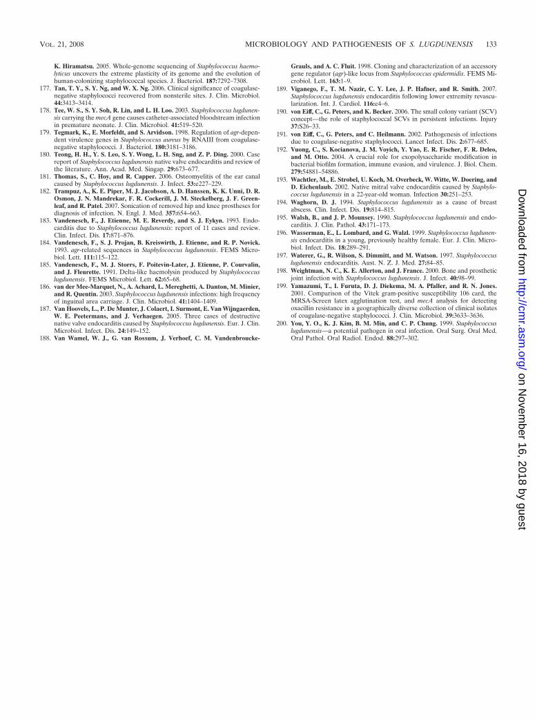
K. Hiramatsu. 2005. Whole-genome sequencing of Staphylococcus haemo-lyticus uncovers the extreme plasticity of its genome and the evolution ofhuman-colonizing staphylococcal species. J. Bacteriol. 187:7292–7308.
177. Tan, T. Y., S. Y. Ng, and W. X. Ng. 2006. Clinical significance of coagulase-negative staphylococci recovered from nonsterile sites. J. Clin. Microbiol.44:3413–3414.
178. Tee, W. S., S. Y. Soh, R. Lin, and L. H. Loo. 2003. Staphylococcus lugdunen-sis carrying the mecA gene causes catheter-associated bloodstream infectionin premature neonate. J. Clin. Microbiol. 41:519–520.
179. Tegmark, K., E. Morfeldt, and S. Arvidson. 1998. Regulation of agr-depen-dent virulence genes in Staphylococcus aureus by RNAIII from coagulase-negative staphylococci. J. Bacteriol. 180:3181–3186.
180. Teong, H. H., Y. S. Leo, S. Y. Wong, L. H. Sng, and Z. P. Ding. 2000. Casereport of Staphylococcus lugdunensis native valve endocarditis and review ofthe literature. Ann. Acad. Med. Singap. 29:673–677.
181. Thomas, S., C. Hoy, and R. Capper. 2006. Osteomyelitis of the ear canalcaused by Staphylococcus lugdunensis. J. Infect. 53:e227–229.
182. Trampuz, A., K. E. Piper, M. J. Jacobson, A. D. Hanssen, K. K. Unni, D. R.Osmon, J. N. Mandrekar, F. R. Cockerill, J. M. Steckelberg, J. F. Green-leaf, and R. Patel. 2007. Sonication of removed hip and knee prostheses fordiagnosis of infection. N. Engl. J. Med. 357:654–663.
183. Vandenesch, F., J. Etienne, M. E. Reverdy, and S. J. Eykyn. 1993. Endo-carditis due to Staphylococcus lugdunensis: report of 11 cases and review.Clin. Infect. Dis. 17:871–876.
184. Vandenesch, F., S. J. Projan, B. Kreiswirth, J. Etienne, and R. P. Novick.1993. agr-related sequences in Staphylococcus lugdunensis. FEMS Micro-biol. Lett. 111:115–122.
185. Vandenesch, F., M. J. Storrs, F. Poitevin-Later, J. Etienne, P. Courvalin,and J. Fleurette. 1991. Delta-like haemolysin produced by Staphylococcuslugdunensis. FEMS Microbiol. Lett. 62:65–68.
186. van der Mee-Marquet, N., A. Achard, L. Mereghetti, A. Danton, M. Minier,and R. Quentin. 2003. Staphylococcus lugdunensis infections: high frequencyof inguinal area carriage. J. Clin. Microbiol. 41:1404–1409.
187. Van Hoovels, L., P. De Munter, J. Colaert, I. Surmont, E. Van Wijngaerden,W. E. Peetermans, and J. Verhaegen. 2005. Three cases of destructivenative valve endocarditis caused by Staphylococcus lugdunensis. Eur. J. Clin.Microbiol. Infect. Dis. 24:149–152.
188. Van Wamel, W. J., G. van Rossum, J. Verhoef, C. M. Vandenbroucke-
Grauls, and A. C. Fluit. 1998. Cloning and characterization of an accessorygene regulator (agr)-like locus from Staphylococcus epidermidis. FEMS Mi-crobiol. Lett. 163:1–9.
189. Viganego, F., T. M. Nazir, C. Y. Lee, J. P. Hafner, and R. Smith. 2007.Staphylococcus lugdunensis endocarditis following lower extremity revascu-larization. Int. J. Cardiol. 116:e4–6.
190. von Eiff, C., G. Peters, and K. Becker. 2006. The small colony variant (SCV)concept—the role of staphylococcal SCVs in persistent infections. Injury37:S26–33.
191. von Eiff, C., G. Peters, and C. Heilmann. 2002. Pathogenesis of infectionsdue to coagulase-negative staphylococci. Lancet Infect. Dis. 2:677–685.
192. Vuong, C., S. Kocianova, J. M. Voyich, Y. Yao, E. R. Fischer, F. R. Deleo,and M. Otto. 2004. A crucial role for exopolysaccharide modification inbacterial biofilm formation, immune evasion, and virulence. J. Biol. Chem.279:54881–54886.
193. Wachtler, M., E. Strobel, U. Koch, M. Overbeck, W. Witte, W. Doering, andD. Eichenlaub. 2002. Native mitral valve endocarditis caused by Staphylo-coccus lugdunensis in a 22-year-old woman. Infection 30:251–253.
194. Waghorn, D. J. 1994. Staphylococcus lugdunensis as a cause of breastabscess. Clin. Infect. Dis. 19:814–815.
195. Walsh, B., and J. P. Mounsey. 1990. Staphylococcus lugdunensis and endo-carditis. J. Clin. Pathol. 43:171–173.
196. Wasserman, E., L. Lombard, and G. Walzl. 1999. Staphylococcus lugdunen-sis endocarditis in a young, previously healthy female. Eur. J. Clin. Micro-biol. Infect. Dis. 18:289–291.
197. Waterer, G., R. Wilson, S. Dimmitt, and M. Watson. 1997. Staphylococcuslugdunensis endocarditis. Aust. N. Z. J. Med. 27:84–85.
198. Weightman, N. C., K. E. Allerton, and J. France. 2000. Bone and prostheticjoint infection with Staphylococcus lugdunensis. J. Infect. 40:98–99.
199. Yamazumi, T., I. Furuta, D. J. Diekema, M. A. Pfaller, and R. N. Jones.2001. Comparison of the Vitek gram-positive susceptibility 106 card, theMRSA-Screen latex agglutination test, and mecA analysis for detectingoxacillin resistance in a geographically diverse collection of clinical isolatesof coagulase-negative staphylococci. J. Clin. Microbiol. 39:3633–3636.
200. You, Y. O., K. J. Kim, B. M. Min, and C. P. Chung. 1999. Staphylococcuslugdunensis—a potential pathogen in oral infection. Oral Surg. Oral Med.Oral Pathol. Oral Radiol. Endod. 88:297–302.
VOL. 21, 2008 MICROBIOLOGY AND PATHOGENESIS OF S. LUGDUNENSIS 133
on Novem
ber 16, 2018 by guesthttp://cm
r.asm.org/
Dow
nloaded from