host-pathogen interactions in gram-positive bacteriosis … · host-pathogen interactions in...
TRANSCRIPT
HOST-PATHOGEN INTERACTIONS
IN GRAM-POSITIVE BACTERIOSIS IN FARMED TURBOT
(Scophthalmus maximus)
SÓNIA ALEXANDRA DA ROCHA DIAS GOMES
Tese de doutoramento em Ciência Animal
Especialidade em Morfologia e Fisiologia
2012
2
SÓNIA ALEXANDRA DA ROCHA DIAS GOMES
INTERAÇÃO PATOGÉNIO-HOSPEDEIRO NA BACTERIOSE PROVOCADA POR BACTÉRIAS GRAM-POSITIVAS NO
RODOVALHO (Scophthalmus maximus)
Tese de Candidatura ao grau de Doutor em Ciência Animal, Especialidade em Morfologia e Fisiologia submetida ao Instituto de Ciências Biomédicas Abel Salazar da Universidade do Porto.
Orientadora – Doutora Maria de Fátima Rodrigues Moutinho Gärtner Categoria – Professora Catedrática Afiliação – Instituto de Ciências Biomédicas Abel Salazar da Universidade do Porto.
3
SÓNIA ALEXANDRA DA ROCHA DIAS GOMES
HOST-PATHOGEN INTERACTIONS IN GRAM-POSITIVE BACTERIOSIS IN FARMED TURBOT (Scophthalmus maximus)
Dissertation submitted for the acquisition of Doctoral degree of Animal Science, Expertise in Morphology and Physiology at Institute of Biomedical Sciences Abel Salazar of Porto University.
Orientator – Doctor Maria de Fátima Rodrigues Moutinho Gärtner Category – Cathedratic Professor Affiliation – Institute of Biomedical Sciences Abel Salazar - Porto University.

4
“You learn that time isn't something you can turn back,
Therefore you must plant your own garden and decorate your own soul,
Instead of waiting for someone to bring you flowers.
And you learn that you really can endure.
You really are strong.
And you can go so farther than you thought you could go.
And that life really has a value.
And you have value within the life.
And that our gifts are betrayers,
And make us lose
The good we could conquer,
If it wasn't for the fear of trying.”
In Minstrel (William Shakespeare)
5
Acknowledgements I feel much honour to express my deep sense of gratitude to my supervisor, Prof.
Doctor Fátima Gärtner, for her encouraging attitude and keen interest throughout the
course of my studies, as well as suggestions, cooperation, guideline and kind gesture.
I am also thankful to Prof. Doctor Berta Martins for her help, kind cooperation and
assistance especially during several stages of my work. Special thanks for giving me the
opportunity of teaching some of my knowledge during some classes of Immunopathology.
I would also like to thank Prof. Doctor António Afonso for giving me the chance of
working and developing the immunobiology lab at CIIMAR.
Special gratitude to all my friends and colleagues, for their kind assistance and
help during the course of my research work.
For this accomplishing words are lacking to express my feelings and indebtedness
to my beloved mother, maternal grandparents, sister, Alexandre, my girls and boy, for
their endless prayers, cooperation and encouragement.
I am meticulously thankful and grateful to my beloved and affectionate ones,
whose hands were always raised for my well being and success...
This study was supported by the Portuguese Foundation for Science and Technology
(FCT; SFRH/BD/19133/2004).
6
Published works or in publication
Afonso, A., Gomes, S., Silva, J., Marques, F. & Henriques, M. (2005). Side-effects in sea
bass (Dicentrarchus labrax L.) due to intraperitoneal vaccination against vibriosis and
pasteurellosis. Fish & Shellfish Immunology, 19: 1-16.
Gomes, S., Afonso, A. & Gärtner, F. (2006). Fish vaccination against infections by
Streptococcal species and the particular case of Lactococcosis. Revista Portuguesa de
Ciências Veterinárias, 101: 25-35 (attached).
Dias, S., Ozório, R.O.A., Gonçalves, J., Gärtner, F. & Afonso, A. (2011). Dietary L-
Carnitine supplementation protected turbot (Scophthalmus maximus) against
Streptococcus parauberis infection. Journal of Applied Aquaculture, 23: 299-303
(attached).
Oral Presentations:
Dias, S. (2008). Fish tuberculosis – a case study of host/pathogen interaction. Oral
presentation at the International Conference on Fish Diseases and Fish Immunology,
Reykjavik, Iceland.
Dias, S. (2008). L-carnitine – a potential immunostimulant for turbot (Scophthalmus
maximus)? Oral presentation at the “III Encontro Nacional de Pós-Graduação em
Ciências Biológicas”, Faro, Portugal.
Dias, S. (2007). “Tuberculose em peixes – um caso de interação patogénio/hospedeiro”.
Oral presentation at “I Congresso de Análises Clínicas e Saúde Pública”, INSA, Porto,
Portugal.
Gomes, S. (2004). The leucocyte response of sea bass (Dicentrarchus labrax L.) to
intraperitoneal vaccination against vibriosis and pasteurellosis. Oral presentation at the
12th Annual Meeting of the Portuguese Society of Animal Pathology jointly with the 16th
Annual Meeting of the Spanish Society of Veterinary Pathology, Famalicão, Portugal.
7
Posters:
Melo, M., Afonso, A., Dias, S. (2006). Production of a polyclonal antibody anti-
Mycobacterium marinum. Poster at “IV Encontro do CIMAR Laboratório Associado”,
Peniche, Portugal.
Dias, S. (2006). Fish tuberculosis: a case of host/pathogen interaction. Poster at “IV
Encontro do CIMAR Laboratório Associado”, Peniche, Portugal.
Gomes, S. (2005). Streptococcosis and Mycobacteriosis in farmed turbot (Scophthalmus
maximus). Poster at the “III Encontro do CIMAR Laboratório Associado”, Lisboa, Portugal.
8
Index
Abbreviations ___________________________________________________________ 10
SUMÁRIO ______________________________________________________________ 11
SUMMARY _____________________________________________________________ 14
CHAPTER 1 _____________________________________________________________ 16
Introduction_________________________________________________________ 16 FISH TUBERCULOSIS _________________________________________________________________ 19 HOST-PATHOGEN INTERACTIONS ______________________________________________________ 21
CHAPTER 2 _____________________________________________________________ 24
Peritoneal leucocyte population in Scophthalmus maximus __________________ 24 MATERIAL AND METHODS ____________________________________________________________ 24 RESULTS AND DISCUSSION ____________________________________________________________ 26
CHAPTER 3 _____________________________________________________________ 29
M. marinum’s growth behavior _________________________________________ 29 MATERIAL AND METHODS ____________________________________________________________ 29 RESULTS AND DISCUSSION ____________________________________________________________ 31
CHAPTER 4 _____________________________________________________________ 34
Turbot inflammatory response__________________________________________ 34 MATERIAL AND METHODS ____________________________________________________________ 34 RESULTS AND DISCUSSION ____________________________________________________________ 36
CHAPTER 5 _____________________________________________________________ 42
Turbot infection trial __________________________________________________ 42 MATERIAL AND METHODS ____________________________________________________________ 42 RESULTS AND DISCUSSION ____________________________________________________________ 45
CHAPTER 6 _____________________________________________________________ 49
In vitro studies with M. marinum ________________________________________ 49 MATERIAL AND METHODS ____________________________________________________________ 49 RESULTS AND DISCUSSION ____________________________________________________________ 52
CHAPTER 7 _____________________________________________________________ 57
In vitro studies with S. parauberis _______________________________________ 57 MATERIAL AND METHODS ____________________________________________________________ 57 RESULTS AND DISCUSSION ____________________________________________________________ 59
CHAPTER 8 _____________________________________________________________ 62
Conclusions _________________________________________________________ 62

9
CHAPTER 9 _____________________________________________________________ 65
Future Perspectives ___________________________________________________ 65
CHAPTER 10 ____________________________________________________________ 66
References __________________________________________________________ 66
CHAPTER 11 ____________________________________________________________ 72
Attachments ________________________________________________________ 72
10
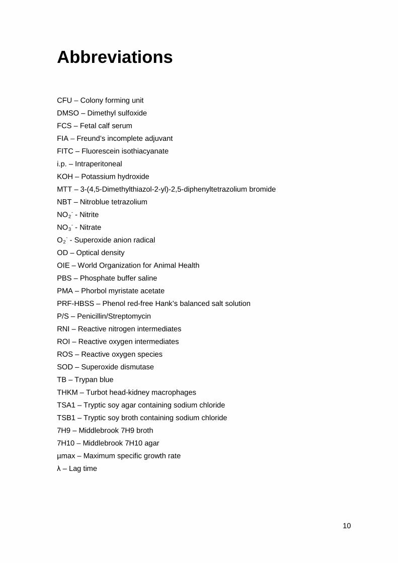
Abbreviations
CFU – Colony forming unit
DMSO – Dimethyl sulfoxide
FCS – Fetal calf serum
FIA – Freund’s incomplete adjuvant
FITC – Fluorescein isothiacyanate
i.p. – Intraperitoneal
KOH – Potassium hydroxide
MTT – 3-(4,5-Dimethylthiazol-2-yl)-2,5-diphenyltetrazolium bromide
NBT – Nitroblue tetrazolium
NO2- - Nitrite
NO3- - Nitrate
O2- - Superoxide anion radical
OD – Optical density
OIE – World Organization for Animal Health
PBS – Phosphate buffer saline
PMA – Phorbol myristate acetate
PRF-HBSS – Phenol red-free Hank’s balanced salt solution
P/S – Penicillin/Streptomycin
RNI – Reactive nitrogen intermediates
ROI – Reactive oxygen intermediates
ROS – Reactive oxygen species
SOD – Superoxide dismutase
TB – Trypan blue
THKM – Turbot head-kidney macrophages
TSA1 – Tryptic soy agar containing sodium chloride
TSB1 – Tryptic soy broth containing sodium chloride
7H9 – Middlebrook 7H9 broth
7H10 – Middlebrook 7H10 agar
µmax – Maximum specific growth rate
λ – Lag time
11
SUMÁRIO A produção de rodovalho (Scophthalmus maximus) é afetada, na sua maioria pela
streptococcose e pela tuberculose. Após o aparecimento de uma epizootia de tuberculose
numa produção de rodovalho em Portugal, o estudo desta patologia, bem como da sua
interação com o rodovalho foram o ponto de partida desta longa jornada. Quer a
streptococcose, quer a tuberculose são causadas por bactérias Gram-positivas. Os seus
agentes etiológicos: o Streptococcus parauberis e a Mycobacterium marinum, foram
descritos. A taxa máxima de crescimento específico e o tempo de crescimento máximo
foram estimados por contagem direta e através da leitura das absorvâncias. Os valores
da taxa máxima de crescimento variaram entre 0.01 e 0.0336 h-1 nas estirpes de M.
marinum (MM-SP e MM-PT, respetivamente) e 0.2463h-1 na amostra de S. parauberis.
Os resultados relacionados com o tempo de crescimento confirmaram os anteriores,
mostrando que as estirpes de M. marinum são de crescimento lento, quando comparadas
com a do S. parauberis. Para conhecermos a reação imune perante um agente
patogénico ativo ou inativo, temos sempre que conhecer a população leucocitária em
repouso. Por isso, após injeção de um tampão fosfato (PBS) na cavidade peritoneal do
rodovalho, observámos que os resultados não foram diferentes daqueles que haviam sido
documentados para outras espécies de peixes, mostrando-se que a população de
macrófagos era a mais abundante e a de neutrófilos a menos abundante. A nível
citoquímico podemos usar a esterase e a peroxidase como marcadores de macrófagos e
neutrófilos, respetivamente, uma vez que reagem positivamente a cada uma delas
individualmente. A avaliação da resposta inflamatória do rodovalho é uma mais valia no
estudo da interação patogénio/hospedeiro. Esta resposta imune foi estudada através da
resposta peritoneal leucocitária, tendo-se observado uma resposta significativa ao
bacterin da M. marinum. Em relação à MM-PT inativada, os neutrófilos mostraram um
aumento significativo inicial, no entanto, tal resposta padronizada não foi observada em
relação à MM-SP inativada. A resposta do rodovalho ao adjuvante foi característica de
uma resposta inflamatória que persiste ao longo do tempo, mostrando que este agente
flogístico é de difícil degradação pelo rodovalho. A infeção experimental in vivo é de
importância crucial para os imunologistas e patologistas. O desenvolvimento da doença,
a oscilação nas populações leucocitárias e os efeitos observados são relevantes no
estudo do agente patogénico e da sua patogenia. O rodovalho mostrou-se sensível à
infeção experimental com as duas estirpes de M. marinum (MM-PT e MM-SP), tendo-se
observado que os macrófagos foram a população leucocitária predominante. No ensaio
da infeção, observou-se que no grupo infectado com a M. marinum ocorreu um aumento
12
inicial no número de macrófagos e de neutrófilos. Os primeiros aumentaram até 17 vezes
nos casos mais severos. Até aos 90 dias do ensaio, durante a resposta inflamatória não
específica, observou-se um pico leucocitário ao dia 4, com ausência de granulomas, mas
sendo os macrófagos as células predominantes. Contudo, não foram encontradas úlceras
cutâneas, alterações de pigmentação ou exoftalmia uni- ou bilateral. No rim-anterior
foram encontrados aglomerados bacterianos durante um maior período de tempo,
quando comparado com o baço ou o exsudado peritoneal. Após a análise dos resultados
obtidos a partir do estudo destas duas estirpes bacterianas, realizámos um ensaio mais
preciso para estudar os seus efeitos na função e integridade dos macrófagos do rim-
anterior do rodovalho. Esses ensaios basearam-se na deteção da produção do anião
superóxido. As estirpes MM-PT e MM-SP são capazes de induzir uma “explosão
respiratória” nos macrófagos do rim-anterior do rodovalho. Contudo, a resposta sofreu um
aumento quando a MM-PT foi previamente inativada, e um decréscimo quando a MM-SP
se encontrava inativada. Tais diferenças não fazem parte de um cenário normal,
observando-se que a MM-PT inibe a libertação do anião superóxido para o espaço
extracelular, quando comparado com a MM-SP. A citometria de fluxo é uma das
tecnologias mais usadas, quando se estudam as populações celulares nos mamíferos e
em algumas espécies de peixes. A população leucocitária do rodovalho mostrou ser uma
forte candidata para ser estudada através da citometria de fluxo e o azul de tripano
mostrou ser um bom agente para eliminar a fluorescência extracelular dos nossos
estudos. Constatou-se que a espécie bacteriana intracelular (MM-PT) exibe um padrão
de fagocitose diferente, quando comparada com a extracelular (S. parauberis). Porém, a
estirpe Espanhola: MM-SP, comporta-se como o S. parauberis, sugerindo que poderá
existir uma influência geográfica na infeção e modelo de virulência evidenciados pelas
mesmas espécies bacterianas. A origem geográfica é muito importante e até mesmo
considerada um aspeto fundamental na distinção e caracterização de diversas espécies.
Durante alguns ensaios experimentais, encontrámos certos resultados intrigantes a
respeito das duas espécies bacterianas estudadas. Foram realizadas várias abordagens,
para analisar em maior profundidade estas diferenças, nomeadamente estudos da
explosão respiratória e da libertação de intermediários nitrogenados dos macrófagos do
rim-anterior do rodovalho, após estimulação bacteriana. A estirpe Portuguesa da M.
marinum é semelhante à descrita na literatura para as espécias micobacterianas.
Todavia, a estirpe Espanhola comportou-se como se se tratasse de uma bactéria
extracelular, tal como o S. parauberis. Talvez tenha ocorrido uma separação destas duas
estirpes de M. marinum no passado e elas tivessem que ter desenvolvido diversas
adaptações para sobreviverem. Em detrimento da nossa preocupação, aliada ao facto da
imunologia e da vacinologia de peixes, apesar de em progresso, estarem apenas no

13
começo, estes ensaios preliminares são de uma importância crucial por forma a dar
continuidade aos trabalhos vindouros.
14
SUMMARY Turbot’s (Scophthalmus maximus) production is mainly affected by streptococcosis
and fish tuberculosis. The tuberculosis outbreak in a Portuguese turbot’s farm helped the
study of this pathogen and its interaction with turbot and was responsible for the beginning
of these works. Streptococcosis and tuberculosis are caused by Gram-positive bacteria.
Their etiological agents are: Streptococcus parauberis and Mycobacterium marinum,
respectively and both were described. The maximum specific growth rates and lag times
were estimated by direct viable count and absorbance data. The maximum specific growth
rates varied between 0.01 and 0.0336 h-1 in both M. marinum strains (MM-SP and MM-
PT, respectively) and 0.2463 h-1 in S. parauberis. Results concerning lag times confirmed
the previous ones, showing that M. marinum strains are slow growers, when compared to
S. parauberis. To describe turbot’s immune response against an active or an inactive
pathogen, we have to know turbot’s unstimulated leucocyte population. So, after the
intraperitoneal injection of phosphate buffer saline (PBS), we have observed that results
were not different from those previously described for other fish species, showing that
macrophages were the predominant cell type and neutrophils the less abundant one.
Citochemically, we can use both esterase and peroxidase to distinguish between
macrophages and neutrophils, because they reacted positively to each of them
(macrophages to esterase and neutrophils to peroxidase). The evaluation of turbot’s
inflammatory response is worthwhile in the study of the interaction between the pathogen
and its host. This immune response was studied observing the kinetics in the peritoneal
leucocyte population of turbot and it was observed a significant response to inactivated M.
marinum. When inactivated MM-PT was injected, neutrophils showed an increase at the
beginning, but this typical response was not observed when inactivated MM-SP was
injected. Turbot’s immune response to adjuvant was consistent to an inflammatory
response persistent during the period studied. This showed that adjuvant was hardly
degraded by fish. The in vivo experimental infection is very important for immunologists
and pathologists. The development of a disease, the oscillation between the leucocyte
populations and its observed effects, are relevant in the study of the pathogen and its
pathogenicity. Turbot was shown to be sensitive to the experimental infection with both
strains of M. marinum (MM-PT and MM-SP) and macrophages were the predominant cell
type. The group infected with M. marinum showed an increase in the numbers of
macrophages and neutrophils. The first increased up to 17 times in the most severe
cases. Till 90 days post-infection it was observed high levels of leucocytes at day 4, but
with no granulomas. Macrophages were the predominant cell type, however we could not
15
find skin granulomas, pigmentation alteration, uni- or bilateral exophthalmia. We observed
bacterial aggregations in the head-kidney, during a higher time period, when compared to
the spleen or the peritoneal exudates. After the analysis of the achieved results, a more
precise study was conducted to observe their effects in the function and integrity of
turbot’s head-kidney macrophages. These studies were related to the detection of
superoxide anion production. MM-PT and MM-SP strains are capable of inducing a
respiratory burst in turbot’s head-kidney macrophages. However, the response was higher
when MM-PT was previously inactivated and led to a decrease when MM-SP was
inactivated. Those differences were not typical. MM-PT was shown to inhibit the release of
superoxide anion to the extracellular space, when compared to MM-SP. Flow cytometry is
one of the most used technologies when we are studying the cellular population in
mammals and some fish species. Turbot’s leucocyte population showed to be a strong
candidate to be studied using this technique and trypan blue was a good agent to
eliminate the extracellular fluorescence of our studies. We have observed that MM-PT
showed a different pattern of phagocytosis, when compared to S. parauberis. However,
MM-SP behaved as the previous bacterial species, suggesting that it may exists a
geographic influence in the infection and virulence patterns evidenced by these two M.
marinum strains. The geographic origin is very important and is even considered a
fundamental issue in the distinction and characterization of several species. Among
several experimental trials we came across with some intriguing results, concerning these
two bacterial species. We have made different approaches to analyze these differences
mainly in respiratory burst studies and in the release of nitrogen intermediates from
turbot’s head-kidney macrophages after bacterial stimulation. The Portuguese strain of M.
marinum is similar to that encountered and described in literature for mycobacterial
species. However, MM-SP behaved as if it was an extracellular bacteria, like S.
parauberis. It may have occurred a separation between the two M. marinum strains in the
past, and they have developed different survival strategies. It is well known that fish
immunology and vaccinology are in progress and these preliminary assays are very
important and a wonderful tool for the forthcomings works.
16
CHAPTER 1 Introduction
World aquaculture has grown dramatically in the last 50 years. From a production
of less than one million tonnes in the early 1950s, production in 2009 was reported to
have risen to 55.1 million tonnes, being the marine aquaculture responsible for more than
36% of the total value (FAO, 2010). However, the total fisheries captures have not
decreased to levels wanted (FAO, 2010). If we look more deeply in values related to
flatfish production, we can see that from 2000 to 2008, there was an increase of more
than 122 000 tonnes, being Spain and China the leading producers. Among all the flatfish
species, turbot (Scophthalmus maximus) was considered the most important one (FAO,
2010).
Turbot (S. maximus) is a flatfish species that belongs to the Scophthalmidae
family. It is a demersal fish that preferentially lives in marine or brackish waters of
the North Atlantic, Baltic and the Mediterranean Sea. When looking on an anatomically
basis, we see that turbot is a marine teleost with an oval body, almost circular in shape.
As a flatfish it has got a dorsoventral symmetry: on its dorsal or ocular side, there is some
pigmentation and we can find both eyes, because turbot’s right eye migrates during
metamorphosis. Its ventral side is also known as the blind one. Turbot has not got scales
on the skin and its dorsal fin begins near the above eye. Its mouth has an arc form, due to
its movement during metamorphosis. The swimbladder only exists during its larval stage,
disappearing later on, when turbot becomes a benthonic species. It lives between 20 and
100m below water (Carvalho and Diniz, 1998; Owen, 2011).
Why producing turbot? Well, among several fish species, turbot (S. maximus) is an
economically important rearing species. Turbot aquaculture began in the 1970s in
Scotland (UK) and it was subsequently brought to France and to Spain. At first, the
number of facilities in Spain was rather limited due to the scarcity of juveniles. The
technological development of juvenile production changed this. At the beginning of the
1990s, there were already 16 producers in Spain. A significant crisis in turbot aquaculture
occurred in 1992; there was an increase of 52 percent in production but the industry
lacked a consolidated commercial marketing network. Another factor that contributed to
this crisis was that farms were small and had very high production costs. This crisis
caused the closure of some farms. From that moment onwards a reorganization of the
17
sector began, which gave rise to a growth both in production and in the number of
countries where turbot was farmed. Spain, with its highly suitable oceanographic
conditions, is now the major producer worldwide but turbot is also currently farmed in
Denmark, Germany, Iceland, Ireland, Italy, Norway, Wales (UK), France and Portugal,
and was previously reared in the Netherlands. Turbot has also been introduced to other
regions (notably Chile in the late 1980s), and more recently, China (FAO, 2010). Besides
commercial investment to improve facilities or built new farms, other decisive factors have
assisted in the consolidation and development of this species production. These have
included the production of dry feed and the development of vaccines for the most
important diseases affecting turbot (FAO, 2010).
During the last decades, we have witnessed the dissemination of several diseases
among aquacultures and Portugal was not an exception. It is well known that conditions in
fish farms lead to the appearance of severe epizootic outbreaks, not only due to
overcrowding, but also due to intensive farming conditions (Jacobs et al, 2009). Moreover,
intensive rearing of aquatic species in aquaculture facilities provides an opportunity for the
amplification of both native and exotic diseases (Jacobs et al, 2009). According to the
World Organization for Animal Health (OIE), an emerging disease is defined as “...a
recently admitted serious illness, whose aetiology can, or not, have already been
established, and which is likely to be propagated within a population or between
populations” (Renault and Guichard, 2007; Saulnier et al, 2007).
Fish diseases have been characterized and it was observed that some of the
diseases affecting fresh water fish species productions were also considered as emerging
diseases in marine fish species (Toranzo, 2005). Fish are continuously exposed to viral
and bacterial pathogens, however, in this thesis, we will only focus on the bacterial
pathogens associated to outbreaks in aquacultures.
When we look at bacterial diseases, we need to distinguish between Gram-
negative and Gram-positive bacteria. Turbot production is affected by some Gram-
negative bacterial species, such as: Lestionella anguillarum (formerly known as Vibrio
anguillarum), the etiological agent of vibriosis; Aeromonas salmonicida subsp
salmonicida, the etiological agent of furunculosis; Pseudomonas anguiliseptica, the
etiological agent of “winter disease” or pseudomonadiasis, as well as Tenacibaculum
maritimus (formerly known as Flexibacter maritimus), the etiological agent of
flexibacteriosis. Besides these bacterial species, turbot production can also be affected by
some Gram-positive species. Among these we can point out: Streptococcus parauberis,
the etiological agent of streptococcosis and Mycobacterium marinum, the etiological agent
of mycobacteriosis or fish tuberculosis (Toranzo, 2005).
18
More than 150 species of salt water and freshwater fish are affected by
mycobacteriosis or fish turberculosis (Toranzo, 2005) and its main etiological agent is M.
marinum (as mentioned before). However, M. fortuitum and M. chelonae are also capable
of inducing tuberculosis in fish (Toranzo, 2005). The first report of a mycobacterial
infection in fish has been attributed to Bataillon et al (1897), who isolated acid-fast bacilli
from a tuberculous lesion in a common carp. Mycobacteria are disseminated in the
environment, so fish are continuously exposed to these bacterial agents. Why some fish
develop disease and others don’t? Well, with all the conditions observed in aquacultures,
such as: overcrowding, poor nutrition, deteriorated water quality and advanced age, fish
become more susceptible to opportunistic agents (Toranzo, 2005). Turbot was not an
exception and it showed to be sensible to these agents during its entire production cycle.
Streptococcal diseases are also responsible for important epizootic outbreaks in
freshwater fish facilities, as well as, in saltwater farms (Toranzo, 2005). Streptococcus
parauberis, classically known as the etiological agent of mastitis in cattle, was first
described in 1958, affecting a culture of rainbow trout in Japan (Toranzo, 2005). In 1993,
was responsible for an important disease outbreak in turbot in Galicia (NW Spain)
(Toranzo, 2005). Streptococcosis was considered to be the cause of heavy economic
losses, because affected fish became unmarketable due to their poor external
appearance (Toranzo, 2005; Toranzo et al, 2009).
Turbot is susceptible to streptococcosis at all stages of the production cycle. This
fish pathogen forms a biochemically and antigenically homogenous group, being that an
advantage when developing a vaccine against it (Toranzo et al, 2009). The vaccines
developed against streptococcosis only where effective when administered
intraperitoneally (Toranzo et al, 2009). With the development of these vaccines, turbot
production became more protected, but even though sensible to other diseases
outbreaks, such as mycobacteriosis. And for this special one, there are no vaccines
available to prevent it (Toranzo et al, 2009), which makes turbot susceptible to this
disease during its production life cycle.
In 2004, in a Portuguese Turbot Aquaculture, located in the north of the country,
an outbreak of mycobacteriosis was witnessed and followed. For obvious reasons, this
Farm will not be identified in this thesis, but was the motor that led to its development.
After the observation of a mycobacteriosis infection in the turbot farm, and using
the peritoneal cavity as a model, we tried to induce an experimental inflammation and
infection by two strains of M. marinum: a Portuguese and a Spanish one, both isolated
from farm outbreaks. For a better comparison, in vitro studies were also conducted.
We will now provide an overview of each theme that will be discussed during this
thesis. On Chapter 2 we will provide the characterization of the resting peritoneal
19
population of turbot, essential for the forthcoming works. On Chapter 3 we will
characterize M. marinum, on Chapter 4 we will discuss the experimental inflammation
trial, on Chapter 5 the experimental infection trial and on Chapter 6 we will discuss the in
vitro studies conducted with M. marinum. Finally on Chapter 7 we will correlate the
behaviour of M. marinum with S. parauberis and the results achieved will be discussed on
Chapter 8.
The results of this thesis, as well as others related to some of the themes
discussed, have been documented in a number of publications, which will be attached at
the end.
FISH TUBERCULOSIS
Fish tuberculosis is caused by a bacteria belonging to the genus Mycobacterium.
Mycobacteria are Gram-positive, usually acid and alcohol fast at some stages of its
growth, non-motile, do not form capsules, endospores or conidia, rarely exhibit grossly
visible aerial hyphae, produce oxidatively acid from sugars and with some exception of
those that do not grow in vitro, can be divided into rapid- and slow-growing strains
(Godfellow and Wayne, 1983). The genus was established by Lehmann and Neumann in
1896 (Godfellow and Wayne, 1983), because it included tubercle and leprosy bacilli, being
considered atypical or nontuberculous (Chemlal et al, 2002), but it now encompasses 41
approved species (Godfellow and Wayne, 1983). The past 40 years have witnessed a
marked change in the outlook of bacterial systematic, due to an introduction of new and
more exacting biochemical, chemical, genetical and numerical methods, which have led to
rapidly changing views of how bacteria ought to be classified and identified (Alkhodair et
al, 2010).
Tuberculosis in fish is a subacute to chronic wasting disease capable of affecting
fresh and salt water fishes (Saulnier et al, 2007). It typically includes granulomas in the
spleen, kidney and liver. External manifestations are scale loss, hemorrhagic lesion
(Alkhodair et al, 2010). As a chronic progressive disease, affected fish may lose their
appetite, appear debilitated and emaciated, have impaired growth and become more
susceptible to infection by opportunistic bacteria (Heckert et al, 2001).
M. marinum is a pathogen of poikilothermic hosts, such as frogs and fish (Prouty
et al, 2003), being closely related to M. tuberculosis. This has been confirmed using gas
chromatography of fatty acids and alcohols, DNA-DNA hybridization and 16S rRNA gene
sequence analysis (Tonjum et al, 1998). Moreover, it is a photochromogenic organism
20
that causes disease in fish and self-limiting skin granulomas in man. Its natural reservoir
appears to be water however, results published by Le Dantec et al (2002), showed that
mycobacteria are highly resistant to chloride disinfection. They have shown to be able to
replicate in biofilms, and solid-liquid interfaces may be regarded as sites of selective
enrichment for these bacteria. On a primary isolation, it grows readily at 30º C but not at
37º C, but after several subcultures it can become adapted to growth at the mentioned
temperature, as stated by Godfellow, but also confirmed by us (Godfellow and Wayne,
1983). Its optimal temperature growth may be the primary reason why it only causes
lesions on the extremities of humans (EL-ETR et al, 2001). M. marinum has a generation
time of 4 hours compared to 20 hours of M. tuberculosis, being a biosafety level two
organism (EL-ETR et al, 2001). M. marinum is an intracellular pathogen, capable of
developing several survival strategies in a normally hostile environment. It can escape
from its vesicles into the cytoplasm of the host cell, be resistant to the lysosomal
compartment that does not become lysosomal in nature (Decostere et al, 2004).
Pathogenic mycobacterial species are believed to survive by use of the latter strategy,
although the mechanisms by which these bacteria avoid phagosome-lysosome fusion
appear to be different from those of other organisms studied (Decostere et al, 2004).
Barker and colleagues (Decostere et al, 2004) showed that live M. marinum, like other
pathogenic mycobacterial species, reside in a compartment which is not acidified and
does not contain lysosomal enzymes. These bacteria are capable of replication and
persistence in cultured macrophage cell lines (Decostere et al, 2004).
Patients with swimming-pool granuloma are usually tuberculin positive, probably
by the Koch reaction. Indeed the lesions caused may be a continuous response to a few
extremely superficial bacilli (Alkhodair et al, 2010).
Aetiological factors related with the mycobacterial granuloma are: chemical
constituents, virulence, hypersensivity (Decostere et al, 2004). A granuloma may be
defined as an inflammatory lesion in which there is a conglomeration of cells of the
mononuclear phagocyte series. Granulomas may be classified as: non-toxic, due to a
chemically and biologically inert substance such as carbon particles; toxic, due to certain
substances such as colloidal silica or asbestos, which act by increasing the permeability
of the lysossomal membrane; immunological, mediated by humoral antibody, cell
mediated immunity or both. The type of granuloma is consistent to the predominant cell
type encountered (Decostere et al, 2004) and there are several structures of granulomas,
such as: organized or unorganized granulomas. These structures are related to the
presence or absence of concentrically arranged or whorled structures (Houben et al,
2009). However, this organization is not always observed in mycobacterial granulomas
and never dominates the entire granuloma. The unorganized granulomas have several
21
variants and represent the form and distribution of the antigen, the number, size, density
and uniformity of the microorganism (Houben et al, 2009).
When inside the mononuclear phagocytes, what happens to mycobacteria is very
important, mainly because it is related to the pathogenesis of tuberculosis (Houben et al,
2009). Mycobacteria multiply inside mononuclear phagocytes in susceptible animals, as
well as other opportunistic pathogens (Houben et al, 2009). But, what we cannot forget is
that mycobacterial inhibition or death in a granuloma may be regarded as an extracellular
process mediated by the accumulation of high concentrations of products from the
mononuclear phagocyte’s metabolism, as well as from its degeneration or secretion
(Houben et al, 2009).
There is not a vaccine available against fish tuberculosis. However, Pasnik and
Smith (2005) developed a DNA vaccine encoding a potent immunostimulator, which
elicited high levels of protective immune responses in hybrid striped bass (Morone
saxatilis x M. crysops). The degree of protection was dose- and route-dependent, so it
became impracticable, but was an important step in the development of a protective
vaccine against fish tuberculosis.
HOST-PATHOGEN INTERACTIONS
Charles Chapin once wrote that “as it takes two to make a quarrel, so it takes two
to make a disease, the microbe and its host” and he could not be more right. During the
first pages of this thesis we mentioned our host: turbot and our pathogen: M. marinum, so
now we will try to correlate both.
Organisms, such as fish, have an important immune system capable of destroying
any microorganism and preventing its entrance. Pathogens, in turn, possess self-
defending systems, that will overcome the innate and adaptive host immune responses.
The non specific immune response is very important, and it was considered
essential when fighting against pathogens, because the adaptive immune system is
limited, not only due to the poikilothermic nature of fish, but also due to their few repertoire
of antibodies, deficit in proliferation, maturation and memory existent in their lymphocytes
(Ellis, 2001; Uribe et al, 2011). Fish have got physical barriers (skin, scales, mucus) and
chemicals barriers (lysozyme, lectins) which prevent the entrance and attachment of
pathogens in their cells. The protective effect of mucus has been established in turbot
(Fouz, 1990) but the exact mechanisms involved in this protection are not known.
Lysozyme is an enzyme capable of affecting the peptidoglycan layer of bacterial cell walls,
22
and was also found in fish mucus, in peritoneal macrophages and blood neutrophils (Ellis,
2001). Phagocytosis, is one of the most important activity of the immune system of fish, as
it is not dependent of temperature. The main cells involved in this defence mechanism are
neutrophils and macrophages. They remove bacteria by the production of reactive oxygen
species, such as superoxide anion, during respiratory burst (Uribe et al, 2011).
The complement will have an important function in activating cellular defences. It is
a system of serum proteins and can be activated in three ways: the alternative pathway,
the classical pathway and the lectin pathway. The first is considered very important in fish,
because it is found in high doses in fish serum, when compared to mammals (Ellis, 1999;
Nakao et al, 2011). By releasing components (such as C3b and C5a), it can activate the
recruitment of phagocytes (Ellis, 2001).
However, primary immune defences can be deficient and pathogen may be able to
enter in the host cell and there, it needs to survive against the innate and adaptive
immune system of the organism invaded. The site of infection and the type of pathogen is
clearly, important to decide which immune response will be effective.
Pathogens are extremely diverse, but based on the pathogenesis of infection and
on the immune response elicited in the host, they can be classified as intracellular and
extracellular.
Bacteria causing extracellular infections, like S. parauberis, are preferentially
localized in body fluids and other extracellular spaces, being killed if captured by
phagocytes. Therefore, the host defence against these bacteria is related with the
activation of the non-classical complement, neutralisation by antibodies, release of
cytokines, inflammation and efficient phagocytosis (Ellis, 2001). The key mechanism and
importance demonstrated by the last in the defence against infection, was first proposed
by Metchnikoff, more than 100 years ago.
Intracellular bacteria, as the one used in our studies (M. marinum), induce a
chronic inflammatory response on the host. These kinds of bacteria survive and replicate
inside host cells, making their internal environment, their “home”. Intracellular bacteria are
able to survive due to their ability of interfering with bactericidal activities inside the host
cell. Professor Kaufmann has enumerated some of these mechanisms, such as inhibition
of fusion of lysossomes with the phagossome, a common strategy demonstrated by
mycobacteria (Kaufmann, 1989). Moreover, after infecting macrophages, T lymphocytes
are responsible for their recognition and development of several protective responses.
However, macrophages protect microorganisms from these immune responses, making
the eradication of the pathogen far more complicated. During the last years, investigations
were conducted and Galluzzi et al (2011) correlated the autophagy with bacterial invasion.
Autophagy may be associated with the engulfment of intracellular components by double-

23
membrane organelles, known as phagossomes. Later on, by the action of lysossomes
these components will be degraded. In stress conditions, cells may activate this process
of autophagy, though contributing to the clearance of bacteria inside host cells (Galluzzi et
al, 2011).
24
CHAPTER 2 Peritoneal leucocyte population in Scophthalmus maximus
The initial investigation in this thesis concerned the study of the resting peritoneal
leucocyte population of turbot (S. maximus).
The use of the peritoneal model (Silva et al, 1989) was advantageous because the
inflammatory and infection events that occur inside it could be studied both quantitatively
and qualitatively, in contrast to what happens in solid organs. Moreover, the leucocyte
population within the peritoneal cavity is dispersed and can be easily processed for
cytological analysis.
MATERIAL AND METHODS
Turbot, weighting 65±5.0 g (from Piscicultura Marinha do Rio Alto – A. Coelho &
Castro, Lda, in Póvoa do Varzim, Portugal) were used. Fish were kept at 18-22º C in a
recirculating aerated salt water (35‰) system at Centro Interdisciplinar de Investigação
Marinha e Ambiental (CIIMAR, Porto, Portugal). Water quality was maintained with
mechanical and biological filtration and fish were fed ad libitum on commercial pelleted
feed (SORGAL, S.A.; Ovar, Portugal).
Turbot were divided into 2 groups of 40 fish each. Each group was intraperitoneally
injected (i.p.; 100 µl) with Phosphate Buffer Saline (PBS: 1.47 mM KH2PO4, 7.82 mM
Na2HPO4, 0.169 mM NaCl, 13.21 mM Na2CO3; pH 7.2), after being anaesthetized with
0,03% (v/v) ethylene glycol monophenyl ether (Merck).
The peritoneal response was observed by studying quantitatively the leucocytes at
12, 24 and 48 hours, as well as, 4, 7, 15, 30, 60 and 90 days after i.p. injection.
Leucocytes were collected from groups of 10 fish (5 per each group). Turbot were
killed by an overdose of anesthesia (0.06% (v/v)). Peritoneal exudates were collected
using a previously described technique (Afonso et al, 1997; Afonso et al, 1998). Briefly,
PBS was injected into the peritoneal cavity (0.5 mL per fish). Fluid containing the
25
peritoneal cells was then collected and maintained on ice until processing for cytospins
preparations and for cytochemical analysis by electron microscopy.
CYTOSPINS PREPARATIONS
Cytospins preparations of peritoneal leucocyte suspensions were made using a
Cellspin I apparatus. Cytospins were fixed with formol-ethanol (10% of 37% formaldehyde
in absolute ethanol) for one minute and stained with Wright’s stain (Haemacolor Merck).
For peroxidase detection, the Antonow’s technique (Afonso et al, 1998) was used.
Differential counts of macrophages, neutrophils and other small mononuclear cells were
made under x100 oil immersion. Preliminary studies developed in other fish species
(Afonso et al, 1997; do Vale et al, 2002), confirmed that the population of small
mononuclear cells is almost entirely composed by lymphocytes, but by light microscopy it
is difficult to differentiate between lymphocytes and other mononuclear cells such as small
monocytes and thrombocytes. Thus, these were classified together as small mono cells.
To overcome the weight variation between individuals, leucocyte numbers were converted
to leucocytes per gram of body weight.
CYTOCHEMICAL ANALYSIS
Peritoneal leucocyte suspensions collected, were then fixed in 5% Gluteraldehyde
in PBS and processed for electron microscopy. Briefly, after fixation, the peritoneal
exudates were washed in 0.1M Cacodylate Buffer (pH 7.3) during the course of one hour.
Later on, they were post-fixed in 1% Osmium Tetroxide in Cacodylate Buffer for 3 hours at
room temperature, incubated during 30 minutes in 0.5% Uranyl Acetate and then
embedded in Epon. Ultrathin sections were obtained using an Ultramicrotome, contrasted
with Uranyl Acetate and Lead Citrate (Silva et al, 1987) and examined with a JEOL JEM
100CXII Electron Microscope. Detection of peroxidase activity by electron microscopy was
carried out by the method of Robbins et al (1971). The technique described by Robinson
and Karnovsky (1983) was used for the detection of esterase.
26
RESULTS AND DISCUSSION
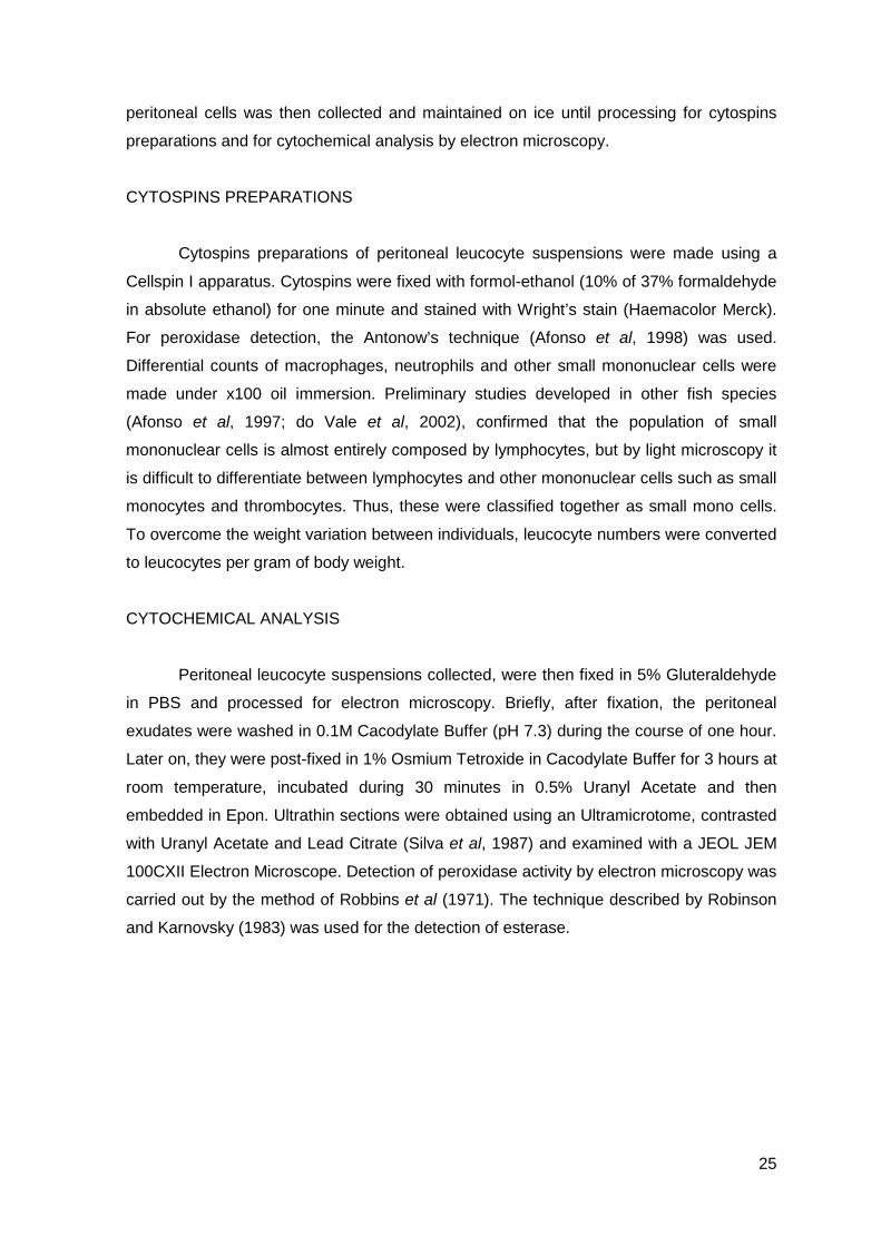
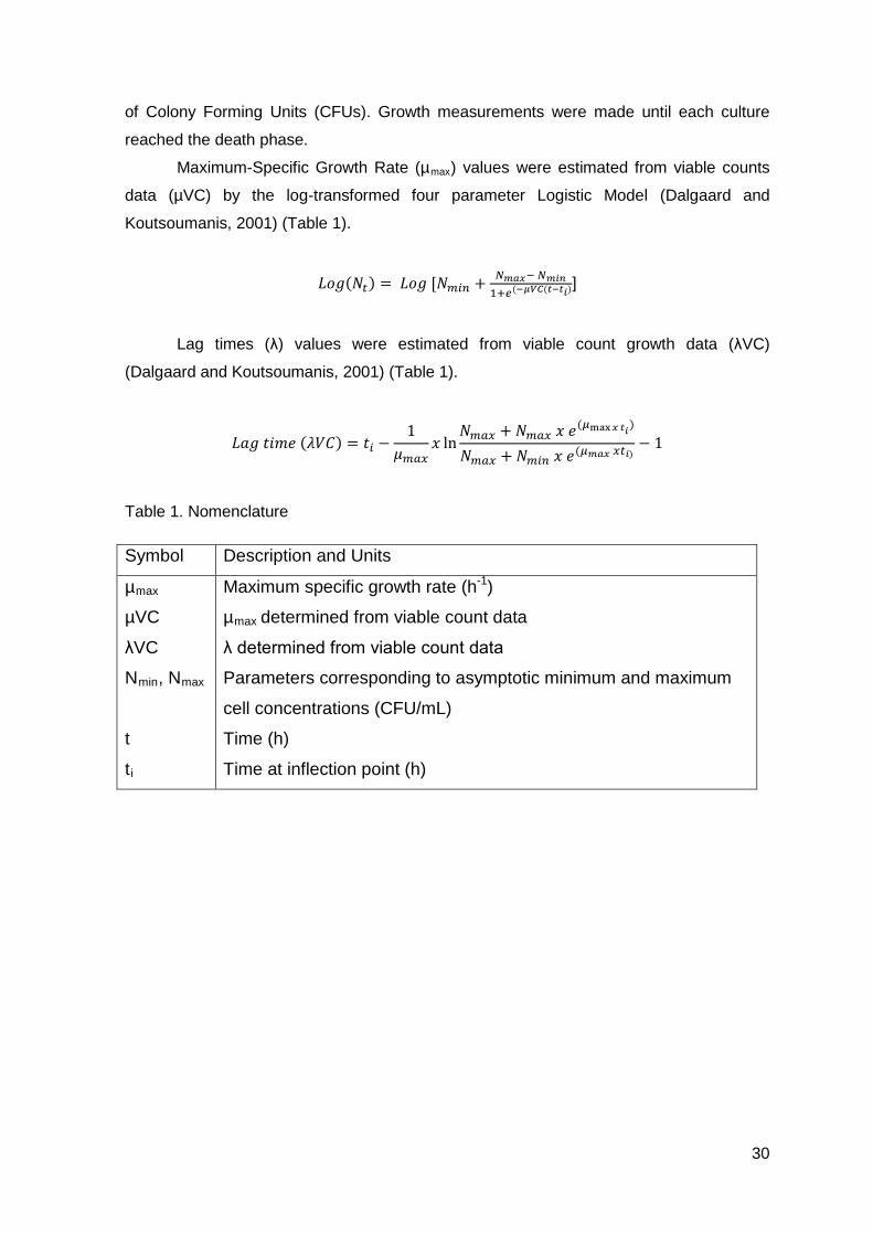
Figure 1. Citological preparation from normal turbot peritoneal washes stained by
haematoxylin-eosin (M – macrophage; N – neutrophil; SMC – small mono cell).
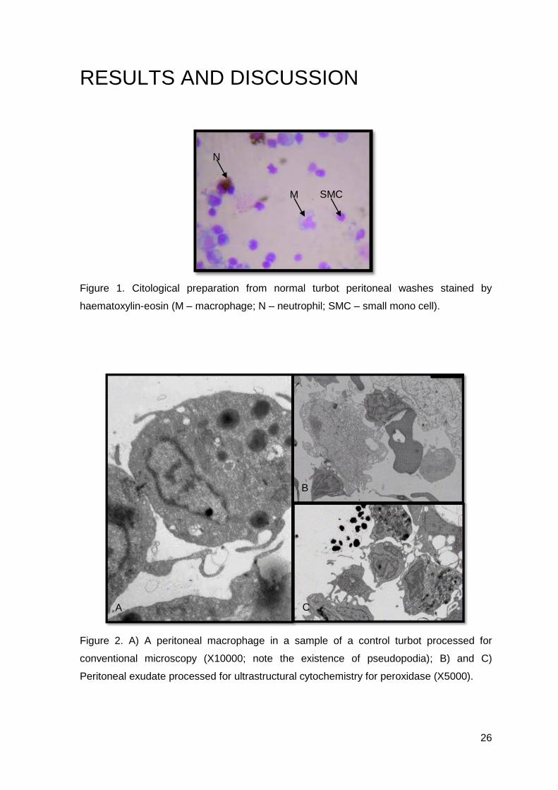
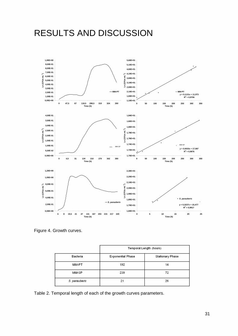
Figure 2. A) A peritoneal macrophage in a sample of a control turbot processed for
conventional microscopy (X10000; note the existence of pseudopodia); B) and C)
Peritoneal exudate processed for ultrastructural cytochemistry for peroxidase (X5000).
A C
B
M SMC
N
27
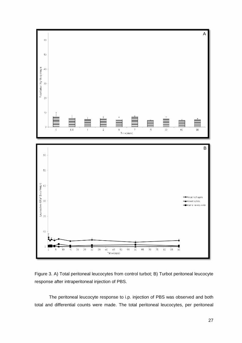
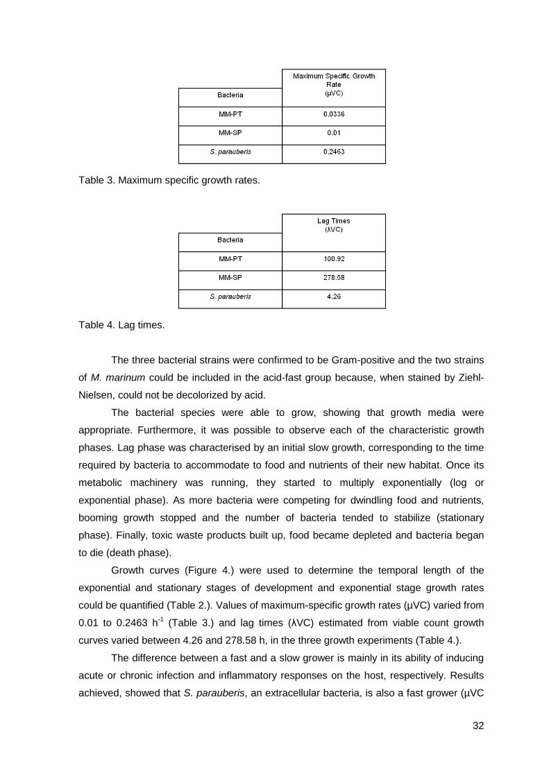
Figure 3. A) Total peritoneal leucocytes from control turbot; B) Turbot peritoneal leucocyte
response after intraperitoneal injection of PBS.
The peritoneal leucocyte response to i.p. injection of PBS was observed and both
total and differential counts were made. The total peritoneal leucocytes, per peritoneal
A
B
28
cavity, of control fish were 2.14±1.10x10⁶, which corresponded to 3.81±1.82x10⁴ per gram
of body weight (Figure 3.A). Differential leucocyte counts were: macrophages 53.20%,
small mono cells 40.80% and neutrophils 6% (Figure 3.B). The control group showed
small insignificant variations over time (Figures 3.A and 3.B).
As observed, the relative percentages of each leucocyte type, in the unstimulated
peritoneal leucocyte population was not different from those previously described for other
fish species (Afonso et al, 1997; Afonso et al, 1998), as well as in recent mammals
studies (Pires et al, 2008). In resting conditions, the concentrations of the two professional
phagocytes were very dissimilar. Macrophages were the predominant phagocyte and
neutrophils the less abundant one.
Peritoneal leucocytes from turbot are mainly composed by macrophages, which
can be observed in our cytological preparation (Figure 1.). They are round in shape, with a
basophilic cytoplasm, their nucleus is in general eccentric and occasionally horse-shoe
shaped. Besides macrophages, we could also find neutrophils, which reacted positively to
peroxidase. These are round and large, with fine neutrophilc granules in the cytoplasm,
their nucleus is small, rod-shaped and occasionally segmented (Figure 1.). The other cell
type, which we named small mono cells were mainly composed by small round cells, with
a large nucleus, which occupies almost all the cytoplasm (Figure 1.). When observing
electron microscopy photos we observed that macrophages were negative for peroxidase
(Figures 2.B and 2.C) and positive for esterase (electron photography not included) and
neutrophils were positive for peroxidase (Figures 2.B and 2.C) and negative for esterase
(electron photography not included). Moreover, we could also observe the existence of a
vacuolated cytoplasm in macrophages, which is a common feature among this cell type
(Figure 2.A). When regarding their morphological features, we observed that, once again,
turbot leucocytes were not different from other fish leucocytes (Afonso et al, 1997; do Vale
et al, 2002; Tavares-Dias et al, 2005). Data concerning cytochemistry studies was in
accordance with previous documented studies (Afonso et al, 1998; do Vale et al, 2002).
Moreover, flatfish neutrophils seem to have similar fibrillar granules to those firstly
described for amphibians and mammals (Hine and Wayne, 1988) and this similarity may
be explained on a common ancestor basis.
These results strongly show that even if there could be doubts in identifying
peritoneal turbot leucocytes, we can do it using cytochemical methods such as the
identification of peroxidase or esterase, because they are a distinctive feature between
macrophages and neutrophils.
29
CHAPTER 3 M. marinum’s growth behavior
When studying a relationship between a host and a pathogen, we need to
characterize each of them individually and only then, establish their interaction.
We have obtained the growth curve for M. marinum (Portuguese and Spanish
strains) and for S. parauberis (for comparison).
MATERIAL AND METHODS
We used, as mentioned, two bacterial species, in a total of three strains.
One strain of M. marinum was kindly supplied by Dr. Nuno M.S. dos Santos
(Institute for Molecular and Cell Biology, IBMC, Porto, Portugal) (dos Santos et al, 2002),
and we named it Portuguese M. marinum (MM-PT). The other was kindly supplied by Dr.
Jesús Romalde (University of Santiago de Compostela, USC, Spain), and we named it
Spanish M. marinum (MM-SP). The last one had already been biochemically
characterised. Both bacteria were thawed from a frozen stock (-70º C) and platted onto
Middlebrook 7H10 Agar (7H10, Difco). Cultures were incubated one week at 30º C, and
used to inoculate in 100 mL Middlebrook 7H9 Broth (7H9, Difco) in 500 mL Erlenmeyer
flasks. Ziehl-Nielsen stain (Merck, Germany) was used for presumptive identification of
each culture.
S. parauberis strain was kindly supplied by Dr. Jesús Romalde (USC, Spain)
(Romalde et al, 1999). It was thawed from a frozen stock (-70º C) and plated onto Tryptic
Soy Agar (TSA, Difco) supplemented with sodium chloride (NaCl) to a final concentration
of 1% (TSA1). Cultures were incubated 48 hours at 22º C and used to inoculate in 100 mL
Tryptic Soy Broth (TSB, Difco), supplemented with NaCl to a final concentration of 1%
(TSB1) in 500 mL Erlenmeyer flasks, after a preliminary confirmation of the purity of the
culture, using the Gram-Hucker Stain (Panreac Química S.A., Barcelona, Spain).
In order to perform growth measurements, two times per day, each bacterial
density was measured by spectrophotometry at 550 nm (Jenway 6405 UV/VIS) and serial
dilutions in 7H9 (M. marinum) or TSB1 (S. parauberis) were made to estimate the number
30
of Colony Forming Units (CFUs). Growth measurements were made until each culture
reached the death phase.
Maximum-Specific Growth Rate (µmax) values were estimated from viable counts
data (µVC) by the log-transformed four parameter Logistic Model (Dalgaard and
Koutsoumanis, 2001) (Table 1).
𝐿𝑜𝑔(𝑁𝑡) = 𝐿𝑜𝑔 [𝑁𝑚𝑖𝑛 + 𝑁𝑚𝑎𝑥− 𝑁𝑚𝑖𝑛1+𝑒(−𝜇𝑉𝐶(𝑡−𝑡𝑖)
]
Lag times (λ) values were estimated from viable count growth data (λVC)
(Dalgaard and Koutsoumanis, 2001) (Table 1).
𝐿𝑎𝑔 𝑡𝑖𝑚𝑒 (𝜆𝑉𝐶) = 𝑡𝑖 −1
𝜇𝑚𝑎𝑥𝑥 ln
𝑁𝑚𝑎𝑥 + 𝑁𝑚𝑎𝑥 𝑥 𝑒(𝜇max𝑥 𝑡𝑖)
𝑁𝑚𝑎𝑥 + 𝑁𝑚𝑖𝑛 𝑥 𝑒(𝜇𝑚𝑎𝑥 𝑥𝑡𝑖)− 1
Table 1. Nomenclature
Symbol Description and Units
µmax
µVC
λVC
Nmin, Nmax
t
ti
Maximum specific growth rate (h-1)
µmax determined from viable count data
λ determined from viable count data
Parameters corresponding to asymptotic minimum and maximum
cell concentrations (CFU/mL)
Time (h)
Time at inflection point (h)
31
RESULTS AND DISCUSSION
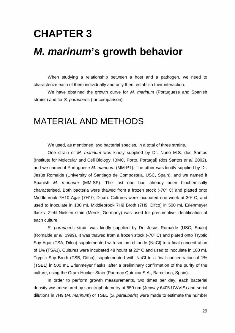
Figure 4. Growth curves.
Table 2. Temporal length of each of the growth curves parameters.
0,00E+00
1,00E-01
2,00E-01
3,00E-01
4,00E-01
5,00E-01
6,00E-01
7,00E-01
8,00E-01
9,00E-01
1,00E+00
0 47,5 67 118,5 298,5 310 324 350Time (h)
Ln (C
FUs
mL-1
)
MM-PTy = 0,1131x + 11,973
R2 = 0,97061,10E+01
1,60E+01
2,10E+01
2,60E+01
3,10E+01
3,60E+01
4,10E+01
4,60E+01
5,10E+01
5,60E+01
0 50 100 150 200 250 300 350Time (h)
Ln (C
FUs
mL-1
)
MM-PT Linear (MM-PT)
0,00E+00
5,00E-02
1,00E-01
1,50E-01
2,00E-01
2,50E-01
3,00E-01
3,50E-01
4,00E-01
0 4,5 31 134 210 270 342 350Time (h)
Ln (C
FUs
mL-1
)
MM-ESy = 0,0033x + 17,087
R2 = 0,9876
1,70E+01
1,72E+01
1,74E+01
1,76E+01
1,78E+01
1,80E+01
1,82E+01
1,84E+01
0 50 100 150 200 250 300 350Time (h)
Ln (C
FUs
mL-1
)
MM-ES Linear (MM-ES)
0,00E+00
2,00E-01
4,00E-01
6,00E-01
8,00E-01
1,00E+00
1,20E+00
6 8 19,5 21 47 131 167 203 215 217 220Time (h)
Ln (C
FUs
mL-1
)
S. parauberisy = 0,3237x + 15,477
R2 = 0,99171,60E+01
1,70E+01
1,80E+01
1,90E+01
2,00E+01
2,10E+01
2,20E+01
2,30E+01
0 5 10 15 20 25Time (h)
Ln (C
FUs
mL-1
)
S. parauberisLinear (S. parauberis)
MM-SP MM-SP
32
Table 3. Maximum specific growth rates.
Table 4. Lag times.
The three bacterial strains were confirmed to be Gram-positive and the two strains
of M. marinum could be included in the acid-fast group because, when stained by Ziehl-
Nielsen, could not be decolorized by acid.
The bacterial species were able to grow, showing that growth media were
appropriate. Furthermore, it was possible to observe each of the characteristic growth
phases. Lag phase was characterised by an initial slow growth, corresponding to the time
required by bacteria to accommodate to food and nutrients of their new habitat. Once its
metabolic machinery was running, they started to multiply exponentially (log or
exponential phase). As more bacteria were competing for dwindling food and nutrients,
booming growth stopped and the number of bacteria tended to stabilize (stationary
phase). Finally, toxic waste products built up, food became depleted and bacteria began
to die (death phase).
Growth curves (Figure 4.) were used to determine the temporal length of the
exponential and stationary stages of development and exponential stage growth rates
could be quantified (Table 2.). Values of maximum-specific growth rates (µVC) varied from
0.01 to 0.2463 h-1 (Table 3.) and lag times (λVC) estimated from viable count growth
curves varied between 4.26 and 278.58 h, in the three growth experiments (Table 4.).
The difference between a fast and a slow grower is mainly in its ability of inducing
acute or chronic infection and inflammatory responses on the host, respectively. Results
achieved, showed that S. parauberis, an extracellular bacteria, is also a fast grower (µVC
33
= 0.2463 h-1; λVC = 4.26 h). This is in accordance with the temporal length of each growth
phase, as they take less time to be achieved and less time in duration, as well as with
previous detailed works conducted by Dalgaard and Koutsoumanis (2001). Moreover,
these results show that, S. parauberis mainly induces an acute inflammatory response on
the host. In contrast, the other two strains of M. marinum are acid-fast bacilli and are
known as intracellular pathogens. Comparing the results achieved, with those from S.
parauberis, we observe that these bacteria are slow growers, not only by the maximum-
specific growth rates, but also by the lag time’s results. Once again, and based in these
results, M. marinum is capable of developing a chronic inflammatory on the host.
All these results are in accordance to that described in literature (Kaufmann, 1992;
Ellis, 1999, 2001, Galluzzi et al, 2011). As a slow grower, M. marinum studies its host and
the best entrance to survive and replicate. This intracellular pathogen has chosen
macrophages as its home and survives because it is difficult for the immune system of the
host to recognize where the invaders are and kill them. There is no doubt that is a well
developed and clever way of surviving within the host and that is why these pathogens
subsist. In comparison, extracellular bacteria, as S. parauberis, as they live in the
extracellular space have more difficulties in circumventing host’s immune system and so
they need to be fast growers, to replicate as fast as possible and establish the disease.
What seemed to be a routine experiment, the determination of bacterial growth
curves, turned to the beginning of an interesting issue. In fact, results showed that MM-SP
appeared to be slower grower than MM-PT, not only by the values obtained for the
maximum-specific growth rate and lag times, but also because it took more time to reach
the stationary phase. This kind of behavior has not been documented elsewhere and
works documented later in this thesis are important in supporting these differences.
34
CHAPTER 4 Turbot inflammatory response
In order to evaluate the phenomena associated with turbot inflammation, we
decided to perform an inflammatory trial using M. marinum inactivated, a control group
and an adjuvant group, so as to characterise turbot peritoneal leucocyte response to
phlogistic agents.
MATERIAL AND METHODS
We have used turbot, weighting 65±5.0 g (from Piscicultura Marinha do Rio Alto –
A. Coelho & Castro, Lda; Póvoa de Varzim, Portugal). Fish were kept in the same
conditions as mentioned before (Chapter 2 – Material and Methods).
The bacterial cultures (MM-PT and MM-SP) were inactivated. Briefly, M. marinum
was thawed form a frozen stock (-70º C) previously prepared and inoculated onto 7H10.
Cultures were grown during 1 week and ressuspended into 7H9. The bacterial density
was measured by spectrophotometry at 520 nm and dilution (in 7H9) were made until the
chosen number of CFUs (predicted by the curve obtained previously, corresponding to
200 µg of protein/mL was achieved (Chapter 3). Then, cultures were inactivated by
adding 8% formol and left at 4º C during 3 consecutive days. Later on, it was washed in
PBS by centrifuging five consecutive times (3900 rpm during 20 minutes). Between each
wash the supernatant was discarded and the pellet ressuspended in PBS. Finally, PBS
with 15% of glycerol was added and stored at -70º C for posterior use. Freund’s
Incomplete Adjuvant (FIA, Sigma), was also used as a phlogistic agent.
Turbot were divided into 4 groups of 80 fish each. Briefly, a non-injected, control
and 3 other groups. All fish were starved 24 h prior to inflammatory trial. After
anaesthetizing turbot (0.03% (v/v)), groups were i.p. injected (100 µL) with the following
substances:
Group I – PBS (Control)
Group II – Inactivated MM-PT
35
Group III – Inactivated MM-SP
Group IV – FIA
As mentioned in Chapter 2, the acute phase of the inflammatory response was
evaluated by studying quantitatively the peritoneal leucocytes at 12, 24 and 48 hours after
i.p. injection. To evaluate the chronic response, peritoneal leucocytes were collected at 4,
7, 15, 30 and 60 days after i.p. injection.
Leucocytes were collected from groups of 10 fish for each time period, following
the method described before (Chapter 2, Material and Methods). Differential counts of
macrophages, neutrophils and other small mononuclear cells (small mono cells) were
made under a x100 oil immersion. Once again, to overcome weight variation between
individuals, leucocyte numbers were converted to leucocytes per gram of body weight.
At each collection time, internal organs were observed. Sixty days after i.p.
injection of the several phlogistic agents, internal organs (kidney, liver, spleen, intestine)
and gills of one fish per group randomly chosen, were collected and fixed in formol 4% for
histological analysis. Tissues samples were embedded in paraffin wax and 5 µm sections
were cut with a rotary microtome (American Optical, Buffalo, NY). After dewaxing, the
sections were stained with haematoxylin/eosin. Further tissue sections were evaluated for
the presence and type of pathological lesions.
36
RESULTS AND DISCUSSION
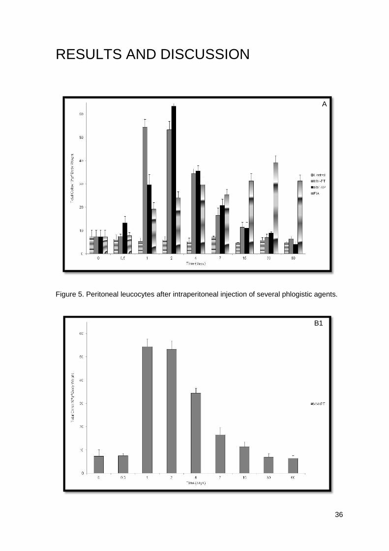
Figure 5. Peritoneal leucocytes after intraperitoneal injection of several phlogistic agents.
A
B1
37
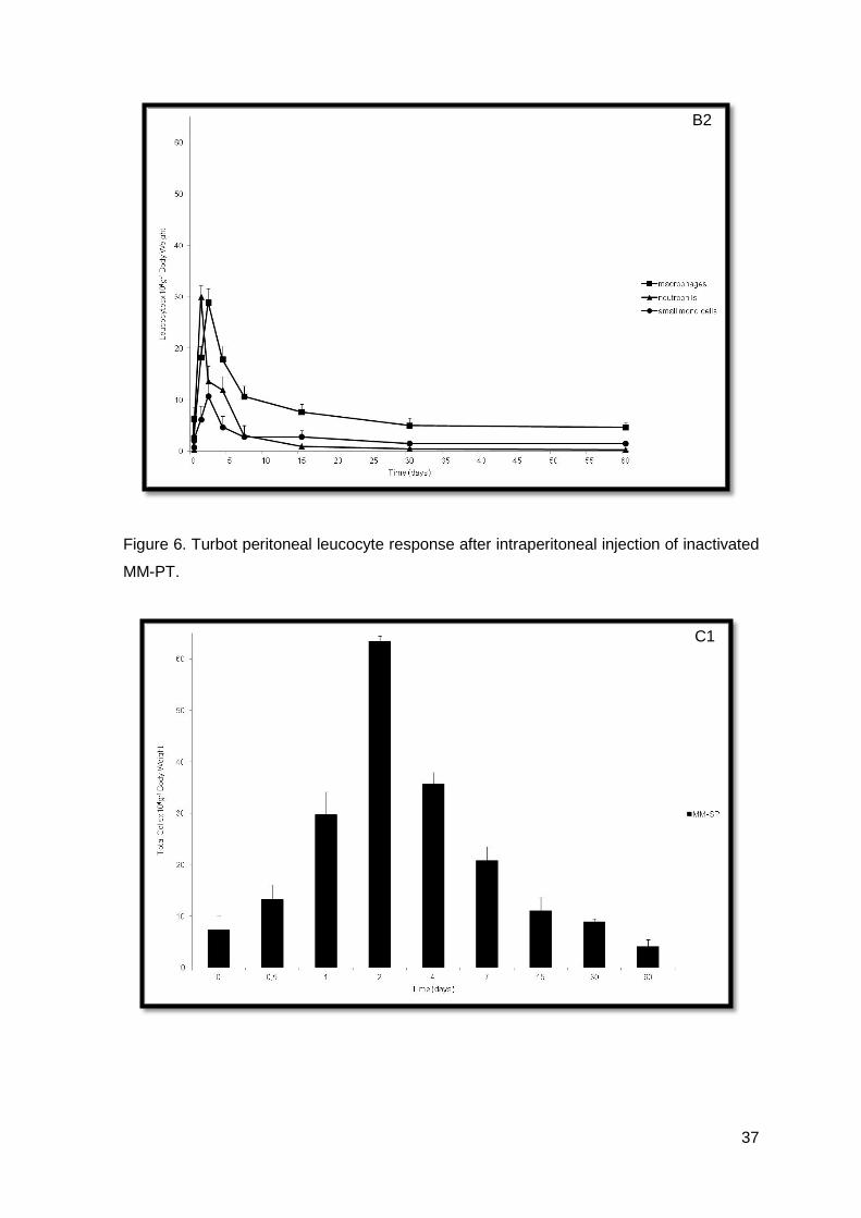
Figure 6. Turbot peritoneal leucocyte response after intraperitoneal injection of inactivated
MM-PT.
B2
C1
38
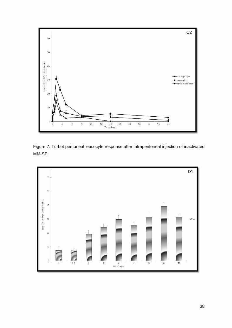
Figure 7. Turbot peritoneal leucocyte response after intraperitoneal injection of inactivated
MM-SP.
C2
D1
39
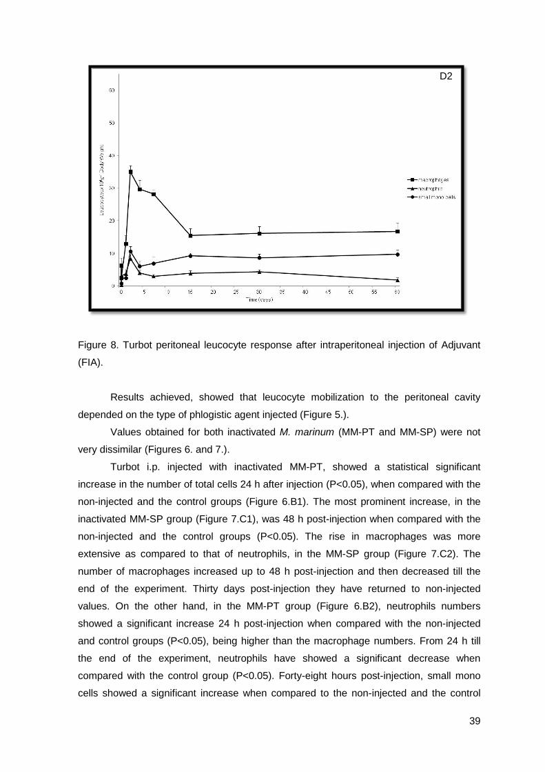
Figure 8. Turbot peritoneal leucocyte response after intraperitoneal injection of Adjuvant
(FIA).
Results achieved, showed that leucocyte mobilization to the peritoneal cavity
depended on the type of phlogistic agent injected (Figure 5.).
Values obtained for both inactivated M. marinum (MM-PT and MM-SP) were not
very dissimilar (Figures 6. and 7.).
Turbot i.p. injected with inactivated MM-PT, showed a statistical significant
increase in the number of total cells 24 h after injection (P<0.05), when compared with the
non-injected and the control groups (Figure 6.B1). The most prominent increase, in the
inactivated MM-SP group (Figure 7.C1), was 48 h post-injection when compared with the
non-injected and the control groups (P<0.05). The rise in macrophages was more
extensive as compared to that of neutrophils, in the MM-SP group (Figure 7.C2). The
number of macrophages increased up to 48 h post-injection and then decreased till the
end of the experiment. Thirty days post-injection they have returned to non-injected
values. On the other hand, in the MM-PT group (Figure 6.B2), neutrophils numbers
showed a significant increase 24 h post-injection when compared with the non-injected
and control groups (P<0.05), being higher than the macrophage numbers. From 24 h till
the end of the experiment, neutrophils have showed a significant decrease when
compared with the control group (P<0.05). Forty-eight hours post-injection, small mono
cells showed a significant increase when compared to the non-injected and the control
D2
40
groups (P<0.05). But this was followed by a decrease till the end of the experiment
(Figures 6.B2 and 7.C2).
The adjuvant group (Figure 8.) could be characterized by a fluctuation in the
number of leucocytes over time. Again, there was a significant increase in the number of
total cells 4 days post-injection, when compared with the non-injected and the control
groups (P<0.05), and has maintained the same pattern till the end of the experiment,
without achieving till 60 days post-injection, the resting values. The observed increases
were mainly due to the increment in the number of macrophages (Figure 8.D2). The
neutrophils and the small mono cells populations were the most constants (Figure 8.D2).
As showed, data observed during the inflammatory trial was also very interesting,
because MM-PT had a characteristically neutrophilic response and MM-SP did not.
Moreover, turbot, in contrast to that observed in other fish species (Afonso et al, 1998; do
Vale et al, 2002; Afonso et al, 2005) and in mammals (Pires et al, 2008), did not seem to
be capable of developing an early neutrophilic response, during an inflammatory status, in
the group i.p. injected with inactivated MM-SP. Moreover, macrophages appeared to be
the most sensitive cell type in presence of phlogistic agents, making them good indicators
of inflammation, as demonstrated by the increase in macrophage numbers up to 17 times
in the most severe cases, usually 48 hours post-injection. There was also maintenance of
macrophage values, up to 90 days in turbot injected with adjuvant, showing that a chronic
inflammation had been established, as in accordance to what was observed in other fish
species (Afonso et al, 2005). It seems feasible to suggest that inactivated M. marinum
acted as transient inflammatory stimulants and the adjuvant, which is hardly degraded by
fish, acted as an inflammatory stimulant, as it was capable of inducing and perpetuating
the inflammatory reaction. However, differences were observed in the MM-SP group and
as we are using the same host (turbot), there should be a strain variation not only related
to the incoming of cells to the peritoneal cavity, as well as in their number. Moreover, it is
well known the important function developed by macrophages during an inflammation
event (Kolaczkowska et al, 2007) and data achieved in this study clearly shows that fish
behaves in the same manner.
Several specimens were opened and it was not found any adhesions or
granulomas inside the peritoneal cavity. However, we observed a slight alteration in the
kidney’s cellular population where there was a proliferation of leucocytes.
Some of the results achieved were not in accordance to what was described in
other fish species, such as sea bass or rainbow trout (Afonso et al, 1998; do Vale et al,
2002; Afonso et al, 2005), where the neutrophilic population was considered a good
indicator of inflammation. Our findings suggest that in turbot, macrophages are the

41
population responsible for this indication, when looking at “our” strain of M. marinum (MM-
PT).
42
CHAPTER 5 Turbot infection trial
The study of the inflammatory response in turbot peritoneal cavity is the first step
among others not less, nor more important. The in vivo experimental infection is of crucial
interest for immunologists and pathologists. The development of disease, the oscillation of
leucocyte populations and the effects observed are of major importance to study the
pathogen, the disease and then work on a way to accomplish the task of developing a
bacterin/vaccine.
MATERIAL AND METHODS
For this assay, turbot, weighting 100±5.0 g, were supplied by Aquacria (Torreira,
Portugal). Fish were kept in the same conditions, as mentioned before (Chapter 2,
Material and Methods). All fish were starved 24 h prior to experimental infection.
The two strains of M. marinum (MM-PT and MM-SP) were thawed from a frozen
stock (-70º C) and inoculated onto 7H10. Cultures were grown for one week at 30º C and
ressuspended into 7H9. The bacterial density was measured by spectrophotometry at 520
nm and dilutions (in 7H9) were made until 107 CFUs/mL were achieved (corresponding to
approximately 200 µg of protein/mL). Actual CFUs counts, used as experimental infection
dose were confirmed, by viable counts of dilutions plated on 7H9 plates and incubated
during one week at 30º C.
Turbot were divided into four groups of 60 fish each. After anaesthetizing turbot
(0.03% (v/v)), groups were i.p. injected (100 µL) like the scheme below:
Group I – Non-injected group
Group II - Control; injected with PBS
Group III – injected with MM-PT
Group IV – injected with MM-SP
43
Groups III and IV were infected and put into two different tanks in an “isolated
infection room”1.
Mortalities were monitored during the entire assay (6 months) and dead fish were
examined for the reisolation of the inoculated bacterial strain from the head-kidney, by
streaking it directly onto 7H9. Shape of colonies and Ziehl-Nielsen stain (Merck) were
used for presumptive identification.
Each turbot collected (8 fish per group) was bled from the caudal vein. The blood
was allowed to clot for 15 min, centrifuged (2100xG for 10 min) and the serum collected.
Serum was stored at 4º C for one day before use. In some cases, serum was heat-
inactivated at 50º C for 30 min.
Then, their peritoneal leucocytes (8 fish per group) were collected at 12, 24, 48
hours and 4, 7, 15, 30, 60 and 90 days. Turbot were killed with an overdose of anesthesia
(0.06% (v/v)).
Peritoneal exudates were collected using a previously described technique
(Chapter 2, Material and Methods) and three sets of cytospins of peritoneal leucocyte
suspensions were made using a Cellspin I apparatus. The first set of dry cytospins were
fixed with formol-ethanol for one minute and stained with Wright’s stain. For peroxidase
detection, the Antonow’s technique was used (Chapter 2, Material and Methods). The
second set of cytospins was stained with Gram-Hucker stain (Deltalab, Barcelona, Spain)
and the third were stained with Ziehl-Nielsen technique, using acid Fucsin (Merck) and
contrasted with methylene blue.
Differential counts of macrophages, neutrophils and small mono cells were made
under x100 oil immersion. Once agains, to overcome weight variation between individuals,
leucocyte numbers were converted to leucocytes per gram of body weight.
At each collection time, internal organs were observed. Internal organs (kidney,
liver, spleen and intestine) and gills, from half of the sample, were collected and fixed in
formol 4% for histological analysis. Tissues samples were embedded in paraffin wax and
5 µm sections were cut with a rotary microtome (American Optical, Buffalo, NY). After
dewaxing, the sections were stained with haematoxylin/eosin.
The number of CFU of each strain injected, MM-PT and MM-SP, in spleen and
head-kidney of infected turbot, was determined, by serial diluting and plating the tissue
homogenates, from 4 fish per sample, onto 7H10 medium. Results are expressed as the
growth index, which represents the difference between the log10 mean of CFU at day 90
and the log10 mean of CFU at day 7. The CFU number in the peritoneal cavity was
1 The “infected room” had a “pediluvium” at the entrance before and after the door, with diluted hypochloride, and before entering, specific boots, white laboratory coat and latex gloves were obligatory.

44
determined in the same way as with the tissue homogenates, but using the collected
lavage fluid.
45
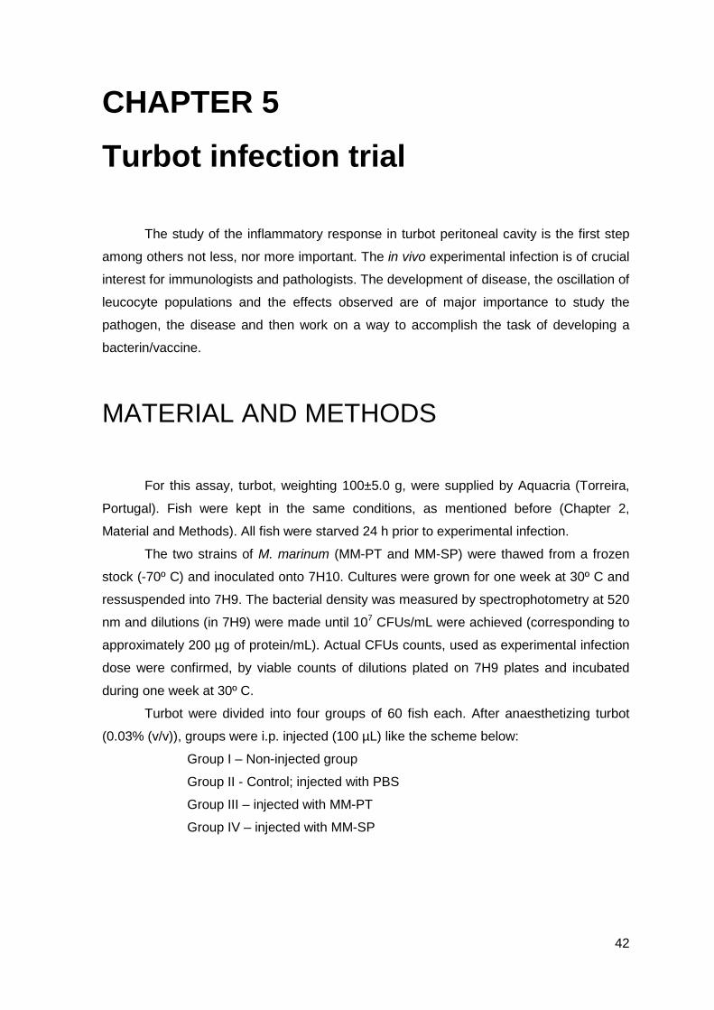
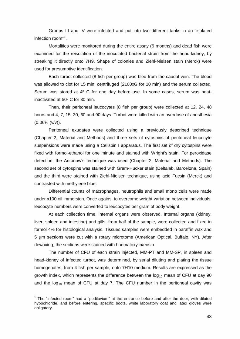
RESULTS AND DISCUSSION
A
B
46
Figure 9. A) Peritoneal leucocytes after intraperitoneal injection of PBS, MM-PT and MM-
SP; B) Turbot peritoneal leucocyte response after intraperitoneal infection with MM-PT; C)
Turbot peritoneal leucocyte response after intraperitoneal infection with MM-SP.
Table 5. Presence (+) or absence (-) of M. marinum in several tissues collected during the
experiment.
Table 6. Growth index in turbot i.p. infected with M. marinum.
C
47
The two strains of M. marinum (MM-PT and MM-SP) were consistent with the
results achieved (Figures 9.B and 9.C).
Both groups of M. marinum, showed a statistical significant increase in the number
of total cells 4 days post-infection (P<0.05), when compared with the non-injected and the
control groups (Figure 9.A). The rise in the number of macrophages was more extensive
as compared to that of neutrophils (Figure 9.B and 9.C). The number of macrophages
increased up to 4 days post-infection and then decreased till the end of the experiment.
The increase observed 30 days post-infection in the MM-PT group was not statistically
significant (Figure 9.B). Sixty days post-infection leucocyte numbers have returned to
values observed in the non-injected group (Figures 9.B and 9.C). The number of
neutrophils or small mono cells did not show any statistically significant difference, during
the 90 trial days (Figures 9.B and 9.C).
Gross examination of turbot infected with M. marinum (MM-PT and MM-SP) did
not reveal any abnormality, such as bleeding on internal organs, in contrast to the results
showed by in vivo infection (Decostere et al, 2004; Gauthier and Rhodes, 2009).
However, histopathological examination showed the existence of bacteria in several
tissues.
Briefly, 12 h after i.p. infection, no bacteria were detected in liver, kidney, spleen
and gills. However, in MM-PT group we observed bacteria in the spleen that persisted till
24 h post-infection, when it was observed for the first time in turbot infected with MM-SP.
Bacteria were observed in the kidney of turbot infected with MM-PT and MM-SP, 24 h
post-infection and persisted more 6 days. Moreover, bacteria were only observed in gills
48 h post-infection in both groups: MM-PT and MM-SP (Table 5.).
Till the end of the experimental trial, no bacteria were detected in heart and/or skin.
In the control group, bacteria did not grow, showing that animals were not
contaminated and validating our experiment. Till 7 days post-infection there was an
increase in the number of CFUs in each tissue and exudates. However, these values
began to decrease till 60 days post-infection, indicating that after 7 days post-infection,
turbot was able to control the infection, even though without complete elimination of the
bacteria during the period studied. These data clearly show that turbot may be capable of
releasing the bacteria to the medium, as described by Avendaño-Herrera et al (2006),
however, attempts to recover them were not successful.
Growth index in turbot i.p. infected with MM-PT was higher than in turbot i.p.
infected with MM-SP, showing that MM-PT proliferates more extensively in turbot, than
MM-SP (Table 6.).
48
During the course of infection, the infecting organism (pathogen), uses host’s
resources in order to multiply, which in turn will interfere with normal functions developed
by the host, and may lead to chronic wounds, grangrene, loss of an infected limb or even
death (Bassett et al, 2003). As observed previously, reaction to phlogistic agents was
more severe, because microorganisms were inactivated and could not interfere with host’s
resistance mechanisms, when compared to an experimental infection.
In this trial, turbot was shown to be sensitive to the experimental infection by M.
marinum, and recovery of bacteria from organs and peritoneal exudates was described.
Moreover, we observed that head-kidney tended to exhibit bacterial aggregations for a
longer time period, when compared to spleen and the peritoneal exudate. Even still,
typical infection signs were not encountered till 60 days post-infection and that may be
related to non-specific immunossuppression in turbot, by mycobacterial infection
(Appelberg et al, 1989). Avendaño-Herrera et al (2006) showed that turbot is capable of
releasing bacteria to the water, but we have analysed water and no mycobacteria could
be recovered (data not shown). Differences between MM-PT and MM-SP were only
encountered when calculating growth index, where we have observed that it was higher in
turbot i.p. infected with MM-PT, than in turbot i.p. infected with MM-SP. This showed that
MM-PT was capable of proliferating more extensively in turbot, than MM-SP. As described
elsewhere (Olsson et al, 1998), tests were made to evaluate the persistence of a bacteria
inside the host, by infecting its intestines. However, due to etiological and deontological
reasons, these trials were done conducted.
During the infection trial, we have calculated the difference between weights and
we have obtained a decrease in weight till 4 days post-infection, in groups i.p. infected
with M. marinum, but not statistically significant (data not shown). However, the decrease
was in accordance with Stamm & Brown (2004), who also observed that mycobacterial
infection was almost always accompanied by weight loss. We have also noticed that fish
tended to separate from each other, becoming solitary individuals in the corner holding
facilities. Once again, this behaviour is in accordance with Decostere et al (2004).
49
CHAPTER 6 In vitro studies with M. marinum
After the inflammatory and infection trials, we have decided to study the effect of
two strains of M. marinum on the function and integrity of turbot (S. maximus) head-kidney
macrophages. During this chapter, we will detect the production of superoxide anion and
not only due to the fact that counting the number of engulfed particles under a
microscope, to evaluate phagocytosis, is tedious, laborious and time consuming, but also
because mycobacteria have a high capacity to aggregate, we decided to develop a flow
cytometric assay to quantify phagocytosis.
MATERIAL AND METHODS
Turbot weighting 200±20 g, were purchased from a commercial fish farm located in
the North of Portugal. Fish were kept in the same conditions as mentioned before
(Chapter 2 – Material and Methods). All fish were maintained for at least 15 days and
starved 24h prior to cell collection. Turbot were bled from the caudal vein. The blood was
allowed to clot for 15 min, centrifuged (2100xG for 10 min) and the serum collected.
Serum was stored at 4º C for one day before use. In some cases, serum was heat-
inactivated at 50º C for 30 min.
Two virulent strains of M. marinum (MM-PT and MM-SP) were thawed from a
frozen stock (Chapter 4, Material and Methods).
Turbot were killed by an overdose of anesthesia (0.06% (v/v)). Then, the head-
kidney was aseptically dissected, as previously described (Chung and Secombes, 1988)
and pushed through a 100 µm nylon mesh with L-15 medium containing 2% Fetal Calf
Serum (FCS, Gibco), 100 u/mL Penicillin/Streptomycin (P/S, Gibco) and 20 u/mL heparin
(Sigma). The resultant suspension was layered onto a 30-45% discontinuous Percoll
gradient previously set up. The gradient was then centrifuged at 1000xG for 40 min at 4º
C, and the leucocytes were recovered at the 30-45% Percoll interface. The macrophage
enriched isolated cells (THKM) were counted in a haemocytometer, adjusted to 107
cells/mL in L-15, 5% FCS, P/S, heparin and plated in 96 well plates (100 µL/well). Viability
50
of cells monitored by means of trypan blue exclusion assay was above 92%, in all
preparations. After overnight at 20º C, non-adherent cells were discarded by carefully
washing the cultures twice with phenol red-free Hank’s Balanced Salt Solution (PRF-
HBSS, Gibco).
DETECTION OF EXTRACELLULAR SUPEROXIDE ANION
The method based on the reduction of ferrocytochrome C was used for the
detection of extracellular superoxide anion (Secombes, 1990). Monolayers of THKM were
washed twice with PRF-HBSS and 100 µL of live or inactivated MM-PT and MM-SP in
ferrocytochrome C solution (2 mg ferrocytochrome C mL-1 PRF-HBSS) were added. Serial
dilutions ranged from 3.1 x 10⁷ to 10 x 108 bacteria mL-1. Ferrocytochrome C solution
containing 1 µg mL-1 phorbol myristate acetate (PMA, Sigma) was added to the positive
control, as a soluble stimulant of the respiratory burst. Wells with THKM and
ferrocytochrome C were used as negative control, for spontaneous and background
production of superoxide anion. Ferrocytochrome C with bacteria or PMA and 300 units of
superoxide dismutase mL-1 (SOD, Sigma) was used to confirm the specificity of the
reaction. Bacteria previously opsonised with 1:500 diluted serum for 30 min were used in
some samples.
For each sample, 4 wells of macrophages per fish were assayed. Plates were read
immediately after addition of the bacteria and/or reagents to the THKM monolayers and
reading were then taken every 60 s for 60 min, on a multiscan spectrophotometer
(BioTek, Power Wave 440) at 550 nm. Data were expressed at the Vmax rate of the
response in mOD min-1 (optical density, OD). Data using opsonised bacteria were
expressed as an index (opsonisation index) relative to results using unopsonised bacreria
and allowed pooling of data.
DETECTION OF INTRACELLULAR SUPEROXIDE ANION
For the detection of intracellular superoxide anion, the method of the reduction of
the redox dye nitroblue tetrazolium (NBT) was used. Briefly, NBT was dissolved at 1
mg.mL-1 in PRF-HBSS, and PMA added at 1 µg.mL-1. SOD was also added to the same
solution, as a negative control. THKM were washed twice with PRF-HBSS, 100 µL of live
or inactivated MM-PT and MM-SP in a NBT solution, containing 2.5 x 10⁸ bacteria.mL-1
were added. Microplates were incubated for 60 min. Medium was then removed and cells
fixed in 70% methanol and allowed to air dry. The formazan in each well was dissolved in
120 µL 2M potassium hydroxide (KHO) and 140 µL dimethyl sulfoxide (DMSO, Fluka),
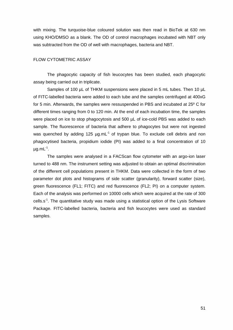
51
with mixing. The turquoise-blue coloured solution was then read in BioTek at 630 nm
using KHO/DMSO as a blank. The OD of control macrophages incubated with NBT only
was subtracted from the OD of well with macrophages, bacteria and NBT.
FLOW CYTOMETRIC ASSAY
The phagocytic capacity of fish leucocytes has been studied, each phagocytic
assay being carried out in triplicate.
Samples of 100 µL of THKM suspensions were placed in 5 mL tubes. Then 10 µL
of FITC-labelled bacteria were added to each tube and the samples centrifuged at 400xG
for 5 min. Afterwards, the samples were ressuspended in PBS and incubated at 25º C for
different times ranging from 0 to 120 min. At the end of each incubation time, the samples
were placed on ice to stop phagocytosis and 500 µL of ice-cold PBS was added to each
sample. The fluorescence of bacteria that adhere to phagocytes but were not ingested
was quenched by adding 125 µg.mL-1 of trypan blue. To exclude cell debris and non
phagocytised bacteria, propidium iodide (PI) was added to a final concentration of 10
µg.mL-1.
The samples were analysed in a FACScan flow cytometer with an argo-ion laser
turned to 488 nm. The instrument setting was adjusted to obtain an optimal discrimination
of the different cell populations present in THKM. Data were collected in the form of two
parameter dot plots and histograms of side scatter (granularity), forward scatter (size),
green fluorescence (FL1; FITC) and red fluorescence (FL2; PI) on a computer system.
Each of the analysis was performed on 10000 cells which were acquired at the rate of 300
cells.s-1. The quantitative study was made using a statistical option of the Lysis Software
Package. FITC-labelled bacteria, bacteria and fish leucocytes were used as standard
samples.
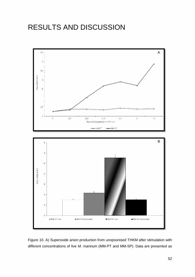
52
RESULTS AND DISCUSSION
Figure 10. A) Superoxide anion production from unopsonised THKM after stimulation with
different concentrations of live M. marinum (MM-PT and MM-SP). Data are presented as
A
B
53
Vmax rates of ferrocytochrome C reduction (Vmax for PMA was 5.66 mOD.min-1); B)
Superoxide anion production from THKM, after stimulation with live or inactivated M.
marinum (MM-PT and MM-SP). Data are presented as Vmax of ferrocytochrome C
reduction, expressed as the means ± S.E.M. of 3 fish.
Figure 11. NBT reduction from THKM, after stimulation with live or inactivated M. marinum
(MM-PT and MM-SP). Data are presented as the means ± S.E.M. of 3 fish (OD for PMA
stimulated macrophages was 0.66).
In both assays, SOD and PMA were used as negative and positive controls,
respectively, and validated our experiment.
Using a concentration of 109 bacteria, the effect of challenging turbot head-kidney
macrophages with MM-PT and MM-SP, elicited a respiratory burst (Figure 10.), being that
in accordance with Stave et al (1984), Bandín et al (1995) and Chaves-Pozo et al (2005).
MM-SP elicited a higher response, when compared to MM-PT (P<0.05).
In the detection of intracellular superoxide anion (Figure 11.), we observed that
there was a higher increase in its production in THKM stimulated with MM-PT, when
compared to the response elicited by THKM stimulated with MM-SP (P<0.05), being that
in accordance to Salinas et al (2006).
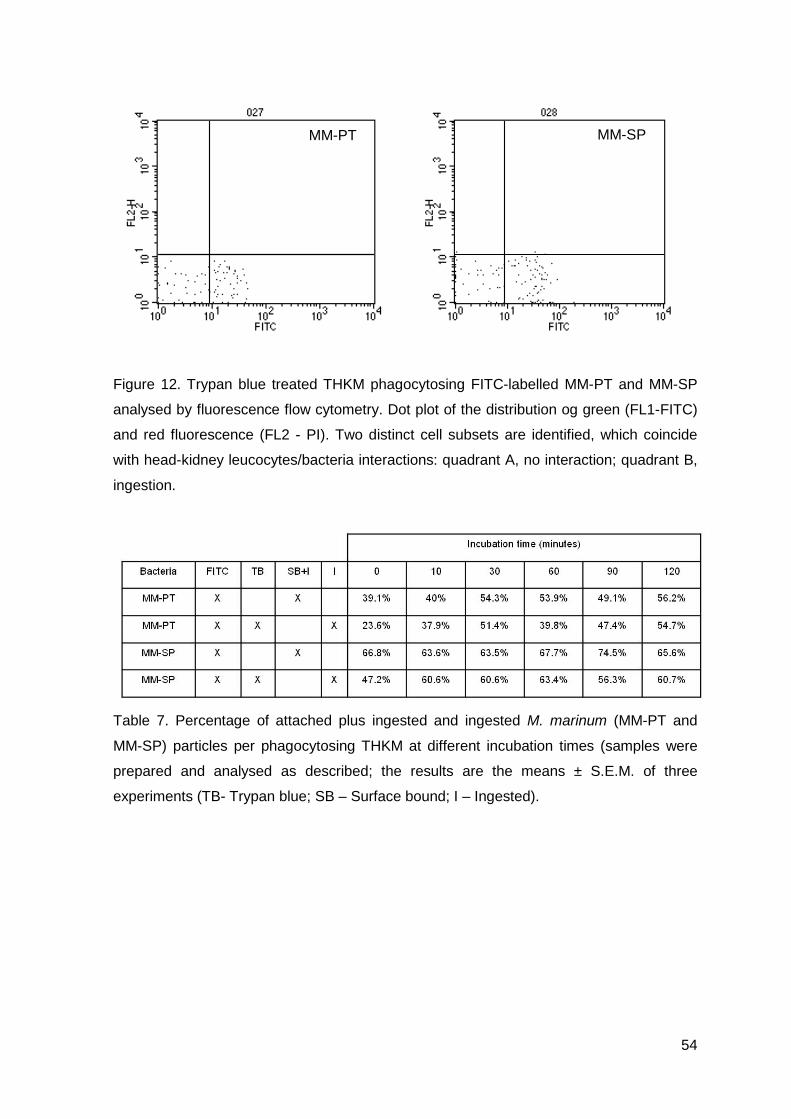
54
Figure 12. Trypan blue treated THKM phagocytosing FITC-labelled MM-PT and MM-SP
analysed by fluorescence flow cytometry. Dot plot of the distribution og green (FL1-FITC)
and red fluorescence (FL2 - PI). Two distinct cell subsets are identified, which coincide
with head-kidney leucocytes/bacteria interactions: quadrant A, no interaction; quadrant B,
ingestion.
Table 7. Percentage of attached plus ingested and ingested M. marinum (MM-PT and
MM-SP) particles per phagocytosing THKM at different incubation times (samples were
prepared and analysed as described; the results are the means ± S.E.M. of three
experiments (TB- Trypan blue; SB – Surface bound; I – Ingested).
MM-PT MM-SP
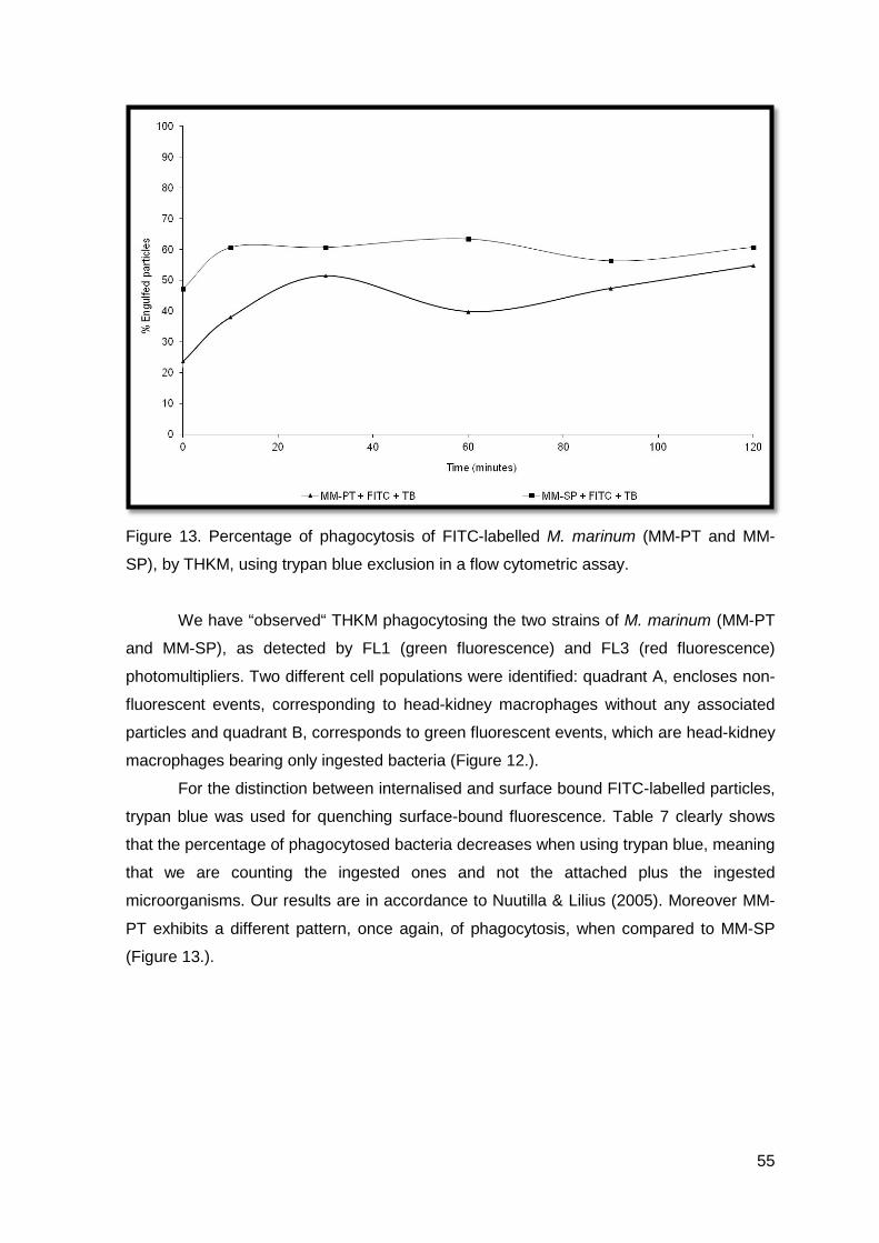
55
Figure 13. Percentage of phagocytosis of FITC-labelled M. marinum (MM-PT and MM-
SP), by THKM, using trypan blue exclusion in a flow cytometric assay.
We have “observed“ THKM phagocytosing the two strains of M. marinum (MM-PT
and MM-SP), as detected by FL1 (green fluorescence) and FL3 (red fluorescence)
photomultipliers. Two different cell populations were identified: quadrant A, encloses non-
fluorescent events, corresponding to head-kidney macrophages without any associated
particles and quadrant B, corresponds to green fluorescent events, which are head-kidney
macrophages bearing only ingested bacteria (Figure 12.).
For the distinction between internalised and surface bound FITC-labelled particles,
trypan blue was used for quenching surface-bound fluorescence. Table 7 clearly shows
that the percentage of phagocytosed bacteria decreases when using trypan blue, meaning
that we are counting the ingested ones and not the attached plus the ingested
microorganisms. Our results are in accordance to Nuutilla & Lilius (2005). Moreover MM-
PT exhibits a different pattern, once again, of phagocytosis, when compared to MM-SP
(Figure 13.).
56
The response of turbot to a mycobacterial infection was observed and we have
also conducted studies to observe the influence of the two strains of M. marinum (MM-PT
and MM-SP) in the homeostasis of turbot head-kidney macrophages. Results presented
show that early contact of macrophages with MM-PT or MM-SP induced respiratory burst
release of reactive oxygen intermediates, including superoxide anion. Moreover, there
was also a more intense response elicited by live microorganisms, than inactivated ones,
observed when turbot head-kidney macrophages were stimulated with MM-SP. This is in
accordance to studies developed in other fish species (Stave et al, 1984; Bandín et al,
1995; Chaves-Pozo et al, 2005). However, when stimulating with MM-PT, inactivated
bacteria elicited higher superoxide anion production by macrophages when compared to
the response of the live pathogen, in accordance to Salinas et al (2006). These results
were further confirmed when intracellular superoxide anion was performed to both MM-PT
and MM-SP. It is evident, by results, that MM-PT is capable of inhibiting and MM-SP of
stimulating the release of superoxide anion to the extracellular space.
It seems feasible to suggest that the Portuguese strain of M. marinum does not
inhibit the respiratory burst, and is capable to stimulate bacterial enzymes, such as
superoxide dismutase, thus augmenting the production of hydrogen peroxide.
Intracellular pathogens, as M. marinum, are considered virulent due to their ability
of resist host microbicidal mechanisms (Gupta, 2012), however, during the course of my
thesis, some inconsistencies were found. These continued even in the cytometric
analysis. Once again, MM-PT exhibited a different pattern of phagocytosis, when
compared to MM-SP.
57
CHAPTER 7 In vitro studies with S. parauberis
Results achieved during the course of this Thesis, showed that the two strains of
M. marinum, behave in different ways. As intracellular bacteria, but isolated from different
fish farms, we have decided to compare them with an extracellular bacteria, S. parauberis.
This Gram-positive bacterium is also the etiological agent of severe disease in turbot, as
mentioned before.
Studies of the respiratory burst and the release of nitrogen intermediates from
THKM after bacterial stimulation were conducted.
MATERIAL AND METHODS
Turbot weighting 200±20 g, were purchased from a commercial fish farm located in
the North of Portugal. Fish were kept in the same conditions as mentioned before
(Chapter 2 – Material and Methods). All fish were maintained for at least 15 days and
starved 24h prior to cell collection.
M. marinum strains (MM-PT and MM-SP) were thawed from frozen stocks
(Chapter 4 and 5, Material and Methods), inactivated and live, respectively.
A S. parauberis strain, was thawed from a frozen stock (Chapter 3, Material and
Methods) and inoculated onto TSA1. Cultures were grown during 48 h at 22º C and
ressuspended into TSB1. The bacterial density was measured by spectrophotometry at
550 nm and dilutions, in TSB1, were made until 107 CFUs.mL-1 were achieved. Then, one
part of the culture was inactivated, as previously described (Chapter 4, Material and
Methods).
Turbot were killed with an overdose of anesthesia (0.06% (v/v)) and head kidney
was aseptically removed and processed as previously described (Chapter 6.1, Material
and Methods).
Detection of extracellular and intracellular superoxide anion was conducted as
described in Chapter 6.1 (Material and Methods).

58
Nitrite concentration in the medium was measured by a microtitre assay method
described elsewhere (Ding et al, 1988). Briefly, 50 µL of each bacterial sample live and
inactivated (109 bacteria.mL-1) were added to monolayers with THKM and incubated with
equal volume of Griess reagent (1% sulphanilamide; 0.1% naphthylethylene diamine
dihydrochloride; 2.5% phosphoric acid; Sigma) at room temperature for 30 min. The
absorbance at 550 nm was determined using BioTek. Nitrite concentration was
determined by using sodium nitrite as a standard.
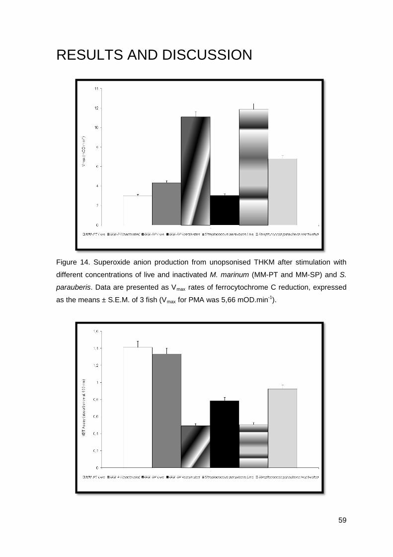
59
RESULTS AND DISCUSSION
Figure 14. Superoxide anion production from unopsonised THKM after stimulation with
different concentrations of live and inactivated M. marinum (MM-PT and MM-SP) and S.
parauberis. Data are presented as Vmax rates of ferrocytochrome C reduction, expressed
as the means ± S.E.M. of 3 fish (Vmax for PMA was 5,66 mOD.min-1).
60
Figure 15. NBT reduction from THKM, after stimulation with live or inactivated M. marinum
(MM-PT and MM-SP) and S. parauberis. Data are presented as the means ± S.E.M. of 3
fish (OD for PMA stimulated macrophages was 0.66).
Table 8. Results concerning nitrite concentration in MM-PT, MM-SP and S. parauberis.
After observing the date presented, we can say that MM-PT, when inactivated
elicited a higher, although not statistically significant response (P<0.05) than live bacteria.
In contrast, live MM-SP exhibit a similar behaviour of that observed for S. parauberis,
eliciting a statistically significant higher response (P<0.05), when compared to its
inactivated form (Figure 14.).
The results concerning the detection of intracellular superoxide anion, showed
that, there was an increase in its production in THKM stimulated with live MM-PT, in
comparison with the inactivated one, although not being statistically significant (P>0.05).
In contrast, MM-SP behaved like S. parauberis (Figure 15.).
Once again, when looking at the results concerning nitrite concentration, MM-SP
behaved in a different pattern, when compared to MM-PT (Table 8.).
Release of reactive oxygen intermediates from phagocytes in response to bacterial
pathogens is well known in fish, and it is generally accepted that live microorganisms may
elicit a more intense response than inactivated ones (Stave et al, 1984). In our study, this
was observed when turbot macrophages were stimulated with S. parauberis. However,
when stimulating with M. marinum (MM-PT strain), it was observed that inactivated
bacteria elicited higher superoxide anion production by macrophages when compared to
the response of the live pathogen. These results were further confirmed when detection of
intracellular superoxide anion was performed to both pathogenic bacteria. The
phenomenon evidenced by M. marinum has also been noticed in mammalian studies
using the intracellular pathogen M. lepraemurium (Brett and Butler, 1988; Mor et al, 1988;
61
Ellis, 1999). It is evident that M. marinum is capable of inhibiting and S. parauberis of
stimulating the release of superoxide anion to the extracellular space. Results achieved in
the detection of intracellular superoxide anion, suggest that M. marinum does not inhibit
the respiratory burst. The response was greater to M. marinum than to S. parauberis.
Once could say that it is capable of stimulating bacterial enzymes such as superoxide
dismutase, thus augmenting the production of hydrogen peroxide. Bacteria which survive
within macrophages may be capable of stimulating the burst by binding to the
macrophage through its hydrophobic surface and enter the cell at a later stage when the
burst is exhausted, as shown for Renibacterium salmoninarum (Bandín et al, 1993;
Bandín et al, 1995, Ellis, 1999), but this does not seem to be the case for M. marinum.
62
CHAPTER 8 Conclusions Studies concerning host-pathogen interactions are vast and the best way to
accomplish them is subjective. During the development of this thesis, we have tried to
maintain a connection line, and sometimes that was not very easy. The epizootic outbreak
of fish tuberculosis, observed and witnessed in a Portuguese farm, was the motor for the
development of these studies. Fish, as other animals, are difficult to maintain in sterile
environments, and external influences are always interfering with experimental results.
Hence we did lots of experiments and some of them were only important for validation of
previous results.
During the last decades, we have observed great improvements in fish
immunopathology field. However, we have considered that, fish
mycobacteriosis/tuberculosis studies lacked some of the initial information regarding
turbot, the host, and M. marinum, the pathogen. Moreover, the initial processes of
infection are very important in studies of host-pathogen interactions. Not only they are
capable of determining the establishment of a disease, but also its eradication, because
successful pathogenic bacterium need to multiply at a degree, so as to subsist in the host
or be transmitted effectively.
Results achieved during this thesis led to the following conclusions:
℘ Turbot peritoneal cavity, in resting conditions is composed by two professional
phagocyte populations: macrophages, the predominant leucocyte (53.20%) and
neutrophils, the less abundant one (6%).
℘ Turbot leucocytes are similar to those from other fish species and mammals.
Macrophages are round in shape, their cytoplasm is basophilic and nucleus is
eccentric and sometimes horse-shoe shaped. Ultrastructurally, there have
pseudopodia and are peroxidase negative and esterase positive. Neutrophil is
large, round and contains granules in its cytoplasm. Neutrophil’s nucleus is small,
rod-shaped and sometimes even segmented. Cytochemically, neutrophils are
peroxidase positive and esterase negative. The small mono cell is mainly
composed by small round cells, with a large nucleus, occupying almost all the
cytoplasm.
63
℘ We can identify the different types of peritoneal leucocytes using cytochemical
methods such as for the presence of peroxidase or esterase, because they are a
distinctive feature between macrophages and neutrophils.
℘ The two strains of M. marinum which were isolated from two mycobacteriosis
outbreaks: one in Portugal (MM-PT; dos Santos et al, 2002) and the other in Spain
(MM-SP, not in publication) were studied. An extracellular bacterium, also isolated
from an epizootic outbreak in Galicia (Spain), was used as control: S. parauberis
(Toranzo et al, 1994). S. parauberis was confirmed to be Gram-positive bacteria
and a fast-grower and M. marinum strains, confirmed to be slow growers.
℘ In the determination of bacterial growth curves, MM-SP appeared to be slower
grower than MM-PT.
℘ Turbot is not capable of developing an early neutrophilic response, during an
inflammatory status, in the group i.p. injected with inactivated MM-SP.
℘ Turbot’s macrophages appeared to be the most sensitive cell type in presence of
phlogistic agents, making them good indicators of inflammation. Inactivated M.
marinum acted as transient inflammatory stimulants and the adjuvant, which is
hardly degraded by fish, acted as an inflammatory stimulant, as it was capable of
inducing and perpetuating the inflammatory reaction.
℘ In turbot, macrophages are the population responsible for the indication of the
presence of an inflammatory status.
℘ When turbot is experimentally infected with M. marinum, they tend to separate
from each other, becoming solitary individuals in the corner holding facilities.
℘ Turbot is sensitive to the experimental infection by M. marinum, and recovery of
bacteria from organs and peritoneal exudates was accomplished. Turbot head-
kidney tends to exhibit bacterial aggregations for a longer time period, when
compared to spleen and the peritoneal exudate. However, typical infection signs
were not encountered till 60 days post-infection and that may be related to non-
specific immunossuppression in turbot, by mycobacterial infection.
℘ MM-PT is capable of proliferating more extensively in turbot, than MM-SP.
℘ Early contact of macrophages with MM-PT or MM-SP induces respiratory burst
release of reactive oxygen intermediates, including superoxide anion. There is a
more intense response elicited by live microorganisms, than inactivated ones.
However, when stimulating with MM-PT, inactivated bacteria elicited higher
superoxide anion production by macrophages when compared to the response of
the live pathogen. MM-PT is capable of inhibiting and MM-SP of stimulating the
release of superoxide anion to the extracellular space.
64
℘ In the cytometric analysis MM-PT exhibits a different pattern of phagocytosis,
when compared to MM-SP.
℘ MM-PT has a similar behaviour to any mycobacterial species, however, MM-SP
seems to behave as an extracellular species, like S. parauberis.
We can suggest that geographic coherence, coupled with environmental
influences, is able to generate patterns in M. marinum capable of inducing differences in
their mode of action. Moreover, the Portuguese virulent M. marinum strain may be
indicated as a possible vaccine component, because is capable of inducing a significant
leucocytic response in turbot peritoneal population. Without being too speculative, and
due to the existence of a close relationship between M. marinum and M. tuberculosis,
works should be conducted so as to determine a possible role of M. marinum as a
candidate in the fight against Human Tuberculosis...
65
CHAPTER 9 Future Perspectives
Works conducted in this thesis have opened a new window for future
investigations. The two different behaviours witnessed between the spanish and the
Portuguese strains of M. marinum, have led to the possibility of an environmental
influence among this bacterial species. Such a difference may be considered in the area
of immunopathology and also in the wide area of the vaccines industries.
Is the environment or the geographic origin, important to take into account when
studying a specific bacterial species?
May that be considered a possible reason why some vaccines do not act as they
should?
Lots of experimental works can be developed and future is and cannot be a barrier
to conquer any innovation in this area...tuberculosis is a bacterial disease and needs to be
controlled...fish, in particular, turbot, may be a good tool and the beginning of that battle...
66
CHAPTER 10 References Acosta, F et al (2006). Complement consumption by Photobacterium damselae subsp.
piscicida in seabream, red porgy and sea bass normal and immune serum. Effect of the
capsule on the bactericidal effect. Fish & Shellfish Immunology, 20: 709-717.
Afonso, A et al (1997). The leucocyte population of the unstimulated peritoneal cavity of
rainbow trout (Oncorhynchus mykiss). Fish & Shellfish Immunology, 7: 335-348.
Afonso, A et al (1998). Neutrophil and macrophage responses to inflammation in the
peritoneal cavity of rainbow trout, Oncorhynchus mykiss. Diseases of Aquatic Organisms,
34: 27-37.
Afonso, A et al (1998). Uptake of neutrophils and neutrophilic components by
macrophages in the inflamed peritoneal cavity of rainbow trout (Oncorhynchus mykiss).
Fish & Shellfish Immunology, 8: 319-338.
Afonso, A et al (2005). Side-effects in sea-bass (Dicentrarchus labrax L.) due to
intraperitoneal vaccination against vibriosis and pasteurellosis. Fish & Shellfish
Immunology, 49: 447-454.
Alkhodair, R et al (2010). Fish tank granuloma: misdiagnosed as cutaneous leishmaniasis.
International Journal of Dermatology, 49: 53-55.
Appelberg, R et al (1989). Induction of non-specific immunosuppression in mice by
mycobacterial infections and its relationship to macrophage activation. Scandinavian
Journal of Immunology, 30: 165-174.
Aronson, J (1926). Spontaneous tuberculosis in salt water fish. Journal of Infectious
Diseases, 39: 314-320.
Avendaño-Herrera, R et al (2006). A challenge model for Tenacibaculum maritimum
infection in turbot, Scophthalmus maximus (L.). Journal of Fish Diseases, 29: 371-374.
67
Bandín, I et al (1993). Interaction between rainbow trout macrophages and Renibacterium
salmoninarum in vitro. Fish & Shellfish Immunology, 3: 25-33.
Bandín, I et al (1995). Effects of serum factors on the survival of Renibacterium
salmoninarum within rainbow trout macrophages. Diseases of Aquatic Organisms,23:
221-227.
Bassett, C et al (2003). Innate immunity and host-pathogen interaction. Vaccine, 21: S12-
S23.
Bataillon, E et al (1897). Un nouveau type de tuberculose. Comptes Rendus de la Societè
de Biologie, 4: 446.
Brett, SJ and Butler, R (1988). Interactions of Mycobacterium lepraemurium with resident
peritoneal macrophages: phagocytosis and stimulation of the oxidative burst. Clinical
Experimental Immunology, 71: 32-38.
Carvalho, V. and Diniz, M (1998). Pregado (Rodovalho) (Scophthalmus maximus). In:
Manual de Aquacultura. M. A. R. Henriques, Ed.. Porto, Portugal: 188-189.
Chaves-Pozo, E et al (2005). Early innate immune response and redistribution of
inflammatory cells in the bony fish gilthead seabream experimentally infected with Vibrio
anguillarum. Cell and Tissue Research, 320: 61-68.
Chemlal, K et al (2002). Characterization of an unusual Mycobacterium: a possible
missing link between Mycobacterium marinum and Mycobacterium ulcerans. Journal of
Clinical Microbiology, 40: 2370-2380.
Chung, S and Secombes, CJ (1988). Analysis of events occurring within teleost
macrophages during the respiratory burst. Comparative Biochemistry and Physiology,
89B: 539-544.
Dalgaard, P and Koutsoumanis, K (2001). Comparison of maximum specific growth rates
and lag times estimated from absorbance and viable count data by different mathematical
models. Journal of Microbiology Methods, 43: 183-196.
68
Decostere, A et al (2004). Piscine mycobacteriosis: a literature review covering the agent
and the disease it causes in fish and humans. Veterinary Microbiology, 99: 159-166.
Ding, A et al (1988). Release of reactive nitrogen intermediates and reactive oxygen
intermediates from mouse peritoneal macrophages. The Journal of Immunology, 141:
2407--2412.
do Vale, A et al (2002). The professional phagocytes of sea bass (Dicentrarchus labrax L.)
cytochemical characterisation of neutrophils and macrophages in the normal and inflamed
peritoneal cavity. Fish & Shellfish Immunology, 13: 183-198.
dos Santos, NMS et al (2002). Mycobacterial infection in farmed turbot Scophthalmus
maximus. Diseases of Aquatic Organisms, 52: 87-91.
EL-ETR, S et al (2001). Fish monocytes as a model for mycobacteral host-pathogen
interactions. Infection and Immunity, 69: 7310-7317.
Ellis, A (1999). Immunity to bacteria in fish. Fish & Shellfish Immunology, 9: 291-308.
Ellis, A (2001). Innate host defence mechanisms of fish against viruses and bacteria.
Developmental and Comparative Immunology, 25: 827-839.
FAO (2010). The State of World Fisheries and Aquaculture 2010, Rome, Italy.
Fouz, B (1990). Antibacterial action of the mucus of turbot. Bulletin of the European
Association of Fish Pathologists, 10: 56-59.
Galluzzi, L et al (2011). Autophagy and innate immunity ally against bacterial invasion.
The EMBO Journal, 30: 3213-3214.
Gauthier, DT and Rhodes, MW (2009). Mycobacteriosis in fishes: a review. The
Veterinary Journal, 180: 33-47.
Godfellow, M. and Wayne, LG (1983). Taxonomy and nomenclature. In: Biology of the
Mycobacteria - Physiology, Identification and Classification; Chapter 10, Academic Press,
New York, USA.
69
Gupta, A (2012). Mycobacterium tuberculosis: immune evasion, latency and reactivation.
Immunobiology, 217: 363-374.
Heckert, R et al (2001). Detection of a new Mycobacterium species in wild striped bass in
the Chesapeake Bay. Journal of Clinical Microbiology, 39: 710-715.
Hine, PM and Wain, JM (1988). Observations on the granulocyte peroxidase of teleosts: a
phylogenetic perspective.
Houben, EN et al (2009). Differential expression of a virulence factor in pathogenic and
non-pathogenic mycobacteria. Molecular Microbiology, 72: 41-52.
Jacobs, J et al (2009). Influence of nutritional state on the progression and severity of
mycobacteriosis in striped bass Morone saxatilis. Diseases of Aquatic Organisms, 87:
183-197.
Kaufmann, S (1989). Immunity to bacteria and fungi. In: Current Opinion in Immunology,
1: 431-440.
Kaufmann, S et al (1992). Immunity to intracellular bacteria. Memórias do Instituto
Oswaldo Cruz, 87: 91-94.
Le Dantec, C et al (2002). Chlorine disinfection of atypical mycobacteria isolated from a
water distribution system. Applied and Environmental Microbiology, 68: 1025-1032.
Mor, N et al (1988). Mycobacterium lepraemurium activates macrophages but fails to
trigger release of superoxide anion. Journal of Immunology, 140: 3956-3961.
Nakao, M et al (2011). The complement system in teleost fish: Progress of post-homolog-
hunting researches. Developmental and Comparative Immunology, 35: 1296-1308.
Nuutilla, J and Liliua, EM (2005). Flow cytometric quantitative determination of ingestion
by phagocytes needs the distinguishing of overlapping populations of binding and
ingesting cells. Cytometry Part A, 65: 93-102.
Olsson, JC et al (1998). Survival, persistence and proliferation of the fish pathogen Vibrio
anguillarum in juvenile turbot (Scophthalmus maximus) intestines and faeces. Journal of
fish Diseases, 21: 1-9.
70
Owen, R (2011). On the Anatomy of Vertebrates. Cambridge University Press.
Parry, R. et al (1965). A rapid and sensitive assay of muramidase. Proceedings of the
Society for Experimental Biology and Medicine, 119: 384-387.
Pasnik, D and Smith, S (2005). Immunogenic and protective effects of a DNA vaccine for
Mycobaterium marinum in fish. Veterinary Immunology and Immunopathology, 103: 195-
206.
Pires, MA et al (2008). Comparison of different animal models in the study of
inflammation. Experimental Pathology and Health Sciences, 2: 23-26.
Prouty, M et al (2003). Zebrafish - Mycobacterium marinum model for mycobacterial
pathogenesis. FEMS Microbiology Letters, 225: 177-182.
Renault, T and Guichard, B (2007). Risk factors for emerging infectious diseases in
aquaculture. INRA Production Animales, 20: 219-222.
Robbins, D et al (1971). A fine structural cytochemical localization of peroxidase activity in
rat peritoneal cells, mononuclear cells, eosinophils and mast cells. Journal of
Histochemistry and Cytochemistry, 19: 517-575.
Robinson, J and Karnovsky, M (1983). Ultrastructural localization of several phosphatases
with cerium 1.2. Journal of Histochemistry and Cytochemistry, 31: 1197-1208.
Romalde, J et al (1999). Genetic analysis of turbot pathogenic Streptococcus parauberis
strains by ribotyping and random amplified polymorphic DNA. FEMS Microbiology Letters,
179: 297-304.
Salinas, I et al (2006). Effect of heat-inactivated fish and non-fish derived probiotics on the
innate immune parameters of a teleost fish (Sparus aurata L.). Veterinary Immunology
and Immunopathology, 111: 279-286.
Saulnier, D et al (2007). Emerging diseases in organisms of aquaculture interest: some
scenarios ilustrated with examples. INRA Production Animales, 20: 207-212.
71
Secombes, C (1990). Isolation of salmonid macrophages and analysis of their killing
activity. Techniques in fish Immunology, 1: 137-154.
Silva, M et al (1987). In vivo killing and degradation of Mycobacterium aurum within
mouse peritoneal macrophages. Infection and Immunity, 55: 2006-2016.
Silva, M et al (1989). Neutrophil-macrophage cooperation in the host defence against
mycobacterial infections. Microbial Pathogenesis, 6: 369-380.
Stamm, L and Brown, E (2004). Mycobacterium marinum: the generalization and
specialization of a pathogenic mycobacterium. Microbes Infection, 6: 1418-1428.
Stave, J et al (1984). Factors affecting the chemiluminescent response of fish phagocytes.
Journal of Fish Biology, 25: 197-206.
Tavares-Dias, M et al (2005). Peripheral blood cells of the armored catfish Hoplosternum
littorale HANCOCK, 1828: a morphological and cytochemical study. Brazilian Journal of
Morphological Sciences, 22: 215-220.
Tonjum, T et al (1998). Differentiation of Mycobacterium ulcerans, M. marinum and M.
haemophilum: mapping of their relationships to M. tuberculosis by fatty acid profile
analysis, DNA-DNA hybridization, and 16S rRNA gene sequence analysis. Journal of
Clinical Microbiology, 36: 918-925.
Toranzo, A (2005). A review of the main bacterial fish diseases in mariculture systems.
Aquaculture, 246: 37-61.
Toranzo, A et al (1994). Streptococcosis in cultured turbot caused by an Enterococcus-
like bacterium. Bulletin of the European Association of Fish Pathologists, 14: 19-23.
Toranzo, A et al (2009). Present and future of aquaculture vaccines. Options
Méditerranéennes, 86: 155-176.
Uribe, C et al (2011). Innate and adaptive immunity in teleost fish: a review. Veterinarni
Medicina, 10: 486-503.
Yano, T (1992). Assays of haemolytic complement activity. In: Techniques in Fish
Immunology, Chapter 13: 131-141. J. S. et al ed. SOS Publications.
72
CHAPTER 11 Attachments Turbot is affected by streptoccosis during all the production cycle. However, as it
has been said, there is already an available vaccine to prevent this disease. At the
beginning of this Thesis, we thought it would be important to make a review concerning
some important cases etiological diseases, capable of affecting turbot, like lactococcosis.
Moreover, and knowing that weight and survival are extremely important when we are
dealing with an enterprise, like an aquaculture, the effect of L-carnitine supplemented in
turbot’s diet was also evaluated.
R E V I S T A P O R T U G U E S A
CIÊNCIAS VETERINÁRIASDE
Fish vaccination against infections by Streptococcal species and the particular case of Lactococcosis
Vacinação em peixes contra infecções por espécies de Streptococcus e ocaso particular da Lactococcose
Sónia Gomes1,2*, António Afonso1,2 e Fátima Gartner2,3
1CIIMAR - Centro Interdisciplinar de Investigação Marinha e Ambiental, Rua dos Bragas, 286, 4050-123 Porto2ICBAS - Instituto de Ciências Biomédicas Abel Salazar, Largo do Prof. Abel Salazar, 2, 4099-003 Porto
3IPATIMUP - Instituto de Patologia e Imunologia Molecular da Universidade do Porto, Rua Dr. Roberto Frias, 4200-465 Porto
Summary: Lactococcus garvieae, a species of theStreptococose-ceae family, is the etiological agent of lactoco-cose. Lactococose has become a worldwide disseminated infec-tion reported in a large number of fish specimens, includingsalmonids. The increased seriousness of the outbreaks after afirst contact is well recognized, being the pathology accompa-nied by intraocular haemorrhage, anorexia, acute meningitis aswell as "oculo-splanchnic dissociation". Treatment is in mostcases unsatisfactory and the overall recommendation is todestroy the diseased stock. Immunoprophylaxis seems to be apromising field in the prevention of lactococcosis, especially infreshwater fish, which are the ones experiencing the highestwater temperatures, during summer months. Moreover, vaccina-tion has been proved to be a highly effective method controllingdiseases in salmonid industry, mainly located in Europe,America and Japan. Although there has been a development ofstudies on vaccination against lactococcosis, its future stillseems to be uncertain, because major efforts need to be done toachieve an effective and long prophylactic method against thisdisease.
Keywords: Streptococcosis, Lactococcosis, Lactococcus garvieae
Resumo: O Lactococcus garvieae, pertencente à famíliaEstreptococose, é o agente etiológico da lactococose. A lacto-cocose é uma infecção que se tem vindo a disseminar porinúmeras regiões do Globo, afectando várias espécies de peixes,incluindo salmonídeos. Sendo reconhecido o aumento da gravi-dade dos surtos após um primeiro contacto, a patologia é normalmente evidenciada pelo aparecimento de hemorragiasintra-oculares, anorexia, meningite aguda, bem como por "dis-sociação óculo-esplâncnica". Os tratamentos terapêuticos nãosão na sua maioria satisfactórios, e a recomendação aquando deum surto baseia-se na destruição do stock que evidencia adoença. Assim, a imunoprofilaxia parece ser o campo de escolhana prevenção da lactococcose, especialmente nos peixes deágua-doce que vivem em ambientes onde se observam tempera-turas mais elevadas, durante os meses de Verão. A vacinaçãotem provado ser um método eficaz na protecção contra doençasem salmoniculturas, na sua maioria localizadas na Europa,América e Japão. Apesar de já terem sido realizados inúmerosestudos na área da vacinação contra a lactococcose, o seu futuro
é ainda incerto, havendo ainda um longo caminho a percorreraté que se encontre um método profiláctico eficaz e duradoiro.
Palavras-chave: Estreptococose, Lactococose, Lactococcusgarvieae
Introduction
Fish are a heterogeneous group of animals comprisingmore than 21 700 species (Fänge, 1994). The presenceof a circulatory system filled with blood containingred and white cells (erythrocytes and leukocytes), is afeature shared by all vertebrates and probably inheritedfrom extinct Pre-Cambrian proto-vertebrates (Fänge,1994).
In fish as in mammals, immune cells and lympho-haematopoietic tissues and organs are responsible forthe defence mechanisms in the host against infection.One example is the appearance of an inflammatorystatus. This results in specific morphological andchemical changes in tissues and cells (Suzuki and Iida,1992).
An efficient clearance and degradation system maybe worthwhile during pathogen invasion, which other-wise would lead to severe inflammation and eventuallydeath (Dalmo et al., 1997). This can be termed "innateimmunity" or "non-specific defence mechanisms",which are activated immediately after infection andrapidly control the replication of the infectingpathogen (Medzhitov and Janeway, 2000). On theother hand, there is a more complex system involvedin the immune response: "adaptive immunity" or "spe-cific defence mechanism". In the adaptive immunesystem, the T-cell receptor and the B-cell receptor aregenerated somatically, during the development of Tand B cells, in a way that endows each lymphocytewith a structurally unique receptor (Medzhitov andJaneway, 2000). Moreover, the random generation of a
ARTIGO DE REVISÃO
25
*Correspondence: [email protected]: (+351) 223 401 800
highly diverse repertoire of antigen receptors allowsthe adaptive immune system to recognize virtually anyantigen (Medzhitov and Janeway, 2000).
Prophylactic methods against fish diseases
During the last decades, there has been a continuousgrowth of aquaculture industry all over the world(Hastein, 1995). The most significant growth has beenseen in carp production as well as in the production ofother cyprinids and even more so for carnivorous fishspecies such as salmonids, which are farmed in inten-sive conditions (Gudding et al., 1999). However, anyintensive bioproduction – whether on land or at sea –will experience disease problems (Hastein, 1995).Infectious diseases that occur as sporadic events inwild fish populations may cause high mortalititieswhen appearing in intensive fish farming (Gudding etal., 1999). For the aquaculture industry to prosper inthe future it is a prerequisite that the losses caused bydiseases and the use of antibiotics are kept to a mini-mum. Immunoprophylaxis recognized by stimulationof non-specific and specific immunity is consequentlythe method adopted for developing aquaculture into asustainable bioproduction in the aquatic ecosystems.
Fish can be immunized in three ways: by injectionpreferably intraperitoneally, by immersion, usually bydipping the fish in a diluted vaccine solution, or byoral administration along with food. These methodshave different advantages and disadvantages withrespect to the level of protection, side effects, practi-cability and cost-efficiency. Injection and immersionare the two major methods that have been developedfor use at an industrial scale and in commercial pro-duction of salmonids and both vaccination proceduresare established practices and integrated as part of theproduction routines.
There is still a limited understanding of the mecha-nisms involved in antigen uptake and presentation afterimmersion and oral vaccination (Nakanishi and Ototake,1997; Quentel and Vigneulle, 1997; Moore et al., 1998).Even so, the first encounter with pathogens occursthrough the mucosal surfaces and the indisputable presenceof a mucosal associated immune system makes antigendelivery through the mucosal surfaces a viable approach.The mucosal immunity of fish is still a poorly exploredfield of research, although indications are available forthe existence of this type of immunity (Rombout andJoosten, 1992). Especially for fish living in a pathogen--rich aquatic environment, the existence of a mucosalimmune system may be very important. In fact, com-pared with higher vertebrates fish have their skin as anextra mucosal barrier. However, an important criterionfor the existence of a mucosal immune system is thesecretion of antigen-specific antibodies at mucosal sur-faces (Rombout and Joosten, 1992).
Oral vaccination research has so far focused on pro-tecting the antigen from digestion and decomposition
during passage through the stomach and anterior partof the gut. The ultimate goal has been to stimulate theimmunocompetent tissues in the posterior part of theintestine. Different approaches have been tried, one ofthe latest using antigens encapsulation in alginate andliposome microcapsules (Joosten et al., 1997).
The optimal way of developing an effective vaccineentails the identification of the pathogens’ key viru-lence factors. Further, the induction of the immuneresponse must be optimized so that the vaccinated ani-mal develops a protective immunity against thepathogen (Gudding et al., 1999).
Vaccines and adjuvants
Inactivated vaccines (bacterins)
A common approach in vaccine production is toinactivate the pathogen and toxins by physical (e.g.UV and heat) or by chemical (e.g. kill by formol)means so that it is no longer capable of replication inor damaging the host. It is critically important tomaintain the structure of epitopes on antigens duringinactivation. Administration of these bacterins is doneby injection or immersion. Provided that the serotypesused for vaccine preparation cover the field of virulentstrains and that the vaccines are used correctly, thesevaccines are usually effective and give negligible sideeffects (Stevenson, 1997; Toranzo et al., 1997). Mostvaccines used in aquaculture to date have been inacti-vated, bacterial vaccines (Gudding et al., 1999).
Live vaccines
Live, attenuated vaccines should potentially havemany advantages in aquaculture (Benmansour and deKinkelin, 1997). Vaccination with a live vaccine is inreality an infection (with an attenuated strain), and ifvaccinated fish sheds the vaccine strain, an effectivedissemination of the antigen in the production wouldtake place over an extended time period. Live vaccinesalso have the advantage of stimulating the cellularbranch of the immune system (Marsden et al., 1996).Finally, attenuated vaccines have some economicaladvantages in terms of simple delivery and low doserequirements due to multiplication in the fish(Gudding et al., 1999). However, the risk of reversionto virulence is still poorly understood and there seemsto be a general view that the risks associated with livevaccines in aquaculture, whether produced by conven-tional methods or by recombinant techniques shouldbe heavily scrutinized.
Vaccines based on DNA-technology
In recent years, several vectors have been used forcost-effective production of sufficient quantities ofprotective antigens by recombinant DNA technology.
RPCV (2006) 101 (557-558) 25-35Gomes S e Afonso A
26
In aquaculture, research on recombinant vaccines hasfocused on viral vaccines. Genetic immunizationusing naked DNA is the most recent approach in vac-cine design (Babuik et al., 1996). This technology isbased on the observations that skeletal muscle cellsinjected with purified plasmid DNA express plasmi-dencoded proteins. DNA vaccines have advantagesover conventional vaccines. In mammals, the specificimmune response after DNA vaccination encompassesantibodies, T-helper cells, as well as cytotoxic cells.However, before DNA vaccines are applied in com-mercial enterprises in aquaculture, safety for fish,environment and the consumer have to be addressed.As the DNA-sequence encodes only a single viral gene,there should be no possibility of reversion to virulence, which is a critical factor in relation to envi-ronmental safety in aquaculture (Gudding et al., 1999).
Adjuvants in the vaccines formulation
In most vaccines for animals, adjuvants are a crucialingredient for efficacy (Gudding et al., 1999). Briefly,these substances will enhance the immune response inorder to make antigens more effective by magnifyingthe immune pathways and also will be helpful increa-sing the action of small doses of antigens to enhancethe immune response to levels that can prevent disea-ses in fish culture (Anderson, 1997). In salmonids, asingle injection of vaccines with aluminium-basedadjuvants or glucans induced an acceptable protectionagainst furunculosis, however, the duration was rela-tively short (Gudding et al., 1999). In this fish group,various oil-adjuvants induced better and more long-lasting protection than aluminium and glucan againstfurunculosis in challenge and field trials (Midtlyng,1996; Midtlyng et al., 1996a; Midtlyng et al., 1996b).Oil-adjuvants are now used extensively in commercialinjectable bacterins. A double-adjuvant principle com-bines glucans and oil-adjuvants and thus induces bothshort- and long-term protection (Gudding et al.,1999). Adjuvants in injectable vaccines generally cre-ate local reactions with granulomas at the injectionsite. The reactions may vary from mild to severe. Oil-adjuvants generally create the most severe reactions(peritoneal granulomas and pigmentation) and on afew occasions they have affected growth rate of thefish as well as the quality of the final product (Poppeand Breck, 1997; Midtlyng and Lillehaug, 1998;Afonso et al., 2004). There is little doubt thatimmunoprophylaxis may largely reduce the risks forlarge scale animal suffering, caused by disease epi-zootics in fish farming. Side effects due to handlingand anaesthesia, which are required to vaccinate fishby injection, can be minimized. While the use of adju-vanted vaccines may cause some undesirable sideeffects, they are nevertheless acceptable from an ani-mal welfare point of view, especially when taking intoaccount the consequences of leaving the fish unpro-
tected against disease. At present, the reduction of vac-cine side effects by maintenance of durable immunityconstitutes the presently dominant challenge for fishvaccinology (Midtlyng, 1997).
The ability of fish vaccines to induce a protectiveimmunity is for the most part based on experimentalchallenge studies and/or field experiments. However,the virulence mechanisms for some fish pathogenshave been elucidated, and methods for studying theantibody response and even the cellular response tosome protective antigens have been described(Gudding et al., 1999). Consequently data have beengenerated to describe the relationship between specificparameters of the immune response and protection(Reitan and Secombes, 1997). The environmental tem-perature has an important influence on the develop-ment of immunity in fish (Gudding et al., 1999). Theproduction of antibodies in Atlantic salmon vaccinatedand reared at temperatures of 2 0C were generally lowerthan in fish vaccinated at 10 0C (Eggset et al., 1997).
Once an effective vaccine has been developed on alaboratory scale, it must be scaled up for production(Ward, 1982). Regulations set up by the licensingauthorities ensure that only vaccines, which are safeand effective, are launched on to the market (Ward,1982). Briefly, after an initial field-testing, developedin populations of fish, there will be a submission for aProduct License (Ward, 1982). However, when analy-zing this subject on a fish farmer point of view, otherfactors are also taken into account. For the fish farmer,vaccination is a matter of economy, the use of vaccinesat relatively low cost should increase profitability bypreventing loss of fish (Horne and Robertson, 1987).
Bacteriosis caused by streptococcalspecies
Bacterial infections in aquaculture were mainly origi-nated by Gram-negative microorganisms. However, inthe last years, due to unknown reasons, infections pro-voked by Gram-positive bacteria have developed, beingnowadays one of the main problems in the aquacultureindustries (Padrós and Furones, 2002). The Gram-posi-tive cocci are grouped together based on their Gram-stain reaction, thick cell wall composition, and spheri-cal shape. In this review we will focus on theStreptococcaceae family. Two genera of cocci areincluded in this family: Streptococcus and Diplococcus,being primarily differentiated on the basis of arrange-ment. The primary division of the Streptococcaceaeinto two physiological groups is based on the differencein fermentation products namely homofermentativeand heterofermentative. The first is further divided onthe basis of solubility in surface tension reducents intothe genera Diplococcus (soluble) and Streptococcus(not soluble) (Frobisher, 1954).
RPCV (2006) 101 (557-558) 25-35Gomes S e Afonso A
27
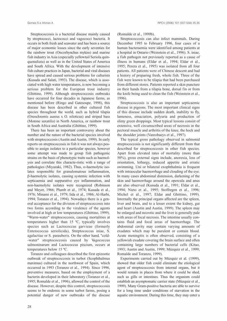
Streptococcosis is a bacterial disease mainly causedby streptococci, lactococci and vagococci bacteria. Itoccurs in both fresh and seawater and has been a sourceof major economic losses since the early seventies forthe rainbow trout (Oncorhynchus mykiss) and marinefish industry in Asia (especially yellowtail Seriola quin-queradiata) as well as in the United States of Americaand South Africa. With the development of intensivefish culture practices in Japan, bacterial and viral diseaseshave spread and caused serious problems for culturists(Kusuda and Salati, 1993). The disease, which is asso-ciated with high water temperatures, is now becoming aserious problem for the European trout industry(Ghittino, 1999). Although streptococcosis outbreakshave occurred for four decades in Japanese farms, asmentioned before (Ringo and Gatesoupe, 1998), thisdisease has been described in other cultured fishspecies throughout the world, such as hybrid tilapia(Oreochromis aureus x O. niloticus) and striped bass(Morone saxatilis) in North America, or rainbow troutin South Africa and Australia (Ghittino, 1999).
There has been an important controversy about thenumber and the nature of the bacterial species involvedwith streptococcosis (Austin and Austin, 1999). In earlyreports on streptococcosis in fish it was not always pos-sible to assign isolates to a particular species, howeversome attempt was made to group fish pathogenicstrains on the basis of phenotypic traits such as haemol-ysis and correlate this characte-ristic with a range ofpathologies (Miyazaki, 1982). Thus, α-haemolytic iso-lates responsible for granulomatous inflammation, β-haemolytic isolates, causing systemic infection withsepticaemia and suppurative eye inflammation, andnon-haemolytic isolates were recognized (Robinsonand Meyer, 1966; Plumb et al., 1974; Kusuda et al.,1976; Minami et al., 1979; Iida et al., 1986; Al-Harbi,1994; Toranzo et al., 1994). Nowadays there is a gen-eral acceptance for the division of streptococcosis intotwo forms according to the virulence of the agentsinvolved at high or low temperatures (Ghittino, 1999)."Warm-water" streptococcosis, causing mortalities attemperatures higher than 15 ºC, typically involvesspecies such as Lactococcus garvieae (formerlyEnterococcus seriolicida), Streptococcus iniae, S.agalactiae or S. parauberis. On the other hand, "cold--water" streptococcosis caused by Vagococcussalmoninarum and Lactococcus piscium, occurs attemperatures below 15 ºC.
Toranzo and colleagues described the first epizooticoutbreak of streptococcosis in turbot (Scophthalmusmaximus) cultured in the northwest of Spain, whichoccurred in 1993 (Toranzo et al., 1994). Since 1996,preventive measures, based on the employment of abacterin developed in their laboratory (Toranzo et al.,1995; Romalde et al., 1996), allowed the control of thedisease. However, despite this control, streptococcosisseems to be endemic in some turbot farms, posing apotential danger of new outbreaks of the disease
(Romalde et al., 1999b).Streptococcosis can also infect mammals. During
December 1995 to February 1996, four cases of ahuman bacteraemia were identified among patients ata hospital in Ontario (Weinstein et al., 1996). S. iniae,a fish pathogen not previously reported as a cause ofillness in humans (Eldar et al., 1994; Eldar et al.,1995; Perera et al., 1995) was isolated from all fourpatients. All patients were of Chinese descent and hada history of preparing fresh, whole fish. Three of thefish were known to be tilapia that had been purchasedfrom different stores. Patients reported a skin punctureon their hands from a tilapia bone, dorsal fin or fromthe knife being used to clean the fish (Weinstein et al.,1996).
Streptococcosis is also an important septicaemicdisease in pigeons. The most important clinical signsof this disease include sudden death, inability to fly,lameness, emaciation, polyuria and production ofslimy green droppings. Most typical lesions consist ofextensive, well circumscribed areas of necrosis in thepectoral muscle and arthritis of the knee, the hock andthe shoulder joints (Vanrobaeys et al., 1997).
The typical gross pathology observed in salmonidstreptococcosis is not significantly different from thatdescribed for streptococcosis in other fish species.Apart from elevated rates of mortality (more than50%), gross external signs include, anorexia, loss oforientation, lethargy, reduced appetite and erraticswimming. Uni or bilateral exophthalmia is frequentwith intraocular haemorrhage and clouding of the eye.In many cases abdominal distension, darkening of theskin and haemorrhage around the opercula and anusare also observed (Kusuda et al., 1991; Eldar et al.,1994; Nieto et al., 1995; Stoffregen et al., 1996;Michel et al., 1997; Eldar and Ghittino, 1999).Internally the principal organs affected are the spleen,liver and brain, and to a lesser extent the kidney, gutand heart (Austin and Austin, 1999). The spleen maybe enlarged and necrotic and the liver is generally palewith areas of focal necrosis. The intestine usually con-tains fluid and focal areas of haemorrhage. Theabdominal cavity may contain varying amounts ofexudates which may be purulent or contain blood.Acute meningitis is often observed, consisting of ayellowish exudate covering the brain surface and oftencontaining large numbers of bacterial cells (Kitao,1993; Austin and Austin, 1999; Múzquiz et al., 1999;Romalde and Toranzo, 1999).
Experiments carried out by Múzquiz et al. (1999),showed that older fish could eliminate the etiologicalagent of streptococcosis from internal organs, but itwould remain in places from where it could be shed,such as gills or intestines. Thus the organism couldestablish an asymptomatic carrier state (Múzquiz et al.,1999). Many Gram-positive bacteria are able to survivefor a long time under conditions of starvation in theaquatic environment. During this time, they may enter a
RPCV (2006) 101 (557-558) 25-35Gomes S e Afonso A
28
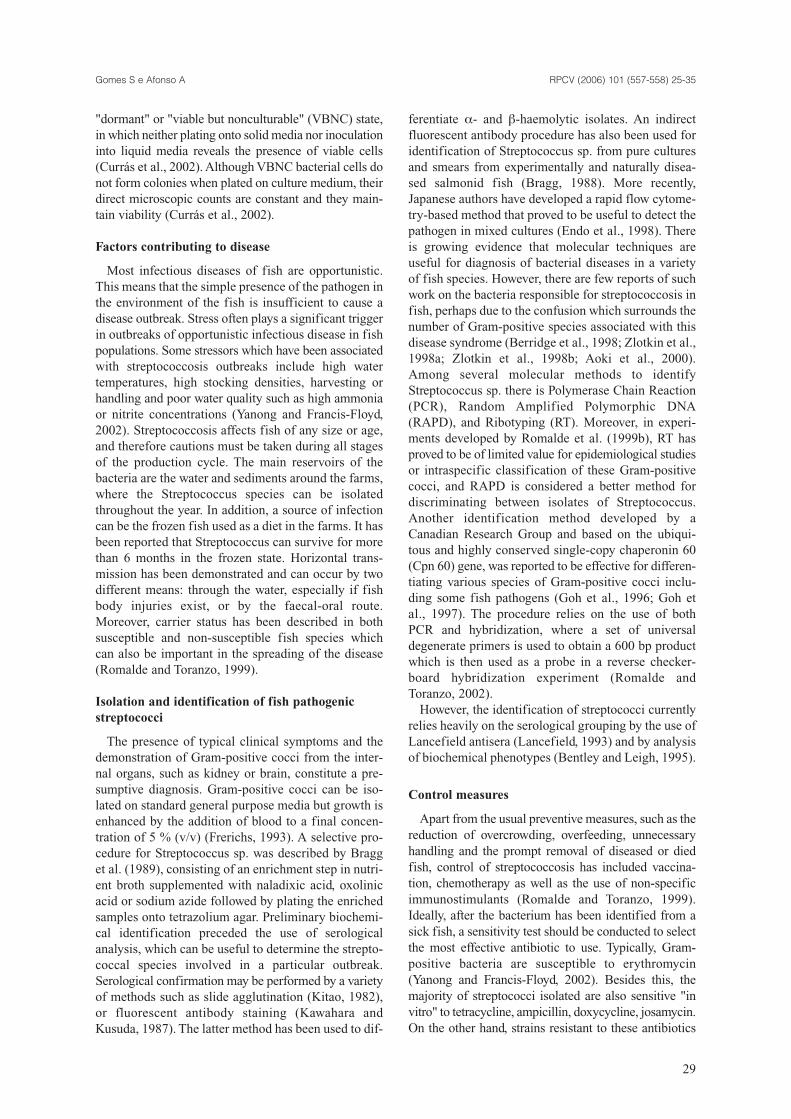
"dormant" or "viable but nonculturable" (VBNC) state,in which neither plating onto solid media nor inoculationinto liquid media reveals the presence of viable cells(Currás et al., 2002). Although VBNC bacterial cells donot form colonies when plated on culture medium, theirdirect microscopic counts are constant and they main-tain viability (Currás et al., 2002).
Factors contributing to disease
Most infectious diseases of fish are opportunistic.This means that the simple presence of the pathogen inthe environment of the fish is insufficient to cause adisease outbreak. Stress often plays a significant triggerin outbreaks of opportunistic infectious disease in fishpopulations. Some stressors which have been associatedwith streptococcosis outbreaks include high watertemperatures, high stocking densities, harvesting orhandling and poor water quality such as high ammoniaor nitrite concentrations (Yanong and Francis-Floyd,2002). Streptococcosis affects fish of any size or age,and therefore cautions must be taken during all stagesof the production cycle. The main reservoirs of thebacteria are the water and sediments around the farms,where the Streptococcus species can be isolatedthroughout the year. In addition, a source of infectioncan be the frozen fish used as a diet in the farms. It hasbeen reported that Streptococcus can survive for morethan 6 months in the frozen state. Horizontal trans-mission has been demonstrated and can occur by twodifferent means: through the water, especially if fishbody injuries exist, or by the faecal-oral route.Moreover, carrier status has been described in bothsusceptible and non-susceptible fish species whichcan also be important in the spreading of the disease(Romalde and Toranzo, 1999).
Isolation and identification of fish pathogenicstreptococci
The presence of typical clinical symptoms and thedemonstration of Gram-positive cocci from the inter-nal organs, such as kidney or brain, constitute a pre-sumptive diagnosis. Gram-positive cocci can be iso-lated on standard general purpose media but growth isenhanced by the addition of blood to a final concen-tration of 5 % (v/v) (Frerichs, 1993). A selective pro-cedure for Streptococcus sp. was described by Bragget al. (1989), consisting of an enrichment step in nutri-ent broth supplemented with naladixic acid, oxolinicacid or sodium azide followed by plating the enrichedsamples onto tetrazolium agar. Preliminary biochemi-cal identification preceded the use of serological analysis, which can be useful to determine the strepto-coccal species involved in a particular outbreak.Serological confirmation may be performed by a varietyof methods such as slide agglutination (Kitao, 1982),or fluorescent antibody staining (Kawahara andKusuda, 1987). The latter method has been used to dif-
ferentiate α- and β-haemolytic isolates. An indirectfluorescent antibody procedure has also been used foridentification of Streptococcus sp. from pure culturesand smears from experimentally and naturally disea-sed salmonid fish (Bragg, 1988). More recently,Japanese authors have developed a rapid flow cytome-try-based method that proved to be useful to detect thepathogen in mixed cultures (Endo et al., 1998). Thereis growing evidence that molecular techniques areuseful for diagnosis of bacterial diseases in a varietyof fish species. However, there are few reports of suchwork on the bacteria responsible for streptococcosis infish, perhaps due to the confusion which surrounds thenumber of Gram-positive species associated with thisdisease syndrome (Berridge et al., 1998; Zlotkin et al.,1998a; Zlotkin et al., 1998b; Aoki et al., 2000).Among several molecular methods to identifyStreptococcus sp. there is Polymerase Chain Reaction(PCR), Random Amplified Polymorphic DNA(RAPD), and Ribotyping (RT). Moreover, in experi-ments developed by Romalde et al. (1999b), RT hasproved to be of limited value for epidemiological studiesor intraspecific classification of these Gram-positivecocci, and RAPD is considered a better method fordiscriminating between isolates of Streptococcus.Another identification method developed by aCanadian Research Group and based on the ubiqui-tous and highly conserved single-copy chaperonin 60(Cpn 60) gene, was reported to be effective for differen-tiating various species of Gram-positive cocci inclu-ding some fish pathogens (Goh et al., 1996; Goh etal., 1997). The procedure relies on the use of bothPCR and hybridization, where a set of universaldegenerate primers is used to obtain a 600 bp productwhich is then used as a probe in a reverse checker-board hybridization experiment (Romalde andToranzo, 2002).
However, the identification of streptococci currentlyrelies heavily on the serological grouping by the use ofLancefield antisera (Lancefield, 1993) and by analysisof biochemical phenotypes (Bentley and Leigh, 1995).
Control measures
Apart from the usual preventive measures, such as thereduction of overcrowding, overfeeding, unnecessaryhandling and the prompt removal of diseased or diedfish, control of streptococcosis has included vaccina-tion, chemotherapy as well as the use of non-specificimmunostimulants (Romalde and Toranzo, 1999).Ideally, after the bacterium has been identified from asick fish, a sensitivity test should be conducted to selectthe most effective antibiotic to use. Typically, Gram-positive bacteria are susceptible to erythromycin(Yanong and Francis-Floyd, 2002). Besides this, themajority of streptococci isolated are also sensitive "invitro" to tetracycline, ampicillin, doxycycline, josamycin.On the other hand, strains resistant to these antibiotics
RPCV (2006) 101 (557-558) 25-35Gomes S e Afonso A
29
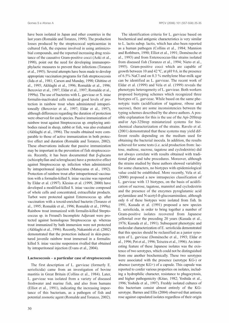
have been isolated in Japan and other countries in thelast years (Romalde and Toranzo, 1999). The productionlosses produced by the streptococcal septicaemias incultured fish, the expense involved in using antimicro-bial compounds, and the reported increasing drug resis-tance of the causative Gram-positive cocci (Aoki et al.,1990), point out the need for developing immunopro-phylactic measures to prevent these infections (Toranzoet al., 1995). Several attempts have been made to developappropriate vaccination programs for fish streptococcosis(Iida et al., 1981; Carson and Munday, 1990; Ghittino etal., 1995; Akhlaghi et al., 1996; Romalde et al., 1996;Bercovier et al., 1997; Eldar et al., 1997; Romalde et al.,1999a). The use of bacterins with L. garvieae or S. iniaeformalin-inactivated cells rendered good levels of pro-tection in rainbow trout when administered intraperi-toneally (Bercovier et al., 1997; Eldar et al., 1997),although differences regarding the duration of protectionwere observed for each species. Passive immunization ofrainbow trout against Streptococcus sp. employing anti-bodies raised in sheep, rabbit or fish, was also evaluated(Akhlaghi et al., 1996). The results obtained were com-parable to those of active immunization in both protec-tive effect and duration (Romalde and Toranzo, 2002).These observations indicate that passive immunizationmay be important in the prevention of fish streptococco-sis. Recently, it has been documented that β-glucans(schizophyllan and scleroglucan) have a protective effectagainst Streptococcus sp. infection when administeredby intraperitoneal injection (Matsuyama et al., 1992).Protection of rainbow trout after intraperitoneal vaccina-tion with a formalin-killed S. iniae vaccine was reportedby Eldar et al. (1997). Klesius et al. (1999, 2000) havedeveloped a modified-killed S. iniae vaccine composedof whole cells and concentrated, extracellular products.Turbot were protected against Enterococcus sp. aftervaccination with a toxoid-enriched bacterin (Toranzo etal., 1995; Romalde et al., 1996; Romalde et al., 1999a).Rainbow trout immunized with formalin-killed Strepto-coccus sp. in Freund’s Incomplete Adjuvant were pro-tected against homologous Streptococcus sp. whereastrout immunized by bath immersion were not protected(Akhlaghi et al., 1996). Recently, Nakanishi et al. (2002)demonstrated that the protection induced in skin-punc-tured juvenile rainbow trout immersed in a formalin-killed S. iniae vaccine suspension rivalled that obtainedby intraperitoneal injection (Evans et al., 2004).
Lactococcosis – a particular case of streptococcosis
The first description of L. garvieae (formerly E.seriolicida) came from an investigation of bovinemastitis in Great Britain (Collins et al., 1984). Later,L. garvieae was isolated from a variety of diseasedfreshwater and marine fish, and also from humans(Elliot et al., 1991), indicating the increasing impor-tance of this bacterium, as a pathogen of fish andpotential zoonotic agent (Romalde and Toranzo, 2002).
The identification criteria for L. garvieae based onbiochemical and antigenic characteristics is very similarto L. lactis subsp. lactis, which has also been reportedas a human pathogen (Collins et al., 1984; Mannionand Rothburn, 1990; Elliot et al., 1991; Doméneche etal., 1993) and from Enterococcus-like strains isolatedfrom diseased fish (Toranzo et al., 1994; Nieto et al.,1995). Gram-positive cocci which are capable ofgrowth between 10 and 42 ºC, at pH 9.6, in the presenceof 6.5% NaCl and on 0.3 % methylene blue-milk agarcan be identified as L. garvieae. The recent work ofEldar et al. (1999) and Vela et al. (1999) reveals thephenotypic heterogeneity of L. garvieae. Both workersproposed biotyping schemes which recognized threebiotypes of L. garvieae. While based on the same phe-notypic traits (acidification of tagatose, ribose andsucrose), there are some inconsistencies between thetyping schemes described by the above authors. A pos-sible explanation for this is the use of the Api-20Strepand/or Api-32Strep miniaturized systems for bio-chemical characterization of the strains. Ravelo et al.(2001) demonstrated that these systems may yield dif-ferent results depending on the medium used forobtaining the bacterial inocula. In addition, the resultsachieved for some tests (i.e. acid production from: lac-tose, maltose, sucrose, tagatose and cyclodextrin) didnot always correlate with results obtained with tradi-tional plate and tube procedures. Moreover, althoughthe strains studied by these authors showed variabilityfor some characters, no biotypes with epidemiologicalvalue could be established. More recently, Vela et al.(2000) proposed a new intraspecies classification of L. garvieae with 13 biotypes, on the basis of acidifi-cation of sucrose, tagatose, mannitol and cyclodextrinand the presence of the enzymes pyroglutamic acidarylamidase and N-acetyl-ß-glucosaminidase, althoughonly 6 of these biotypes were isolated from fish. In1991, Kusuda et al. (1991) proposed a new species E. seriolicida, in order to bring together a number ofGram-positive isolates recovered from Japanese yellowtail over the preceding 20 years (Kusuda et al.,1976; Kusuda et al., 1991). Subsequent phenotypic andmolecular characterization of E. seriolicida demonstratedthat this species should be reclassified as a junior syno-nym of L. garvieae (Doméneche et al., 1993; Eldar etal., 1996; Pot et al., 1996; Teixeira et al., 1996). An inter-esting feature of these Japanese isolates was the exis-tence of two serotypes, which could not be distinguishedfrom one another biochemically. These two serotypeswere associated with the presence (serotype KG-) orabsence (serotype KG+) of a capsule. This capsule wasreported to confer various properties on isolates, includ-ing a hydrophilic character, resistance to phagocytosis,and higher pathogenicity (Kitao, 1982; Yoshida et al.,1996; Yoshida et al., 1997). Freshly isolated cultures ofthis bacterium consist almost entirely of the KG-serotype. Barnes and Ellis (2004) observed that antiserarose against capsulated isolates regardless of their origin
RPCV (2006) 101 (557-558) 25-35Gomes S e Afonso A
30
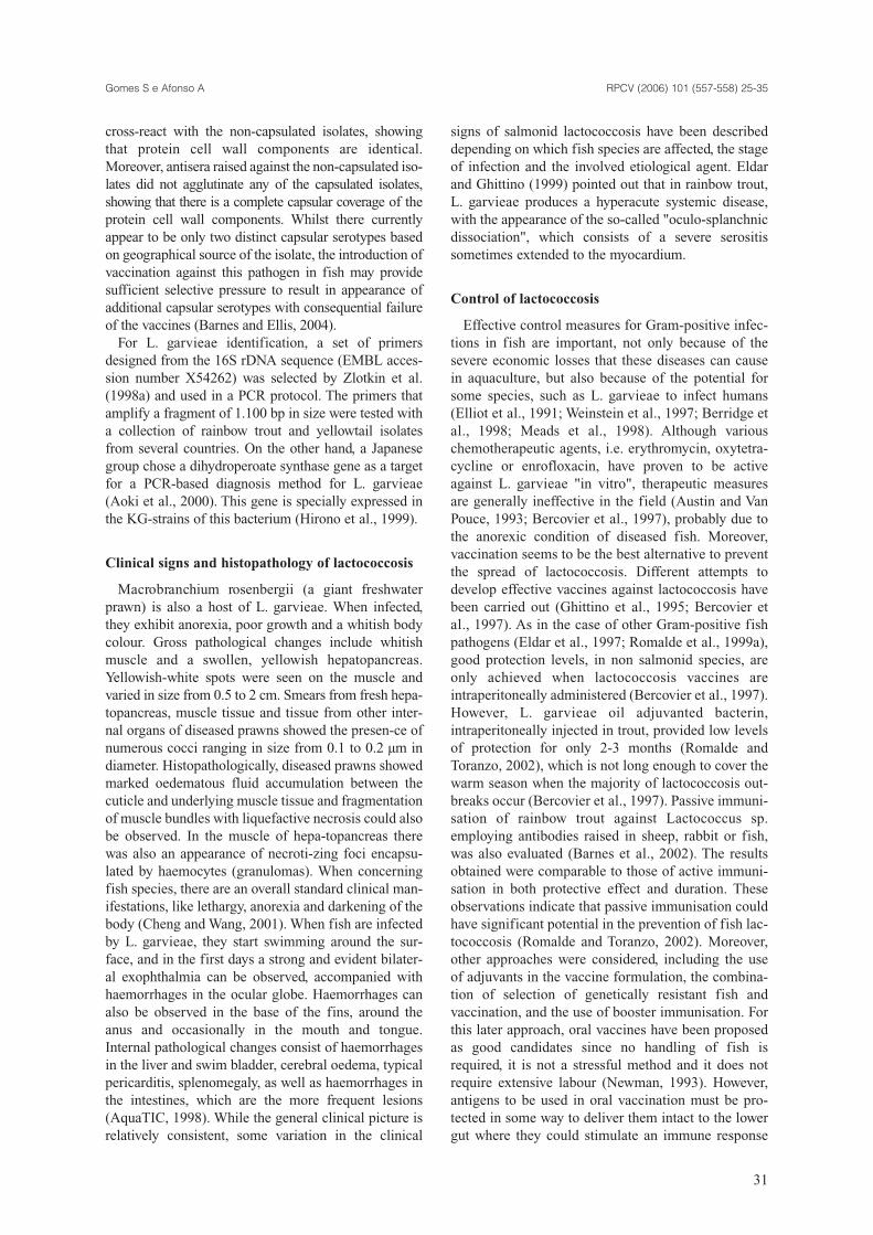
cross-react with the non-capsulated isolates, showingthat protein cell wall components are identical.Moreover, antisera raised against the non-capsulated iso-lates did not agglutinate any of the capsulated isolates,showing that there is a complete capsular coverage of theprotein cell wall components. Whilst there currentlyappear to be only two distinct capsular serotypes basedon geographical source of the isolate, the introduction ofvaccination against this pathogen in fish may providesufficient selective pressure to result in appearance ofadditional capsular serotypes with consequential failureof the vaccines (Barnes and Ellis, 2004).
For L. garvieae identification, a set of primersdesigned from the 16S rDNA sequence (EMBL acces-sion number X54262) was selected by Zlotkin et al.(1998a) and used in a PCR protocol. The primers thatamplify a fragment of 1.100 bp in size were tested witha collection of rainbow trout and yellowtail isolatesfrom several countries. On the other hand, a Japanesegroup chose a dihydroperoate synthase gene as a targetfor a PCR-based diagnosis method for L. garvieae(Aoki et al., 2000). This gene is specially expressed inthe KG-strains of this bacterium (Hirono et al., 1999).
Clinical signs and histopathology of lactococcosis
Macrobranchium rosenbergii (a giant freshwaterprawn) is also a host of L. garvieae. When infected,they exhibit anorexia, poor growth and a whitish bodycolour. Gross pathological changes include whitishmuscle and a swollen, yellowish hepatopancreas.Yellowish-white spots were seen on the muscle andvaried in size from 0.5 to 2 cm. Smears from fresh hepa-topancreas, muscle tissue and tissue from other inter-nal organs of diseased prawns showed the presen-ce ofnumerous cocci ranging in size from 0.1 to 0.2 µm indiameter. Histopathologically, diseased prawns showedmarked oedematous fluid accumulation between thecuticle and underlying muscle tissue and fragmentationof muscle bundles with liquefactive necrosis could alsobe observed. In the muscle of hepa-topancreas therewas also an appearance of necroti-zing foci encapsu-lated by haemocytes (granulomas). When concerningfish species, there are an overall standard clinical man-ifestations, like lethargy, anorexia and darkening of thebody (Cheng and Wang, 2001). When fish are infectedby L. garvieae, they start swimming around the sur-face, and in the first days a strong and evident bilater-al exophthalmia can be observed, accompanied withhaemorrhages in the ocular globe. Haemorrhages canalso be observed in the base of the fins, around theanus and occasionally in the mouth and tongue.Internal pathological changes consist of haemorrhagesin the liver and swim bladder, cerebral oedema, typicalpericarditis, splenomegaly, as well as haemorrhages inthe intestines, which are the more frequent lesions(AquaTIC, 1998). While the general clinical picture isrelatively consistent, some variation in the clinical
signs of salmonid lactococcosis have been describeddepending on which fish species are affected, the stageof infection and the involved etiological agent. Eldarand Ghittino (1999) pointed out that in rainbow trout,L. garvieae produces a hyperacute systemic disease,with the appearance of the so-called "oculo-splanchnicdissociation", which consists of a severe serositissometimes extended to the myocardium.
Control of lactococcosis
Effective control measures for Gram-positive infec-tions in fish are important, not only because of thesevere economic losses that these diseases can causein aquaculture, but also because of the potential forsome species, such as L. garvieae to infect humans(Elliot et al., 1991; Weinstein et al., 1997; Berridge etal., 1998; Meads et al., 1998). Although variouschemotherapeutic agents, i.e. erythromycin, oxytetra-cycline or enrofloxacin, have proven to be activeagainst L. garvieae "in vitro", therapeutic measuresare generally ineffective in the field (Austin and VanPouce, 1993; Bercovier et al., 1997), probably due tothe anorexic condition of diseased fish. Moreover,vaccination seems to be the best alternative to preventthe spread of lactococcosis. Different attempts todevelop effective vaccines against lactococcosis havebeen carried out (Ghittino et al., 1995; Bercovier etal., 1997). As in the case of other Gram-positive fishpathogens (Eldar et al., 1997; Romalde et al., 1999a),good protection levels, in non salmonid species, areonly achieved when lactococcosis vaccines areintraperitoneally administered (Bercovier et al., 1997).However, L. garvieae oil adjuvanted bacterin,intraperitoneally injected in trout, provided low levelsof protection for only 2-3 months (Romalde andToranzo, 2002), which is not long enough to cover thewarm season when the majority of lactococcosis out-breaks occur (Bercovier et al., 1997). Passive immuni-sation of rainbow trout against Lactococcus sp.employing antibodies raised in sheep, rabbit or fish,was also evaluated (Barnes et al., 2002). The resultsobtained were comparable to those of active immuni-sation in both protective effect and duration. Theseobservations indicate that passive immunisation couldhave significant potential in the prevention of fish lac-tococcosis (Romalde and Toranzo, 2002). Moreover,other approaches were considered, including the useof adjuvants in the vaccine formulation, the combina-tion of selection of genetically resistant fish and vaccination, and the use of booster immunisation. Forthis later approach, oral vaccines have been proposedas good candidates since no handling of fish isrequired, it is not a stressful method and it does notrequire extensive labour (Newman, 1993). However,antigens to be used in oral vaccination must be pro-tected in some way to deliver them intact to the lowergut where they could stimulate an immune response
RPCV (2006) 101 (557-558) 25-35Gomes S e Afonso A
31
(Horne, 1997). Preliminary results obtained byRomalde et al. (2004) showed that antigen encapsula-tion in alginate appears to be a promising method fororal vaccination of trout against L. garvieae, at least asa booster immunisation procedure.
Two decades have elapsed since bacteriophages(phages) were reassessed scientifically as a therapeuticand prophylactic agent for bacterial infections (Nakaiand Park, 2002). Potential advantages of phage treat-ment over chemotherapy are: 1) the narrow host rangeof phages, indicating that the phages do not harm thenormal intestinal microflora; and 2) the self-perpetuatingnature of phages in the presence of susceptible bacteria,indicating the superfluousness of multiple administra-tions (Smith and Huggins, 1982; Barrow and Soothill,1997). However, experiments using phage therapy areso far scarce. "In vitro" studies revealed that the L.garvieae phages are tolerant to physicochemical factorssuch as water temperature ranging from 5 to 37 ºC andsalinity varying from distilled water to double-strengthartificial seawater (Nakai et al., 1999). Moreover, it wasdemonstrated of that intraperitoneal injection of thephage in treating young yellowtail intraperitoneallyinjected with L. garvieae was effective. The resultsshow that phage administration might be useful as atherapeutic measure at an early stage of systemic infec-tion or with low burdens of bacterial infection in yel-lowtail (Nakai et al., 1999). If phages are introduced byoral administration into the intestine of fish where viru-lent L. garvieae cells adhere or commence to multiply,it is expected that the phage will attack the pathogen(Nakai et al., 1999).
The knowledge of a novel vaccine against lactococco-sis is worthwhile, not only to prevent the disseminationof such an infection, but also to prevent the destructionof cultured stocks of rainbow trout, among others. Theprolonged destruction of the stocks will lead in a forth-coming future to the closure of several aquacultures.
We all hope that from now on, there will be anincrease in the importance given to this emergingpathological problem, which seems to be hidden byother neither less nor more important bacterial diseases.
Bibliography
Afonso A, Gomes SA, da Silva J, Marques F and Henrique M (2004). Side-effects in sea bass (Dicentrachus labrax L.) due to intraperitoneal vaccination against vibriosis and pas-teurellosis. Fish & Shellfish Immunology (in press),
Akhlaghi M, Munday BL and Whittington RJ (1996). Comparison of passive and active immunization of fish against streptococcosis (enterococcosis). Journal of Fish Diseases, 19: 251-258.
Al-Harbi AL (1994). First isolation of Streptococcus sp. from hybrid tylapia (Oreochromis niloticus x O. aureus) in Saudi Arabia. Aquaculture, 128: 195-201.
Anderson DP (1997). Adjuvants and Immunostimulants for
Enhancing Vaccine Potency in Fish. In: Adjuvants and Immunostimulants for Enhancing Vaccine Potency in FishDev. Biol. Stand., 257-265.
Aoki T, Takami K and Kitao T (1990). Drug resistance in a non-hemolytic Streptococcus sp isolated from cultured yellowtail Seriola quinqueradiata. Diseases of Aquatic Organisms, 8: 171-177.
Aoki T, Park C-I, Yamashita H and Hirono I (2000). Species-specific polymerase chain reaction primers for Lactococ-cus garvieae. Journal of Fish Diseases, 23: 1-6.
AquaTIC RR (1998). La Estreptococosis de la Trucha enEspaña.www.revistaaquatic.com/aquatic/art.asp?t=h&c=21, 2: Austin B and Van Pouce A (1993). A gram-positive"lactic-acid" bacterium causing steady mortalities in farmed rainbow trout, Oncorhynchus mykiss Walbaum, in the UK. Bulletin of the European Association of Fish Pathologists, 13: 114-118.
Austin B and Austin DA (1999). Bacterial Fish Pathogens: Disease in Farmed and Wild Fish. In: Bacterial Fish Pathogens: Disease in Farmed and Wild FishB Austin and DA Austin. Ellis Honwood Ltd.,
Babuik LA, van Drunen Littel-van den Hurk S, Tikoo PJ and Liang X (1996). Veterinary Immunology and Immuno-pathology, 54: 355.
Barnes AC and Ellis AE (2004). Role of capsule in serotypic differences and complement fixation by Lactococcus garvieae. Fish & Shellfish Immunology, 16: 207-214.
Barnes AC, Guyot C, Hansen BG, Horne MT and Ellis AE (2002). Antibody increases phagocytosis and killing of Lactococcus garvieae by rainbow trout (Oncorhynchus mykiss, L.) macrophages. Fish & Shellfish Immunology, 12: 181-186.
Barrow PA and Soothill JS (1997). Bacteriophage therapy and prophylaxis: rediscovery and renewed assessment of poten-tial. Trends in Microbiology, 5: 268-271.
Benmansour A and de Kinkelin P (1997). Fish Vaccinology In: Developments of Biological Standardization, 153.
Bentley RW and Leigh JA (1995). Development of PCRbased hybridization protocol for identification of strepto-coccal species. Journal of Clinical Microbiology, 33: 1296-1301.
Bercovier H, Ghittino C and Eldar A (1997). Immunization with bacterial antigens: infections with streptococci and relatedorganisms. Dev Biol Stand, 90: 153-160.
Berridge BR, Fuller JD, Azavedo J, Low DE, Bercovier H and Frelier PF (1998). Development of specific nested oligonu-cleotide PCR primers for the Streptococcus iniae 16S-23S ribossomal DNA intergenic spacer. Journal of Clinical Microbiology, 36: 2778-2781.
Bragg RR (1988). The indirect fluorescent antibody tech-nique for the rapid identification of streptococcosis of rain-bow trout (Salmo gairdneri). Onderstepoort J. Vet. Res., 55: 59-61.
Bragg RR, Todd JM, Lordan SM and Combrink ME (1989). A selective procedure for the field isolation of pathogenic Streptococcus spp. of rainbow trout (Salmo gairdneri). Onderstepoort J. Vet. Res., 56: 179-184.
Carson J and Munday B (1990). Streptococcosis-an emerging disease in aquaculture. Australian Aquaculture, 5: 32-33.
Cheng W and Wang C (2001). The susceptibility of the giant freshwater prawn Macrobrachium rosenbergii to Lacto-coccus garvieae and its resistance under copper sulfate stress. Diseases of Aquatic Organisms, 47: 137-144.
RPCV (2006) 101 (557-558) 25-35Gomes S e Afonso A
32
Collins MD, Farrow JAE, Phillips BA and Kandler O (1984). Streptococcus garviae and Streptococcus plantarum sp. nov. Journal of General Microbiology, 129: 3427-3431.
Currás M, Magariños B, Toranzo AE and Romalde Jl (2002). Dormancy as a survival strategy of the fish pathogen Streptococcus parauberis in the marine environment. Diseases of Aquatic Organisms, 52: 129-136.
Dalmo RA, Ingebrigtsen K and Bogwald J (1997). Non-spe-cific defence mechanisms in fish, with particular reference to the reticuloendothelial system (RES). Journal of Fish Diseases, 20: 241-273.
Doméneche A, Prieta J, Fernandez-Garayzabal JF, Collins MD, Jones D and Domínguez L (1993). Phenotypic and phylogenetic evidence for a close relationship between Lactococcus garvieae and Enterococcus seriolicida. Microbiologia, 9: 63-68.
Eggset G, Mikkelsen H and Killie J-E, A. (1997). Immunocompetence and duration of immunity against Vibrio salmonicida and Aeromonas salmonicida and after vaccination of Atlantic salmon (Salmo salar L.) at low and high temperatures. Fish & Shellfish Immunology, 7: 247-260.
Eldar A and Ghittino C (1999). Lactococcus garvieae and Streptococcus iniae infections in rainbow trout Oncorhynchus mykiss: similar, but different diseases. Diseases of Aquatic Organisms, 36: 227-231.
Eldar A, Bejerano Y and Bercovier H (1994). Streptococcusshiloi and Streptococcus difficile: two new streptococcal species causing meningoencephalitis in fish. Current Microbiology, 28: 139-143.
Eldar A, Horovitcz A and Bercovier H (1997). Development and efficacy of a vaccine against Streptococcus iniae infec-tion in farmed rainbow trout. Vet Immunol Immunopathol, 56: 175-183.
Eldar A, Goria M, Ghittino C, Zlotkin A and Bercovier H (1999). Biodiversity of Lactococcus garvieae strains iso-lated from fish in Europe, Asia, and Australia. Applied and Environmental Microbiology, 65(3): 1005-1008.
Eldar A, Frelier PF, Assenta L, Varner PW, Lawhon S and Bercovier H (1995). Streptococcus shiloi, the name for an agent causing septicemic infection in fish, is a junior synonym of Streptococcus iniae. International Journal of Systematic Bacteriology, 45: 840-842.
Eldar A, Ghittino C, Asanta L, Bozzetta E, Gloria M, Prearo M and Bercovier H (1996). Enterococcus seriolicida is a junior synonym of Lactococcus garvieae, a causative agent of septicemia and meningoencephalitis in fish. Current Microbiology, 32: 85-88.
Elliot JA, Collins MD, Pigott NE and Facklam RR (1991). Differentiation of Lactococcus lactis and Lactococcus garvieae from human by comparison of whole-cell protein patterns. Journal of Clinical Microbiology, 29: 2731-2734.
Endo H, Nakayama J, Ushio H, Hayashi T and Watanabe E (1998). Application of flow cytometry for rapid detection of Lactococcus garvieae. Applied Biochemistry and Biotechnology, 75: 295-306.
Evans JJ, Klesius PH and Shoemaker CA (2004). Efficacy of Streptococcus agalactiae (group B) vaccine in tilapia (Oreochromis niloticus) by intraperitoneal and bath immer-sion administration. Vaccine (in Press),
Fänge R (1994). Blood cells, haemopoiesis and lym-phomyeloid tissues in fish. Fish & Shellfish Immunology, 4: 405-411.
Frerichs GN (1993). Isolation and identification of fish
bacterial pathogens. In: Isolation and identification of fish bacterial pathogens. Blackwell Scientific Publications, 257-286.
Frobisher M (1954). Spherical Bacteria. In: Spherical Bacteria.W. B. Saunders Company, 442-450.
Ghittino C (1999). Las Estreptococosis en los peces. Revista AquaTIC, 6:
Ghittino C, Prearo M, Bozzetta E and Eldar A (1995). Pathological characterization of the causative agent of fish streptococcosis in Italy and vaccination trials in rainbow trout. Boll.Soc. Ital. Pathol. Ittica, 16: 2-12.
Goh SH, Potter S, Wood JO, Hemmingsen SM, Reynolds RP and Chow AW (1996). HSP60 gene sequences as universal targets for microbial species identification: studies with coagulase-negative staphylococci. Journal of Clinical Microbiology, 34: 818-823.
Goh SH, Santucci Z, Kloos WE, Faltyn M, George CG, Driedger D and Hemmingsen SM (1997). Identification of Staphylococcus species ans subspecies by the chaperonin 60 gene identification method and reverse checkerboard hybridisation. Journal of Clinical Microbiology, 35: 3116-3121.
Gudding R, Lillehaug A and Evensen O (1999). Recent develop-ments in fish vaccinology. Veterinary Immunology and Immunopathology, 72: 203-212.
Hastein T (1995). Sustainable Fish Farming. In: Sustainable Fish FarmingH Reinersten and H Haaland. A.A. Balkema, 183.
Hirono I, Yamashita H, Park C, Yoshida T and Aoki T (1999). Identification of genes in a KG- phenotype of Lactococcus garvieae, a fish pathogenic bacterium, whose proteins react with anti-KG- rabbit serum. Microbial Pathogenesis, 27: 407-417.
Horne MT (1997). Technical aspects of the administration of vaccines. In: Technical aspects of the administration of vaccines, 79-89.
Horne MT and Robertson DA (1987). Economics of vaccina-tion against Enteric Redmouth disease of salmonids. Aquaculture of Fisheries Management, 18: 131-137.
Iida T, Wakabayashi H and Egusa S (1981). Vaccination for control of stretococcal disease in cultured yellowtail. Fish Pathology, 16: 201-206.
Iida T, Furukawa K, Sakai M and Wakabayashi H (1986). Non-haemolytic Streptococcus isolated from the brain of the vertebral deformed yellowtail. Fish Pathology, 21: 33-38.
Joosten PHM, Tiemersma E, Threels A, CaumartinDhieux C and Rombout JHWM (1997). Oral vaccination of fish against Vibrio anguillarum using alginate microparticles. Fish & Shellfish Immunology, 7: 471-485.
Kawahara E and Kusuda R (1987). Direct fluorescent anti-body technique for diagnosis of bacterial diseases in e el. Nippon Suisan Gakkaishi, 53: 395-399.
Kitao T (1982). The methods for detection of Streptococcussp. causative bacteria of streptococcal disease of cultured yellow-tail (Seriola quinqueradiata). Fish Pathology, 17: 17-26.
Kitao T (1993). Streptococcal infections. In: Streptococcal infections. Blackwell Scientific Publications, 196-210.
Klesius PH, Shoemaker CA and Evans JJ (1999). Efficacy of a killed Streptococcus iniae vaccine in tilapia (Oreochromis niloticus). Bulletin of the European Association of Fish Pathologists, 19: 38-41.
RPCV (2006) 101 (557-558) 25-35Gomes S e Afonso A
33
Klesius PH, Shoemaker CA and Evans JJ (2000). Efficacy of a single and combined Streptococcus iniae isolate vaccine administered by intraperitoneal and intramuscular routes in tilapia (Oreochromis niloticus). Aquaculture, 188: 237-246.
Kusuda R and Salati F (1993). Major bacterial diseases affecting mariculture in Japan. Annual Review of Fish Diseases, 69-85.
Kusuda R, Kawai K, Toyoshima T and Konatsu I (1976). A new pathogenic bacterium belonging to the genus Streptococcus, isolated from epizootic of cultured yellow-tail. Bulletin of the European Japanese Society Science and Fisheries, 42: 1345-1352.
Kusuda R, Kawai K, Salati F, Banner CR and Fryer JL (1991). Enterococcus seriolicida sp nov. a fish pathogen. International Journal of Systematic Bacteriology, 41: 406-409.
Lancefield RC (1993). A serological differentiation of human and other groups of haemolytic streptococci. Journal of Experimental Medicine, 57: 571-595.
Mannion PT and Rothburn MM (1990). Diagnosis of bacterialendocarditis caused by Streptococcus lactis and assisted by immunoblotting of serum antibodies. Journal of Infections, 21: 317-318.
Marsden MJ, Vaughan LM, Foster TJ and Secombes CJ (1996). Infection and Immunity, 64: 3863.
Matsuyama H, Mangindaan REP and Yano T (1992). Protective effect of schizophylian and scleroglucan against Streptococcus sp. infection in yellowtail (Seriola quin-queradiata). Aquaculture, 101: 197-203.
Meads SV, Shotts EB and Maurer J (1998). Biochemical and molecular typing of Streptococcus iniae isolates from fish and human cases. Proceedings of the 23rd Annual Eastern Fish Health Workshop. Plymouth,M.A., USA. 31
Medzhitov R and Janeway C (2000). Innate immunity. The New England Journal of Medicine, 343: 338-344.
Michel C, Nougayrède P, Eldar A, Sochon E and de Kinkelin P (1997). Vagococcus salmoninarum, a bacterium of patho-logical significance in rainbow trout Oncorhynchus mykiss farming. Diseases of Aquatic Organisms, 30: 199-208.
Midtlyng PJ (1996). A field study on intraperitoneal vaccination of Atlantic salmon (Salmo salar L.) against furunculosis. Fish & Shellfish Immunology, 6: 553-565.
Midtlyng PJ (1997). Vaccinated fish welfare: protection versus side-effects. In: Vaccinated fish welfare: protection versus side-effectsDevelopments in biological standardi--zation, 371-379.
Midtlyng PJ and Lillehaug A (1998). Growth of Atlantic salmon Salmo salar after intraperitoneal administration of vaccines containing adjuvants. Diseases of Aquatic Organisms, 32: 91-97.
Midtlyng PJ, Reitan LJ and Speilberg L (1996a). Experimental studies on the efficacy and side-effects of intraperitoneal vaccination of Atlantic salmon (Salmo salar L.) against furunculosis. Fish & Shellfish Immunology, 6: 335-350.
Midtlyng PJ, Reitan LJ, Lillehaug A and Ramstad A (1996b). Protection, immune response and side effects in Atlantic salmon (Salmo salar,L.) vaccinated against furunculosis by different procedures. Fish & Shellfish Immunology, 6:599-613.
Minami T, Nakamura M, Ikeda Y and Ozaki H (1979). A bea-hemolytic Streptococcus isolated from cultured yellowtail. Fish Pathology, 14: 33-38.
Miyazaki T (1982). Pathological study on streptococcicosis histopathology of infected fishes. Fish Pathology, 17: 39-47.Moore JD, Ototake M and Nakanishi T (1998). Fish & Shellfish Immunology, 8: 393.
Múzquiz JL, Royo FM, Ortega C, Blas I, Ruiz I and Alonso JL (1999). Pathogenicity of streptococcosis in rainbow trout (Oncorhynchus mykiss): Dependence on age of diseased fish. Bull. Eur. Ass. Fish Pathol., 19(3): 114-119.
Nakai T and Park SC (2002). Bacteriophage therapy of infec-tious diseases in aquaculture. Research in Microbiology, 153: 13-18.
Nakai T, Sugimoto R, Park KH, Matsuoka S, Mori K, Nishioka T and Maruyama K (1999). Protective effects of bacteriophage on experimental Lactococcus garvieaeinfection in yellowtail. Diseases of Aquatic Organisms, 37: 33-41.
Nakanishi T and Ototake M (1997). In: Developments in Biological Standardization, 59.
Nakanishi T, Kiryu I and Ototake M (2002). Development of a new vaccine delivery method for fish: percutaneous administation by immersion with application of a multiple-puncture instrument. Vaccine, 20: 3764-3769.
Newman SG (1993). Bacterial vaccines for fish. Annual Review of Fish Diseases, 3: 145-185.
Nieto JM, Devesa S, Quiroga I and Toranzo AE (1995). Pathology of Enterococcus sp. infection in farmed turbot, Scophthalmus maximus L. Journal of Fish Diseases, 18: 21-30.
Padrós F and Furones MD (2002). Patología bacteriana en piscicultura. Actualidad, 13-21.
Perera RP, Johnson SK, Collins MD and Lewis DH (1995). Streptococcus iniae associated with mortality of Tilapia nilotica x T. aurea hybrids. Journal of Aquatic Animal Health, 6: 335-340.
Plumb JA, Schachte JH, Gaines JL, Peltier W and Carrol B (1974). Streptococcus sp. from marine fishes along the Alabama and northwest Florida coast of the gulf of Mexico. Transactions of the American Fisheries Society, 103: 358-361.
Poppe TT and Breck O (1997). Pathology of Atlantic salmon Salmo salar intraperitoneally immunized with oil-adjuvantedvaccine. A case report. Diseases of Aquatic Organisms, 29: 219-226.
Pot B, Devriese LA, Ursi D, Vandamme P, Haesebrouck F and Kersters K (1996). Phenotypic identification and differen-tiation of Lactococcus strains isolated from animals. Systematic and Applied Microbiology, 19: 213-222.
Quentel C and Vigneulle M (1997). In: Developments of Biological Standardization, 69.
Ravelo C, Magariños B, Romalde Jl and Toranzo AE (2001). Conventional versus miniaturized systems for the phenotypic characterization of Lactococcus garvieae strains. Bulletin of the European Association of Fish Pathologists, 21: 136-143.
Reitan LJ and Secombes CJ (1997). In: Developments of Biological Standardization, 293.
Ringo E and Gatesoupe FJ (1998). Lactic acid bacteria in fish: a review. Aquaculture, 160: 177-203.
Robinson JA and Meyer FP (1966). Streptococcal fish pathogen. Journal of Bacteriology, 92: 512.
Romalde Jl and Toranzo AE (1999). Streptococcosis of Marine Fish. In: Streptococcosis of Marine Fish.
RPCV (2006) 101 (557-558) 25-35Gomes S e Afonso A
34
International Council for the Exploration of the Sea, 56.Romalde JL and Toranzo AE (2002). Molecular Approaches
For The Study and Diagnosis of Salmonid Streptococcosis. In: Molecular Approaches For The Study and Diagnosis of Salmonid Streptococcosis, 56.
Romalde JL, Silva R and Toranzo AL (1996). Long-Lasting Protection Against Turbot Streptococcosis Obtained with a Toxoid-Enriched Bacterin.
Romalde Jl, Magariños B and Toranzo AE (1999a). Prevention of streptococcosis in turbot by intraperitoneal vaccination: a review. Journal of Applied Ichtyology, 15: 153-158.
Romalde JL, Magarinos B, Villar C, Barja JL and Toranzo AE (1999b). Genetic analysis of turbot pathogenic Streptococcus parauberis strains by ribotyping and random amplified polymorphic DNA. FEMS Microbiol Lett, 179: 297-304.
Romalde JL, Luzardo-Alvárez A, Ravelo C, Toranzo AE and Blanco-Méndez J (2004). Oral immunization using algi-nate microparticles as a useful strategy for booster vaccination against fish lactococcosis. Aquaculture, 236: 119-129.
Rombout JHWM and Joosten EPHM (1992). Mucosal immunity in fish. In: Mucosal immunity in fish., 1-12.
Smith HW and Huggins MB (1982). Successful treatment of experimental Escherichia coli infections in mice using phage: its general superiority over antibiotics. Journal of General Microbiology, 128: 307-318.
Stevenson RMW (1997). In: Developments of Biological Standardization, 117.
Stoffregen DA, Backman SC, Perham RE, Bowser PR and Babish JG (1996). Initial disease report of Streptococcus iniae infection in hybrid striped (sunshine) bass and suc-cessful therapeutic intervention with the fluoroquinolone antibacterial enrofloxacin. Journal of World Aquacult. Soc., 27: 420-434.
Suzuki Y and Iida T (1992). Fish granulocytes in the process of inflammation. Annual Review of Fish Diseases, 1: 149-160.
Teixeira LM, Merquior VLC, Vianni MCE, Carvalho MGS, Fracalanzza SEL, Steigerwalt AG, Brenner DJ and Facklam RR (1996). Phenotypic and genotypic characterization of atypical Lactococcus garvieae strains isolated from water buffalos with subclinical mastitis and confirmation of L. garvieae as a senior subjective synonym of Enterococcus seriolcida. International Journal of Systematic Bacteriology, 46: 664-668.
Toranzo AE, Santos Y and Barja JL (1997). In: Developments Biological Standardization, 93.
Toranzo AE, Devesa S, Heinen P, A., R, Núñez S and Barja JL (1994). Stretococcosis in cultured turbot caused by an Enterococcus-like bacterium. Bulletin of the European Association of Fish Pathologists, 14: 19-23.
Toranzo AE, Devesa S, Romalde JL, Lamas J, Riaza A, Leiro J and Barja JL (1995). Efficacy of intraperitoneal and immersion vaccination against Enterococcus sp. infection in tubot. Aquaculture, 134: 15-27.
Vanrobaeys M, De Herdt P, Ducatelle R, Creten W and Haesebrouck F (1997). Extracellular proteins and virulence in Streptococcus bovis isolates from pigeons. Veterinary Microbiology, 59: 59-66.
Vela AI, Vásquez J, Gibello A, Blanco MM, Moreno MA, Liébana P, Albendea C, Alcalá B, Mendez A, Domínguez L and Fernández-Garayzábal JF (2000). Phenotypic and genetic characterization of Lactococcus garvieae isolated in Spain from Lactococcosis outbreaks and comparison with isolates of other countries and sources. Journal of Clinical Microbiology, 38: 3791-3795.
Vela I, Liebana P, Albendea C, Gibello A, Blanco M, Moreno M, Cutúli M, García JA, Doméneche A, Domínguez L, Vázquez J and Fernandéz-Garayzával JF (1999). Caracterización fenotípica y molecular de Lactococcus garvieae. Proceedings of the XIII Congreso Nacional de Microbiologia de la Sociedad Española de Microbiologia. Granada, Spain. 47
Ward PD (1982). The development of bacterial vaccines. Microbial Diseases of Fish, 47-58.
Weinstein M, Low D and McGeer A (1996). Invasive infec-tion due to Streptococcus iniae: a new or previously unrecognised disease - Ontario1995-1996. In: Invasive infection due to Streptococcus iniae: a new or previously unrecognised disease - Ontario1995-1996. Publisher, 129-132.
Weinstein MR, Litt M, Kertesz DA, Wyper P, Rose D, Coulter M, NcGeer A, Facklam R, Ostach D, Willey BM, Borczyk A and Low DE (1997). Invasive infection due to a fish pathogen, Streptococcus iniae. New Engl. J. Med., 337: 589-594.
Yanong RPE and Francis-Floyd R (2002). Streptococcal infections of fish. Report from University of Florida.
Yoshida T, Endo M, Sakai M and Inglis V (1997). A cell cap-sule with possible involvement in resistance to opsonophagocytosis in Enterococcus seriolicida from yellowtail, Seriola quinqueradiata. Diseases of Aquatic Organisms, 29: 233-235.
Yoshida T, Eshima T, Wada Y, Yamada Y, Kakizaki E, Sakai M, Kitao T and Inglis V (1996). Phenotypic variation asso-ciated with an antiphagocytic factor in the bacterial fish pathogen Enterococcus seriolicida. Diseases of Aquatic Organisms, 25: 81-86.
Zlotkin A, Hersho H and Eldar A (1998a). Possible transmissionof Streptococcus iniae from wild fish to cultured marine fish. Applied and Environmental Microbiology, 64: 4065-4067.
Zlotkin A, Eldar A, Ghittino C and Bercovier H (1998b). Identification of Lactococcus gravieae by PCR. Journal of Clinical Microbiology, 36: 983-985.
RPCV (2006) 101 (557-558) 25-35Gomes S e Afonso A
35
Journal of Applied Aquaculture, 23:299–303, 2011Copyright © Taylor & Francis Group, LLCISSN: 1045-4438 print/1545-0805 onlineDOI: 10.1080/10454438.2011.626361
Dietary L-Carnitine Supplementation ProtectedTurbot (Scophthalmusmaximus) Against
Streptococcus parauberis Infection
S. DIAS1, R. O. A. OZÓRIO2, J. GONÇALVES1,2, and F. GÄRTNER1,3
1Instituto de Ciências Biomédicas Abel Salazar, ICBAS,Universidade do Porto, Porto, Portugal
2CIMAR/CIIMAR, Centro Interdisciplinar de InvestigaçãoMarinha e Ambiental,Universidade do Porto, Porto, Portugal
3Instituto de Patologia e Imunologia Molecular daUniversidade do Porto,IPATIMUP, Porto, Portugal
The protective effect of dietary L-carnitine supplementation wasaccessed in turbot (Scophthalmusmaximus) challenged withStreptococcus parauberis. Infected fish fed 240mg L-carnitineshowed significantly (P < 0.01) higher survival (78%) thaninfected fish fed 40mg L-carnitine (11%). When dietary L-carnitinesupplementation was suspended, a significant increase in mortal-ity was observed in infected fish. The present report documentsthat dietary L-carnitine fed on a continuous basis may act as animmunostimulant in turbot.
KEYWORDS L-carnitine, turbot, immunostimulant
INTRODUCTION
In Portugal, turbot (Scophthalmusmaximus) aquaculture accounts for morethan 13% of the total marine aquaculture production (Food and AgricultureOrganization 2002). Streptococcus spp. are responsible for more deaths inmarine aquaculture than any other gram-positive bacterial group (Groberg,Rohovec, & Fryer 1983; Abruzzini, Ingram, & Clem 1982; Toranzo et al.
The authors would like thank Dr. Jesús Romalde from University of Santiago deCompostela for supplying the S. parauberis (RA 83.1) strain.
Address correspondence to S. Dias, Instituto de Ciências Biomédicas Abel Salazar, ICBAS,Universidade do Porto, Largo Professor Abel Salazar, 2, Porto, 4099-003, Portugal. E-mail:[email protected]
299
Dow
nloa
ded
by [
Sóni
a D
ias]
at 0
1:07
21
Nov
embe
r 20
11
300 S. Dias et al.
1994). Streptococcus parauberis, for example, was responsible for heavyeconomic losses in turbot aquaculture in Galicia, Spain, in 1993.
L-carnitine, a multi-physiological bioactive additive, is not only knownto act as a growth-enhancer, but also appears to protect fish against xeno-biotic compounds and variations in water quality. It also increases immuno-competence against several bacterial diseases (Harpaz 2005; Ozorio 2009).
Deufel (1990) reported that leucocytes in human subjects, includingperipheral blood mononuclear cells, are rich in carnitine, suggesting thatL-carnitine and its congeners may modulate immune networks. That is inagreement with Arafa et al. (2003), who observed an increase of immuneresponse in rats following dietary L-carnitine supplementation.
The objective of this study was to evaluate the protective effectof dietary carnitine in turbot experimentally infected with Streptococcusparauberis and to determine if dietary supplementation should be on acontinuous basis.
MATERIALS AND METHODS
Turbot weighing 60 ± 5.0 g (from PisciculturaMarinha do Rio Alto, A.Coelho & Castro, Lda, Póvoa do Varzim, Portugal) were kept at 18–22◦C ina recirculating aerated saltwater (35%) system at Centro Interdisciplinar deInvestigação Marinha e Ambiental (CIIMAR) in Porto, Portugal. Water qual-ity was maintained with mechanical and biological filtration. Turbot weredivided into four groups of 60 fish each (30 fish for each of two replicatedgroups) and were fed daily ad libitum for 16 weeks on one of two nutri-tionally identical diets (crude protein, 39%; crude fat, 13%; gross energy,19.5 kJ/g), differing only in dietary L-carnitine content (40 or 240 mg/kgdiet). The duration of the feeding trial was chosen as the necessary timefor the dietary L-carnitine groups (L-C) to develop different body carnitinelevels. After 16 weeks, the two dietary treatment groups were sub-dividedinto two groups each: 40 mg L-C/not infected (G1), 40 mg L-C/infected(G2), 240 mg L-C/not infected (G3), and 240 mg L-C/infected (G4). Fishfrom G2 and G4 were challenged with Streptococcus parauberis (strain RA83.1). Fish were starved for 24 h, anesthetized with 0.03% of ethylene glycolmonophenyl ether (Merck), and intraperitoneally injected with 100 µL of aS. parauberis suspension containing 108CFUs/mL.
To determine the importance of continuing to feed L-carnitine, sevendays post-infection G3 and G4 were each sub-divided into two sub-groups,one that continued to receive L-carnitine at 240 mg/kg diet (G3, G4) and onefor which L-carnitine supplementation was suspended (G5, G∧). G5 (240 mgL-C suspended/not infected) was derived from G3, while G6 (240 mg L-Csuspended/infected) was derived from G4.
Mortalities were monitored for 18 days post-infection, and dead fishwere examined for re-isolation of the inoculated strain from the anterior
Dow
nloa
ded
by [
Sóni
a D
ias]
at 0
1:07
21
Nov
embe
r 20
11
Dietary L-Carnitine in Turbot 301
kidney by streaking it directly onto TSA1. Gram-Hucker stain (PanreacQuímica, SA; Barcelona, Spain) was used for presumptive identification.
The L-carnitine content of the diets and muscle samples of the dif-ferent treatments was determined by a radiochemical method, whichis based on the conversion of L-carnitine into [3H] acetylcarnitine bycarnitine-O-acetyltransferase (McGarry & Foster 1976).
The potential protective effect of L-carnitine against Streptococcusparauberis infection was calculated as relative percent survival (Amend1981): RPS = [1- (% 240 mg L-C mortality/% 40 mg L-C mortality)] × 100.
Statistical analysis of carnitine intake, muscle carnitine content, andcumulative mortality between treatment groups were performed by one-way ANOVA using a computer software program (STATISTICA; StatSoft).If significant differences were detected, paired comparisons between treat-ments were analyzed by Tukey’s tests. Values were considered significantlydifferent at P < 0.05.
RESULTS
The dietary L-carnitine intake and its effect on muscle L-carnitine content ispresented in Figure 1. Muscle carnitine was positively correlated to dietaryL-carnitine level (P < 0.001). Groups fed 240 mg L-carnitine/kg diet hadsignificantly higher cumulative L-carnitine intake (81 mg) than the ones fed40 mg L-carnitine/kg (14 mg). L-carnitine content in the muscle was 92 and124 mg/kg muscle for groups fed 40 mg and 240 mg L-carnitine/kg diet,respectively (P < 0.001).
Cumulative mortalities are shown in Figure 2. Eight days post-infection,mortalities began in fish fed 40 mg L-carnitine (G2), peaking at day 11.In groups G4 and G6, mortalities began day 11 post-infection and stopped
20
40
60
80
100
120
140
160
Muscle contentIntake
40 mg/kg
240 mg/kg
Car
nitin
e (m
g/kg
) b
a
b
a
P < 0.001
FIGURE 1 Dietary carnitine intake and muscle carnitine content in turbot juveniles fed 40 or240 mg L-carnitine/kg diet. Bars not sharing a common superscript letter are significantlydifferent.
Dow
nloa
ded
by [
Sóni
a D
ias]
at 0
1:07
21
Nov
embe
r 20
11

302 S. Dias et al.
0
10
20
30
40
50
60
70
80
90
100
1 2 3 4 5 6 7 8 9 10 11 12 13 14 15 16 17 18Post-infection (days)
Cu
mu
lati
ve m
ort
alit
y (%
)
G1
G2
G3
G4
G5
G6
FIGURE 2 Cumulative mortality (%) of turbot challenged intraperitoneally with S. parauberisafter treatment with dietary L-carnitine supplements. GI: 40 mg L-C/not infected; G2: 40 mgL-C/infected; G3: 240 mg L-C/not infected; G4: 240 mg L-C/infected; G5: 240 mg L-Csuspended/not infected; and G6: 240 mg L-C suspended/infected.
by day 12 and 13 post-infection, respectively. All deaths were confirmedto be caused by S. parauberis by TSA1 plating and by Gram-Hucker’sstain.
Significant protection resulting from the diets containing 240 mg/kgL-carnitine was observed when compared with fish fed 40 mg L-carnitine(P < 0.004). When dietary L-carnitine supplementation was suspended(G5 and G6), fish showed a reduction in protection. Overall, infected fishfed 240 mg L-carnitine showed significantly (P < 0.01) higher relative per-cent survival (78%) relative to infected fish fed 40 mg L-carnitine (11%).No mortalities were recorded in non-infected groups.
DISCUSSION
Experiments carried out with L-carnitine have shown contradictory results,probably related to the complexity of the immune system (Athanassakisetal. 2001) interacting with the dosage of L-carnitine, the route of administra-tion, and/or duration of the trial. Under the conditions of our study, dietaryL-carnitine supplementation was effective at increasing body L-carnitine con-centration, which in turn had a positive effect on the immune system ofturbot, resulting in improved resistance to S. parauberis infection. That is inagreement with Shug and Gravenstein (1996); Mast, Buyse, and Goddeeris(2000); and Janssens et al. (2000) in work on the effects of oral L-carnitinetreatment in pigeons.
Dow
nloa
ded
by [
Sóni
a D
ias]
at 0
1:07
21
Nov
embe
r 20
11
Dietary L-Carnitine in Turbot 303
Mortality rate associated with S. parauberis infection increased signifi-cantly when L-carnitine feeding was suspended, indicating a protective effectonly as long as L-carnitine is continuously supplemented in fish diets. Theseimmunostimulant properties of L-carnitine in turbot should be evaluated atan industrial scale so as to confirm this protective effect.
REFERENCES
Abruzzini, A. F., L. O. Ingram, and L. W. Clem. 1982. Temperature-mediated pro-cesses in teleost immunity: homeoviscous adaption in teleost lymphocytes.Proc. Soc. Exper. Biol. Med. 169:12–18.
Amend, D. F. 1981. Potency testing of fish vaccines.Develop. Biol. Stand. 49:47–454.Arafa, H. M., A. R. Abd-Allah, M. A. El-Mandy, L. A. Ramadan, and F. M. Hamada.
2003. Immunomodulatory effects of L-carnitine and q10 in mouse spleenexposed to low frequency high-intensity magnetic field. Toxicol. 187:171–181.
Athanassakis, I., M. Mouratidou, P. Sakka, A. Evangeliou, M. Spilioti and S.Vassiliadis. 2001. L-carnitine modifies the humoral immune response in miceafter in vitro or in vivo treatment. Int. Immunopharmacol. 1:1,813–1,822.
Deufel, T. 1990.Determination of L-carnitine in biological fluids and tissues.J. Clin.Chem. Clin. Biochem. 28:307–311.
U.S. Food and Agriculture Organization (FAO). 2002. The state of the world fisheriesand aquaculture. Rome, Italy: FAO.
Groberg, J., J. S. Rohovec, and J. L. Fryer. 1983. The effects of water temperatureon infection and antibody formation induced by Vibrioanguillarum in juvenilecoho salmon (Oncorhynchuskisutch). J. World Maricult. Soc. 14:240–248.
Harpaz, S. 2005. L-carnitine and its attributed functions in fish culture and nutrition—A review. Aquacult. 249:3–21.
Janssens, G. P. J., J. Mast, B. M. Goddeeris, E. Cox, M. Hesta, and R. O. M. deWilde. 2000. Enhanced specific antibody response to bovine serum albumin inpigeons due to L-carnitine supplementation. Brit. Poult. Sci. 41:448–453.
Mast, J., J. Buyse, and B. M. Goddeeris. 2000. Dietary L-carnitine supplementationincreases antigen-specific immunoglobulin G production in broiler chicks. Brit.J. Nutr. 83:161–166.
McGarry, J. D., and D. W. Foster. 1976. An improved and simplified radioiso-topic assay for the determination of free and esterifiedcarnitine. J. Lipid Res.17:277–281.
Ozório, R. O. A. 2009. Dietary L-carnitine supplementation to cultivated fish: Amini-review. Curr.Nutr. Food Sci. 5:40–48.
Shug, A. L., and S. Gravenstein. 1996. Method of stimulating antibody formation.U.S. Patent 5:457,569.
Toranzo, A. E., S.Devesa, P.Heinen, A.Riaza, S.Núñez, and J. L. Barja. 1994.Streptococcosis in cultured turbot caused by an Enterococcus-like bacterium.Bull.Eur. Assoc. Fish Pathol. 14:19–23.
Dow
nloa
ded
by [
Sóni
a D
ias]
at 0
1:07
21
Nov
embe
r 20
11