hospital for special surgery: what's the diagnosis - case 47
DESCRIPTION
What’s the Diagnosis? is a means for you to test your orthopaedic, rheumatologic and radiology/imaging knowledge. Monthly, new cases will be presented as unknowns. The answers will be available and indexed so that should you want to search on cases representative of a specific topic, you can do so. The cases are from the records of HSS and the teaching files of the Department of Radiology and Imaging. The cases are intended to be representative and informative demonstrating the comprehensive care of Orthopaedics, Rheumatology, Radiology and Imaging and related services at HSS. We know you like to be challenged and hope this section meets your expectationsTRANSCRIPT

1What’s the Diagnosis – Case 47

2What’s the Diagnosis – Case 47
3What’s the Diagnosis – Case 47
4What’s the Diagnosis – Case 47

5What’s the Diagnosis – Case 47

6What’s the Diagnosis – Case 47

7What’s the Diagnosis – Case 47
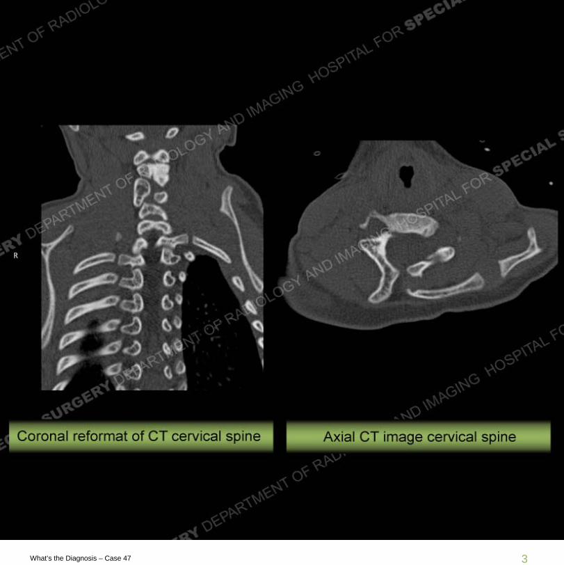
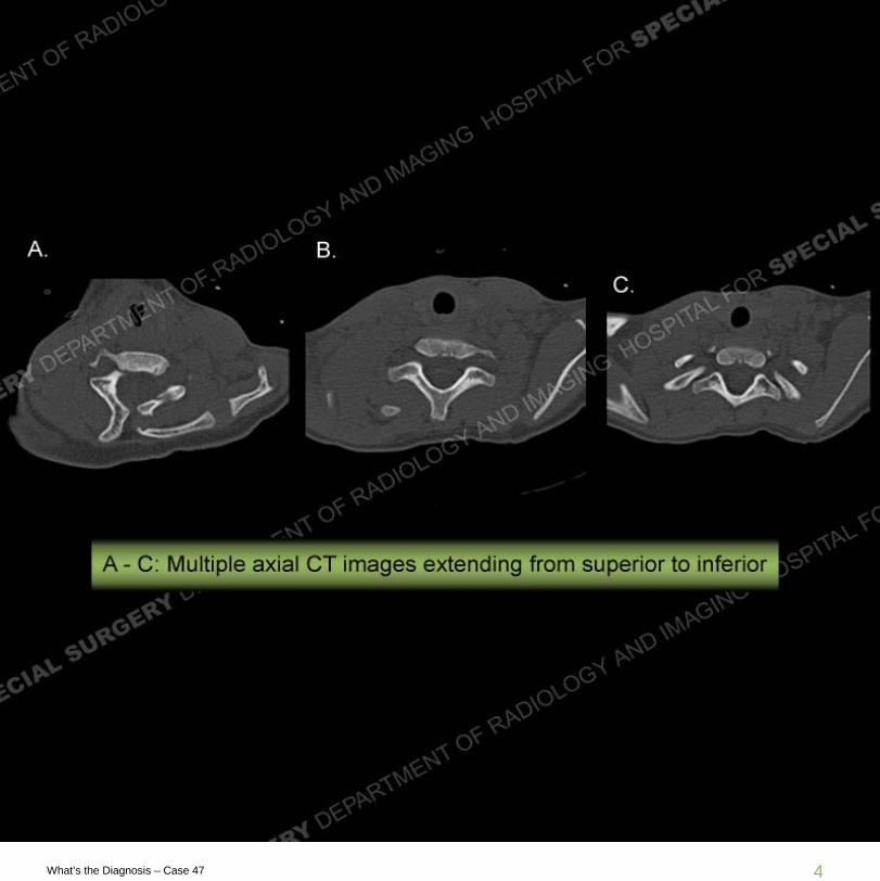
Findings
Multiple modalities demonstrate elevation of the left scapula with an abnormal bone emanating from the cervical spine and extending towards the left scapula. Fusion anomalies are seen of the cervical spine. There is a non osseous connection seen between the bone emanating from the cervical spine towards the left scapula. This is demonstrated on the CT by the absence of bone and on the MRI by high signal extending between the additional bone and the native scapula.

8What’s the Diagnosis – Case 47

9What’s the Diagnosis – Case 47

10What’s the Diagnosis – Case 47

11What’s the Diagnosis – Case 47

12What’s the Diagnosis – Case 47
13What’s the Diagnosis – Case 47
Diagnosis: Sprengel’s Deformity
Sprengel’s deformity is a congenital deformity yielding an elevation and medial rotation of the scapula related to a failure of the normal caudal migration of the scapula. In approximately 30% of the deformities, an omovertebral bone is present that extends from the posterior elements of the cervical spine to the native scapula. This may be connected directly to the scapula by osseous bridging or by non-osseous (cartilage or fibrous tissue) bridging as in this case. The deformity is most typically seen in the setting of a Klippel Feil (KF) syndrome where there is fusion of two or more cervical vertebrae. KF often has associated other vertebral anomalies, a webbed neck, cervical ribs, and cardiac/pulmonary/renal/ and GI anomalies.

14What’s the Diagnosis – Case 47
Resources
Resnick and Kransdorf. Bone and Joint Imaging. 2005.
http://www.wheelessonline.com/ortho/sprengels_deformity.