hormone-induced protein phosphorylation. ii. localization ... · indicate that the 29,000-dalton...
TRANSCRIPT

Hormone-induced Protein Phosphorylation. II. Localization
to the Ribosomal Fraction from Rat Exocrine Pancreas
and Parotid of a 29,000-dalton Protein Phosphorylated In 5itu
in Response to Secretagogues
STEVEN D. FREEDMAN and JAMES D. JAMIESON The Section of Cell Biology, Yale University School of Medicine, New Haven, Connecticut 06510
ABSTRACT In the preceding paper, we demonstrated that the endogenous phosphorylation of a protein with a molecular weight of 29,000 was enhanced by various secretagogues in rat pancreatic and parotid Iobules, the phosphorylation of this protein correlating both temporally and in a dose-dependent fashion with secretory protein discharge. In the present study, we established a specific methodology to characterize this phosphoprotein. Once established, this 29,000-dalton phosphoprotein was then followed selectively and quantitatively throughout subcellular fractionation procedures. Analysis of two-dimensional polyacrylamide gels dem- onstrated that proteins with similar mobilities (Mr 29,000; pl > 8.4) were affected by cholecys- tokinin octapeptide and isoproterenol in rat pancreatic and parotid Iobules, respectively, suggesting that the same 29,000-dalton phosphoprotein was covalently modified in both tissues. Cellular fractionation studies using differential velocity and sucrose density gradient centrifugation revealed that the 29,000-dalton phosphoprotein copurified with the rough microsomal fraction of pancreas and was highly enriched in ribosomal fractions of both pancreas and parotid. Electrophoresis in two dimensions confirmed that the 29,000-dalton polypeptide that was resolved directly from stimulated cells and from ribosomal fractions exhibited a common mobility, and apparent identity of the species was strongly suggested when the 29,000-dalton polypeptides from both sources were compared by peptide mapping following limited digestion with Staphylococcus aureus V8 protease. This phosphoprotein was tentatively identified as ribosomal protein S6 after analysis by pH 8.6/4.2 two-dimensional PAGE.
In the previous paper (l), we reported that the endogenous phosphorylation of a protein with an Mr of 29,000 was mark- edly stimulated in response to secretagogue application in intact cells from both the rat exocrine pancreas and parotid and was dephosphorylated upon termination of secretagogue effects. To gain additional insight into the role that this phos- phoprotein may. play in secretagogue action, we here describe the subcellular localization as well as the characterization of this protein by two-dimensional PAGE and limited proteolytic digestion using Staphylococcus aureus V8 protease. The results indicate that the 29,000-dalton protein whose phosphorylation is stimulated by hormone application in pancreatic and parotid lobules is localized to a highly enriched ribosomal fraction and is likely the small ribosomal subunit protein $6. A preliminary note on this research has been published (2).
MATERIALS AND METHODS
Mater ia ls
All chemicals used were of reagent grade and were obtained from the following sources: isoproterenol hydrochloride, deoxycholic acid, and Nonidet P-40 from Sigma Chemical Co. (St. Louis, MO); ampholytes from Bio-Rad Laboratories (Richmond, CA). All other reagents were obtained as described previously (l).
M e t h o d s
SAMPLE PREPARATION: 32el-labeled rat pancreatic and parotid lobules were prepared and incubated for 30 rain at 37°C in the presence or absence of secretagogues as described in the previous paper (1) with the modification that the reactions were terminated by the addition of 80/d of 10% SDS. The samples were then boiled and sonicated as previously described and analyzed by two- dimensional PAGE as described below.
CELL FRACTEONATION PROCEDURES: 40--80 lobules of parotid or pan-
THE JOURNAL O~ CELt BIOLOGY- VOLUME 95 DECEMBER 1982 909-917 © The Rockefeller University Press • 0021-9525/82/12/0909/09 $1.00 909

creas were placed in 2 ml of oxygenated Krebs-Ringer-HEPES medium (KRH) (104 mM NaCI, 5 mM KCI, 1.2 mM MgSO4, 2.0 mM CaCI2, 2.5 mM glucose, 25 mM HEPES, pH 7.4, 0.2 mg/ml soybean trypsin inhibitor) containing 5 mCi '~EPi in a 25-ml Erlenmeyer flask and incubated for 45 min under an O2 phase at 37°C with shaking at 150 oscillations/rain. The lobules were then washed with KRH and incubated for an additional 30 min in the presence of I nM cholecystokinin octapeptide (CCK-8) or 1 #M isoproteranol for the pancreas or parotid, respec- tively, and immediately placed in an ice bath to terminate the reaction. All subsequent steps were carried out at 4°C. The :nP~-labeled lobules were rinsed three times in 4 ml of 0.3 M sucrose containing 0.1 mg/ml soybean trypsin inhibitor (STI) and homogenized at a final tissue concentration of 1/10 (wt/vol) using four strokes (~ 10 s/stroke) in a Brendler type homogenizer (0.10--0.15 mm clearance) driven at 2,200 rpm. In the case of the pancreas, the fractionation procedure of Jamieson and Palade (3) as modified by Ronzio (4) was used to prepare nuclear (PI), zymogen granule (P2), mitochondrial (P3), microsomal (P4), and soluble fractions ($4). Rough and smooth microsomes were prepared by the sandwich gradient technique of Tartakoff and Jamieson (5) as modified by Scheele et al. (6). Ribosomal fractions were prepared in the case of parotid from a crude microsomal fraction and in the ease of pancreas from P4 as follo@s. The parotid crude microsomal fraction was obtained by centrifuging a tissue homogenate at 1,000 g,v for 10 min (to pellet nuclei and cellular debris) and subjecting the resulting supernatant fraction to centrifugation at 100,000 g,v for 60 rain to pellet the crude microsomal fraction. Microsomal fractions in each case were resuspended in 10 mM HEPES, pH 7.4, containing 1 mM MgC12 and 0.1 mg/ml STI and clarified by the addition of 5% deoxycholate at a final concen- tration of 0.5% (2:1 [wt/wt], deoxycholate to protein). This solution was layered over a 2.0 M sucrose cushion containing 0.5% deoxycholate and 0.1 mg/ml STI, and the ribosomal fraction was pelleted by centrifugation for 12 h at 100,000 gay. All fractions were resuspended in 0.3 M sucrose containing 0.1 mg/ml STI and adjusted to the same protein concentration as described previously (1).
PAOE: We employed three different two-dimensional PAGE systems. In the first system, the SDS-solubilized :3ZPi-labeled lobules (~200 #g of protein) were placed in a solution containing 5.4% Nonidet P-40 (NP-40), 6.4 M urea, 3.4% ~- mercaptoethanol, and 1.3%, pH 3-10, ampholytes so that the final ratio of NP-40 to SDS was exactly 8:1. This mixture was then subjected to isoelectric focusing (IEF) in the first dimension and SDS PAGE in the second dimension according to Ames and Nikaido (7), except that slab gels were employed in both dimensions.
In the other two-dimensional gel systems, 3-ZPi-labeled lobules or ribosomal fractions were sonicated in KRH or homogenized in 0.3 M sucrose, respectively, and the ribosomes were extracted with acetic acid according to the method of Hardy et al. (8). The samples were subjected to electrophoresis in urea-polyacryl- amide gels at pH 5.0 in the first dimension and to 11% SDS PAGE in the second dimension according to the procedure of Gorenstein and Warner (9), both dimensions employing slab gels. Additionally, the two-dimensional PAGE tech- nique of Lastick and McConkey (10) was used which produces a standardized polypeptide map for ribosomal proteins (I 1).
One-dimensional SDS PAGE was performed as described previously (l). For autoradiography, dried gels were placed on Kodak XRP-I x-ray film with DuPont Intensifying screens (DuPont Instruments, Inc., Wilmington, PA) for 12 to 24 h to detect those proteins which incorporated radioactive phosphate.
Peptide mapping after limited proteolysis was performed according to Cleve- land et al. (12) as modified by Huttner and Greengard (13), except that the bands of interest were electrophoresed in the presence of 5-15 #g of S. aureus V8 protease (SAP) at 40 V for ~18 h.
o r r l e a METHODS: RNA was measured by the technique of Fleck and Munro (14) as modified by Blobel and Potter (15). Lipids were extracted from cellular fractions with 20 vol of chloroform-methanol 2:1 (vol/vol) and purified according to Folch et al. (16). Phospholipid phosphorus was determined according to Ames (17).
followed selectively and quantitatively throughout fractiona- tion procedures and localized to a specific subcellular organdie.
On one-dimensional SDS polyacrylamide gels, more than one protein may comigrate at the same position. Therefore techniques providing high resolution of 29,000-dalton polypep- tides were employed as shown in Figs. 1 and 2, using two- dimensional procedures. In the first approach to this problem, 32pi-labeled pancreatic and parotid lobule proteins were ana-
FIGURE 1 Autoradiographs of two-dimensional polyacrylamide gels of 32P~-Iabeled pancreatic Iobules incubated with either KRH alone or I nM CCK-8 for 30 rain at 37°C. Proteins were separated in the first dimension by isoelectric focusing (IEF) and in the second dimension by SDSPAGE. The arrows indicate the location of the 29,000-dalton protein in this and all subsequent figures. All samples applied to the gels have been normalized to protein. In the control gel (upper panel), the tracking dye was run off the gel.
RESULTS
Two-dimensional Gel Analysis of Proteins Phosphorylated In Situ in Response to Hormone Application
In the previous paper (l), we reported that the endogenous phosphorylation of principally one polypeptide band with an apparent Mr of 29,000 was markedly enhanced in pancreatic and parotid lobules in response to secretagogues. The purposes of the present paper are twofold. The first is to establish techniques to be used to characterize the 29,000-dalton protein. Second, with the characterization of this species, the secret- agogue-affected 29,000-dalton phosphoprotein can then be
910 t h e JOURNAL OF CELL BIOLOGY - VOLUME 95, 1982
FIGURE 2 Autoradiographs of two-dimensional polyacrylamide gels of 32P~-Iabeled parotid Iobules incubated with either KRH alone or with 1 #M isoproterenol for 30 rain at 37°C. Proteins were separated in the first dimension by isoelectric focusing (IEF) and in the second dimension by SDS PAGE.

lyzed by IEF-SDS where the proteins are separated in the first dimension by charge using an isoelectric focusing gradient (pH 4-8.7) and in the second dimension by molecular weight in SDS polyacrylamide gels. In Fig. 1, secretagogue-dependent effects can be observed on the phosphorylation of a basic protein from pancreatic lobules with an M~ of 29,000. Since the presence of this band at the origin of the IEF gel (where the sample was loaded) may be due to incomplete solubilization rather than to a basic pI, the sample was also polymerized uniformly throughout a disk gel and then subjected to two- dimensional IEF-SDS PAGE. The results attained were qual- itatively similar to those in Fig. 1 (data not shown).
32Pi-labeled parotid lobule proteins were also analyzed with this technique (Fig. 2). Isoproterenol enhanced the phospho- rylation of a protein with a mobility in this gel system similar to that seen for the 29,000-dalton polypeptide in the pancreas in Fig. l. In the parotid, this phosphoprotein shows a streak toward the acidic end of the gel which might signify multiple phosphorylation sites on the molecule (increasing number of phosphate groups imparting a more negative charge on the protein) or perhaps poor resolution in the first dimension. It should be noted that in the pancreas (Fig. 1) and the parotid preparations (Fig. 2), the phosphorylation of other proteins including species seen at Mr <20,000 was also affected by secretagogue applications. These effects were not consistently observed.
The fact that basic proteins are poorly resolved with this IEF-SDS gel system and that there is an unsettling streaking over a broad pH range of amplified phosphorylation made it important to employ a second two-dimensional gel system for analysis of the 29,000-dalton phosphoprotein(s). For this pur- pose, we used a two-dimensional gel electrophoresis procedure which separates proteins in the first dimension on the basis of charge in a pH-5.0 urea-polyacrylamide gel and by molecular weight on SDS polyacrylamide gels in the second dimension as in the previously described two-dimensional gel system. Although this system clearly resolves basic proteins derived from purified ribosomes (vida infra), quantitatively only a small amount of material from lobule preparations exhibited both mobility in this gel system and enhanced phosphorylation in comparison to the level of increased phosphorylation shown in one-dimensional gels (data not shown). This is apparently due to incomplete solubilization of whole tissue fragments. None- theless, IEF and especially pH-5 urea-polyacrylamide two- dimensional gel electrophoresis provide means to separate the 29,000-dalton polypeptide from other phosphorylated proteins in subcellular fractions. As will be shown, subsequent peptide mapping of the 29,000-dalton phosphoprotein will furnish the rationale for examining selectively and quantitatively the phos- phorylation observed in one-dimensional SDS gels.
Subce l lu la r Loca l iza t ion o f the 29,000-
da l ton Phosphopro te in
To determine the subcellular localization of the 29,000-dal- ton protein phosphorylated in intact cells, 32Pi-labeled pan- creatic lobules stimulated with 1 nM CCK-8 for 30 min at 37°C were homogenized and fractionated as described in Ma- terials and Methods. Stimulation of the tissue by hormone was found not to affect qualitatively the fractionation procedure (data not shown). Initially, the magnitude of dephosphoryla- tion and the extent of its selectivity as a result of tissue homogenization and fractionation was assessed. This study was
carried out in the following manner. Approximately 80 pan- creatic lobules, which represent about one-half of a rat pan- creas, were labeled for 45 min with 32Pi and subsequently incubated for 30 min at 37°C with 1.0 nM CCK-8. Two lobules were then removed, treated with SDS, sonicated, and boiled. The rest of the preparation was homogenized and fractionated at 4°C as described in Materials and Methods, each final fraction as well as aliquots of intermediate supernatants being retained. An aging experiment was performed at 4°C, each sample being solubilized with 1.8% SDS immediately, 2 h later, and 4 h later, and subsequently analyzed on one-dimensional SDS polyacrylamide gels. Quantification of the 29,000-dalton phosphoprotein by scintillation spectrometry revealed an ap- proximate fivefold loss of radioactivity from the phosphopro- tein in the homogenate compared to that initially present in intact lobules. Dephosphorylation was not inhibited by the use of sodium fluoride or pyrophosphate (50 mM each). The activation of phosphatase activity occurred primarily during the homogenization procedure and was nonselective in that the radioactivity in all gel bands decreased in parallel. No further dephosphorylation of the 29,000-dalton protein occurred in homogenates and postnuclear supernatants up to 4 h at 4°C subsequent to this step. However, the postzymogen granule supernatant showed a 22.5% decrease in 32pi incorporated into the 29,000-dalton species over a 4 h period at 4°C (decreasing linearly over time). This effect was reduced to 10.3% per 4 h in the postmitochondrial supernatant. The microsomal, postmi- crosomal supernatant, and all subsequent subcellular fractions exhibited no phosphatase activity over 4 h at 4°C. These data provide a means to quantitatively correct for the decreases in incorporated 3~pi in the 29,000-dalton species throughout the fractionation scheme. In parotid lobules treated in the same way, no loss of 32pi from proteins was detectable after homog- enization or during subsequent fractionation steps.
In Fig. 3, the distribution of the phosphoproteins in the various subcellular fractions from pancreatic lobules as ana- lyzed by one-dimensional SDS-PAGE is shown. We found that changing the pH of the 9% lower gel from 8.80 to 8.95 provided higher resolution and afforded a separation of distinct polypeptides of mobility corresponding to 29,000 daltons. In this figure, examination of the Mr 29,000 region of the gel in the homogenate reveals a doublet. The upper band, which exhibits secretagogue amplified phosphorylation, is enriched in P4, the total microsomal fraction, and is recovered essentially quantitatively in P5, the rough microsomal fraction (the smooth microsomal fraction contained principally a 68,000-dalton phosphoprotein [data not shown]). The lower phosphorylated band in the 29,000-dalton region of the gel is predominantly in $4, the soluble fraction, and was secretagogue unresponsive. This more refined system will form the basis of further analysis.
FIGURE 3 Autoradiograph of pancreatic fractions. 32pi-la- beled pancreatic Iobules were fractionated as described in Materials and Methods and 30 /Lg of protein from each frac- tion analyzed by one-dimen- sional SDS PAGE with autora- diography. Horn, total homog- enate; P1, nuclear fraction; P2, zymogen granule fraction; P3,
mitochondrial fraction; P4, total microsomes; P5, rough microsomal fraction; and $4, soluble fraction.
FREEDMAN AND JAMIESON Hormone-Induced Protein Phosphorylation. II. 911
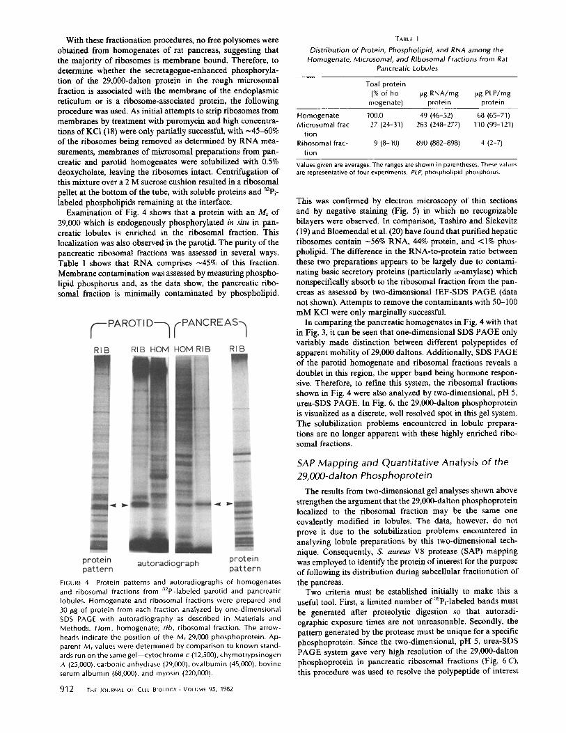
With these fractionation procedures, no free polysomes were obtained from homogenates of rat pancreas, suggesting that the majority of ribosomes is membrane bound. Therefore, to determine whether the secretagogue-enhanced phosphoryla- tion of the 29,000-dalton protein in the rough microsomal fraction is associated with the membrane of the endoplasmic reticulum or is a ribosome-associated protein, the following procedure was used. As initial attempts to strip ribosomes from membranes by treatment with puromycin and high concentra- tions of KC1 (18) were only partially successful, with ~45-60% of the ribosomes being removed as determined by RNA mea- surements, membranes of microsomal preparations from pan- creatic and parotid homogenates were solubilized with 0.5% deoxycholate, leaving the ribosomes intact. Centrifugation of this mixture over a 2 M sucrose cushion resulted in a ribosomal pellet at the bottom of the tube, with soluble proteins and 32Pi- labeled phospholipids remaining at the interface.
Examination of Fig. 4 shows that a protein with an M~ of 29,000 which is endogenously phosphorylated in situ in pan- creatic lobules is enriched in the ribosomal fraction. This localization was also observed in the parotid. The purity of the pancreatic ribosomal fractions was assessed in several ways. Table I shows that RNA comprises -45% of this fraction. Membrane contamination was assessed by measuring phospho- lipid phosphorus and, as the data show, the pancreatic ribo- somal fraction is minimally contaminated by phospholipid.
TABLE I
Distribution of Protein, Phospholipid, and RNA among the Homogenate, Microsomal, and Ribosomal Fractions from Rat
Pancreatic Lobules
Toal protein (%of ho- #g RNA/mg #g PLP/mg
mogenate) protein protein
100.0 49 (46-52) 68 (65-71) 27 (24-31) 263 (248-277) 110 (99-121)
Homogenate Microsomal frac-
tion Ribosomal frac-
tion 9 (8-10} 890 (882-898) 4 (2-7)
Values given are averages. The ranges are shown in parentheses. These values are representative of four experiments. PLP, phospholipid phosphorus.
This was confirmed by electron microscopy of thin sections and by negative staining (Fig. 5) in which no recognizable bilayers were observed. In comparison, Tashiro and Siekevitz (19) and Bloemendal et al. (20) have found that purified hepatic ribosomes contain -56% RNA, 44% protein, and <1% phos- pholipid. The difference in the RNA-to-protein ratio between these two preparations appears to be largely due to contami- nating basic secretory proteins (particularly a-amylase) which nonspecifically absorb to the ribosomal fraction from the pan- creas as assessed by two-dimensional IEF-SDS PAGE (data not shown). Attempts to remove the contaminants with 50-100 mM KC1 were only marginally successful.
In comparing the pancreatic homogenates in Fig. 4 with that in Fig. 3, it can be seen that one-dimensional SDS PAGE only variably made distinction between different polypeptides of apparent mobility of 29,000 daltons. Additionally, SDS PAGE of the parotid homogenate and ribosomal fractions reveals a doublet in this region, the upper band being hormone respon- sive. Therefore, to refine this system, the ribosomal fractions shown in Fig. 4 were also analyzed by two-dimensional, pH 5, urea-SDS PAGE. In Fig. 6, the 29,000-dalton phosphoprotein is visualized as a discrete, well resolved spot in this gel system. The solubilization problems encountered in lobule prepara- tions are no longer apparent with these highly enriched ribo- somal fractions.
FIGURE 4 Protein patterns and autoradiographs of homogenates and ribosomal fractions from a2P~-Iabeled parotid and pancreatic Iobules. Homogenate and ribosomal fractions were prepared and 30 p,g of protein from each fraction analyzed by one-dimensional SDS PAGE with autoradiography as described in Materials and Methods. Horn, homogenate; rib, ribosomal fraction. The arrow- heads indicate the position of the M, 29,000 phosphoprotein. Ap- parent M, values were determined by comparison to known stand- ards run on the same gel--cytochrome c (12,500), chymotrypsinogen A (25,000), carbonic anhydrase (29,000), ovalbumin (45,000), bovine serum albumin (68,000), and myosin (220,000).
SAP M a p p i n g and Quan t i t a t i ve Analysis o f the
29,000-dal ton Phosphopro te in
The results from two-dimensional gel analyses shown above strengthen the argument that the 29,000-dalton phosphoprotein localized to the ribosomal fraction may be the same one covalently modified in lobules. The data, however, do not prove it due to the solubilization problems encountered in analyzing lobule preparations by this two-dimensional tech- nique. Consequently, S. aureus V8 protease (SAP) mapping was employed to identify the protein of interest for the purpose of following its distribution during subcellular fractionation of the pancreas.
Two criteria must be established initially to make this a useful tool. First, a limited number of 32Pi-labeled bands must be generated after proteolytic digestion so that autoradi- ographic exposure times are not unreasonable. Secondly, the pattern generated by the protease must be unique for a specific phosphoprotein. Since the two-dimensional, pH 5, urea-SDS PAGE system gave very high resolution of the 29,000-dalton phosphoprotein in pancreatic ribosomal fractions (Fig. 6 C), this procedure was used to resolve the polypeptide of interest
912 T.E JOURNAL OF CELL BIOLOGY • VOLUME 95, 1982

FIGURE 5 Electron micrograph of the pancreatic ribosomal fraction. For negative staining, the ribosomal fraction was suspended in 10 mM HEPES, pH 7.4, plus 5 mM MgCI2 at a protein concentration of 0.01 mg/ ml. Carbon films were prepared on freshly cleaved mica and trans- ferred to 300-mesh copper grids. The sample was adsorbed to the film for 10 s, rinsed in buffer alone, stained with 0.5% uranyl acetate, and the grid was examined in a Siemens 101 electron microscope at 80 kV.
from other contaminating phosphoproteins. For the purposes of peptide mapping, the radioactively labeled spot was excised from gels of this type and subjected to limited proteolytic digestion with 15 #g of SAP. Lane E in Fig. 7 illustrates that primarily three peptide fragments with approximate molecular weights of 15,000, 10,000, and 5,000 are generated from the labeled 29,000-dalton ribosomal protein with 32Pi ratios of approximately 17:1:1, respectively. In addition, 32Pi-labeled material in excess of 45,000 daltons is observed which may represent small peptides that bind SDS poorly and hence migrate slowly in this 15% gel. Further studies showed that digestion with 10 ~tg of SAP resulted in maximal proteolytic processing of the phosphoprotein with no further change in the ratio or number of fragments being produced with up to 50 ~tg of SAP or longer electrophoresis times (data not shown).
SAP digestion of the phosphoprotein which migrates slightly faster than the 29,000-dalton polypeptide of interest in one- dimensional SDS gels and which is enriched in the soluble fraction of the pancreas (see Fig. 3, lane $4) is shown in lane G. A different pattern is seen with this protein (which when analyzed by IEF-SDS gel electrophoresis exhibits an acidic pI), indicating the uniqueness of the SAP phosphopeptide pattern of the band of interest. Thus, the two criteria mentioned above have been satisfied.
Since excision of the 29,000-dalton band from one-dimen- sional SDS gels followed by scintillation counting affords a
,32 method for quantifying the amount o f " P~ incorporated into it while the SAP procedure provides identification of this phos-
phoprotein, these two techniques were combined. To obtain adequate separation of the two bands in the 29,000-dalton region so that the upper secretagogue-responsive species could be reliably harvested, not only was the pH of the lower gel raised to 8.95, but the tracking dye was allowed to run off the gel and electrophoresis continued for an additional 30 min.
In Fig. 7 we see phosphopeptide analysis of the upper secretagogue-responsive 29,000-dalton polypeptide in one-di- mensional SDS gels of intact pancreatic lobules (lane A), homogenates of lobules (lane B), the microsomal fraction (lane C), and ribosomal fractions (lane D). These results demonstrate that the same phosphoprotein that was affected by hormones in situ is localized to these subcellular fractions. Additionally, the SAP map of these fractions is strikingly similar to that of purified ribosomes separated on the pH 5 urea-SDS PAGE system (Fig. 7, lane E). Whether the additional minor phos- phopeptides observed in Fig. 7, lanes A-D, represent other phosphorylation sites on the 29,000-dalton protein or are de- rived from other contaminating proteins which comigrate with the secretagogue-responsive 29,000-dalton protein is not known. Fig. 7 also shows that the general dephosphorylation that occurs upon homogenization of the lobules affects all phosphopeptides equally. SAP analysis of the 29,000-dalton phosphoprotein from '32Pi-labeled parotid lobules and ribo- somes (the upper band of the doublet in Fig. 4) produces the same pattern as observed for the exocrine pancreas (data not shown). Taken together, these results strongly suggest that the same phosphoprotein is in fact being followed throughout cell
FRE[OMAN AN[) ]AMfESON Hormone-Induced Protein Phosphorylation. II. 913

FIGURE 6 Two-dimensional PAGE of the 32P-labeled ribosomal fractions shown in Fig. 4. The 32P~-Iabeled ribosomal fractions were analyzed in the first dimension in pH-5.0 urea-polyacrylamide gels and in the second dimension by SDS PAGE. (A) Coomassie Blue pattern of the pancreatic ribosomal fraction. (B) Coomassie Blue pattern of the parotid ribosomal fraction. (C) Autoradiograph of the pancreatic ribosomal fraction. (D) Autoradiograph of the parotid ribosomal fraction. Diagonal arrows indicate the 29,000- dalton protein of interest.
fractionation. With the use of the SAP method for identifying the secretagogue-enhanced phosphoprotein, we could now, in conjunction with this higher resolution one-dimensional SDS PAGE system, quantitate the distribution of the 29,000-dalton phosphoprotein during cell fractionation.
The amounts of RNA and of 3ZPi in the 29,000-dalton phosphoprotein in the various subcellular fractions is shown quantitatively by the histogram in Fig. 8. The distribution of the 29,000-dalton phosphoprotein (the values of which were corrected for the phosphatase activity present in the postzy- mogen granule and mitochondrial supernatants) is closely cor- related with the amount of RNA in each cell fraction. In this study, recoveries were 89% for protein, 97% for RNA, and 94% for 32Pi in the 29,000-dalton phosphoprotein. The nonparallel recovery of protein and RNA suggests that the RNA values may be overestimated, most likely due to a small fraction of the protein being hydrolyzed by KOH during the RNA assay and thus becoming acid soluble along with the RNA. Thus, the subcellular distribution of the 29,000-dalton phosphopro- tein which copurifies with RNA can be quantitatively ac- counted for, its identity being confirmed by SAP analysis.
914 T,E IOURNAL OF CELL BIOLOGY. VOLUME 95, 1982
Identification of the Phosphorylated Ribosomal Protein
To identify the ribosomal protein that is phosphorylated in a hormone-dependent fashion, the 32Pi-labeled pancreatic ri- bosomal fraction was analyzed by the two-dimensional PAGE technique of Lastick and McConkey (10). A standard number- ing system for eucaryotic ribosomal proteins has been pub- lished for this gel system (11). The protein pattern for the pancreatic ribosomal fraction shown in Fig. 9 A is qualitatively similar to other published ribosomal patterns (see reference 11), allowing each spot to be identified with known ribosomal proteins. The autoradiograph in Fig. 9 B clearly shows only one phosphorylated species which is indicated by a dotted line on the Coomassie-Blue-stained gel and which coelectro- phoreses with ribosomal protein $6. SAP digestion of the phosphoprotein excised from this gel (Fig. 7, lane F) produces the same pattern as the one shown in Fig. 7, lane E, derived from the two-dimensional, pH 5, urea-SDS polyacrylamide gel system except that the labeled material at the top of the gel is not seen. Thus, the protein whose phosphorylation is secre-

tagogue modulated in situ is ribosomal protein $6, based on these data.
Further examination of Fig. 9 reveals that, while the majority of the radioactivity overlies ribosomal protein $6, a tail extends towards the anode. The orientation of the spot towards the origin may reflect an anodal shift in both dimensions due to the reduction in positive charge by multiple phosphate residues. These observations are consistent with the characteristic shape of phosphorylated $6 after two-dimensional PAGE described for many other tissues (for review see reference 21), strengthen- ing the identity of the phosphoprotein described in this study as ribosomal protein $6. In addition, other investigators have
found that $6 is the only ribosomal protein phosphorylated in vivo (21).
D I S C U S S I O N
In the preceding paper (1), we demonstrated that the phospho- rylation of the 29,000-dalton protein in intact cells from both the parotid and exocrine pancreas is specifically enhanced in response to secretagogue application, the incorporated s2Pi being metabolically stable. The present study has shown that this same phosphoprotein is localized to a highly enriched ribosomal fraction. The rapidity with which this protein was phosphorylated as well as the fact that cycloheximide had no affect on the enhancement of its phosphorylation by CCK-8 or isoproterenol in the exocrine pancreas and parotid, respectively (data not shown), implies that the increase in 32p~ incorporation is not due to an increase in the net synthesis of the 29,000- dalton protein.
In many eucaryotic cell systems, the phosphorylation of the 40S ribosomal protein $6 with a reported Mr of 29,000 to 34,000 has been shown to be stimulated by a variety of agents such as hormones, growth factors, cAMP, viral infection,
24"4 Iq~ F264
20- -220 -:]3
16- r q -176 r",
< 1 o Z "0 rr 12- -132 o
8' -88 £ 4" ~ -44 ~'~
FIGURE 7 Phosphopeptide pattern of the 29,000-dalton phospho- protein from a2P~-labeled pancreatic lobules and subcellular frac- tions after l imited proteolytic digestion with SAP. The fractions used were from the same experiment used for quantif ication in Fig. 8, the band for proteolytic digestion being taken from a duplicate one- dimensional SDS polyacrylamide gel. Lanes: A, lobule; B, homoge- nate; C, microsomes; D, ribosomal fraction; E, phosphorylated 29,000-dalton protein excised from a two-dimensional, pH-5.0 urea- SDS PAGE system; F, ribosomal protein $6 obtained by the two- dimensional PAGE technique of Lastick and McConkey (10); and G, acidic phosphoprotein from postmicrosomal supernatant (54).
Horn P1 P2 P3 P4 P5 $4
FIGUR[ 8 Distribution of RNA and 32p, in the 29,000-dalton phos- phoprotein in subcellular fractions from rat pancreas. RNA was determined in each subcellular fraction and is expressed as the total amount of RNA (milligrams) per subcellular fraction (open bars). In the same experiment, the 29,000-dalton phosphoprotein was excised from the one-dimensional SDS polyacrylamide gels and quantif ied by scintil lation spectrometry. The radioactivity which represented 52 /~g of protein per lane on the gel was corrected for the total amount of protein per subcellular fraction and values are expressed as cpm incorporated (stippled bars).
FIGURE 9 Two-dimensional elec- trophoretic analysis of the 32pi-la- beled pancreatic ribosomal frac- tion. (A) Coomassie Blue pattern. (B) Autoradiograph. Electrophoresis was from anode to cathode: from left to right in the first dimension and from top to bottom in the sec- ond dimension. The arrow indicates the position of ribosomal protein $6. The dotted line shows the posi- t ion of the radioactive spot relative to the protein staining.
FREEDMAN AND JAMIESON Hormone-Induced Protein Phosphorylation. II. 915

changes in cell cycle in culture, and during liver regeneration as reviewed in Wool (21). In addition, Schubart et al. (22) have shown that 32Pi incorporation into a 28,000-dalton ribosomal protein is enhanced after a l-min incubation of insulinoma cells with glucagon. This phosphorylation correlated with an increase in intracellular cAMP but not with insulin secretion. The similarities between ribosomal protein $6 described in the literature and the 29,000-dalton phosphoprotein examined in the present study, i.e., the kinetics of the phosphorylation of these two proteins, the metabolic stability of the phosphate group, comigration of these polypeptides after two dimensional gel electrophoresis, subcellular localization, and dependence on both cAMP-dependent and -independent protein kinases (see also following paper), would strongly suggest that these two proteins are identical.
In addition, we have demonstrated that this Mr 29,000 ribosomal protein is the same protein whose phosphorylation is hormonally regulated in intact acinar cells, based on the following data. First, SAP mapping of the 29,000-dalton phos- phoprotein from lobules, subcellular fractions, and from the ribosomal protein $6 obtained from the Lastick and McConkey two-dimensional gel system (10) all produce very similar pat- terns. Second, the 32Pi incorporated into the 29,000-dalton ribosomal protein can be quantitatively accounted for through- out the subcellular fractionation procedure. Taken together, these data demonstrate that the phosphorylation of ribosomal protein $6 is modulated in a secretagogue-dependent fashion in pancreatic and parotid acinar ceils.
On two-dimensional gels, as many as six forms of ribosomal protein $6 have been reported which contain increasing num- bers of phosphoserine residues (21). Fig. 7 shows that three phosphopeptides are generated after SAP digestion of the 29,000-dalton protein from pancreatic ribosomes phosphoryl- ated in situ, indicating that more than one site is phosphorylated on the 29,000-dalton molecule. The commonality of hormone and growth factor stimulation of the phosphorylation of $6 in many different cell types would suggest that it may also occur in response to hormones in exocrine cells as well. In agreement with this idea, secretagogue stimulation of the endogenous phosphorylation of the same 29,000-dalton protein was ob- served in both parotid and pancreatic acinar ceils, implying that this covalent modification may be a general response in a variety of hormonally stimulated exocrine tissues.
The role of the phosphorylation of the 29,000-dalton poly- peptide in secretagogue action is presently unknown. Although it is recognized that the pancreas is capable of undergoing chronic changes in the composition of proteins secreted, these effects presumably being modulated at the level of biosynthesis (23), the question of short-term changes in enzyme and zymo- gen composition is controversial. Iacino et al. (24) have found that, in an in vitro preparation of rabbit pancreas, cholecysto- kinin-pancreozymin caused a significant change in the com- position of '~S-methionine-labeled secretory proteins within 25--40 min. Whether these results are due to discharge from different pools of secretory prote!ns within the cell(s) under basal and stimulated conditions or reflect the modulation of translation of specific mRNAs is not known. The localization of the 29,000-dalton phosphoprotein to the ribosomal fraction would suggest that, if hormones do indeed regulate some aspect of translation, then protein phosphorylation may be involved.
Alternatively, in addition to regulating secretory protein discharge, both cholecystokinin and secretin produce hypertro- phy as well as hyperplasia in adult rat pancreas, their effects
916 T.E JOURNAL OF CELL BIOLOGY-VOLUME 95, 1982
being synergistic (25, 26). These same effects have also been well documented in rat parotid glands in response to chronic administration of isoproterenol (27). These trophic effects, which vary in magnitude and specificity during postnatal de- velopment in the pancreas (28), emphasize the fact that hor- mones are multiphasic in their actions. Thus, the phosphoryl- aLlan of the M~ 29,000 species may regulate some aspect of pancreatic and parotid growth analogous to the proposed role of ribosomal protein $6 phosphorylation in mRNA discrimi- nation and translational control of protein production (21, 22, 29-33).
This research was supported by National Institutes of Health grant AM-17389 and National Research Service Award grant GM-07223.
Received for publication 29 March 1982, and in revised form 17 August 1982.
REFERENCES
1, Freedman, S. D., and J. D. Jamieson. 1982. Hormone-induced protein phosphorylatinn. I. The relationship between secretagogue action and endogenous protein phosphorylation in intact cells from the exocrine pancreas and parotid. J. Cell Biol. 95:903-908.
2. Freedman, S. D., and J. D. Jamieson. 1981. Ribosomal localization of a 29,000 dalton protein whose phosphorylatioo is stimulated in situ by secretagogues in the rat exocrine pancreas and parotid. J. Cell Biol. 91(2, Ft. 2):213a (Abstr.).
3. Jamieson, J. D., and G. E Palade. 1967, Intraccllular transport of secretory proteins in the pancreatic exocrine cell. II. Transport to the condensing vacuoles and zymogen granules. J. Cell Biol. 34:577-596.
4. Ronzio, R. A. [973. Glycoprotein synthesisin the adult pancreas. 1. Subcellular distribution of nridine diphosphate galactose: glycopsotein galactosyltransferase and thiamine pyro- phosphate phnsphohydrolase. Biochim. Biophys. Acta. 313:286-295.
5. Tartakoff, A. M., and J. D. Jamieson. 1974. Subeellular fractionation of the pancreas. Methods EnzymoL 31:41-59.
6. Scheele, G. A., G. E. Palade, and A. M. Tartakoff. 1978. Cell fractiooation studies on the guinea pig pancreas. Redistribution of exocrine proteins during homogenization. J. Cell BioL 78:110-130.
7. Ames, G. F., and K. Nikaido. 1976. Two dimensional gel electrophoresis of membrane proteins. Biochemistry 15:616-625.
8. Hardy, S. J. S., C, G. Kurland, P. Voynow, and G. Morn. 1969. The ribosomal proteins of Escherichia coil. I. Purification of the 30S ribosomal proteins. Biochemistry. 8:2897-2905.
9. Gorenstein, C., and J. R. Warner. 1976. Coordinate regulation of the synthesis of eukaryotic ribosomal proteins. Proc. Natl. Acad. ScL U. S. A. 73:1547-1551.
10. Lastick, S. M., and E. H, McConkey. 1976. Exchange and stability of HeLa ribosomal proteins in viva. Z Biol. Chem. 251:2867-2875.
I I. McConkey, E. H., H. Bielka, J. Gordon, S. M. Lastick, A. Lin, K. Ogata, J.-P. Reboud, J A. Traugh, R. R. Traut, J. R. Warner, H. Welfle, and 1. G. Wool. 1979. Proposed uniform nomenclature for mammalian ribosomal proteins. Mot. Gen. Genet. 169:1-6.
12. Cleveland, D. W., S. G. Fischer, M. W. Kirsehner, and U. K. Laemmli, 1977. Peptide mapping by limited proteolysis in sodium dodecyl sulfate and analysis by gel electropho- resis. J. Biol. Chem. 252:1102-1106.
13. Huttner, W. B., and F. Greengard. 1979, Multiple phosphorylation sites in protein 1 and their differential regulation by cyclic AMP and calcium. Proc. Natl. Acad Sci. U. S. A. 76:5402-5406.
14. Fleck, A., and H. N. Munro. 1962. The precision of ultraviolet absorption measurements in the Schmidt-Thannhanser procedure for nucleic acid estimation. Biochim. Biophys. Acta. 55:571 580.
15. Blobel, G., and V. R. Potter. 1968. Distribution of radioactivity between the acid-soluble pool and the pools of RNA in the nuclear, nonsedimentable and ribosome fractions of rat liver after a single injection of labeled orotic acid. Biochim. Biophys. Acta. 166:48-57.
16. Folch. J,, M. Lees, and G. H. Sloane-Stanley. 1957. A simple method for the isolation and purification of total lipides from animal tissues. J. Biol. Chem. 226:497-509.
17. Ames, B. N. 1966. Assay of inorganic phosphate, total phosphate and phosphatases. Methods EnzymoL 8:115 119.
18. Blobel, G., and B. Dobberstein. 1975. Transfer of proteins across membranes. 1I. Recoo- stitution of functional rough microsomes from heterologous components..L Cell Blot 67:852-862.
19. Tashiro, Y., and P. Siekevitz. 1965. Ultracentrifagal studies on the dissociation of hepatic ribosomes..L Mol. BioL I 1:149-165.
20. Bloemendal, H., E. L. Benedetti, and W. S. Bout. 1974. Preparation and characterization of free and membrane-bound polysomes. Methods EnzymoL 30:313-327.
2 I. Wool, I. G. 1979. The structure and function of eukaryotic ribosomes. A nnu. Rev. Biochem 48:791-754.
22. Schubart, U. K., S. Shapiro, N. Fleiseher, and O. M. Rosen. 1977. Cyclic adenosine 3',5"- monophnsphate-mediated insulin secretion and ribosomal protein phosphorylation in a hamster islet cell tumor. ,L Biol. Chem. 252:92-101.
23. Reboud, J. P., G. Marchis-Mouren, L. Pasera, A. Cozzone, and P. Dcsnuelle. 1966. Adaptation de la vitesse de l'amylase pancreatique de du chymotrypsinogene a des regimes reiches en amidon ou en proteines. Biochem. Biophys. Acta. 117:351-367.
24. lacino, D., G. A. Scheele, and C. Liebow. 1980. Secretory response of the rabbit pancreas to cholecystokinin stimulation. Am. J. Physiol. 239:G247-G254.
25. Dembinski, A. B., and L. R. Johnson. 1980. Stimulation of pancreatic growth by secretin. caerulein and pentagastrin. Endocrinology, 106:323 328.
26. Folsch, U. R., K. Winckler, and K. G. Wormsley. 1978. Influence of repeated administra- tion ofcholecystokinin and secretin on the pancreas of the rat. Stand. J. Gastroent. 13:663- 671.
27. Selye, H., R. Veilleux, and M. Cantin. 1961. Excessive stimulation of salivary gland growth by isoproterenol. Science (Wash. D~ C.). 133:44-45.

28. Brants, F., a,ld J. Morisset. 1976. Trophic effect of cholecystokinin pancreozymin on pancreatic acinar cells from rats of different ages. Proc. Soc. Exp. BioL Meal 153:523-527.
29. Smith, C. J., C. S. Rubin, and O. M. Rosen. 1980. Insulin-treated 3T3-LI adipocytes and cell-free extracts derived from them incorporate " P into ribosomal protein $6. Proc. NatL Acad Sci. U. S. A. 77:2641 2645.
30 Smith, C. J.. P J. Wejksnora, J. R. Warner. C. S. Rubin, and O. M. Rosen. 1979. Insulin- stimulated protein phosphorylation in 3T3-LI preadipocytes. Proc. NatL Acad Sci. U. S. A. 76:2725 2729.
31. Rosen, O. M., C. S. Rubin, M. H. Cobb, and C. J. Smith. 1981. Insulin stimulates the phosphorylation of ribosomal protein S6 in a cell-free system derived from 3T3-LI adipocytes. J. Biol. Chem. 256:3630-3633.
32. Halegoua, S., and J. Patrick. 1980. Nerve growth factor mediates phosphorylation of specific proteins. Cell. 22:571-58 I.
33. Thomas, G., M Siegmann, and J. Gordon. 1979. Multiple phospborylation of ribosomal protein $6 during transition of quiescent 3T3 cells into early G~, and cellular compart- mentalization of the phosphate donor. Proc. Nai l Acad Sci. U. S. A. 76:3952-3956.
FREEDMAN ANO JAMI[SON Hormone-Induced Protein Phosphorylation. I I 977