homology modeling and molecular dynamics simulations of ...vuir.vu.edu.au/33937/1/muc9-mhc21june10...
TRANSCRIPT

Homology modeling and molecular dynamicssimulations of MUC1-9/H-2Kb complex suggest
novel binding interactions
Athanassios Stavrakoudis,1 Ioannis G. Tsoulos,2 Katalin Uray,3
Ferenc Hudecz3,4 and Vasso Apostolopoulos5
1 Department of Economics, University of Ioannina, Ioannina, Greece2 Department of Communications, Informatics and Management, Technological Educa-tional Institute of Epirus, Arta, Greece3 Research Group of Peptide Chemistry, Hungarian Academy of Sciences, Budapest, Hun-gary4 Institute of Chemistry, Eotvos Lorand University, Budapest, Hungary5 Immunology and Vaccine Laboratory, Centre for Immunology, The Macfarlane BurnetInstitute for Medical Research and Public Health, Melbourne, Australia
Correspondence:Athanassios Stavrakoudis, Department of Economics, University of Ioannina, GR-451
10, Ioannina, Greece, tel: +30 265 100 5935, fax: +30 265 100 5092, email: [email protected],web: http://stavrakoudis.econ.uoi.gr
Vasso Apostolopoulos, Immunology and Vaccine Laboratory, Centre for Immunology,The Macfarlane Burnet Institute for Medical Research and Public Health, Melbourne,Australia tel: +61 3928 22111, fax: +61 3928 22100, email: [email protected]
1
Abstract1
Human MUC1 is over-expressed on human adenocarcinomas and has been used as a2
target for immunotherapy studies. The 9-mer MUC1-9 peptide has been identified3
as one of the peptides which binds to murine MHC class I H-2Kb The structure4
of MUC1-9 in complex with H-2Kb has been modeled and simulated with classical5
molecular dynamics, based on the x-ray structure of the SEV9 peptide/H-2Kb com-6
plex. Two independent trajectories with the solvated complex (10 ns in length) were7
produced. Approximately 12 hydrogen bonds were identified during both trajecto-8
ries to contribute to peptide/MHC complexation, as well as 1-2 water mediated9
hydrogen bonds. Stability of complex was also confirmed by buried surface area10
analysis, although the corresponding values were about 20% lower than those of the11
original x-ray structure. Interestingly, a bulged conformation of the peptide’s cen-12
tral region, partially characterized as a β-turn, was found exposed form the binding13
groove. In addition, P1 and P9 residues remained bound in the A and F binding14
pockets, even though there was suggestion that P9 was more flexible. The complex15
lacked the numerous water mediated hydrogen bonds that were present in the refer-16
ence peptide x-ray structure. Moreover, local displacements of residues Asp4, Thr517
and Pro9 resulted in loss of some key interactions with the MHC molecule. This18
might explain the reduced affinity of the MUC1-9 peptide, relatively to SEV9, for19
the MHC class I H-2Kb.20
Keywords: Class I MHC; H-2Kb; homology modeling; molecular dynamics; MUC1;21
tumor22
1

1 Introduction23
Major histocompatibility complex (MHC) proteins bind small peptide fragments de-24
rived from pathogenic proteins and form peptide/MHC (pMHC) complexes (Ragha-25
van et al., 2008). MHC proteins are divided into two classes: class I (MHC-I) and26
class II (MHC-II). The MHC-I consists of a polymorphic transmembrane heavy chain27
and β2-microglobulin, which are non-covalently associated (Zhang et al., 1998). The28
proteolysis of intracellular proteins by the proteasome produces the majority of pep-29
tides suitable for MHC-I binding. In most cases, peptides of 8-10 residues in length30
are found in the binding groove of MHC-I.31
After the first crystal structures of pMHC complexes were available, (Bjorkman32
et al., 1987; Fremont et al., 1992; Matsamura et al., 1992) it was suggested that pep-33
tides bound to MHC-1 with a canonical extended strucure. MHC class I residues34
that form the binding groove are responsible for the specificity of the peptide selec-35
tion. Six (out of 8-10) residues of the peptide sequence are accomodated within the36
A-F binding pockets of the MHC-I protein (Saper et al., 1991). Residues that do37
not participate directly in binding are believed to interact with the TCR.38
Human mucin, MUC1, is a membrane-bound glycoprotein, expressed on the39
surface of epithelial cells. It is often overproduced and/or underglycosylated in ade-40
nocarcinomas (breast, ovary, colon, lung, kidney, etc) and is present in the serum of41
cancer patients. MUC1 is immunogenic in mice and in humans, with both humoral42
and cellular immune responses being induced by MUC1-based vaccine constructs43
(Tang et al., 2008b,a). MUC1 mucin partly consists of a variable number of tandem44
repeats region of the consensus sequence 1PDTRPAPGSTAPPAHGVTSA20 which45
is repeated 40-80 times (Gendler et al., 1988). The majority of anti-MUC1 antibod-46
ies recognize sequences within the SA1PDTRPAP7 region (Price et al., 1991; Xing47
et al., 1991, 1992; Burchell et al., 1989). The SAPDTRPAP (MUC1-9) 9-mer peptide48
was also found to be presented by MHC-1 H-2Kb and to be immunogenic (Apos-49
tolopoulos et al., 1997). MUC1-9 binds with low affinity to H-2Kb (Apostolopoulos50
2

et al., 1997) via a noncanonical mode and it was suggested that the C-terminus of51
the peptide looped out of the peptide binding groove (Apostolopoulos et al., 1998;52
Apostolopoulos and Lazoura, 2004).53
Computer simulation of molecular dynamics is a well established method for54
studying several aspects of biomolecular structure and function (Hansson et al.,55
2002; Karplus, 2003; Aksimentiev et al., 2008; Tantar et al., 2008). In recent years56
such computational approaches have been increasingly incorporated in drug design57
(Galeazzi, 2009), in immunological reasearch (Morikis and Lambris, 2004; Mallik and58
Morikis, 2006; Stavrakoudis, 2010) and also to peptide/MHC complexes (Omasits59
et al., 2008; Knapp et al., 2009). Moreover, biomolecular modeling can complement60
experimental studies (van Gunsteren et al., 2008) and can elucidate dynamics of61
immunological synapse (Wan et al., 2008), allows to study the dynamics of a peptide62
bound to antibody (Tatsis et al., 2009; Stavrakoudis, 2009b), could be used to model63
disulphide peptide complexed proteins (i.e. C8γ (Stavrakoudis, 2009a)) or even more64
excitingly to help in clinical decision making (Sadiq et al., 2008).65
Modeling of the MUC1-9 peptide with both murine and human MHC class I,66
H-2Kb and HLA-A2 respectively have been previously performed (Apostolopoulos67
et al., 1998), based on a simulated annealing protocol and high temperature molec-68
ular dynamics (Chelvanayagam et al., 1996). That work was a considerable progress69
in our knowledge of peptide/MHC interactions in the MUC1-9 case and provided a70
possible structural explanation of the antibody binding of MUC1 peptides presented71
by the MHC molecules. However, modern progress in computational biophysics, ac-72
companied with the big enhancement of available computer power, can be utilized to73
further improve the computer-generated model of the MUC1-9 peptide complexed74
the the MHC class I H-2Kb.75
Here, we present a homology modeling and molecular dynamics approach of76
MUC1-9 (SAPDTRPAP) in complex with MHC class I H-2Kb. Since the initial77
conformation was modeled rather than taken from an x-ray structure, we chose78
3

to perform two indepent simulation runs, to obtain more robust results. Long-79
run dynamics, inclusion of the whole MHC molecule and explicit representation of80
solvent have been utilized in order to more accurate picture the MUC1-9 structure81
and interactions with the MHC molecule. Such approach has been suggested to82
give more reliable results in MD investigations (van Gunsteren et al., 2008; Omasits83
et al., 2008). Our results suggest that this was a beneficial approach in the current84
study, and has given insights into the peptide binding mode of the MUC1-9.85
2 Methods86
Initial coordinates for the SEV9/MHC complex were downloaded from Protein Data87
Bank (Berman et al., 2002), access code: 1kpv.88
The original peptide from sendai-virus, FAPGNYPAL was mutated to SAPDTR-89
PAP, whilst MHC molecule remained untouched. The SEV9 peptide was selected90
from other canditates due to its homology with the MUC1-9 peptide. Pro residue91
homology in positions P3 and P7 was also crucial for selection. Since the backbone92
dihedral angle φ of Pro residue is restrained, it is preferable to choose a peptide93
that has the same residue in these positions. Ideally, it would be perfect to also94
have alignment for position P9, however there was no such option. Topology and95
force field parameters for all atoms were assigned from the CHARMM22-CMAP96
parameter set (Mackerell et al., 2004; MacKerell et al., 2004). It has been noted97
that addition of cross terms with CMAP potential improves the system parametriza-98
tion and helps to avoid undesired backbone helical transitions (Buck et al., 2006;99
Stavrakoudis, 2008).100
Hydrogen atoms were added with the VMD program (Humphrey et al., 1996)101
and its autopsf utility. Protonation status of Histidine side chains were determined102
with the REDUCE program (Word et al., 1999). The peptide/MHC complex was103
centered in a rectagular box with dimensions 95.7×88.3×102.9A3. The box was filled104
4

with TIP3P water molecules and neutralized with the addition of 26 Na+ and 20105
Cl− ions respectively, to approximate a 0.1 mM ion concetration. Crystallographic106
water molecules (345) were also included in the model. The final system contained107
24429 water molecules. Total number of atoms of the entire system were 80598.108
Non-bonded van der waals interactions were gradually turned off at a distance109
between 12 and 14 A (Yonetani, 2006). Long range electrostatics were calculated110
with the PME method (Darden et al., 1993). Non-bonded forces and PME elec-111
trostatics were computed every second step. Pair list was updated every 10 steps.112
Bonds to hydrogen atoms were constrained with the SHAKE method allowing a 2 fs113
time step for integration. The system was initially subjected to energy minimization114
with 5 000 steps. The temperature of the system was then gradually increased to 310115
K, with Langevin dynamics using the NVT ensemble, during a period of 3 000 steps,116
by stepwise reassignment of velocities every 500 steps. The simulation was continued117
at 310 K for 100 000 steps (200 ps). During minimization and equilibration phases,118
protein backbone atoms (N, Cα, C’, O) and oxygen atoms of crystallographic waters119
were restrained to their initial positions with a force constant of 50 kcalmol −1A−2
.120
The system was equilibrated for further 200 ps with the force constant reduced to121
5 kcalmol−1A−2
. Finally, 400 ps of NVT simulation at 310 K was performed with122
total elimination of the positional restraints. The simulation was passed to the123
productive phase, by applying constant pressure with the Langevin piston method124
(Feller et al., 1995). Velocities were re-initialized and two independent trajectories125
were produced (trA and trB). Pressure was maintained at 1 atm and temperature126
at 310 K. Results are based to a period of 10 ns of this isothermal-isobaric (NPT)127
runs. Shapshots were saved to disk at 1 ps interval for structural analysis.128
The initial structure of the SEV9/MHC complex (PDB code 1kpv) were also129
simulated under identical conditions for comparative analysis (tr0 trajectory).130
Trajectory analysis was performed with Eucb (Tsoulos and Stavrakoudis, 2009)131
and Carma (Glykos, 2006) software packages. Secondary structure analysis was132
5
performed with STRIDE (Frishman and Argos, 1995). Circular data statistics (di-133
hedral angles, etc) were calculated with appropriate corrections (Agostinelli, 2009).134
Structural figures were prepared with PyMOL (www.pymol.org).135
2.1 Burried surface area calculation136
Calculation of buried surface area (BSA) was performed with the NACCESS pro-137
gram (http://www.bioinf.manchester.ac.uk/naccess/), based on the formula:138
BSA = Sp + Sa − Sc (1)
thus as the difference of the surface accessible area of the complex (Sc) from the139
sum of the of surface accessible areas of the peptide (Sp) and MHC molecule (Sa)140
respectively.141
β-turn classifications were based on geometrical characteristics of the backbone142
conformation (Hutchinson and Thornton, 1994). Initially, a β-turn was accepted if143
d(Cαi −Cα
i+3) ≤ 7A and |α(Cαi −Cα
i+1−Cαi+2−Cα
i+3)| < 90◦, where d is the distance144
and a is the dihedral angle between the corresponding atoms. Further classification145
of the β-turn was based on hydrogen bond patterns and backbone dihedral values146
of the i+ 1 and i+ 2 residues.147
In order to indentify isolated (from the bulk) water molecules in the peptide/MHC148
interface the instantaneous water coordination number (Nc) approach (Petrone and149
Garcia, 2004). This method counts the water oxygen atoms within a range (typically150
3.5 A) of any water oxygen atom, which is actually the first hydration shell. The Nc151
can be found between 0 and 15, depending on the local structure of water. In the152
bulk water this number is always greater than 3, while in the protein interior is 0 to153
2. This implies that a water molecule has no other water neighbours and it is inside154
the protein interior. The Nc is measured for all the MD trajectory and isolated155
water molecules are indentified if the Nc value is small for a prolonged period of156
6
time. In the current study, a search of water molecule with Nc ≤ 1 for at least 70%157
of the MD time has been performed.158
2.2 MM-PBSA calculation of ∆Gbinding159
The binding free energy of the association of two molecules (A+B→AB) can be160
estimated, according to the MM-PBSA approach (Kollman et al., 2000; Wan et al.,161
2005), as:162
∆Gbinding = GAB −GA −GB, (2)
where:163
∆Gi = 〈EMM〉+ 〈Gsolv〉 − TS. (3)
In the above equations, 〈.〉 denotes average value for a a set of snapshots alogn a164
molecular dynamics trajectory, while EMM is the molecular mechanics energy of the165
ith molecule in the gas phase, namely the sum of f internal bonded energy (comprising166
bond, angle and dihedral terms), van der Waals and electrostatic interactions. Gsolv167
is the solvation free energy of the ith molecule. This term can be estimated as the168
sum of the electrostatic solvation free energy calculated by the Poisson–Boltzmann169
equation and the non-polar solvation free energy calculated from the SASA.170
Hence, the binding free energy is:171
∆Gi = 〈∆EMM〉+ 〈∆Gsolv〉 − TS. (4)
The average properties can be computed directly from the MD trajectory snap-172
shots. In the current study, the last 5 ns were used, assuming that equilibrium was173
reached after the first 5 ns of the simulation. 5000 structures were utilized for the174
SASA and EMM calculations, while 50 structures (one every 100 frames) were used175
for the calculation of the Gelecsolv with the APBS (Baker et al., 2001; Dolinsky et al.,176
2004) software.177
7
3 Results and Discussion178
3.1 RMSF and RMSD analysis179
Root mean square fluctuations (RMSF) of the Cα atoms of the MHC and peptide180
chains, as well as the time evolution of the root mean square deviation (RMSD) of181
the backbone atoms (N, Cα, C’) of the MHC and peptide chains, during both MD182
trajectories, trA and trB respectively, are shown in Figure 1.183
In both trA and trB cases, RMSF profiles of chains A and B from the MHC184
molecule were almost identical, which indicates the robustness of the study. RMSF185
values were between 0.5 and 2.0 A, which is quite common in similar MD studies186
of protein complexes around equilibrium. Similarly, RMSD time series were also187
very similar for chain A and B, with only a small exception of the trA trajectory:188
RMSD values escaped from stationarity around 4ns in trA, and a small peak of189
RMSD 0.28 A was observed. In general, both trajectories were quite stable, Fig-190
ure 1. Time series of RMSD fluctuated around 1.5–2.0 A for chain A and around191
1.0 A for chain B. If we take into consideration the simulation temperature (310 K)192
these values are considered small, indicating the stability of the complex. Moreover,193
there is strong evidence that the MHC molecule did not undertake significant con-194
formational changes upon mutation of the peptide residues (Fremont et al., 1992;195
Matsamura et al., 1992). This is in accordance with other X-ray studies of the H-196
2Kb MHC class-I molecule with different nonamer peptides in the binding groove.197
These observations corroborate our hypothesis that homology modeling coupled198
with molecular dynamics simulations produces a reliable model of the MUC1-9/H-199
2Kb complex.200
Peptide’s RMSF values of Cα atoms showed an interesting differentation between201
trA and trB trajectories. While values of 0.5-1.0 A were recorded for residues 1-7202
in both cases, trA trajectory showed increased values of 1.5 and 2.0 A for residues203
8 and 9 respectively. In trB trajectory, RMSF remained close to 1.0 A for all204
8

residues. Values in the order of 2.0 A are still considered relatively small, however,205
the differentation is notable. Since this fact was observed in only one of the two206
trajectories, it could be considereded as a relative random effect of the simulation.207
On the other hand, it definitely indicates that the peptide binding to the MHC208
groove is not so tight at the C-terminal region, as previously has been suggested209
(Apostolopoulos et al., 1998; Apostolopoulos and Lazoura, 2004).210
Peptide’s RMSD time series of backbone atoms were very similar in both cases.211
RMSD values ranged between 0.99 and 2.1 A and averaged at 1.46 (0.16) A for212
trA, whilst the RMSD values ranged between 1.04 and 1.69 A and averaged at 1.36213
(0.09) for trB case. There is only a minor difference between these two profiles:214
trB trajectory showed slightly smaller values with smaller standard deviation of the215
time series. This is possibly due to increased fluctuation at the C-terminal end in216
trA . However, as it was previously noted for chains A and B of the MHC molecule,217
RMSD profiles of the peptide corroborate the stability of the trajectories and the218
validity of the proposed model.219
3.2 Peptide backbone dynamics220
Backbone conformations play an important role in peptide/MHC binding (Barinaga,221
1992; Matsamura et al., 1992). Here we present a detailed analysis of the peptide’s222
backbone conformation.223
Figure 2 displays the distribution (Ramachandran map) of the backbone dihedral224
angles φ, ψ of peptide residues in the region 2-8. It is evident that, for most of the225
residues, the backbone dihedrals show very similar distributions in the trA and trB226
trajectories. The only exception comes from the Ala8 residue. As it has been noted,227
the C-terminal residues showed increased mobility (higher RMSF values), and this228
is very well reflected in the distribution of its backbone dihedral angles.229
The initial values of -61◦ and 150◦ of φ, ψ angles of Ala2 were well conserved230
during both MD trajectories. Percentage of dihedral angles from both trA and trB231
9
trajectories within 30◦ of the initial values were found 98% and 80% for φ, ψ angles232
respectively.233
Pro3’s backbone φ, ψ dihedral angles was -60◦ and 146◦ respectively in the initial234
structure. As it was expected, the fluctuation of φ was found rather small, and over235
60% of the frames were found within 15◦ of the initial value (>99% if 30◦ bin is236
taken into consideration). Backbone ψ angle also showed minimal fluctuation and237
more than 80% of the frames in both trA and trB trajectories were found within238
30◦ of the initial value.239
Asp4’s backbone φ, ψ dihedral angles were -120◦ and 153◦ respectively in the240
initial structure. Contrary to the Pro3 case, Asp4 residue experienced a significant241
move to its backbone φ dihedral angle. Time series of this angle fluctuated between242
-30◦ and -122◦ and averaged at -69◦(11◦). Only 35% of the trA frames and 55% of243
the trB frames remained within 30◦ of the initial value. Similarly, backbone ψ angle244
averaged at -35◦(12◦). Thus Asp4 residue showed (in total) an approximately 100◦245
move in backbone dihedral angles. It could be considered that Asp4 represents a246
first differentation between the crystal structure of the reference peptide and the247
MUC1-9 peptide studied here.248
Thr5’s backbone φ, ψ dihedral angles were 74◦ and 48◦ respectively in the initial249
structure of the SEV9 peptide. A positive φ angle, although abnormal in other cases,250
is not uncommon in peptide’s conformation of other peptide/MHC complexes. For251
example φ angle of residue Ser5 was found to be 60◦ in SRDHSRTPM (YEA9)252
peptide (Apostolopoulos et al., 2002). During both trA and trB trajectories, the253
sign of backbone φ dihedral angle of residue Thr5 changed quickly and the residue254
adopted backbone φ angles close to –150◦ (Figure 2). Time series of Thr5’s φ angle255
averaged at -151◦(22◦) in both trA and trB trajectories. Negative values of φ at256
position 5 have also been observed in other crystal structures of peptide/MHC H-257
2Kb complexes. For example, in the SSYRRPVGI peptide from influenza A virus,258
the φ angle of Arg5 was found to be -67◦ (PDB access code 1wbz) (Meijers et al.,259
10

2005). The identical results obtained in both trajectories underline the robustness260
of the found values for Thr5’s φ angle. Backbone dihedral ψ of Thr5 averaged at261
162◦(65◦) and 160◦(49◦) in trA and trB trajectories respectively. Average values are262
approximately 115◦ different from the initial value.263
Arg6’s backbone φ, ψ dihedral angles was -59◦ and 107◦ respectively in the initial264
structure. Similarly to Thr5, backbone dihedral angles were altered during MD265
trajectories. Average values of φ angle were found to be -129◦(14◦) and -128◦(13◦)266
in trA and trB trajectories respectively. Average values of ψ angle were found to267
be 153◦(17◦) and 151◦(12◦) in trA and trB trajectories respectively. Only 47% of268
trajectories frames in trA and 30% in the trB retained backbone dihedrals within269
30◦ of the initial values.270
Pro7’s backbone φ, ψ dihedral angles was -57◦ and 144◦ respectively in the initial271
structure. Average values of φ angle were found to be -49◦(13◦) and -53◦(13◦) in272
trA and trB trajectories respectively. Average values of ψ angle were found to273
be 138◦(21◦) and 143◦(19◦) in trA and trB trajectories respectively. After three274
continuous residues that escaped the initial conformation, Pro7 retained mostly its275
initial structure.276
Ala8’s backbone φ, ψ dihedral angles were -65◦ and 145◦ respectively in the277
initial structure. Ala8’s backbone φ angle averaged at -119◦(28◦) and -118◦(19◦)278
during trA and trB MD trajectories respectively. As it is indicated by the higher279
standard deviation value, and it is also seen in Figure 2, values of φ backbone280
dihedral showed significant more dispersion during trA trajectory than in trB. This281
is in accordance with the higher RMSF value observed for Ala8 in the trA trajetory.282
Backbone dihedral angle ψ was found to be similar to its initial values. Average283
values of ψ angle were found to be 140◦(21◦) and 130◦(15◦) in trA and trB trajectories284
respectively.285
Pro9’s φ dihedral angle remained close to -70◦ (as it is expected from the pro-286
line’s cyclic structure). The original (from the x-ray structure, Leu9) anlge was287
11
-70.9◦. Thus, there was no significant backbone difference in this part of the pep-288
tide.289
Hairpin and β-turn structures in peptides bound to MHC molecules have been290
identified in case of MHC class II molecules (Zavala-Ruiz et al., 2004). However,291
this happens to the peptide’s region that is outside of the binding group. In the292
current study, we have identified a very interesting case of β-turn in the central293
region of the peptide, covering residues Pro3 to Arg6. This sequence has been294
found in β-turn conformation for 50 and 77% of the simulation time, in the trA295
and trB trajectories respectively. We did not recorded any intra-peptide hydrogen296
bond stabilizing this β-turn. Table 1 lists the values of backbone dihedral angles as297
calculated for the central residues of the β-turn, Asp4 and Thr5 respectively. Both298
trA and trB trajectories showed very close values of backbone φ and ψ dihedrals.299
These values differ from the initial values found in the crystal structure of the SEV9300
peptide. However, the common finding from the two independent trajectories (trA301
and trB) corroborate the suggestion that a β-turn around the Asp4-Thr5 region302
exists, at least partially.303
3.3 Interactions between the peptide and the MHC304
The binding mode of nonamer peptides with the H-2Kb MHC class I molecule has305
been investigated in the past. There are numerous studies in the literature (Mat-306
samura et al., 1992; Fremont et al., 1992; Apostolopoulos et al., 2002; Meijers et al.,307
2005; D. H. Fremont and E. A. Stura and M. Matsumura and P. A. Peterson and308
I. A. Wilson, 1995) addressing the principles of peptide anchoring to MHC’s bind-309
ing groove. It is generally assumed that H-2Kb has six binding pockets, A to F,310
that accomodate residues P1,P2,P3,P6,P7 and P9 of nonamer peptides (Matsamura311
et al., 1992; Saper et al., 1991). Residues P4 and P5 do not make direct contacts312
with the MHC molecule and protrude towards the solvent, hence their side chains313
are available for interaction with the TCR. The charge groups of N- and C-terminal314
12

residues make strong interactions with the MHC binding clefts (pockets A and F315
respectively).316
A general view of the peptide/MHC binding motif in shown in Figure 3, whilst317
the peptide’s orientation inside the MHC’s binding is depicted at Figure 4.318
Peptide’s Ser1 (P1) was found to form two stable hydrogen bonds with the319
MHC molecule. Its backbone atoms N and O were found in hydrogen bond state320
with side chains of Glu63A and Tyr159A respectively. These hydrogen bonds were321
conserved, in both trA and trB trajectories, for approximately 91 to 95% of the322
simulation time (Table 2). The distance between Ser1:N and Glu63A side chain323
oxygen atoms, in the initial structure, were found 4.6 and 5.8 A for Oε1 and Oε2324
respectively, which indicates that this strong (charged) hydrogen bond between the325
N-terminal group of the peptide and the side chain of Glu63A was formed during326
the modeling proccess and was not present in the initial structure. Indeed, Glu63A’s327
side chain (atom Oε1) actually was to form a hydrogen bond with Ala2:N atom, in328
the structure of the original peptide (Matsamura et al., 1992). The hydrogen bond329
between Ser1:O and Tyr159A:Oη, on the other hand, was well formed in the initial330
structure (distance 2.67 A) and very well conserved in both MD trajectories (Table331
2). Another hydrogen bond interaction between Ser1 and the MHC molecule was332
present between the side chains of Ser1 and Tyr7A (or Tyr171A for short periods),333
for approximately 95% of the simulation time. This is very interesting, since no334
side-chain interactions have been observed in the x-ray structure of SEV9 peptide335
(Fremont et al., 1992). Thus, overall two to three hydrogen bonds contributed to336
peptide’s binding. These results corroborate the importance of this binding pocket337
in the peptide/MHC binding process.338
Side chain of Glu63A (pocket B) accepted hydrogen bond from Ala2 Nitrogen339
atom (position P2). This interaction was conserved for 93.5% (trA) or 98.6% (trB)340
of the simulation time, and it was well formed in the initial structure (the distance341
between Ala2:N and Glu63A:Oδ1 was found 2.9 A). This finding underlines the342
13

importance of the Glu63A residue, since its negatively charged side chain formed343
two stable hydrogen bonds with the peptide’s backbone amide groups. Side chain344
of Lys66A was found in hydrogen bond state with Ala2:O atom for over 90% of the345
simulation time. The corresponding distance between Lys66A:Nζ and Ala2:O atoms346
in the initial structure was found 2.7 A, indicating the existense of the hydrogen347
bond. Moreover, side chains of Tyr7A and Tyr45A made hydrophobic contacts348
with Ala2’s aliphatic side chain. The above analysis is for the Ala2 interactions is349
almost identical with the x-ray structure of the SEV9 peptide (Fremont et al., 1992),350
indicating the fact the preservation of the Ala2 residue in position P2 (binding351
pocket B) contributed to the retaing of the same peptide/MHC interactions.352
Pro3 (P3) made important hydrophobic interactions with Tyr159A’s side chain.353
Average distance of their side chain centers were found 4.0 A(0.6) or 4.2 A(0.6)354
during trA or trB MD trajectories respectively. For approximately 25% of the time,355
the two side chains were found in parallel orientation forming a stacking interaction.356
It is noted that Tyr159A’s side chain donated a hydrogen bond to Ser1:O, hence357
this MHC residue is considered to contribute significantly to peptide’s binding. The358
original hydrogen bond between Pro’s backbone oxygen atom and Asn70A’s side359
chain was found to be relatively weak during trA and trB MD trajectories: 12.7 and360
27.2% of the frames respectively satisfied the hydrogen bond criteria.361
Central residues Asp4 and Thr5 did not show any significant interactions with362
the MHC’s residues. Only Asp4’s side chain was found hydrogen bonded to Arg62A’s363
side chain for limited period of simulation time, ≈ 15%. Both residues were exposed364
outside of the binding groove.365
Binding pocket C plays an important role in peptide recognition by MHC H-2Kb366
molecules (Molano et al., 1998; D. H. Fremont and E. A. Stura and M. Matsumura367
and P. A. Peterson and I. A. Wilson, 1995; Huard et al., 1997). Peptide’s residue368
Arg6 side chain at position P6, was found to form a strong hydrogen bond with369
Glu24A side chain. Actually, these side chains remained hydrogen bonded the entire370
14

time time in both trA and trB MD trajectories. On the other side, there was no371
backbone interaction with the MHC molecule. However, the ability of the MHC372
molecule to bind different peptide sequences, since the original peptide has Tyr in373
this position (Apostolopoulos et al., 2002), which is a canonical residue at this posi-374
tion for MHC binding. Tyr6 (SEV9 peptide) to Arg6 (MUC1-9 peptide) mutation375
led to some loss of hydrophobic interactions between peptide and MHC molecule, a376
fact that might explain the reduced binding affinity of the MUC1-9 peptide, relative377
to SEV9 peptide. However, the Arg6 remained inside the canonical C-pocket, unlike378
the Arg6 residue in YEA9 peptide (SRDNSRIPM) which utilized the non-canonical379
E binding pocket (Apostolopoulos et al., 2002).380
Residue Pro7, at peptide’s P7 position, had a weak backbone hydrogen bond381
with Tyr117A’s side chain. Occurence was found 28% in trA and only 7% in trB382
trajectories respectively. Given the fact that in crystal structures of peptides bound383
in the H-2Kb molecule, no such hydrogen bond exist (Table 2), the result is not so384
suprising. However, significant hydrophobic interactions with Trp147A and Trp133A385
side chains were found to contribute in peptide/MHC interactions. For example, side386
chain distances between Pro7 and Trp147A varied between 3 and 5 A and averaged387
at 3.6 A (0.2). To a lesser degree, Leu156A and Tyr116A also made hydrophobic388
contacts with side the chain of Pro7.389
Position P8 was occupied by Ala8. The backbone carbonyl group of this residue390
was found to be in hydrogen bond state with Trp147A’s side chain. This is a well391
expected interaction, as it has been found in the crystal structure of the original392
peptide. A relatively weak hydrogen bond was also formed for part of trA trajec-393
tory, between Ala8:N and Glu152A:Oε2. The corresponding distance in the initial394
structure was found to be 5.8 A.395
Finally, residue Pro9 at position P9 (binding pocket F). The C-terminal car-396
boxyl group was found to form two hydrogen bonds (Table 2) with Thr143A and397
Lys146A side chains, for almost all of the simulation time, in both trA and trB tra-398
15

jecoties. The same interactions were also present in the x-ray sructure that served as399
initial point for these calculations. However, the lack of amide hydrogen in proline’s400
structure resulted to the abolishment of a backbone hydrogen bond between peptide401
and the MHC molecule. Thus, the Leu to Pro (SEV9 to MUC1-9 peptide) mutation402
resulted in a small shift of the position of this residue. These subtle changes in403
peptide’s conformation have been shown (Hoare et al., 2008) to affect drastically404
the MHC recognition and might explain to some extent the reduced affinity of the405
MUC1-9 peptide when bound to class I H-2Kb. Pro9’s side chain also made hy-406
drophobic contacts with Val76A, Leu81A and Trp147A side chains. For at least 90%407
of the simulation time, a pair of side chain heavy atoms from these residues were in408
close contact (distance less than 4.5 A) with a side chain heavy atom from Pro9.409
These hydrophobic interactions further stabilized the peptide/MHC interactions,410
and along with the hydrogen bonds strengthen the anchoring role of Pro9.411
Overall, as it can be seen from Figure 6, there were approximately 12 hydrogen412
bonds between the peptide and the MHC molecule, during both MD trajectories.413
This number approximates very well the number of the reported (Fremont et al.,414
1992) hydrogen bonds (11) between the peptide SEV9 and the MHC molecule.415
3.4 Buried Surface Area416
Buried surface area (BSA) is a good indicator of the binding of a ligand into a417
protein (Olsson et al., 2008). Figure 7 shows the time evolution of BSA between418
the peptide and the MHC molecule. BSA flucuated between 666.9 and 1005.6 A419
and averaged at 848.7(47.5) A in the trA trajectory. In the trB case, BSA values420
were found between 656.7 and 999.6 A with mean value of 824.9(52.9) A. As it is421
can be drawn from the graphical representaion of BSA time evolution, and from422
basic statistical analysis, both trajectories showed similar profiles for the calculated423
BSA of peptide/MHC interface. The difference of approximately 25 A2
(3%) in the424
mean values is very small and could be considered to be within expected error. In425
16

a recent experimental re-investigation of BSA of protein x-ray structures (Novotny426
et al., 2007) it was suggested that differences from 50 to 100 A2
in BSA values427
were expected as a measurement error rather than actual difference in BSA. These428
findings corroborate our statement that the peptide/MHC complex was stable and429
that the fluctuations in BSA time series are normal.430
The BSA value in the x-ray structure of the SEV9 peptide was 1076 A2, while431
the BSA value in the MUC1-9/MHC complex after restrained energy minimization432
was found to be 937.5 A2. The loss of approximately 140 A
2can be attributed to433
minor conformational changes that occured during MD run in order for the mutated434
peptide to adapt to the binding groove of the MHC molecule. Considered, however,435
that trajectories were obtained in 310 K, thus the spontaneous thermal moving of436
the atoms resulted in somewhat reduced BSA values.437
Thus, the difference of more than 140 A2
in the BSA of the peptide/MHC in-438
terface, in the SEV9 and MUC1-9 cases, is another indication of the lower binding439
affinity that the MUC1-9 has to the H-2Kb molecule, relatively to the SEV9 peptide.440
4 Concluding remarks441
Homology modeling and molecular dynamics simulations have been used to assess442
the structure of the SAPDTRPAP/H-2Kb complex. Results presented here indicate443
that a stable complex is formed, based on the analysis of two MD trajectories.444
MHC binding pockects A and F interacted closely with the N- and C-terminus445
of the peptide which played an important role in stabilizing the complex. The446
Buried Surface Area of the peptide/H-2Kb interface remained constant during the447
simulation indicating the stability of the complex and its similarity to the initial448
peptide/MHC complex.449
Replacement of Leu with Pro at P9 position did not affect significantly the450
MHC’s binding of the peptide. The C-terminal carboxyl group was found to form451
17
stable hydrogen bonds with the MHC molecule, and the non-polar side chain of Pro452
residue made a number of close contacts with hydrophobic residues of the MHC’s453
F binding pocket. However, the peptide showed relatively increased mobility in the454
C-terminal region, that may affet the strength of the MHC binding.455
A main difference between MUC1-9’s simulated structure and SEV9’s x-ray456
structure was the φ angle of Thr5. A significant transition from +74◦ to ≈ -150◦457
occured. Since it is well known that backbone conformation plays a very important458
role in peptide/MHC recognition (Barinaga, 1992), it is expected that this confor-459
mational transition would alter the MHC’s binding affinity for the peptide, most460
possibly downwards. Moreover, MHC H-2Kb molecules prefer hydrophobic residues461
at position P6 (for nonamer peptides), even though MUC1-9 has Arg in this place.462
This has resulted in a notable alteration of the backabone conformation of the463
central part of peptide and the enhancement of the exposure of the Asp4-Thr5 region464
outside of the MHC’s binding groove. For a considerable amount of simulation time465
this bulged region adopted a β-turn conformation, however without the presence466
of the characteristic hydrogen bond. This had not been noted in previous mod-467
eling studies (Apostolopoulos et al., 1998) and provides a new framework for the468
peptide/MHC interactions.469
Inclusion of explicit water molecules in the current study helped a lot to clarify470
the role of the solvent in peptide/MHC interactions. Water mediated hydrogen471
were found only sparingly and although existed, a clear contribution to the binding472
procces can not be attributed to this kind of interaction.473
Leu to Pro muation at position P9 resulted in slight movement of this residue474
within the F binding pocket. However, this fact, along with the loss of a hydrogen475
bond interaction of the Leu amide hydrogen might be enough reason for observing476
the reduced affinity of the MUC1-9 peptide to H-2Kb binding.477
All of the above observations reflected well in the reduction of the BSA between478
the peptide and the MHC molecule, where a loss of 140 A2
has been measured.479
18

Finaly, it seems that while the MUC1-9 peptide forms stable complex with the H-480
2Kb molecule, it is clear that certain structural reorganization occured and resulted481
in reduced binding affinity.482
Acknowledgements483
NAMD parallel execution have been performed at the Research Center of Sientific484
Simulations (RCSS) of the University of Ioannina. The open source community is485
gratefully acknowledged for proving all the necessary tools (Linux, NAMD, GNU,486
etc) that made this work possible.487
References488
Agostinelli, C., 2009. Circular Statistics with R.489
URL http://cran.r-project.org/web/packages/circular/index.html490
Aksimentiev, A., Brunner, R., Cohen, J., Comer, J., Cruz-Chu, E., Hardy, D., Rajan, A.,491
Shih, A., Sigalov, G., Yin, Y., Schulten, K., 2008. Computer modeling in biotechnology:492
a partner in development. Methods Mol Biol 474, 181–234.493
Apostolopoulos, V., G Chelvanayagam, P. X. X., McKenzie, I. F., 1998. Anti-MUC1 anti-494
bodies react directly with MUC1 peptides presented by class I H2 and HLA molecules.495
J Immunol 161, 767–775.496
Apostolopoulos, V., Haurum, J. S., McKenzie, I. F., 1997. MUC1 peptide epitopes asso-497
ciated with five different H-2 class I molecules. Eur J Immunol 27, 2579–2587.498
Apostolopoulos, V., Lazoura, E., 2004. Noncanonical peptides in complex with MHC class499
I. Expert Rev Vaccines 3, 151–162.500
Apostolopoulos, V., Yu, M., Corper, A. L., Li, W., McKenzie, I. F. C., Teyton, L., Wilson,501
I. A., Plebanski, M., 2002. Crystal structure of a non-canonical high affinity peptide502
complexed with MHC class I: a novel use of alternative anchors. J Mol Biol 318, 1307–503
1316.504
Baker, N. A., Sept, D., Joseph, S., Holst, M., McCammon, J. A., 2001. Electrostatics of505
nanosystems: application to microtubules and the ribosome. Proc Natl Acad USA 98,506
10037–10041.507
Barinaga, M., 1992. Getting some ”backbone”: how MHC binds peptides. Science 257,508
880–881.509
Berman, H. M., Battistuz, T., Bhat, T. N., Bluhm, W. F., Bourne, P. E., Burkhardt,510
K., Feng, Z., Gilliland, G. L., Iype, L., Jain, S., Fagan, P., Marvin, J., Padilla, D.,511
19
Ravichandran, V., Schneider, B., Thanki, N., Weissig, H., Westbrook, J. D., Zardecki,512
C., 2002. The Protein Data Bank. Acta Crystall Sec D: Biol Crystall 58, 899–907.513
Bjorkman, P. J., Saper, M. A., Samraoui, B., Strominger, W. S., Wiley, D. C., 1987.514
Structure of the human class I histocombatibility antigen, HLA-A2. Nature 329, 506–515
512.516
Buck, M., Bouguet-Bonnet, S., Pastor, R., MacKerell, A., 2006. Importance of the CMAP517
correction to the CHARMM22 protein force field: dynamics of hen lysozyme. Biophys-518
ical Journal 90, 36–38.519
Burchell, J., Taylor-Papadimitriou, J., Boshell, M., Gendler, S., Duhig, T., 1989. A short520
sequence, within the amino acid tandem repeat of a cancer-associated mucin, contains521
immunodominant epitopes. Int J Cancer 44, 691–696.522
Chelvanayagam, G., Jakobsen, B., Gao, X., Easteal, S., 1996. Structural comparison of523
major histocompatibility complex class i molecules and homology modelling of five524
distinct human leukocyte antigen-a alleles. Protein Engineering 9, 1151–1164.525
D. H. Fremont and E. A. Stura and M. Matsumura and P. A. Peterson and I. A. Wilson,526
1995. Crystal structure of an H–2Kb–ovalbumin peptide complex reveals the interplay527
of primary and secondary anchor positions in the major histocompatibility complex528
binding groove. Proc Natl Acad Sci USA 92, 2479–2483.529
Darden, T., York, D., Pedersen, L., 1993. Particle mesh ewald: An N log(N) method for530
ewald sums in large systems. J Chem Phys 98, 10089–1092.531
Dolinsky, T. J., Nielsen, J. E., McCammon, J. A., Baker, N. A., 2004. PDB2PQR: an532
automated pipeline for the setup of Poisson–Boltzmann electrostatics calculations. Nucl533
Acids Res 32, W665–W667.534
Feller, S. E., Zhang, Y., Pastor, R. W., Brooks, B. R., 1995. Constant pressure molecular535
dynamics simulation: The Langevin piston method. J Chem Phys B 103, 4613–4621.536
Fremont, D. H., Matsamura, M., Stura, E. A., Peterson, P. A., Wilson, I. A., 1992. Crystal537
structures of two viral peptides in complex with murine MHC class I H-2Kb. Science538
257, 919–927.539
Frishman, D., Argos, P., 1995. Knowledge-based protein secondary structure assignment.540
Proteins: Structure, Function and Genetics 23, 566–579.541
Galeazzi, R., 2009. Molecular Dynamics as a Tool in Rational Drug Design: Current Status542
and Some Major Applications. Current Computer-Aided Drug Design 5, 225–240.543
Gendler, S., Taylor-Papadimitriou, J., Duhig, T., Rothbard, J., Burchell, J., 1988. A highly544
immunogenic region of a human polymorphic epithelial mucin expressed by carcinomas545
is made up of tandem repeats. J Biol Chem 263, 12820–12823.546
Glykos, N. M., 2006. Software news and updates. Carma: a molecular dynamics analysis547
program. J Comput Chem 27, 1765–8.548
Hansson, T., Oostenbrink, C., van Gunsteren, W. F., 2002. Molecular dynamics simula-549
tions. Curr Opin Struct Biol 12, 190–196.550
20
Hoare, H., Sullivan, L., Clements, C., Ely, L., Beddoe, T., Henderson, K., Lin, J., Reid,551
H., Brooks, A., Rossjohn, J., 2008. Subtle Changes in Peptide Conformation Profoundly552
Affect Recognition of the Non-Classical MHC Class I Molecule HLA-E by the CD94–553
NKG2 Natural Killer Cell Receptors. J Mol Biol 377, 1297–1303.554
Huard, R., Dyall, R., Nikolic-Zugic, J., 1997. The critical role of a solvent-exposed residue555
of an MHC class I-restricted peptide in MHC-peptide binding. International Immunol-556
ogy 9, 1701–1707.557
Humphrey, W., Dalke, A., Schulten, K., 1996. VMD: Visual Molecular Dynamics. J Mol558
Graph 14, 33–38.559
Hutchinson, E. G., Thornton, J. M., 1994. A revised set of potentials for beta-turn forma-560
tion in proteins. Protein Sci 3, 2207–22016.561
Karplus, M., 2003. Molecular dynamics of biological macromolecules: a brief history and562
perspective. Biopolymers 68, 350–358.563
Knapp, B., Omasits, U., Frantal, S., Schreiner, W., 2009. A critical cross-validation of564
high throughput structural binding prediction methods for pMHC. J Comp Aid Mol565
Des 23, 301–307.566
Kollman, P., Massova, I., Reyes, C., Kuhn, B., Huo, S., Chong, L., Lee, M., Lee, T.,567
Duan, Y., Wang, W., et al., 2000. Calculating structures and free energies of complex568
molecules: combining molecular mechanics and continuum models. Acc Chem Res 33,569
889–897.570
MacKerell, A. D., Feig, M., Brooks, C. L., 2004. Extending the treatment of backbone571
energetics in protein force fields: Limitations of gas-phase quantum mechanics in re-572
producing protein conformational distributions in molecular dynamics simulations. J573
Comput Chem 25, 1400–1415.574
Mackerell, A. D., Feig, M., Brooks, C. L., 2004. Improved treatment of the protein back-575
bone in empirical force fields. J Am Chem Soc 126, 698–699.576
Mallik, B., Morikis, D., 2006. Applications of Molecular Dynamics Simulations in Im-577
munology: A Useful Computational Method in Aiding Vaccine Design. Curr Proteom578
3, 259–270.579
Matsamura, M., Fremont, D. H., Peterson, P. A., Wilson, I. A., 1992. Emerging principles580
for the recognition of peptide antigens by MHC class I molecules. Science 257, 927–934.581
Meijers, R., Lai, C. C., Yang, Y., Liu, J. H., Zhong, W., Wang, J. H., Reinherz, E. L.,582
2005. Crystal Structures of Murine MHC Class I H-2 Db and Kb Molecules in Complex583
with CTL Epitopes from influenza A Virus: Implications for TCR Repertoire Selection584
and Immunodominance. J Mol Biol 345, 1099–1110.585
Molano, A., Erdjument-Bromage, H., Fremont, D., Messaoudi, I., Tempst, P., Nikolic-586
Zugic, J., 1998. Peptide selection by an MHC H-2Kb class I molecule devoid of the587
central anchor (”C”) pocket. The Journal of Immunology 160, 2815–2823.588
Morikis, D., Lambris, J. D., 2004. Physical methods for structure, dynamics and binding589
in immunological research. Trends Immunol 25, 700–707.590
21

Novotny, M., Seibert, M., Kleywegt, G. J., 2007. On the precision of calculated solvent-591
accessible surface areas. Acta Crystallographica Section D 63, 270–274.592
Olsson, T. S. G., Williams, M. A., Pitt, W. R., Ladbury, J. E., 2008. The thermodynamics593
of protein–ligand interaction and solvation: Insights for ligand design. J Mol Biol 384,594
1002–1007.595
Omasits, U., Knapp, B., Neumann, M., Steinhauser, O., Stockinger, H., Kobler, R.,596
Schreiner, W., 2008. Analysis of key parameters for molecular dynamics of pMHC597
molecules. Molecular Simulation 34, 781–793.598
Petrone, P., Garcia, A., 2004. MHC–peptide binding is assisted by bound water molecules.599
J Mol Biol 338, 419–435.600
Price, M., F., H., O’Sullivan, C., Baldwin, R., Edwards, P., Tendler, S., 1991. Immunolog-601
ical and structural features of the protein core of human polymorphic epithelial mucin.602
Mol Immunology 27, 795–802.603
Raghavan, M., Del Cid, N., Rizvi, S., Peters, L., 2008. MHC class I assembly: out and604
about. Trends in Immunology 29, 436–443.605
Sadiq, S. K., Mazzeo, M. D., Zasada, S. J., Manos, S., Stoica, I., Gale, C. V., Watson,606
S. J., Kellam, P., Brew, S., Coveney, P. V., 2008. Patient-specific simulation as a basis607
for clinical decision-making. Phil Trans R Society A 366, 3199–3219.608
Saper, M. A., Bjorkman, P. J., Wiley, D. C., 1991. Refined Structure of the Human609
Histocompatibility Antigen HLA-A2 at 2.6 A Resolution. J Mol Biol 219, 277–319.610
Stavrakoudis, A., 2008. Molecular dynamics simulations of an apoliprotein derived peptide.611
Chem Phys Lett 461, 294–299.612
Stavrakoudis, A., 2009a. A disulfide linked model of the complement protein C8γ com-613
plexed with C8α indel peptide. J Mol Model 15, 165–171.614
Stavrakoudis, A., 2009b. Computational modeling and molecular dynamics simulations615
of a cyclic peptide mimotope of the CD52 antigen complexed with CAMPATH-1H616
antibody. Mol Sim 36, 127–137.617
Stavrakoudis, A., 2010. Conformational Flexibility in Designing Peptides for Immunology:618
The Molecular Dynamics Approach. Curr Comput-Aided Drug Design 6, in press.619
URL http://www.ncbi.nlm.nih.gov/pubmed/20412039620
Tang, C.-K., Katsara, M., Apostolopoulos, V., 2008a. Strategies used for muc1 im-621
munotherapy: human clinical studies. Expert Rev Vaccines 7, 963–975.622
Tang, C.-K., Katsara, M., Apostolopoulos, V., 2008b. Strategies used for muc1 im-623
munotherapy: preclinical studies. Expert Rev Vaccines 7, 951–962.624
Tantar, A. A., Conilleau, S., Parent, B., Melab, N., Brillet, L., Roy, S., Talbi, E.-L.,625
Horvath, D., 2008. Docking and Biomolecular Simulations on Computer Grids: Status626
and Trends. Cur Comput Aid Drug Des 4, 235–249.627
22
Tatsis, V. A., Tsoulos, I. G., Stavrakoudis, A., 2009. Molecular Dynamics Simulations of628
the TSSPSAD Peptide Antigen in Free and Bound with CAMPATH-1H Fab Antibody629
States: The Importance of the β-Turn Conformation. Int J Pept Res Ther 15, 1–9.630
Tsoulos, I. G., Stavrakoudis, A., 2009. Eucb: a C++ program for trajectory analysis,631
http://stavrakoudis.econ.uoi.gr/eucb.632
van Gunsteren, W. F., Dolenc, J., Mark, A. E., 2008. Molecular simulation as an aid to633
experimentalists. Curr Opin Struct Biol 18, 149–53.634
Wan, S., Coveney, P., Flower, D., 2005. Peptide recognition by the T cell receptor: com-635
parison of binding free energies from thermodynamic integration, Poisson–Boltzmann636
and linear interaction energy approximations. Philosophical Transactions of the Royal637
Society A: Mathematical, Physical and Engineering Sciences 363, 2037.638
Wan, S., Flower, D. R., Coveney, P. V., 2008. Toward an atomistic understanding of the639
immune synapse: large-scale molecular dynamics simulation of a membrane-embedded640
TCR-pMHC-CD4 complex. Mol Immunol 45, 1221–1230.641
Word, J. M., Lovell, S. C., Richardson, J. S., Richardson, D. C., 1999. Asparagine and642
glutamine: using hydrogen atom contacts in the choice of side-chain amide orientation.643
J Mol Biol 285, 1735–1747.644
Xing, P.-X., Prenzoska, J., McKenzie, I., 1992. Epitope mapping of anti-breast and anti-645
ovarian mucin monoclonal antibodies. Mol. Immunology 29, 641–650.646
Xing, P.-X., Reynolds, K., Pietersz, G., McKenzie, I., 1991. Effect of varitions in peptide647
sequence on anti-human milk fat globule membrane antibody reactions. Immunology648
72, 304–311.649
Yonetani, Y., 2006. Liquid water simulation: a critical examination of cutoff length. J650
Chem Phys 124, 204501.651
Zavala-Ruiz, Z., Strug, I., Walker, B. D., Norris, P. J., Stern, L. J., 2004. A hairpin turn652
in a II MHC-bound peptide orients outside the binding groove for T cell recognition.653
Proc Nat Acad Sci USA 101, 13279–13284.654
Zhang, C., Anderson, A., DeLisi, C., 1998. Structural principles that govern the peptide-655
binding motifs of class I MHC molecules. J Mol Biol 281, 929–947.656
Tables657
23
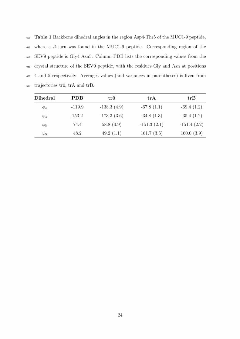
Table 1 Backbone dihedral angles in the region Asp4-Thr5 of the MUC1-9 peptide,658
where a β-turn was found in the MUC1-9 peptide. Corresponding region of the659
SEV9 peptide is Gly4-Asn5. Column PDB lists the corresponding values from the660
crystal structure of the SEV9 peptide, with the residues Gly and Asn at positions661
4 and 5 respectively. Averages values (and variances in parentheses) is fiven from662
trajectories tr0, trA and trB.663
Dihedral PDB tr0 trA trB
φ4 -119.9 -138.3 (4.9) -67.8 (1.1) -69.4 (1.2)
ψ4 153.2 -173.3 (3.6) -34.8 (1.3) -35.4 (1.2)
φ5 74.4 58.8 (0.9) -151.3 (2.1) -151.4 (2.2)
ψ5 48.2 49.2 (1.1) 161.7 (3.5) 160.0 (3.9)
24

Table 2 Hydrogen bond interactions between the SEV9 and MUC1-9 peptides and664
the H-2Kb molecule. Percentage of frames is given, from trajectories tr0, trA and665
trB, that met the geometrical criteria for hydrogen bond interaction. Distance666
between donor-acceptor atoms are taken from the initial structure (PDB column).667
Donor Acceptor PDB (A) tr0 (%) trA (%) trB (%)
Phe1P :N Tyr59A:Oη 4.13 32.4
Phe1P :N Glu63A:Oε1,2 4.61 95.6
Ser1P :N Glu63A:Oε1,2 5.83 93.2 94.1
Tyr159A:Oη Ser1P :O 2.67 59.6 91.4 94.8
Tyr59A:Oη Ser1P :Oγ 5.77 77.7 81.9
Ser1P :Oγ Tyr7A:Oη 5.82 93.2 97.9
Ser1P :Oγ Tyr171A:Oη 5.00 20.6
Ala2P :N Glu63A:Oε1,2 2.90 90.1 93.5 98.6
Lys66A:Nζ Ala2P :O 2.74 76.6 97.3 90.0
Asn70A:Nδ Pro3P :O 3.63 63.4 12.7 27.2
Arg62A:Nη2 Asp4P :Oδ1,2 6.78 19.0
Arg6P :Nη1,2 Glu24A:Oε1,2 5.21 92.4 97.2
Tyr116A:Oη Pro7P :O 4.11 28.2 7.2
Ala8P :N Glu152:Oε1,2 5.87 38.5 5.5
Trp147A:Nε Ala8P :O 2.86 11.7 70.4 98.5
Leu9P :N Asp77A:Oδ1,2 3.02 82.4
Lys146A:Nζ Leu9P :Oτ2 3.00 95.2
Tyr84A:Oη Leu9P :Oτ2 2.84 39.3
Thr143A:Oγ Pro9P :Oτ1,2 2.68 94.1 92.7
Lys146A:Nζ Pro9P :Oτ1,2 3.00 98.6 98.7
25

Figures668
26
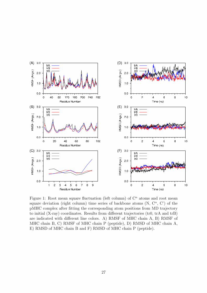
Figure 1: Root mean square fluctuation (left column) of Cα atoms and root meansquare deviation (right column) time series of backbone atoms (N, Cα, C’) of thepMHC complex after fitting the corresponding atom positions from MD trajectoryto initial (X-ray) coordinates. Results from different trajectories (tr0, trA and trB)are indicated with different line colors. A) RMSF of MHC chain A, B) RMSF ofMHC chain B, C) RMSF of MHC chain P (peptide), D) RMSD of MHC chain A,E) RMSD of MHC chain B and F) RMSD of MHC chain P (peptide).
27

Figure 2: Ramachandran plot of backbone dihedral angles of the peptide. Horizon-tal axis is for φ and vertical axis is for ψ angle respectively. The plots representpropability density maps, z-axis is the percentage of frames found within 10◦ di-hedral angle bin. The adjacent colour bar is used to identify regions of low (grey)versus high (blue) populations.
28

Figure 3: A) Ribbon representation of five selective structures of the MUC1-9/H-2Kb
complex (one frame every 2 ns) from trA trajectory, B) Ribbon representation of fiveselective structres of the MUC1-9/H-2Kb complex (one frame every 2 ns) from trBtrajectory, C) Stick representation of the peptide bound in the MHC groove from trAtrajectory, D) Stick representation of the peptide bound in the MHC groove fromtrB trajectory, E) Important hydrogen bond interactions between the pepetide andMHC molecule in the trA trajectory and F) Important hydrogen bond interactionsbetween the peptide and MHC molecule in the trB trajectory. Hydrogens wereomitted from stick represantations. Structures have been fitted to the first frameusing the backbone atoms.
29

Figure 4: Peptide’s (sticks) orientation in MHC (ribbons) binding groove in trA(A) and trB (B) trajectories. Exposure to the solvent of the region Asp4-Thr5,while Arg6 side chain orientates towards the beta-sheet floor, in the interior of thebinding groove of pocket C, in trA (C) and trB (D) trajectories.
30

Figure 5: A) and B) Backbone overlay of the Pro3-Arg6 region of the peptide fromthe trA and trB trajectories respectively. This fragment has been found in β-turnconformation for considerable amount of time. C) Backbone superimposition ofSEV9 peptide (green) from the X-ray structure with representative structures fromtrA (cyan) and trB (orange) trajectories. The differentation of backbone conforma-tion at fragment Asp4-Thr5 is well seen. Side chains of residues 2, 3, 6 and 7 sharecommon orientation towards the MHC binding groove. Interestingly, conformationsof residues at positions 1 and 9 deviate from the original structure.
31

Figure 6: A) Total number of hydrogen bonds between the peptide and the MHCmolecule, as evolved over simulation time. Data were averaged every 10 ps. B)Total number of water mediated hydrogen bonds between the pepetide and MHCmolecule, as evolved over simulation time. Data were taken every 10 ps.
32

Figure 7: Time series of apolar buried surface area (BSA) between the peptide andthe MHC molecule in tr0, trA and trB trajectories.
33