homer2 interacts with plasma membrane ca 2+ atpase and ... · homer2 interacts with plasma membrane...
TRANSCRIPT

Homer2 interacts with Plasma Membrane Ca2+
ATPase and regulates Its Activity
in Mouse Parotid Gland Acinar Cells
Eun Ho Yang
Department of Dental Science
The Graduate School, Yonsei University
Homer2 interacts with Plasma Membrane Ca2+
ATPase and regulates Its Activity
in Mouse Parotid Gland Acinar Cells
Directed by Professor Dong Min Shin
The Master’s Thesis
Submitted to the Department of Dental Science,
The graduate School of Yonsei University
in partial fulfillment of
the requirements for the degree of
Master of Medical Science
Eun Ho Yang
November, 2007
This certifies that the Master’s Thesis
of Eun Ho Yang is approved.
___________________________________________
Thesis Supervisor: Prof. Dong Min Shin
___________________________________________
Thesis Committee: Prof. Syng-Ill Lee
___________________________________________
Thesis Committee: Prof. Jeong Taeg Seo
The Graduate School
Yonsei University
November, 2007

Acknowledgements
I would like to appreciate Professor Dong Min Shin, Vice Director of
Medical Research affairs, Yonsei Health System. He always supported
through out my training and graduate school course with warm concerns and
academic knowledge. I would like to express my deepest appreciations to
Professor Syng-Ill Lee, Director of Brain Korea 21 Project, College of
Dentistry, for thoughtful advice and guide from start to the end of my doctoral
degree. I would like to have a deepest appreciation to Professor Jeong Taeg
Seo, a chairman of Oral Biology. Also, I would like to thank so much to
research assistant Hae Jo. Without his help, I could not accomplish my
experiments effectively. Finally I would like to thank my family, parents,
father-in-law and mother-in-law, my loving wife, Jung Min Park, daughter,
Jung Eun, and son, In Sung.. Whenever I was in difficulty, they always gave
me energy to overcome.

i
Contents
ABSTRACT---------------------------------------------------------------- iv
I. INTRODUCTION------------------------------------------------------ 1
II. MATERIALS AND METHODS------------------------------------ 4
1. Animals------------------------------------------------------------ 4
2. Preparation of parotid acinar cells from WT and Homer 2-/-
--------------------------------------------------------------------- 4
3. Measurement of [Ca2+] i------------------------------------------ 5
4. Immunocytochemistry------------------------------------------- 6
5. Western blot------------------------------------------------------- 7
6. Co-immunoprecipitation---------------------------------------- 8
7. Measurement of [Ca2+]o----------------------------------------- 10
8. Measurement of Ca2+ uptake and release from internal stores
--------------------------------------------------------------------- 11
9. Data analysis and statistics-------------------------------------- 11

ii
III. RESULTS-------------------------------------------------------------- 13
1. Deletion of Homer 2 does not affect polarized expression of
IP3Rs and IP3-mediated Ca2+ release.-------------------------- 13
2. Level of PMCA is selectively increase in the parotid acini of
Homer 2-/- mice.--------------------------------------------------- 14
3. The rates of [Ca2+] efflux are increased in Homer 2-/- cells.
--------------------------------------------------------------------- 17
4. Homer 2 is interacted with PMCA in parotid acinar cells. - 18
IV. DISCUSSION--------------------------------------------------------- 25
V. REFERENCES--------------------------------------------------------- 29
VI. ABSTRACT (in Korean) -------------------------------------------- 34
iii
List of Figures
Fig. 1. Localization of Homer2 and IP3 receptors in parotid gland acinar
cells from WT and Homer 2-/- cells.------------------------------------ 15
Fig. 2. Ca2+ uptake and IP3-mediated Ca2+ release from WT and Homer
2-/- cells.--------------------------------------------------------------------- 16
Fig. 3. Expression of Ca2+ transporters in WT and Homer 2-/- cells.
----------------------------------------------------------------------- 19
Fig. 4. Plasma membrane Ca2+ ATPase activity in WT and Homer 2-/-
cells. ---------------------------------------------------------------------- 20
Fig. 5. Characterization of Ca2+ signaling in WT and Homer 2-/- cells.
---------------------------------------------------------------------- 22
Fig. 6. Alignment of PMCAs with proline-rich motif, PPXXF,
interacting with EVH domain of Homers.----------------------------- 23
Fig. 7. Co-immunoprecipitation between Homer 2 and PMCA.---- 24
iv
ABSTRACT
Homer2 interacts with Plasma Membrane Ca2+ ATPase
and regulates Its Activity
in Mouse Parotid Gland Acinar Cells
Yang, Eun Ho
Department of Dental Science
The Graduate School, Yonsei University
(Directed by Professor Dong Min Shin)
The Homers are scaffold proteins and consist of an N-terminal Ena/VASP
homology 1 (EVH) protein-binding domain and C-terminal leucin
zipper/coiled-coil domain. The EVH domain recognizes the proline-rich
motifs (PPXXF, PPXF, and LPSSP) and binds many Ca2+ signaling proteins
including G protein-coupled receptors (GPCRs), inositol 1,4,5-triphosphate
(IP3) receptors (IP3Rs), ryanodine receptors (RyRs), and TRP channels.
Therefore, Homers critically control Ca2+ signaling and thereby regulate

v
dendritic spine morphogenesis, remodeling of synapses, and synaptic
clustering of CNS neurons, and lead to changes in neuronal transcriptional
activity. However, their role in Ca2+ signaling of non-neuronal cells is not
well known, except that Homer2 tunes GPCRs stimulus intensity by
regulating RGS proteins and PLCβ GAP activities in pancreatic acinar cells.
In the present work, the role of Homer2 in Ca2+ signaling in parotid gland
acinar cells using Homer2-/- mice was investigated with microfluororimeter,
immunofluoroscence, and co-immunoprecipitation. Homer2 showed polarized
luminal localization in these cells, but the deletion of Homer2 did not affect a
localization of IP3Rs or IP3R channel activity. The protein expression level of
plasma membrane Ca2+ ATPase (PMCA) was increased in Homer2-/-, whereas
sarco/endo plasmic reticulum Ca2+ ATPase was not. Moreover, deletion of
Homer2 increased PMCA activity and co-immunoprecipitation showed that
Homer2 interacted with PMCAs. These results suggest that Homer2 may play
an important role in regulation of PMCA expression and PMCA-mediated
Ca2+ signaling in mouse parotid gland acinar cells.
----------------------------------------------------------------------------------------------
Keywords: Homer2; Plasma membrane Ca2+ ATPase; Proline-rich motif;
Parotid Gland Acinar Cells
1
Homer2 interacts with Plasma Membrane Ca2+ ATPase
and regulates Its Activity
in Mouse Parotid Gland Acinar Cells
Eun Ho Yang
Department of Dental Science
The Graduate School, Yonsei University
(Directed by Professor Dong Min Shin)
I. INTRODUCTION
Homers are scaffolding proteins that consists of three members, Homer 1,
Homer 2, and Homer 3, and several splice variants (Fagni et al., 2000;
Szumlinski et al., 2006). Homers are composed of an EVH protein-binding
domain, a coiled-coil multimerization domain, and leucine zipper (Fagni et al.,
2002). The EVH domain is a protein-protein binding module that recognizes
the proline-rich motifs PPXXF, PPXF, and LPSSP (Brakeman et al., 1997;
2
Kato et al., 1998; Tu et al., 1998; Yuan et al., 2003), and binds the GPCR
mGluR1/5, canonical transient receptor potential (TRPC) channels, inositol
1,4,5-triphosphate (IP3) receptors (IP3Rs), ryanodine receptors, and the Shank
family of scaffolding proteins (Tu et al., 1998; Xiao et al., 1998; Tu et al.,
1999; Feng et al., 2002; Shin et al., 2003; Yuan et al., 2003). These binding
receptors are known to consist of a biochemical component in receptor-
mediated Ca2+ signaling.
Intracellular Ca2+ is a common second messenger which has close
relationships to fertilization, muscle contraction, neurotransmitter release,
exocytosis, learning and memory (Berridge et al., 2003). Especially, there is a
close connection between GPCRs and Ca2+ signaling in parotid acinar cell and
these cells also represent an excellent model system not only for the study of
Ca2+ signaling in general but also to investigate cross-talk between cAMP and
Ca2+ signaling (Brini & Carafoli, 2000). Ca2+ release from the endoplasmic
reticulum (ER) leads to activation of store-operated Ca2+ channels in the
plasma membrane, and Ca2+ release and influx increase the intracellular Ca2+

3
concentration ([Ca2+] i). Subsequently, the plasma membrane Ca2+ ATPase
(PMCA) and sarco/endoplasmic reticulum Ca2+ ATPase (SERCA) removes
Ca2+ from the cytosol to reduce Ca2+ toward resting levels until [Ca2+] i
stabilizes at a plateau (Kiselyov et al., 2003). Several previous reports
demonstrated that raising cAMP potentiates Ca2+ release from intracellular
Ca2+ through PKA-mediated phosphorylation of IP3Rs and enhances the rate
of Ca2+ clearance by complex modulation of PMCA activity in parotid acinar
cells (Bruce et al., 2002a; Bruce et al., 2002b). Specially, IP3Rs, which
include proline-rich sequence, can be bound to Homer proteins. Accordingly,
it is possible that Homer proteins has regulated effects for Ca2+ signaling in
cells. However, their role in Ca2+ signaling of non-neuronal cells is not well
known. Therefore, in the present work, the role and functions of Homer 2 in
Ca2+ signaling using parotid acinar cells from wild-type (WT) and Homer 2
mutant (Homer 2-/-) mice was investigated.

4
II. MATERIALS AND METHODS
1. Animals
Wild-type (WT) and Homer 2 mutant (Homer 2-/-) mice have been previously
described (Shin et al., 2003). Homer 2-/- mice have a normal life span similar
to WT littermates. All animal protocols were performed according to
institutional guidelines.
2. Preparation of parotid acinar cells from WT and Homer 2-/-
WT (25-28g) and Homer 2-/- (25-28g) mice were sacrificed by cervical
dislocation. The cells were prepared from the parotids of WT and Homer 2-/-
mice by limited collagenase digestion as previously described (Zeng et al.,
1997). After isolation, the acinar cells were resuspended in an extracellular
physiologic salt solution (PSS), the composition of which was as follows (in
mM): 140 NaCl, 5 KCl, 1 MgCl2, 1 CaCl2, 10 HEPES, and 10 glucose,
adjusted to pH 7.4 with NaOH. The osmolality of the extracellular solution

5
(measured with a FISKE 110 osmometer), was 310 mOsm.
3. Measurement of intracellular calcium concentration ([Ca2+]i)
Cells, which were from both types, were incubated for 40 min in PSS
containing 5 µM Fura-2/AM (Teflabs Inc., Austin, TX) with pluronic F-127
to enhance dye loading. Changes in [Ca2+] i were measured by means of Fura-2
fluorescence, with excitation wavelengths of 340 and 380 nm, and an
emission wavelength of 510 nm at room temperature. Background
fluorescence was subtracted from the raw signals at each excitation
wavelength before calculating the fluorescence ratio as follows: Ratio =
F340/F380. The emitted fluorescence was monitored with a CCD camera
(Photon Technology International Inc., Lawrenceville, NJ) attached to an
inverted microscope. Fluorescence images were obtained at 2 s intervals. All
data are presented as means ± SEM.
6
4. Immunocytochemistry
The immunostaining procedure was described previously (Shin et al., 2003).
In brief, cells from WT and Homer 2-/- mice attached to glass coverslips were
fixed and permeabilized with 0.5 ml of cold methanol for 10 min at –20℃.
After removal of methanol, the cells were fixed with 4% formaldehyde for 20
min at room temperature, followed by permeabilization with 0.05 % Triton X-
100. After removal of methanol or Triton X-100, the cells were washed with
PBS and incubated in 0.5 ml of PBS containing 50 mM glycine for 10 min at
room temperature. This buffer was aspirated and the nonspecific sites were
blocked by 1-h incubation at room temperature with 0.25 ml of PBS
containing 5% goat serum, 1% BSA, and 0.1% gelatin (blocking medium).
The medium was aspirated and replaced with 50 µl of blocking medium
containing control serum or a 1:50 dilution of Ab against Homer 2, or a 1:100
dilution of Abs against IP3R1, 2, or 3. After incubation with the primary Ab
overnight at 4℃ and three washes with the incubation buffer (same as
blocking buffer, but with serum), the Abs were detected with goat anti-rabbit

7
or anti-mouse IgG tagged with fluorescein or rhodamine. Images were
collected with a confocal LSM 510 laser scanning microscope (Zeiss,
Göttingen, Germany).
5. Western blot
Protein extracts were prepared by parotid acini from WT and Homer 2-/- mice
as follows. Pure acinar cells were washed with ice-cold PBS and then
resuspended in lysis buffer containing (in mM): 150 NaCl, 10 Tris (pH 7.8
with HCl), 1 EDTA, 1% NP-40, and 0.1% SDS. Protease and phosphatase
inhibitors were added to the lysis buffer containing (in mM): 2 Na3VO4, 10
NaF, 10 µg/ml aprotinin, 10 µg/ml leupeptine, and 10 µg/ml PMSF. The
samples were then centrifuged at 12,000 rpm for 20 min at 4℃, and separated
by SDS–PAGE. Proteins were probed with a 1:500 dilution of Ab against
SERCA2b; 1:2000 dilution of Abs against IP3R1, 2, and 3; and 1:200 dilution
of Ab against PMCA.
8
6. Co-immunoprecipitation
The co-immunoprecipitation procedure was modified from Shin et al. (2000).
Parotid microsomes of WT mice were prepared by homogenizing a minced
parotid in a buffer containing (in mM): 20 Mops, 250 sucrose, 1 EDTA, 1
MgCl2, 10 benzamidine, and 0.2 PMSF, pH-adjusted to 6.7 with KOH. The
homogenate was centrifuged at 1,200 rpm for 10 min. The supernatant was
collected and centrifuged at 1,800 rpm for 10 min at 4℃. The pellet was
resuspended in the same buffer while avoiding suspension of the hard, white-
colored granular fraction in the bottom of the tube. When needed, the fraction
enriched in secretory granules was collected in homogenization buffer into a
separate tube. To avoid protein degradation by digestive enzymes, IP was
initiated immediately after completion of microsomal preparation. Parotid
microsomes were extracted by a 1-h incubation on ice with a buffer
containing (in mM): 50 Tris (pH 6.8 with HCl), 150 NaCl, 3 EDTA, 2 EGTA,
and 0.5 % Triton X-100 supplemented with protease inhibitors (0.2 mM
PMSF, 10 µg/ml leupeptin, 15 µg/ml aprotinin, 1 mM benzamidine). The
9
lysate was cleared by centrifugation at 7,000 rpm for 10 min. About 300 µl of
the extract was further incubated with 15 µl of Sepharose A beads for 1 h at
4℃ and centrifuged for 2 min at 7,000 rpm to remove the beads. The cleared
supernatant was incubated with 5 µl anti-PMCA, or 5 µl anti-Homer 2 Abs for
30 min before addition of 30 µl Sepharose A beads and an overnight
incubation at 4℃ under gentle agitation. The beads were washed five times
with 0.8 ml lysis buffer and stripped of proteins by boiling in a 50 µl of SDS
sample buffer. To test the effect of the actin cytoskeleton on the binding of the
Homer 2 and PMCA, buffer or 20 µg/ml of the NH2-terminal fragment of
gelsolin was added to equal portions of beads after the second wash. After 20-
min incubation at 4℃, the beads were washed three times with lysis buffer
and the proteins remaining attached to the beads were released by boiling in a
sample buffer. Released proteins were separated by an SDS-PAGE using 7.5%
polyacrylamide gels. The separated proteins were transferred to 0.2 µm
polyvinylidene difluoride membranes, and the membranes were blocked by a
1-h incubation at room temperature in 5% nonfat dry milk in a solution

10
containing 150 mM NaCl, 20 mM Tris (pH 7.5 with HCl), and 0.05% Tween
20 (TTBS). The Homer 2 and PMCA were detected by a 1~2-h incubation of
individual membranes with the respective Abs diluted in TTBS.
7. Measurement of extracellualr calcium concentration ([Ca2+]o)
To measure directly the rate of Ca2+ efflux by PMCA, I measured the
appearance of Ca2+ in the external medium using the procedure modified from
Zhang et al. (1992). Intact parotid acini were washed once and then suspended
in medium containing (in mM): 120 KCl, 20 NaCl, 10 glucose, 0.002 free
acid fura-2/AM, and 10 HEPES, pH-adjusted to 7.4 with KOH. After
initiation of fluorescence recording, 7.5 µM EGTA was added to reduce the
extracellular Ca2+ concentration to ~100 nM. After establishing a baseline leak
for ~1 min, the cells were stimulated with 1 mM carbachol. At the end of
experiment, the signals were calibrated simply by adding 1 mM CaCl2 and
then 1 mM MnCl2 to the medium as previously described (Zhang & Muallem,
1992).

11
8. Measurement of Ca2+ uptake and release from internal stores
IP3-mediated Ca2+ release from internal stores was measured in SLO-
permeabilized cells as described before (Xu et al., 1996). Cells washed with a
high K+, Chelax-treated medium were added to the same medium containing
an ATP regeneration system (comprised of 3 mM ATP, 5 mM MgCl2, 10 mM
creatine phosphate, and 5 U/ml creatine kinase), a cocktail of mitochondrial
inhibitors, 2 µM Fluo3 and 3 mg/ml SLO (Difco). In this medium, the cells
were almost instantaneously permeabilized so that Ca2+ uptake into the ER
could be measured immediately. Uptake of Ca2+ into the ER was allowed to
continue until medium [Ca2+] was stabilized. Then IP3 was added in
increasing concentrations to measure the extent of Ca2+ release and the
potency of IP3 in mobilizing Ca2+ from the ER.
9. Data analysis and statistics
All numeric values are represented as the mean ± S.E. The statistical
significance of the data was determined using Student’s unpaired t-test.

12
Statistical significance was set at p < 0.05 level.

13
III. RESULTS
1. Deletion of Homer 2 does not affect polarized expression of IP3Rs and
IP3-mediated Ca2+ release.
The mGluRs and its related receptors are well known the targeting and
binding receptors with Homer proteins (Tu et al., 1998; Yuan et al., 2003).
Therefore, I first examined the localization and expression of Homer 2 and
IP3Rs in parotid acinar cells of wild-type (WT) and Homer 2-/- mice. In WT
mice, the immunoreactivity of Homer 2 and IP3Rs was primary observed in
the apical pole of parotid acinar cells, whereas this immunoreactivity did not
affect expression and localization of any IP3R isoform except for Homer 2 in
Homer 2-/- mice (Fig. 1). I next examined whether a lack of Homer 2 affects
activity of IP3Rs that regulate the Ca2+ content in the stores by the
concentration of IP3 and [Ca2+] i (Xu et al., 1996; Thrower et al., 2001).
Parotid acinar cells of WT and Homer 2-/- mice are fully permeabilized to
SLO within 10-15 sec and reduced [Ca2+] of the incubation medium to the 50-

14
80 nM range within 2 min of incubation at 37 ℃. Addition of increasing
concentrations of IP3 and the muscarinic agonist carbachol resulted in the
same potency of [Ca2+] increase from stores of SLO-permeabilized WT and
Homer 2-/- cells (Fig. 2A-B). Thus, no compensatory effect in localization and
activity of IP3Rs were observed in Homer 2-/- mice.
2. Level of PMCA is selectively increased in the parotid acini of Homer 2-/-
mice
To establish whether expression of Ca2+ transporters is normal in Homer 2-/-
mice, I examined protein expression by Western blotting using parotid acinar
cells from WT and Homer 2-/- mice. The expression level of PMCA was
significantly increased to 2.5 ± 0.1-fold of WT in Homer 2-/- mice (n = 4, p <
0.001, Fig. 3). However, there was no significant change to SERCA2b
between WT and Homer 2-/- mice (1.2 ± 0.2 fold of WT, n = 4, p = 0.4). The
results in Fig. 1 and Fig. 2 suggest that lack of Homer 2 has no effect on
expression and activity of IP3Rs. Similarly, the levels of IP3Rs were
15
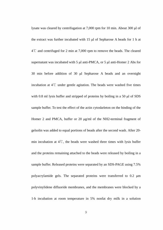
Fig. 1. Localization of Homer2 and IP3 receptors in parotid gland acinar
cells from WT and Homer 2-/- cells.
Parotid acini from WT and Homer 2-/- mice were stained for Homer 2, IP3R1,
IP3R2, and IP3R3. Note lack of effect of Homer 2 deletion on expression and
localization of the IP3Rs.
WT Homer 2-/-
Homer 2
IP3R1
IP3R2
IP3R3
WT Homer 2-/-
Homer 2
IP3R1
IP3R2
IP3R3

16
Fig. 2. Ca2+ uptake and IP3-mediated Ca2+ release from WT and Homer
2-/- cells.
A, Cells from WT and Homer 2-/- mice were permeabilized with SLO and
allowed to reduce [Ca2+] i of the incubation medium to ~75 nM. Next, Ca2+
release was measured by adding increasing concentrations of IP3 (arrows of
solid lines) and carbachol (arrows of dashed lines). B, Summarizes the results
obtained in the experiment with cells prepared from each of WT (solid lines)
and Homer 2-/- (dashed lines) mouse. The results were expressed as the mean
± S.E.
A
Carbachol
IP3
0
160
[Ca2+
] (n
M)
WT160
Carbachol
Homer 2-/-
IP3
0
[Ca2+
] (nM
)
A
Carbachol
IP3
0
160
[Ca2+
] (n
M)
WT160
Carbachol
Homer 2-/-
IP3
0
[Ca2+
] (nM
)
Carbachol
IP3
0
160
[Ca2+
] (n
M)
WT160
Carbachol
Homer 2-/-
IP3
0
[Ca2+
] (nM
)
1 2 3
Carbachol[IP3]
1
(mM)
B
0
25
50
75
100
Homer 2-/-
WT
Ca2+
rel
ease
from
ER
(nM
)
(μμμμM)
1 2 3
Carbachol[IP3]
1
(mM)
B
0
25
50
75
100
Homer 2-/-
WT
Ca2+
rel
ease
from
ER
(nM
)
(μμμμM)
17
unaffected in Homer 2-/- cells (n = 4, data not shown). These finding suggest
that Homer 2 has effect on the Ca2+ signaling by changing of component that
transports Ca2+ into and out of the cytosol.
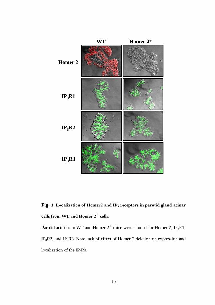
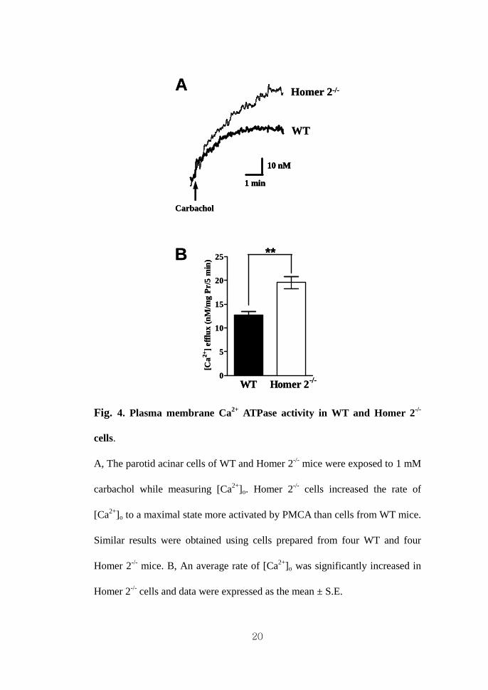
3. The rates of [Ca2+] efflux are increased in Homer 2-/- cells.
The major routes for Ca2+ clearance in non-excitable cells, such as parotid
acinar cells, are believed to be Ca2+ uptake into the ER by the SERCA, Ca2+
efflux across the plasma membrane by the PMCA, and Ca2+ uptake into
mitochondria (Bruce et al., 2002b; Gorr et al., 2005; Homann et al., 2006).
The results in Fig. 2B and Fig. 3 suggest that deletion of Homer 2 has
unaffected on expression and activity of SERCA. Therefore, I measured the
change of [Ca2+]o at low external Ca2+ concentration during stimulation with 1
mM carbachol. As shown in Fig. 4A, [Ca2+]o was significantly increased by
carbachol in both cell types and the change of [Ca2+]o in Homer 2-/- cells was
approximately 1.5-fold more abundant than in WT cells (n = 4, p < 0.01, Fig.
4B). To examine the effect on the Ca2+ clearance by PMCA between WT and

18
Homer 2-/- mice, I performed the experiment following inhibition of SERCA
activity by 100 µM CPA. The increase in [Ca2+] i reached a maximum and then
declined slowly to new steady state, representing a balance of Ca2+ efflux and
Ca2+ influx. Exchange the external solution, addition of 1 mM carbachol and
10 µM atropine in Ca2+ free solution, evoked an immediate clearance of Ca2+
that was primarily because of the PMCA (Fig. 5A). Then, I compared with
slope of Ca2+ clearance during the exchanged the external solution in parotid
acinar cells of WT and Homer 2-/- mice. Under these conditions, Homer 2-/-
cells induced 1.5-fold increase in the slope of Ca2+ clearance (1.5 ± 0.3-fold of
slope, n = 4, p < 0.05, Fig. 5B), indicating that the level of Ca2+ clearance due
to the increase can be responsible for the potentiation of PMCA activity in the
Homer 2-/- mice.
4. Homer 2 is interacted with PMCA in parotid acinar cells.
To identify the specificity of interaction between the PMCA subtypes and
PPXXF motif which was interacted EVH domain of Homers, I investigated

19
Fig. 3. Expression of Ca2+ transporters in WT and Homer 2-/- cells.
A, The level of PMCA and SERCA2b were analyzed in extracts prepared
from parotid acini of four WT and four Homer 2-/- mice. Expression was
analyzed by densitometry. B, Means of protein expression levels between WT
and Homer 2-/- cells. Expression level was significantly increased for PMCA,
except SERCA2b in Homer 2-/- cells. Data were normalized to the expression
level in cells of WT mice and expressed as the mean ± S.E. *** p < 0.001
(compared with WT).
PMCA SERCA2b0.0
0.5
1.0
1.5
2.0
2.5
3.0
***
Rel
ativ
e p
rote
in e
xpre
ssio
n le
vel
A WT Homer 2-/-
SERCA2b
PMCA
BWTHomer 2-/-
PMCA SERCA2b0.0
0.5
1.0
1.5
2.0
2.5
3.0
***
Rel
ativ
e p
rote
in e
xpre
ssio
n le
vel
A WT Homer 2-/-
SERCA2b
PMCAWT Homer 2-/-
SERCA2b
PMCA
SERCA2b
PMCA
BWTHomer 2-/-
WTHomer 2-/-
20
Fig. 4. Plasma membrane Ca2+ ATPase activity in WT and Homer 2-/-
cells.
A, The parotid acinar cells of WT and Homer 2-/- mice were exposed to 1 mM
carbachol while measuring [Ca2+]o. Homer 2-/- cells increased the rate of
[Ca2+]o to a maximal state more activated by PMCA than cells from WT mice.
Similar results were obtained using cells prepared from four WT and four
Homer 2-/- mice. B, An average rate of [Ca2+]o was significantly increased in
Homer 2-/- cells and data were expressed as the mean ± S.E.
A
Carbachol
WT
Homer 2-/-
1 min
10 nM
A
Carbachol
WT
Homer 2-/-
1 min
10 nM
1 min
10 nM
B
WT Homer 2-/-0
5
10
15
20
25 **
[Ca2+
] eff
lux
(nM
/mg
Pr/
5 m
in)B
WT Homer 2-/-0
5
10
15
20
25 **
[Ca2+
] eff
lux
(nM
/mg
Pr/
5 m
in)
21
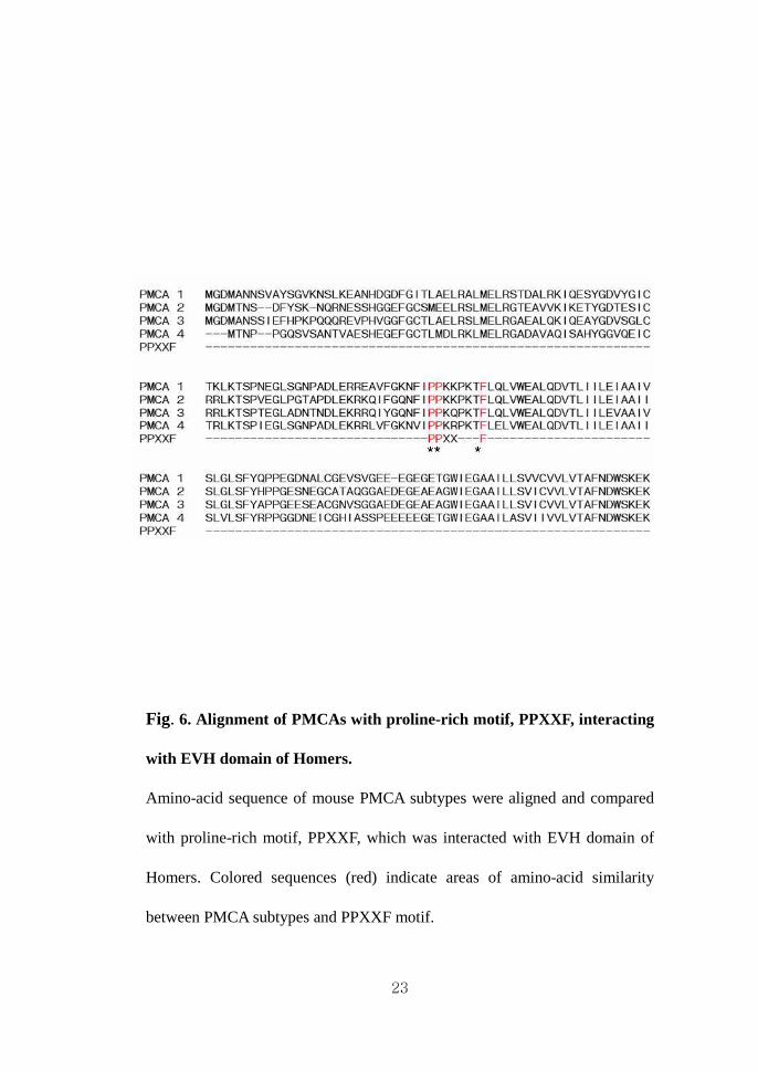
amino-acid sequences (Fig. 6). The amino-terminal 91~98 residues of PMCA
subtypes are identical to the PPXXF motif. This finding suggests that Homer
2 has an interaction with PMCA subtypes. I then examined whether Homer 2
selectively binds to PMCA in parotid acinar cells of WT mice using an in vivo
co-immunoprecipitation assay. After cell lysates were prepared,
immunoprecipitated with anti-PMCA antibodies, and subsequently
immunoblotted with anti-Homer 2 antibodies. As shown in left of Fig. 7, the
PMCA was able to bind to endogenous Homer 2 (left of Fig. 7, fourth lane),
whereas no signal was detected from antibodies non- treated cells (third lanes).
When co-immunoprecipitation assay was performed in reverse with anti-
Homer 2 antibodies followed by immunoblotting with anti-PMCA antibodies,
the result was the same (right of Fig. 7, fourth lane). These results strongly
suggest that the Homer 2 is associated (directly or indirectly) with PMCA
subtypes.

22
Fig. 5. Characterization of Ca2+ signaling in WT and Homer 2-/- cells.
A, [Ca2+] i of parotid acini of four WT and four Homer 2-/- mice were elevated
by 1 mM carbachol and inhibition of SERCA with 100 µM CPA. The increase
in [Ca2+] i maximum and then declined to a new steady state, representing a
balance of Ca2+ efflux and Ca2+ influx. Ca2+ clearance was initiated by
removal of external Ca2+ and addition of 10 µM atropine. B, An average of
Ca2+ clearance rate was significantly increased in Homer 2-/- cells. Data were
normalized to the fold of slope in cells of WT mice and expressed as the mean
± S.E. * p < 0.05, ** p < 0.01 (compared with WT).
Time (sec)0 400 800
0.2
0.3
0.4
0.5
0.6
0.7
0.8
Rat
io (
340/
380)
Regular1 mM Carbachol 100 μμμμM CPACa2+ free + 10 μμμμM Atropine
A
Time (sec)0 400 800
0.2
0.3
0.4
0.5
0.6
0.7
0.8
Rat
io (
340/
380)
Regular1 mM Carbachol 100 μμμμM CPACa2+ free + 10 μμμμM Atropine
Regular1 mM Carbachol 100 μμμμM CPACa2+ free + 10 μμμμM Atropine
Regular1 mM Carbachol 100 μμμμM CPACa2+ free + 10 μμμμM Atropine
A
WT Homer 2-/-0.0
0.5
1.0
1.5
2.0 *
Fol
d of
slo
pe
B
WT Homer 2-/-0.0
0.5
1.0
1.5
2.0 *
Fol
d of
slo
pe
B
23
Fig. 6. Alignment of PMCAs with proline-rich motif, PPXXF, interacting
with EVH domain of Homers.
Amino-acid sequence of mouse PMCA subtypes were aligned and compared
with proline-rich motif, PPXXF, which was interacted with EVH domain of
Homers. Colored sequences (red) indicate areas of amino-acid similarity
between PMCA subtypes and PPXXF motif.

24
Fig. 7. Co-immunoprecipitation between Homer2 and PMCA.
The interaction between PMCA and Homer 2 were co-immunoprecipitated
(IP) using cell lysates, which were prepared from parotid acini of four WT
mice. Left panel showed IP of PMCA and probing for Homer 2, and right
panel showed IP of Homer 2 and probing for PMCA. In IP experiments, Input
donated extract samples used for Western blot, Only Ab donated control IP
using Ab without extract in the IP reaction, –Ab donated control IP using
extract without Ab in the IP reaction, and +Ab donated IP using extract and
Ab in the IP reaction.
IP : PMCA
Homer 2
PMCA
Input Only Ab + Ab
IP : Homer 2
- Ab Input Only Ab - Ab + Ab
IP : PMCA
Homer 2
PMCA
Input Only Ab + Ab
IP : Homer 2
- Ab Input Only Ab - Ab + Ab
25
IV. DISCUSSION
In the present study I demonstrated a novel interaction between the EVH
domain of Homer 2 and proline-rich motif of PMCA using parotid acinar cells
of Homer 2-deficient mice and provided firm evidence in support of a critical
role for Homer 2 in PMCA-mediated Ca2+ signaling using several assays.
Based on the previous reports concerning the modular structure of Homer
proteins in CNS, Homer proteins have ability to bind the GPCR receptors
such as mGluR1/5 and IP3Rs, and act as scaffolding proteins to assemble Ca2+
signaling complexes in cellular microdomains (Kato et al., 1998; Tu et al.,
1998; Xiao et al., 1998; Kammermeier et al., 2000; Fagni et al., 2002). I,
therefore, hypothesized that there is also a functional relationship through the
interaction between Homer 2 and IP3Rs in exocrine cells. First, I found that
Homer 2 and IP3Rs were expressed in the apical pole of parotid acinar cells
using immunocytochemistry. In a variety of exocrine cells, the apical region is
regarded as the “trigger zone” from which Ca2+ waves are initiated (Lee et al.,
26
1997b; Tojyo et al., 1997). Therefore, the apical region seems relative
abundance of Ca2+ release channels and/or expression of the most sensitive
channels. Immunocytochemical studies have reported that the extrame apical
region is highly enriched in all IP3R types (Takemura et al., 1999; Zhang et al.,
1999; Shin et al., 2003). However, I also found that deletion of Homer 2 has
no effect on expression of any IP3R isoform by measurement of Ca2+ uptake
and release from internal stores. The only effect observed is increased PMCA
protein expression in parotid acinar cells of Homer 2-deficient mice. These
Homer 2 effects were originally reported in pancreatic acinar cells by Shin
and his collaborators, but they observed increase of SERCA2b protein
expression and activity in pancreatic acini and brain of Homer 2-deficient
mice (Shin et al., 2003). It might be possible for Homer 2 to play roles via
interactions with several proteins such as PMCA or SERCA2b that are
modulated by the activation of Ca2+ signaling system.
Second, measurements of PMCA activity and co-immunoprecipitation
binding assays confirmed that Homer 2 is interacted with PMCA and
27
increased rate of [Ca2+] i clearance by PMCA in parotid acinar cells of Homer
2-deficient mice. The PMCA is a ubiquitously expressed P-type ATPase with
a high affinity for Ca2+ and has been thought to be the major mechanism for
the maintenance of resting [Ca2+] i (Monteith et al., 1998; Kiselyov et al.,
2003). The PMCA consists of four members, PMCA 1, PMCA 2, PMCA 3,
and PMCA 4, and PMCA isoforms (PMCA 1, 2, and 4, especially PMCA 2)
are expressed in apical region of parotid and submandibular gland cells
(Homann et al., 2006). Furthermore, PMCA and SERCA2a are highly
expressed in the apical region, while SERCA2b is mostly expressed in the
basal pole and basolateral region in salivary gland and pancreatic acinar cells
(Lee et al., 1997a). Therefore, it is possible that the Homer 2 in the apical
region of parotid acinar cells is physically associated with PMCA and
potentially allowing for functional modulation between the proteins in Ca2+
signaling by GPCRs. Sgambato-Faure et al. (2006), recently, reported that
endogenous Ania-3, a member of the Homer 1 family, and Homer 1 are co-
expressed with PMCAs, especially PMCA 2 in the soma and dendrites of rat

28
hippocampal neurons. Similarly, PMCA 2 significantly decreases the
expression levels of mGluR1, IP3R1, and Homer proteins (Homer 1b/c and
Homer 3) in cerebellar Purkinje neurons of PMCA-deficient mice (Kurnellas
et al., 2007). It might be possible that Homer 2 cross-talk with IP3Rs-
mediated Ca2+ signaling via regulation of PMCA, serving as the basis for its
negative regulator effect. This study provides a novel modular model in Ca2+
signaling for examining the role and functions of Homer 2 in the non-
neuronal exocrine cells.

29
V. REFERENCES
Berridge MJ, Bootman MD & Roderick HL. Calcium signalling: dynamics,
homeostasis and remodelling. Nat Rev Mol Cell Biol 2003;4:517-529.
Brakeman PR, Lanahan AA, O'Brien R, Roche K, Barnes CA, Huganir RL &
Worley PF. Homer: a protein that selectively binds metabotropic glutamate
receptors. Nature 1997;386:284-288.
Brini M & Carafoli E. Calcium signalling: a historical account, recent
developments and future perspectives. Cell Mol Life Sci 2000;57:354-370.
Bruce JI, Shuttleworth TJ, Giovannucci DR & Yule DI. Phosphorylation of
inositol 1,4,5-trisphosphate receptors in parotid acinar cells. A mechanism for
the synergistic effects of cAMP on Ca2+ signaling. J Biol Chem
2002a;277:1340-1348.
Bruce JI, Yule DI & Shuttleworth TJ. Ca2+-dependent protein kinase--a
modulation of the plasma membrane Ca2+-ATPase in parotid acinar cells. J
Biol Chem 2002b;277:48172-48181.
Fagni L, Chavis P, Ango F & Bockaert J. Complex interactions between
mGluRs, intracellular Ca2+ stores and ion channels in neurons. Trends
Neurosci 2000;23:80-88.
Fagni L, Worley PF & Ango F. (2002). Homer as both a scaffold and
transduction molecule. Sci STKE 2002; RE8.
Feng W, Tu J, Yang T, Vernon PS, Allen PD, Worley PF & Pessah IN. Homer
regulates gain of ryanodine receptor type 1 channel complex. J Biol Chem
30
2002;277:44722-44730.
Gorr SU, Venkatesh SG & Darling DS. Parotid secretory granules: crossroads
of secretory pathways and protein storage. J Dent Res 2005;84:500-509.
Homann V, Kinne-Saffran E, Arnold WH, Gaengler P & Kinne RK. Calcium
transport in human salivary glands: a proposed model of calcium secretion
into saliva. Histochem Cell Biol 2006;125:583-591.
Kammermeier PJ, Xiao B, Tu JC, Worley PF & Ikeda SR. Homer proteins
regulate coupling of group I metabotropic glutamate receptors to N-type
calcium and M-type potassium channels. J Neurosci 2000;20:7238-7245.
Kato A, Ozawa F, Saitoh Y, Fukazawa Y, Sugiyama H & Inokuchi K. Novel
members of the Vesl/Homer family of PDZ proteins that bind metabotropic
glutamate receptors. J Biol Chem 1998;273:23969-23975.
Kiselyov K, Shin DM & Muallem S. Signalling specificity in GPCR-
dependent Ca2+ signalling. Cell Signal 2003;15:243-253.
Kurnellas MP, Lee AK, Li H, Deng L, Ehrlich DJ & Elkabes S. Molecular
alterations in the cerebellum of the plasma membrane calcium ATPase 2
(PMCA2)-null mouse indicate abnormalities in Purkinje neurons. Mol Cell
Neurosci 2007;34:178-188.
Lee MG, Xu X, Zeng W, Diaz J, Kuo TH, Wuytack F, Racymaekers L &
Muallem S. Polarized expression of Ca2+ pumps in pancreatic and salivary
gland cells. Role in initiation and propagation of [Ca2+] i waves. J Biol Chem
1997a;272:15771-15776.
Lee MG, Xu X, Zeng W, Diaz J, Wojcikiewicz RJ, Kuo TH, Wuytack F,
Racymaekers L & Muallem S. Polarized expression of Ca2+ channels in

31
pancreatic and salivary gland cells. Correlation with initiation and propagation
of [Ca2+] i waves. J Biol Chem 1997b;272:15765-15770.
Monteith GR, Wanigasekara Y & Roufogalis BD. The plasma membrane
calcium pump, its role and regulation: new complexities and possibilities. J
Pharmacol Toxicol Methods 1998;40:183-190.
Sgambato-Faure V, Xiong Y, Berke JD, Hyman SE & Strehler EE. The
Homer-1 protein Ania-3 interacts with the plasma membrane calcium pump.
Biochem Biophys Res Commun 2006;343:630-637.
Shin DM, Dehoff M, Luo X, Kang SH, Tu J, Nayak SK, Ross EM, Worley PF
& Muallem S. Homer 2 tunes G protein-coupled receptors stimulus intensity
by regulating RGS proteins and PLCbeta GAP activities. J Cell Biol
2003;162:293-303.
Shin DM, Zhao XS, Zeng W, Mozhayeva M & Muallem S. The mammalian
Sec6/8 complex interacts with Ca2+ signaling complexes and regulates their
activity. J Cell Biol 2000;150:1101-1112.
Szumlinski KK, Kalivas PW & Worley PF. (2006). Homer proteins:
implications for neuropsychiatric disorders. Curr Opin Neurobiol
2006;16:251-257.
Takemura H, Yamashina S & Segawa A. Millisecond analyses of Ca2+
initiation sites evoked by muscarinic receptor stimulation in exocrine acinar
cells. Biochem Biophys Res Commun 1999;259:656-660.
Thrower EC, Hagar RE & Ehrlich BE. Regulation of Ins(1,4,5)P3 receptor
isoforms by endogenous modulators. Trends Pharmacol Sci 2001;22:580-586.
Tojyo Y, Tanimura A & Matsumoto Y. Imaging of intracellular Ca2+ waves

32
induced by muscarinic receptor stimulation in rat parotid acinar cells. Cell
Calcium 1997;22:455-462.
Tu JC, Xiao B, Naisbitt S, Yuan JP, Petralia RS, Brakeman P, Doan A, Aakalu
VK, Lanahan AA, Sheng M & Worley PF. Coupling of mGluR/Homer and
PSD-95 complexes by the Shank family of postsynaptic density proteins.
Neuron 1999;23:583-592.
Tu JC, Xiao B, Yuan JP, Lanahan AA, Leoffert K, Li M, Linden DJ & Worley
PF. Homer binds a novel proline-rich motif and links group 1 metabotropic
glutamate receptors with IP3 receptors. Neuron 1998;21:717-726.
Xiao B, Tu JC, Petralia RS, Yuan JP, Doan A, Breder CD, Ruggiero A,
Lanahan AA, Wenthold RJ & Worley PF. Homer regulates the association of
group 1 metabotropic glutamate receptors with multivalent complexes of
homer-related, synaptic proteins. Neuron 1998;21:707-716.
Xu X, Zeng W & Muallem S. Regulation of the inositol 1,4,5-trisphosphate-
activated Ca2+ channel by activation of G proteins. J Biol Chem
1996;271:11737-11744.
Yuan JP, Kiselyov K, Shin DM, Chen J, Shcheynikov N, Kang SH, Dehoff
MH, Schwarz MK, Seeburg PH, Muallem S & Worley PF. Homer binds
TRPC family channels and is required for gating of TRPC1 by IP3 receptors.
Cell 2003;114:777-789.
Zeng W, Lee MG, Yan M, Diaz J, Benjamin I, Marino CR, Kopito R,
Freedman S, Cotton C, Muallem S & Thomas P. Immuno and functional
characterization of CFTR in submandibular and pancreatic acinar and duct
cells. Am J Physiol 1997;273:C442-455.
Zhang BX & Muallem S. Feedback inhibition of Ca2+ release by Ca2+ is the

33
underlying mechanism of agonist-evoked intracellular Ca2+ oscillations in
pancreatic acinar cells. J Biol Chem 1992;267:24387-24393.
Zhang BX, Zhao H, Loessberg P & Muallem S. Activation of the plasma
membrane Ca2+ pump during agonist stimulation of pancreatic acini. J Biol
Chem 1992;267:15419-15425.
Zhang X, Wen J, Bidasee KR, Besch HR, Jr., Wojcikiewicz RJ, Lee B &
Rubin RP. (1999). Ryanodine and inositol trisphosphate receptors are
differentially distributed and expressed in rat parotid gland. Biochem J
1999;340 ( Pt 2):519-527.
34
국문요약국문요약국문요약국문요약
생쥐생쥐생쥐생쥐 이하선이하선이하선이하선 세포에서세포에서세포에서세포에서 세포막세포막세포막세포막 칼슘펌프와칼슘펌프와칼슘펌프와칼슘펌프와 결합하결합하결합하결합하여여여여
칼슘펌프칼슘펌프칼슘펌프칼슘펌프 활성도를활성도를활성도를활성도를 조절조절조절조절하는하는하는하는 Homer2.
연세대학교연세대학교연세대학교연세대학교 대학원대학원대학원대학원 치의학과치의학과치의학과치의학과
양양양양 은은은은 호호호호
(지도교수지도교수지도교수지도교수: 신신신신 동동동동 민민민민)
호머들은 지주단백이며 이들은 N-터미널에 Ena/VASP homology 1
(EVH) 단백-결합 도메인과 C-터미널에 leucin zipper/coiled-coiled
도메인들로 구성되어 있다. EVH 도메인은 proline-rich motif (PPXXF,
PPXF, 그리고 LPSSP)를 인식하는데 이를 통해 G-단백 연계
수용체, inositol 1,4,5-triphosphate (IP3) 수용체, ryanodine 수용체, TRP
통로 등과 결합한다. 따라서, 호머들은 세포내 칼슘을 조절하는데,
이와 같은 기전을 이용하여 dendritic spine의 형태형성, 신경에서
시냅스의 개조, 연합신경 중추에서 시냅스의 집중, 신경세포의
유전자 전사활성 등을 조절한다. 그러나 호머들의 비신경
세포에서의 역할은 잘 알려져 있지 않다. 다만 췌장 선세포에서
RGS 단백과 PLCβ의 GAP 활성도를 조절하여 G-단백연계 신호를
조절한다는 보고가 있을 뿐이다. 이에 본 연구에서는 호머 제

35
이형 유전자 결여 생쥐를 이용하여 이하선세포에서 호머 제
이형의 역할을 미세형광분석법, 면역형광염색법, 면역 침강법 등을
통해 알아보고자 하였다. 이하선 세포에서 호머 제 이형은 내강
측으로 발현하고 있었다. 그런데 IP3 수용체의 발현위치와
수용체의 활성도에는 영향을 주지 못 하였다. 호머 제 이형 결여
생쥐에서 소포체 칼슘펌프의 발현에는 변화가 없었으나 세포막
칼슘펌프 (PMCA)의 발현을 증가시켰다. 더불어 호머 제 이형
결여 생쥐에서 세포막 칼슘펌프의 활성도가 증가하였으며,
면역침강 결과는 호머 제 이형과 PMCA가 서로 단백결합을 함을
보여 주었다. 이상의 결과는 생쥐 이하선 세포에서 호머 제
이형은 PMCA의 발현과 PMCA- 연계 칼슘신호 전달에서 중요한
역할을 수행하고 있음을 의미한다.
-------------------------------------------------------------------------------------------
핵심되는 말: 호머 제 이형, 세포막 칼슘펌프, 이하선 선세포