histopathology: colorectal polyps and carcinoma · pdf filelow power view of a tubular...
TRANSCRIPT

Histopathology: Colorectal polypsand carcinoma
These presentations are to help you identify, and to test yourself on identifying, basichistopathological features. They do not contain the additional factual information
that you need to learn about these topics, or necessarily all the images fromresource sessions.
This presentation contains images of basic histopathological features of colorectalpolyps and carcinoma.
Before viewing this presentation you are advised to review relevant histology,relevant sections on neoplasia and gastrointestinal pathology in a pathology
textbook, relevant lecture notes, relevant sections of a histopathology atlas andthe histopathology power point presentation on neoplasia.
Copyright University of Adelaide 2011
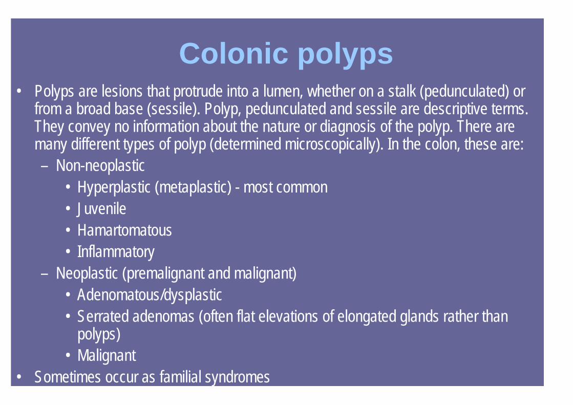
Colonic polyps• Polyps are lesions that protrude into a lumen, whether on a stalk (pedunculated) or
from a broad base (sessile). Polyp, pedunculated and sessile are descriptive terms.They convey no information about the nature or diagnosis of the polyp. There aremany different types of polyp (determined microscopically). In the colon, these are:– Non-neoplastic
• Hyperplastic (metaplastic) - most common• Juvenile• Hamartomatous• Inflammatory
– Neoplastic (premalignant and malignant)• Adenomatous/dysplastic• Serrated adenomas (often flat elevations of elongated glands rather than
polyps)• Malignant
• Sometimes occur as familial syndromes

While squamous dysplasia does not form a mass, glandular dysplasia developing in a lining epithelium often forms apolyp - a protuberance of tissue elevated from the surrounding mucosa. Such dysplastic polyps are often referred to asadenomatous polyps or in the colon, just adenomas. Colorectal adenomas may have a tubular, tubulovillous or villousarchitecture and the dysplasia histologically is graded as mild, moderate or severe. N.B. Adenomas in the GIT aredysplastic and thus premalignant, but adenomas in other sites are not necessarily dysplastic or premalignant e.g.breast, thyroid. Understanding the specific terminology is thus important, as the natural history and potential outcomesof the lesions are different.
Polyp
Normal mucosa
Submucosa

Low power view of a tubular adenomatouspolyp in the colon.
Black star: stalk including submucosa.Note how the glands of the adenomatouspolyp (blue arrows) are elongated andhave a disorganised architecturecompared to the normal colonic glands inthe adjacent mucosa (yellow arrows).Note intact muscularis mucosae: blackarrows

Edge of a tubular adenomatous polyp in the colon (low power). Normal glands are present on the right side of theimage and dysplastic glands on the left (separated by the black line). The dysplastic glands are more elongated,branched and disorganised. There are also fewer goblet cells indicating less differentiation. The yellow lines outlineparts of several glands and indicate (very approximately) the location of their epithelial basement membranes.MM: muscularis mucosae.
MM
MMMM

Compare the epithelial cells from the normal colonic gland on the left to those of the benign neoplastic (dysplastic) gland on the right.Note that the normal epithelial cells have small nuclei that are similar in size to each other (yellow arrows) and only take up about onethird or less of the cell. The well differentiated but neoplastic nuclei (red arrows) are larger and more crowded and take up a largerproportion of the cell. There is only mild nuclear pleomorphism. The basement membranes are outlined in black. Note the capillaries(blue arrows) and plasma cells (black arrows) in the lamina propria.
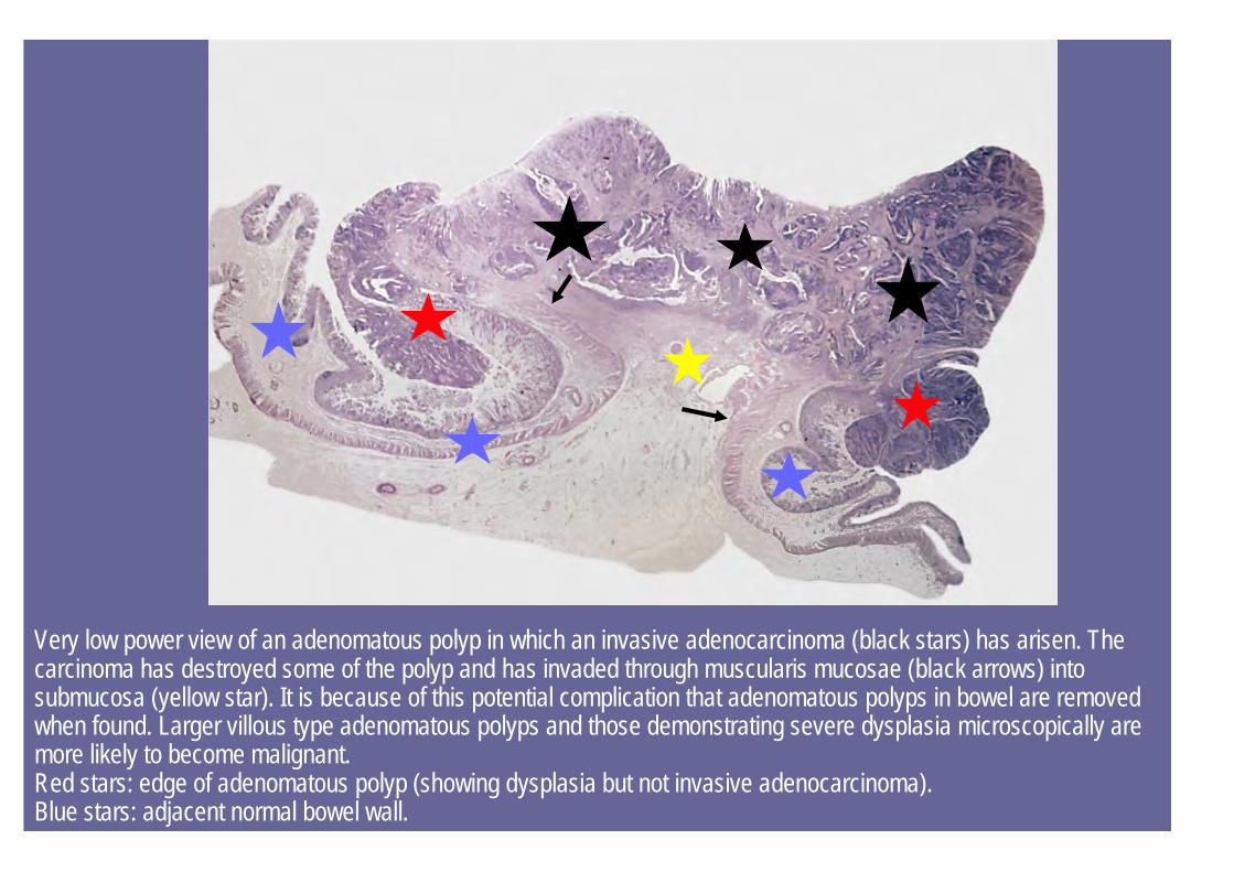
Very low power view of an adenomatous polyp in which an invasive adenocarcinoma (black stars) has arisen. Thecarcinoma has destroyed some of the polyp and has invaded through muscularis mucosae (black arrows) intosubmucosa (yellow star). It is because of this potential complication that adenomatous polyps in bowel are removedwhen found. Larger villous type adenomatous polyps and those demonstrating severe dysplasia microscopically aremore likely to become malignant.Red stars: edge of adenomatous polyp (showing dysplasia but not invasive adenocarcinoma).Blue stars: adjacent normal bowel wall.

Most neoplastic lesions form a mass that is composed not only of neoplastic cells but also supportive stroma, blood vessels andoften chronic inflammatory cells. The image on the left is a macroscopic view of an adenocarcinoma of the colon (red star). Thetumour surface is ulcerated. The image on the right is a very low power histopathological view of the edge of such a lesion. Thetumour is on the left side of the black line and normal colon is on the right side. Note how the tumour cells (seen as dark blue/purpleareas due to their high N:C ratio) invade deeply into the adventitia (A). The eosinophilic staining between the groups of tumour cellsrepresents the tumour stroma. (M: mucosa, MP: muscularis propria). Note that muscularis propria can be seen macroscopically(black arrows, left image) as a pale line, approx. 1mm thick. Even from the macroscopic specimen it can be seen that this lesion is atleast an ACP or Duke’s Stage B.
MP
M
Ulcerated area
A
Invasive tumour
Normal colon
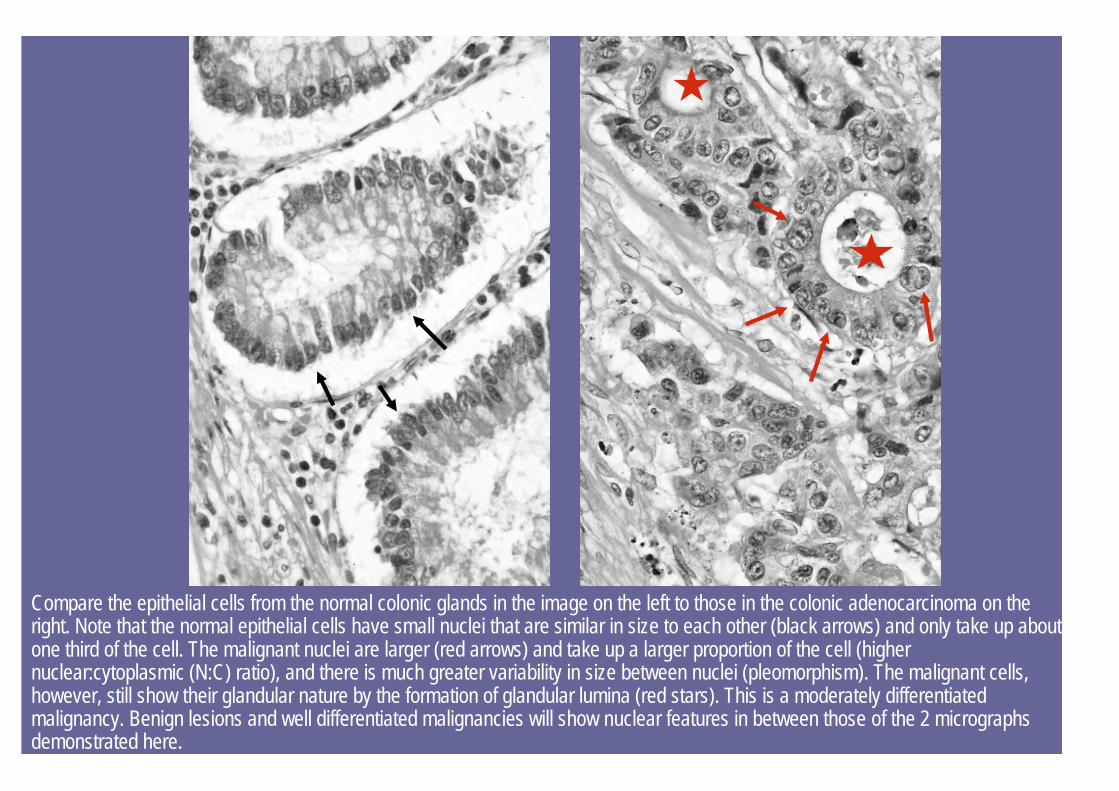
Compare the epithelial cells from the normal colonic glands in the image on the left to those in the colonic adenocarcinoma on theright. Note that the normal epithelial cells have small nuclei that are similar in size to each other (black arrows) and only take up aboutone third of the cell. The malignant nuclei are larger (red arrows) and take up a larger proportion of the cell (highernuclear:cytoplasmic (N:C) ratio), and there is much greater variability in size between nuclei (pleomorphism). The malignant cells,however, still show their glandular nature by the formation of glandular lumina (red stars). This is a moderately differentiatedmalignancy. Benign lesions and well differentiated malignancies will show nuclear features in between those of the 2 micrographsdemonstrated here.
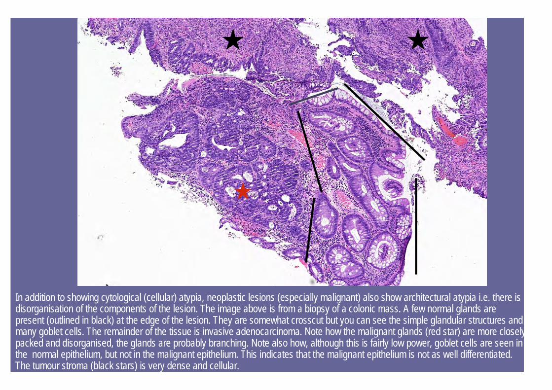
In addition to showing cytological (cellular) atypia, neoplastic lesions (especially malignant) also show architectural atypia i.e. there isdisorganisation of the components of the lesion. The image above is from a biopsy of a colonic mass. A few normal glands arepresent (outlined in black) at the edge of the lesion. They are somewhat crosscut but you can see the simple glandular structures andmany goblet cells. The remainder of the tissue is invasive adenocarcinoma. Note how the malignant glands (red star) are more closelypacked and disorganised, the glands are probably branching. Note also how, although this is fairly low power, goblet cells are seen inthe normal epithelium, but not in the malignant epithelium. This indicates that the malignant epithelium is not as well differentiated.The tumour stroma (black stars) is very dense and cellular.
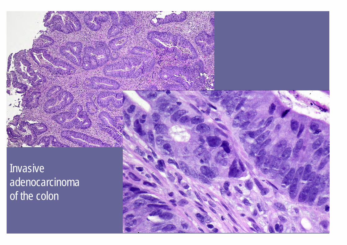
Invasiveadenocarcinoma of the colon

As malignant cells grow they need a blood supply and supportive connective tissue stroma. The stroma that develops looks differentto the normal connective tissue of the region that the tumour invades. The stromal response around the cells of an invasivemalignancy is known as desmoplasia. When florid, this stromal response makes the tumour very firm and pale (e.g. schirrous ductalcarcinoma of the breast). A desmoplastic stromal response is seen here in the invasive colonic adenocarcinoma (image on left) wherethere are more fibroblasts and denser connective tissue (red stars) than in the lamina propria and submucosa (black arrows) ofnormal colon (image on right). Chronic inflammatory cells also infiltrate malignant and often benign lesions.
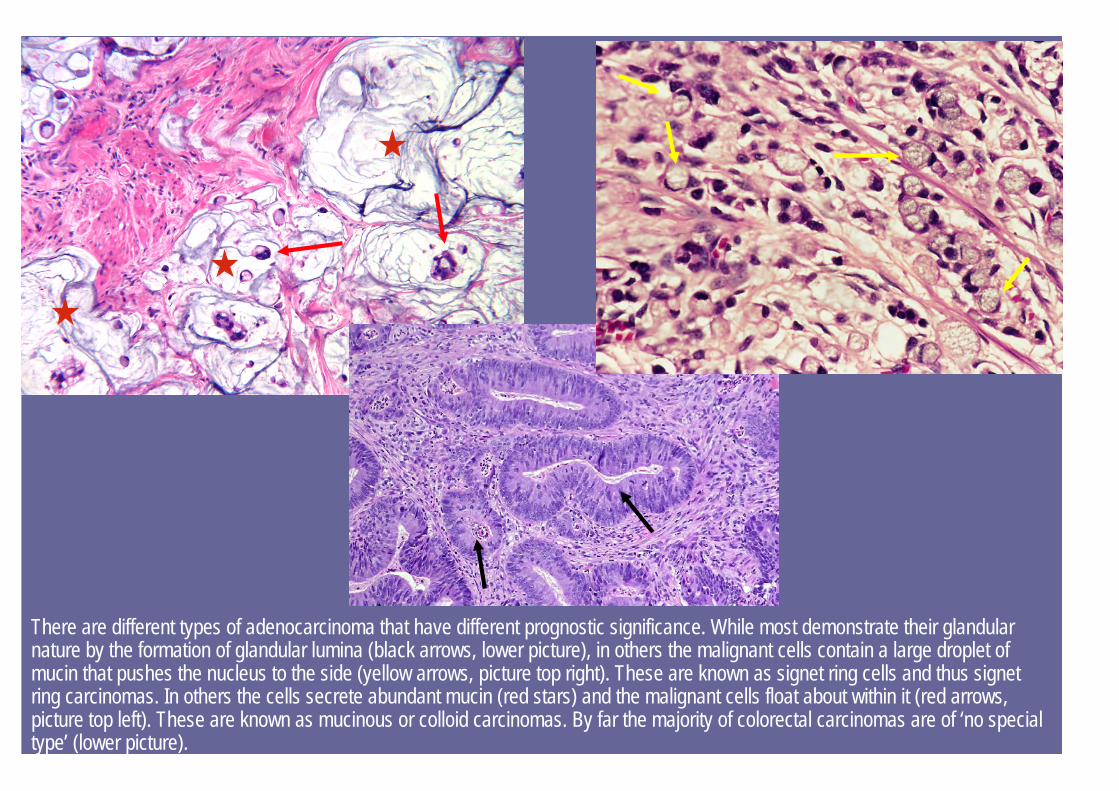
There are different types of adenocarcinoma that have different prognostic significance. While most demonstrate their glandularnature by the formation of glandular lumina (black arrows, lower picture), in others the malignant cells contain a large droplet ofmucin that pushes the nucleus to the side (yellow arrows, picture top right). These are known as signet ring cells and thus signetring carcinomas. In others the cells secrete abundant mucin (red stars) and the malignant cells float about within it (red arrows,picture top left). These are known as mucinous or colloid carcinomas. By far the majority of colorectal carcinomas are of ‘no specialtype’ (lower picture).
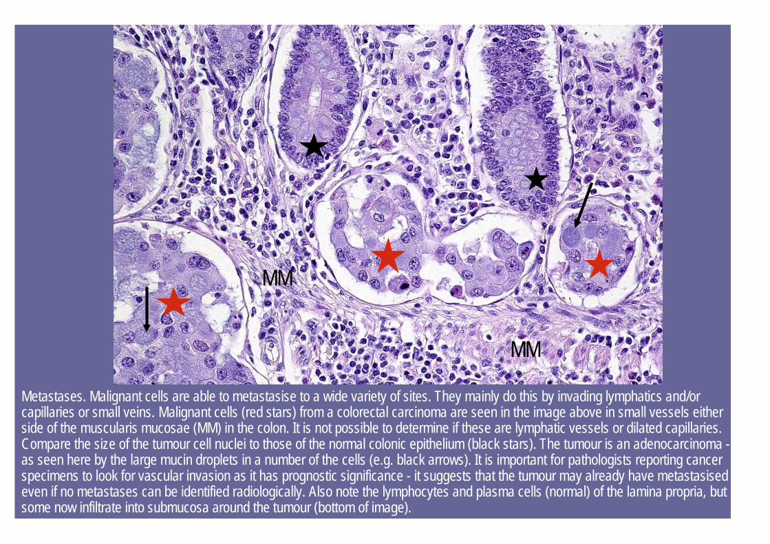
Metastases. Malignant cells are able to metastasise to a wide variety of sites. They mainly do this by invading lymphatics and/orcapillaries or small veins. Malignant cells (red stars) from a colorectal carcinoma are seen in the image above in small vessels eitherside of the muscularis mucosae (MM) in the colon. It is not possible to determine if these are lymphatic vessels or dilated capillaries.Compare the size of the tumour cell nuclei to those of the normal colonic epithelium (black stars). The tumour is an adenocarcinoma -as seen here by the large mucin droplets in a number of the cells (e.g. black arrows). It is important for pathologists reporting cancerspecimens to look for vascular invasion as it has prognostic significance - it suggests that the tumour may already have metastasisedeven if no metastases can be identified radiologically. Also note the lymphocytes and plasma cells (normal) of the lamina propria, butsome now infiltrate into submucosa around the tumour (bottom of image).
MM
MM

Metastatic tumour (black star) colorectal adenocarcinoma in the liver.Yellow star: adjacent liver tissue.
To make a histopathological diagnosis, the pathologist needs to examinemany features of the lesion. In the case of neoplastic or potentiallyneoplastic lesions, they examine the arrangement/architecture andcytological features of the cells and assess whether they are invasive ornot to determine if it is benign or malignant. In difficult cases they mayalso need to look for necrosis, the features of the stroma, or to countmitotic figures. They also need to determine the type of lesion, or its lineof differentiation (glandular, melanocytic, lymphocytic etc) and to grademalignant lesions (depending on the type) as these features alsoinfluence the management of the patient and prognosis. Other prognosticfeatures may also need to be assessed.
Colorectal carcinoma synopticreport
Many features affect the prognosis and management of malignancies. Thepathologist provides much of this information in the pathology report of the surgicallyexcised specimen with lymph nodes. This often detailed information is usuallypresented in a consistent fashion in a synoptic report. These differ somewhatdepending on the malignancy. You don’t need to memorize the details of the followingsynoptic report, which at this stage, is for information only. Note, however, the extentof information that is provided by the pathologist following examination of thespecimen and sections to the referring clinician. Medical students should get into thehabit of reading the pathology reports in patient notes, as doctors you will beexpected to understand them. You should, however, have a general understanding ofthe features that are important in assessing the prognosis and management ofmalignancies (tumour type and subtype, grade, stage, presence of lymphovascularinvasion, other depending on malignancy), including some specific information aboutthe major malignancies.

Nature of specimen: Right hemicolectomy
Tumour site: Ascending colon
Size: 20mm x 25mm
Proximal dilation (obstruction): Absent
Perforation: Absent
Tumour type: Adenocarcinoma NOS
Grade: Moderately differentiated
Margin of tumour: Infiltrative
Inflammatory infiltrate: Peritumoral lymphoid aggregates present
Colorectal carcinoma synopticreport

LOCAL SPREAD:Depth of invasion: Beyond muscularis propriaExcision margins: Proximal and distal margins are free of tumour. Distance of tumour to nearest margin
(distal) 40mmNeural invasion: Absent
DISTANT SPREAD:Lymph nodes: Total number of nodes found: 14Total number of nodes involved by tumour: 2Apical node: InvolvedVascular invasion: PresentMesenteric deposits: AbsentAdjacent bowel: 2x adenomatous polyps 8 and 10mm in dimension, showing moderate dysplasia
Colorectal carcinoma synopticreport
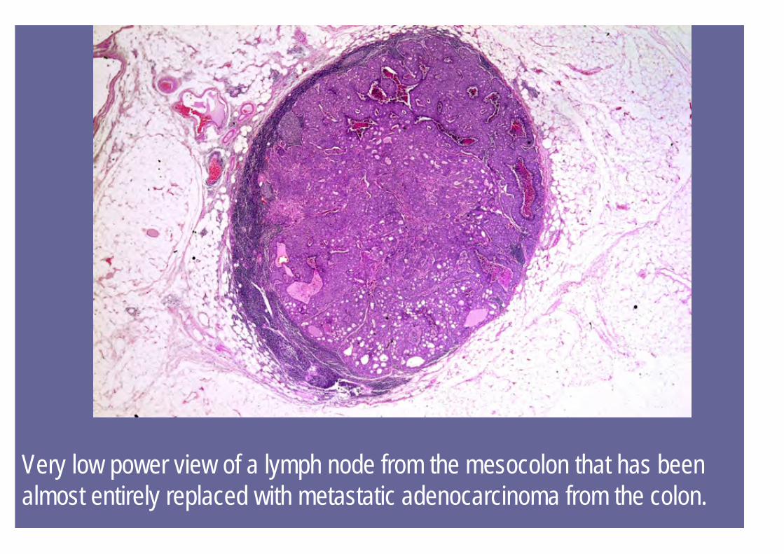
Very low power view of a lymph node from the mesocolon that has beenalmost entirely replaced with metastatic adenocarcinoma from the colon.
ANCILLARY INVESTIGATIONS:Immunohistochemistry studies were performed on paraffinsections of tumour with antibodies to MLH1 and MSH2. There wasno expression of MLH1 gene product by the tumour cells.Comments: Lack of expression of MLH1 as demonstrated byimmunohistochemistry suggests the possibility of underlyingmismatch repair gene abnormality and further gene mutationstudies may be indicated.
Colorectal carcinoma synopticreport
Only in certain cases are such ancillary investigations (previousslide) performed i.e. if there is a suspicion of hereditary non-polyposis colorectal carcinoma (HNPCC). The following slideshows the immunohistochemical staining for MLH1 and MSH2,and a normal H&E of the tumour. The tumour is not expressingthe MLH1 protein, yet the normal epithelial cells are, indicatingthat the tumour cells have a mutation in the encoding gene andthat the patient could have HNPCC.
Hereditary non-polyposis colorectalcarcinoma
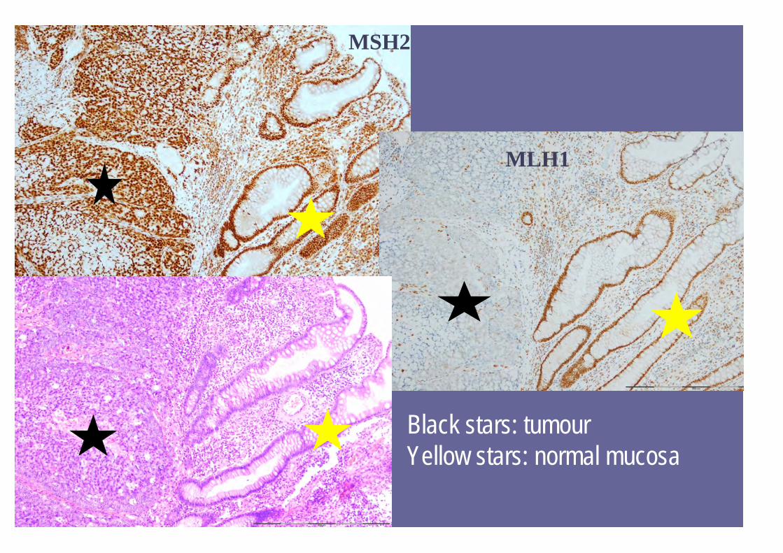
MLH1
MSH2
Black stars: tumourYellow stars: normal mucosa
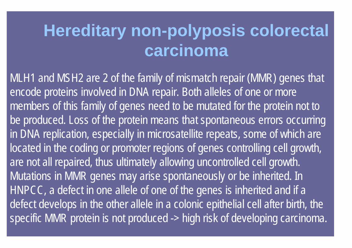
MLH1 and MSH2 are 2 of the family of mismatch repair (MMR) genes thatencode proteins involved in DNA repair. Both alleles of one or moremembers of this family of genes need to be mutated for the protein not tobe produced. Loss of the protein means that spontaneous errors occurringin DNA replication, especially in microsatellite repeats, some of which arelocated in the coding or promoter regions of genes controlling cell growth,are not all repaired, thus ultimately allowing uncontrolled cell growth.Mutations in MMR genes may arise spontaneously or be inherited. InHNPCC, a defect in one allele of one of the genes is inherited and if adefect develops in the other allele in a colonic epithelial cell after birth, thespecific MMR protein is not produced -> high risk of developing carcinoma.
Hereditary non-polyposis colorectalcarcinoma