histology of lymph node - rawalpindi medical university
TRANSCRIPT

Histology of Lymph NodeBy Dr Arsalan Manzoor Mughal

Introduction
• Lymph nodes are bean-shaped, encapsulated structures
• 10 mm by 2.5 cm in size
• distributed throughout the body along the lymphatic vessels
• A total of 400 to 450 lymph nodes are present,

Functions
• defend against the spread of microorganisms
• defend against the spread of tumor cells
• provide enclosed environments for antigen presentation
• development of plasma cells secreting non-IgA antibodies.


Regions
• three regions• Cortex• Paracortex• Medulla

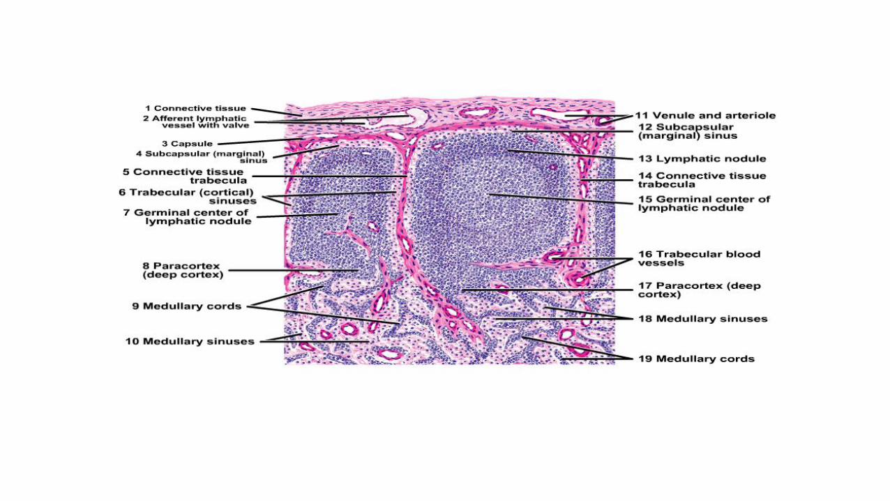
The Cortex

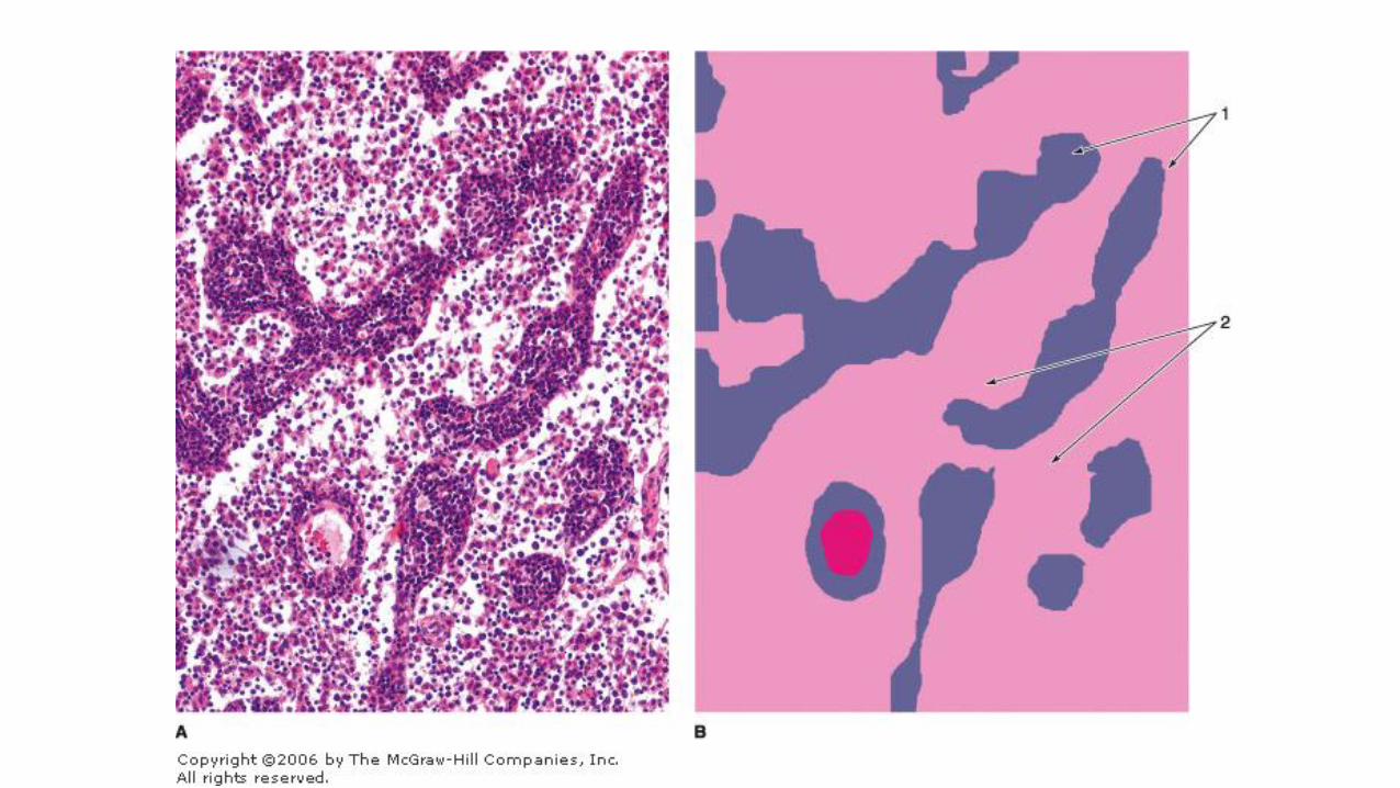
Subcapsular Sinus
• immediately inside the capsule,
• receives lymph from the afferent lymphatics
• From this space cortical sinuses (or trabecular sinuses) branch internally among the lymphoid nodules along trabeculae.
• hese sinuses are lined by a very thin, discontinuous endothelium penetrated by reticulin fibers and processes of dendritic cells.
• Lymph containing antigens, lymphocytes, and APCs passes through these sinuses and percolates easily into the surrounding lymphoid tissue.

Lymphoid nodules
• with or without germinal centers
• consist largely of developing B lymphocytes and occupy much of the cortex not filled with helper T lymphocytes
• Each nodule is organized around the long, interdigitating processes of follicular dendritic cells (FDCs), but these are not readily seen by routine light microscopy.
• Numerous macrophages are also present for removal of newly formed defective B cells which undergo apoptosis.


Paracortex

Paracortex
• The region between the cortex and medulla
• the does not have precise boundaries but can be distinguished from the outer cortex by its lack of nodules
• Unlike the superficial cortex, the paracortex contains lymphoid tissue rich in T cells distinguishable by immunohistochemistry
• specialized postcapillary venules called high endothelial venules (HEVs) represent an important entry point for most (90%) circulating lymphocytes into lymph nodes. Endothelial cells of these vessels become unusually enlarged or cuboidal and express specific apical surface glycoproteins that mediate the tethering and diapedesis of B and T cells from the blood into the paracortex of the lymph node


Medulla

Medullary cords
• branched cordlike masses of lymphoid tissue extending from the paracortex.
• They contain T and B lymphocytes and many plasma cells.

Medullary sinuses
• are dilated spaces lined by discontinuous endothelium that separate the medullary cords.
• The lumens of medullary sinuses include a meshwork of processes from reticular cells, which represent a final lymph filter.
• These sinuses contain many macrophages and sometimes neutrophils if the lymph node is draining an infected region.
• They are continuous with the cortical sinuses and converge at the hilum as the efferent lymphatic vessel


Role of Lymph Nodes in the Immune Response• lymph arriving at a lymph node contains antigens free in solution or
bound to antibodies or complement, still on microorganisms, or already internalized and transported by APCs.
• If draining from an infected or inflamed region, lymph may also contain microorganisms and cytokines. Antigens not yet phagocytosed will be internalized by APCs in the lymph nodes and presented on MHC class II molecules.

Role of Lymph Nodes in the Immune Response• Circulating B and T lymphocytes traffic from node to node, entering
via the lymph or HEVs, where B cells contact antigens on FDCs and T cells sample antigens presented on endritic cells and other APCs.
• Lymphocytes whose receptors recognize such antigens will be activated. B cells will proliferate rapidly in germinal centers of follicles with the help of Th cells, often enlarging the entire lymph node. Activated cytotoxic T cells in the paracortex proliferate to a much lesser extent without forming follicles.

Role of Lymph Nodes in the Immune Response• Many newly made B cells, now activated against a specific antigen,
differentiate as plasma cells and move to the medulla or to downstream sites beyond the lymph node where they produce antibodies.
• Specific Th cells, CTLs, and Tregs also recirculate in the efferent lymph and with the antibodies spread the immune defenses against those microorganisms throughout the body. Both B and T memory cells also move elsewhere in the body, providing long-lived protection and proliferating more rapidly upon subsequent exposure to their specific antigen.

MEDICAL APPLICATION
• Metastatic cancer cells detached from a primary tumor can enter lymphatics and are carried to nearby lymph nodes, especially the sentinel lymph node that is the first one downstream of the region with the tumor.
• Cells from well established tumors are often immunosuppressive themselves and may continue growth as a secondary tumor within lymph nodes.
• During cancer surgery lymph nodes in the lymphatics draining the tumor area are examined by pathologists for the presence of cancer cells. The presence of such metastatic cells in lymph nodes is a key determinant in most staging systems for various types of cancer and an important prognostic indicator.