histology of gills of labeo rohita and hypophthalmichthys molitrix
TRANSCRIPT

~ 1 ~
International Journal of Fisheries and Aquatic Studies 2014; 1(6): 01-06 ISSN: 2347-5129 IJFAS 2014; 1(6): 01-06 © 2013 IJFAS www.fisheriesjournal.com Received: 09-05-2014 Accepted: 21-05-2014 Shivali Singh Department of Zoology and Applied Aquaculture, Barkatullah University, Bhopal (M.P.), India Pinky Kaur Department of Zoology and Applied Aquaculture, Barkatullah University, Bhopal (M.P.), India Correspondence: Pinky Kaur Department of Zoology and Applied Aquaculture, Barkatullah University, Bhopal (M.P.), India
Histology of gills of Labeo rohita and Hypophthalmichthys molitrix infested by monogenean and copepod parasites
Shivali Singh and Pinky Kaur
ABSTRACT The present study was conducted to reveal a histological analysis on the gills of Labeo rohita and Hypophthalmichthys molitrix specimens were observed to reveal the histo-pathological characteristics of the gills of these freshwater carps. The histological examination of the gills of Labeo rohita revealed the proliferation of bronchial tips, shortening and fusion of secondary gill lamellae, desquamation of primary and secondary gill lamellar epithelium, uplifting of respiratory epithelial wall and damaged pillar cells due to monogenetic parasites. Whereas drastic disorganization was observed to be caused due to multiple infestation by protozoan and copepod in gills of Hypophthalmichthys molitrix characterized by revealed the proliferation of bronchial tips, shortening, thinning and deshaping of gill lamellae’s. Keywords: Parasites, Gills, Histology, Carps, Copepod.
1. Introduction Parasites are one of the major groups of organisms that may or may not cause infection in fishes depending on a number of factors. Parasites in fish (endo – and ecto-parasites) are a natural occurrence and common [1]. Fish parasites result in huge economic losses as they increase mortality and also increase farm inputs via increased treatment expenses and cause reduction in growth rate due to the parasitic disease outbreak [2]. Gill parasitic are common on cultured and wild fish. Many of these species have long been recognized to have the potential to affect the growth, fecundity and survival of hosts [3]. As, fish gills are specialized tissues for gas exchange, circulation, ion and acid-base balance, hormone production, and nitrogenous waste secretion [4]. Gill parasites attach to the gills of fish and feed on their host’s blood and tissue [5]. The organs of attachment such as suckers and hooks causes extensive tissue damage and inflammation, and may render fish susceptible to secondary infection by bacteria, fungi, and viruses [6]. Thus, the damage to gill tissue can reduce the ability of the fish to maintain normal oxygen uptake by hindering water flow [5]. According to Abdelhalim [7] extensive tissue damage resulting from the feeding and attachment of these parasites has been reported in several species of fish. Protozoan among other parasites cause immeasurable damage to the fishing industry [8]. Parasites play an important role in determining the health status of the fishes [9]. Therefore present study was undertaken to reveal the effect of different parasites on histology of gills of freshwater carps. 2. Material and Method 2.1 Collection of Host Fishes and parasites Host Fish (Labeo rohita and Hypophthalmichthys molitrix) were collected alive from the Shahpura Lake. Fishes were collected ones in a week for one year (during different seasons i.e. summer, rainy, post-monsoon and winter months). The hosts brought to the laboratory were subjected to a thorough investigation as per the methods employed by Cable [10], Meyer and Olsen [11]. For the purpose of collection of parasites, the gills were taken out and placed in petri dishes, containing normal saline (0.75% NaCl). They were scraped with the help of scalpel to collect the parasites attached to the gills. The parasites collected were examined in live condition.
~ 2 ~
International Journal of Fisheries and Aquatic Studies
2.2 Histological technique Infected gills were immediately fixed in alcoholic Bouin’s fluid for 24 hours. After complete removal of picric acid, the tissue was dehydrated in an ethanol series (30%, 50%, 70%, 90% and 100%), cleared in xylene and processed for preparation of paraffin wax blocks. The tissue was then cut at 4 - 5 μm thickness by rotatory microtome and stained routinely with haematoxylin and eosin (H-E) for histo-pathological examination. Stained histo-pathological sections were examined under Olympus research microscope. Microphotography was taken both at high magnification and low magnification.
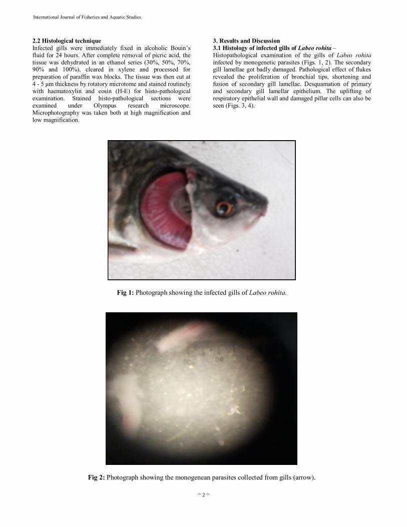
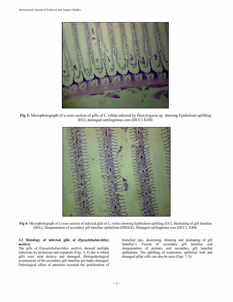
3. Results and Discussion 3.1 Histology of infected gills of Labeo rohita – Histopathological examination of the gills of Labeo rohita infected by monogenetic parasites (Figs. 1, 2). The secondary gill lamellae got badly damaged. Pathological effect of flukes revealed the proliferation of bronchial tips, shortening and fusion of secondary gill lamellae. Desquamation of primary and secondary gill lamellar epithelium. The uplifting of respiratory epithelial wall and damaged pillar cells can also be seen (Figs. 3, 4).
Fig 1: Photograph showing the infected gills of Labeo rohita.
Fig 2: Photograph showing the monogenean parasites collected from gills (arrow).
~ 3 ~
International Journal of Fisheries and Aquatic Studies
Fig 3: Microphotograph of a cross section of gills of L. rohita infected by Dactylogyrus sp. showing Epithelium uplifting (EU), damaged cartilaginous core (DCC) X100.
Fig 4: Microphotograph of a cross section of infected gills of L. rohita showing Epithelium uplifting (EU), Shortening of gill lamellae (SGL), Desquamation of secondary gill lamellae epithelium (DSGLE), Damaged cartilaginous core (DCC) X400.
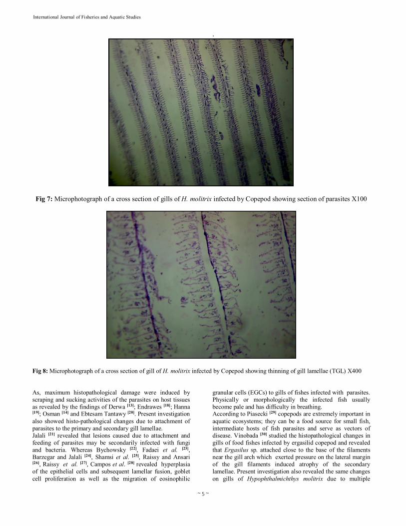
3.2 Histology of infected gills of Hypophthalmichthys molitrix The gills of Hypophthalmichthys molitrix showed multiple infections by protozoan and copepods (Figs. 5, 6) due to which gills were total destroy and damaged. Histopathological examination of the secondary gill lamellae got badly damaged. Pathological effect of parasites revealed the proliferation of
bronchial tips, shortening, thinning and deshaping of gill lamellae’s. Fusion of secondary gill lamellae and desquamation of primary and secondary gill lamellar epithelium. The uplifting of respiratory epithelial wall and damaged pillar cells can also be seen (Figs. 7, 8).

~ 4 ~
International Journal of Fisheries and Aquatic Studies
Fig 5: Photograph showing copepod and protozoan parasites collected from the gills of Hypophthalmichthys molitrix
Fig 6: Photograph showing enlarged view of parasites collected H. molitrix
According to Saleh and El-Nobi [12] infestation due to monogenetic trematodes is one of the most prevalent parasitic agents affecting skin and gills causing irritation and destruction of gills which leading to impairment of breathing as well as tremendous losses in aquaculture. According to Derwa [13]; Osman [14]; Younis [15] and El-Shahat [16] protozoan species Trichodina are extensively isolated from the gills of tilapia and catfish. Osman [14] and El-Shahat [16] isolated Henneguya branchialis from
the gills and suprabranchial organs of cultured catfish. Ronald [17] examined Dactylogyridae is the most common gill parasite in freshwater fish especially young fish. Saleh and El-Nobi [12] collected Monogenea sp. was mainly from Tilapia sp., catfish, few carp sp. and few mullet sp. and stated that young stages of Tilapia were more susceptible to monogenesis (mainly Dactylogyrus and Gyrodactylus) than adult stages.
~ 5 ~
International Journal of Fisheries and Aquatic Studies .
Fig 7: Microphotograph of a cross section of gills of H. molitrix infected by Copepod showing section of parasites X100
Fig 8: Microphotograph of a cross section of gill of H. molitrix infected by Copepod showing thinning of gill lamellae (TGL) X400 As, maximum histopathological damage were induced by scraping and sucking activities of the parasites on host tissues as revealed by the findings of Derwa [13]; Endrawes [18]; Hanna [19]; Osman [14] and Ebtesam Tantawy [20]. Present investigation also showed histo-pathological changes due to attachment of parasites to the primary and secondary gill lamellae. Jalali [21] revealed that lesions caused due to attachment and feeding of parasites may be secondarily infected with fungi and bacteria. Whereas Bychowsky [22], Fadaei et al. [23], Barzegar and Jalali [24], Shamsi et al. [25], Raissy and Ansari [26], Raissy et al. [27], Campos et al. [28] revealed hyperplasia of the epithelial cells and subsequent lamellar fusion, goblet cell proliferation as well as the migration of eosinophilic
granular cells (EGCs) to gills of fishes infected with parasites. Physically or morphologically the infected fish usually become pale and has difficulty in breathing. According to Piasecki [29] copepods are extremely important in aquatic ecosystems; they can be a food source for small fish, intermediate hosts of fish parasites and serve as vectors of disease. Vinobada [30] studied the histopathological changes in gills of food fishes infected by ergasilid copepod and revealed that Ergasilus sp. attached close to the base of the filaments near the gill arch which exerted pressure on the lateral margin of the gill filaments induced atrophy of the secondary lamellae. Present investigation also revealed the same changes on gills of Hypophthalmichthys molitrix due to multiple

~ 6 ~
International Journal of Fisheries and Aquatic Studies infections by protozoan and copepod showed entire changes in histoarchitecture of gills 4. Conclusion Histo-pathological study has been used to diagnose diseases of aquatic organisms throughout the world. Gills are the primary organ which directly reflects the water pollution, contamination and diseases causing factors. The histo-architectural change in gills hinders the oxygen intakes and in the long term causes huge mortality in aquaculture practices. 5. Reference
1. Moyle PB, Cech JJ. Fishes: An introduction to ichthyology. Prentice Hall, Upper Saddle River, New Jersey, 2004, 356.
2. Kayis S, Ozcelep T, Capkin E, Altinok I. Protozoan and metazoan parasites of cultured fish in Turkey and their applied treatments. The Israeli J Aquac Bamidgeh 2009; 61:93-102.
3. Johnson SC, Blaylock RB, Elphick J, Hyatt K. Disease caused by the salmon louse Lepeophtheirus salmonis Copepoda: Caligidae) in wild sockeye salmon (Oncorhynchus nerka ) stocks of Alberni Inlet British Columbia. Can J Fish Aquat Sci 1996, 53:2888-2897.
4. Pelster B, Bagatto B. Respiration. Fish Physiology 2010; 29:289-309.
5. Ojha J, Hughes GM. Effect of branchial parasites on the efficiency of the gills of a freshwater catfsh Wallago attu. J Zool 2001; 255:125-129.
6. Dezfuli B, Sayyaf, Luisa G, Robert K, Paul J, Maurizio M. Immunohistochemistry ultrastructure and pathology of gills of Abramis brama from Lake Mondsee Austria infected with Ergasilus sieboldi (Copepoda). Diseases of Aquatic Organisms 2003; 53:257–262.
7. Abdelhalim AI. Morphology and epidemiology of some parasitic copepods (Poecilostomatoida: Ergasilidae) from British freshwater fish. PhD thesis University of London 1990.
8. Doglel VA, Petrushevski GK, Polyanski YI. Translated by (Kabata) Parasitology of Fishes. Oliver and Boyd Endinburgh and London 1961; 384.
9. Ferguson WH. Gills and pseudobranchs In: Systemic Pathology of Fish A text and Atlas of Comparative Tissue Responses in Diseases of Teleosts. MCK, SH, F 42, 1989.
10. Cable RM. An illustrated laboratory manual of parasitology. Ed 5. Burgess Publishing Minneapolis, Minnesota, 1977.
11. Meyer MC, Olsen OW. Essentials of Parasitology. Ed 2, Wm. C. Brown Co. Iowa, 1975, 1-303.
12. Saleh GA, El-Nobi GA. Prevalence of Monogeniasis in Tilapia fish among different systems of fish management, seasons and fish life stages with special reference to the therapeutic effect of Praziquantel at different temperatures. Zag Vet J 2003; 31(1):37-48.
13. Derwa HIM. Some studies on gill affections of some freshwater fishes M Sc thesis Faculty of Veterinary Medicine Suez Canal University, 1995.
14. Osman HAM. Studies on parasitic gill affections in some cultured freshwater fishes. Master thesis submitted to the Faculty of Veterinary Medicine, Suez Canal University, 2001.
15. Younis AA. Effect of some Ectoparasites on
Reproduction of Oreochromis niloticus Fish with Referring to Treatment. The First International Conference of the Veterinary Research Division, National Research Centre, 15-17 February 2004, 111.
16. El-Shahat RA. Studies on ectoparasites of freshwater fish. Master Thesis submitted to Fac. of Vet. Med., Zagazig University, 2004.
17. Ronald S. Fish pathology. Ed 2, Bailliere Tindall, London, England, 1989.
18. Endrawes MN. Observations on some external and internal parasitic diseases in Nile catfishes. A Master thesis submitted to Dept. of fish diseases and management. Fac of Vet Medicine Zagazig Univ 2001.
19. Hanna MI. Epizootiological studies on parasitic infections in fishes cultured under different fish cultural systems in Egypt. A Master thesis submitted to Dept. of fish diseases. Fac of Vet Med Cairo Univ, 2001.
20. Ebtesam AT. Trial to control the Heterophyid and Echinostomatid Metecercariae Infecting Oreochromis niloticus in Aquaculture by Praziquantel ‘Droncit’. The First International Conference of the Vet Res Division, NRC, 15-17 February 2004, 108-109.
21. Jalali B. Parasites and parasitic diseases of Iran's fresh water fishes Iranian Fishery Institute Publications. Tehran Iran 1997; 312- 407.
22. Bychowsky BE. Monogenetic trematodes of some fish of Iran collected by Pavlowsky EN. Trudi Zoologicheskovo Instituta Akademiya 1949; 8:870-878.
23. Fadaei F, Mokhayer B, Ghorbani H. Identification of fishes and their parasites in Choghakhor Lagoon. J Facul Vet Med Uni Tehran 2001; 56:109-113.
24. Barzegar M, Jalali B. Helminthes Acanthocephala and crustacean parasites of fishes in Vahdat reservoir. Iran J Vet Sci 2004; 2:229-234.
25. Shamsi S, Jalai B, Aghazadeh M. Infection with Dactylogyrus spp among introduced cyprinid fishes and their geographical distribution in Iran. Iran J Vet Res 2009; 10:70-74.
26. Raissy M, Ansari M. Histopathological changes in the gills of naturally infected Capoeta aculeata (Cuvier and Valenciennes, 1844) with parasites. African Journal of Biotechnology 2011; 10(68):15422-15425.
27. Raissy M, Ansari M, Moumeni M. Parasite fauna of Aphanius vladykovi Coad (Osteichthyes: Cyprinodontidae) in Gandoman Lagoon. Comp Parasitol 2011; 78:104-106.
28. Campos CM, Moraes JRE, Moraes FR. Histopathology of gills of Piaractus mesopotamicus (Holmberg, 1887) and Prochilodus lineatus (Valenciennes, 1836) infested by monogenean and myxosporea, caugth in Aquidauana River State of Mato Grosso do Sul, Brazil. Rev Bras Parasitol Vet Jaboticabal 2001; 20(1):67-70.
29. Piasecki W, Goodwin, Andrew E, Eras, Jorge C, Nowak et al. Importance of Copepoda in Freshwater Aquaculture. Zoological Studies 2004; 43:193-205.
30. Vinobada P. Histopathological changes induced by ergasilid copepod infections on the gills of food fish from Batticaloa Lagoon Sri Lanka. Sri lanka J Aquat Sci 2007; 12:77-87.